


 , Jin Zeng 1,3,4,*
1 Department of Urology, The First Affiliated Hospital of Xi’an Jiaotong University, 710061 Xi'an, Shaanxi, China
2 Department of Thoracic Surgery, Tangdu Hospital, Fourth Military Medical University, 710038 Xi'an, Shaanxi, China
3 Key Laboratory for Tumor Precision Medicine of Shaanxi Province, 710061 Xi'an, Shaanxi, China
4 Oncology Research Lab, Key Laboratory of Environment and Genes Related to Diseases, Ministry of Education, 710061 Xi'an, Shaanxi, China
Abstract
Background: Due to its non-invasive and widely applicable features,
photodynamic therapy (PDT) has been a prominent treatment approach against cancer
in recent years. However, its widespread application in clinical practice is
limited by the dark toxicity of photosensitizers and insufficient penetration of
light sources. This study assessed the anticancer effects of a novel
photosensitizer 5-(4-amino-phenyl)-10,15,20-triphenylporphyrin with
diethylene-triaminopentaacetic acid (ATPP-DTPA)-mediated PDT (hereinafter referred to as ATPP-PDT) under the
irradiation of a 450-nm blue laser on colorectal cancer (CRC) in vivo
and in vitro. Methods: After 450-nm blue laser-mediated
ATPP-PDT and the traditional photosensitizer 5-aminolevulinic acid (5-ALA)-PDT
treatment, cell viability was detected through Cell Counting Kit-8 (CCK-8) and
5-ethynyl-2
Keywords
- ATPP-DTPA
- photodynamic therapy
- colorectal cancer
- p38 MAPK
- apoptosis
Colorectal cancer (CRC) is one of the most common digestive tract tumors, which is a general term for malignant lesions of colon or rectal mucosal epithelium under the action of environmental or genetic and other carcinogenic factors [1]. As the second most frequent cause of death from cancer worldwide and the third most common illness overall, accounting for around 10% of all cancer cases, CRC has gained significant attention. It is estimated that there were about 153,020 new cases of CRC, and 52,550 patients may die from the malignancy in 2023 [2, 3]. The reasons for the increase in incidence probably include an aging population and some unhealthy lifestyle choices, such as sedentary behavior, obesity, smoking and excessive drinking [4, 5]. Hereditary factors are also involved in the development of a substantial proportion of all cases of CRC [6]. For most patients with CRC, surgery is still the first choice, but postoperative recurrence and distant metastasis become the main factors restricting the prognosis of patients [7]. With the rapid development of systemic comprehensive therapy such as chemotherapy, targeted therapy and immunotherapy, the prognosis of patients has substantially improved. However, common drugs used in clinic have serious side effects, accompany by drug resistance, which render them ineffective for the treatment of CRC [4, 8, 9]. Therefore, more efficient and safer treatments are urgently needed to improve the survival time and quality of life of CRC patients.
Photodynamic therapy (PDT) is an emerging therapy. After a certain time interval, the photosensitizer is activated by a specific wavelength of light at the desired wavelength, and then react with molecular oxygen to produce reactive oxygen species (ROS), causing cell death [10, 11, 12, 13]. Interestingly, the oxygen environment, photosensitizers and irradiated lasers alone do not have cytotoxicity, and only when they are combined can cause tumor cell death [14]. Actually, compared with the traditional anti-tumor therapy, PDT has its unique advantages: strong selective toxicity to tumor cells, good security for normal organizations, low drug resistance and no long-term side effects [15], which made PDT to become an alternative or supplementary method to treat some malignant tumors, including CRC [16, 17, 18, 19]. However, like most tumor treatment strategies, PDT has its own limitations, such as the phototoxicity of photosensitizers, the expensive light source equipment, the insufficient penetration of light sources in solid tumors and tumor hypoxia, which hinder the clinical promotion of PDT [20, 21]. Therefore, it is urgent to explore a new light source and photosensitizer for PDT.
With regard to photosensitizers, for clinical PDT, the ideal photosensitizers should meet the following standards as far as possible: significant phototoxicity but low dark toxicity, excellent selectivity to target cells, quick excretion from the body, and easy administration through various pathway [22, 23]. 5,10,15,20-tetraphenylporphyrin, nano-materials designed based on its porphyrin ring structure have been emerging in recent years [24, 25, 26]. The 5-(4-aminophenyl)-10,15,20-triphenylporphyrin with diethylene-triaminopentaacetic acid (ATPP-DTPA) in this paper is also modified on the basis of tetraphenyl porphyrin to be closer in clinical application. In terms of light source, the world’s first 450-nm optical surgical system has been developed by our team, and held the China National Medical Products Administration approval (20213010922). Different from the commonly used 630-nm red laser, we chose the 450-nm blue laser for our research, which offers the benefits of excellent tissue solidification, low tissue penetration, and high vaporization efficiency, and is expected to become a new choice for superficial disease surgery [27, 28, 29]. Considering the efficacy and safety of blue laser, our team continued to verify the PDT effect of low-dose 450-nm blue laser on superficial luminal tract tumors. With the combination of the traditional photosensitizer 5-ALA and the 450-nm blue laser, we found a significant therapeutic effect on high-risk human papillomaviruses (HR-HPV) infected cells [30]; additionally, the 450-nm blue laser mediated PDT with the photosensitizer sinoporphyrin sodium (DVDMS) had an obvious inhibitory effect on gastric cancer cells [31]; our self-designed nanomaterial naming HAFeR by encapsulating PSs and RSL3 in metal organic framework (MOF) also demonstrated a significant killing effect on bladder tumor cells combined with 450-nm blue laser [32]. A common result was obtained that 450-nm blue laser showed a better therapeutic efficacy and biosafety for PDT compared to 630-nm red laser for superficial malignancies. In this study, we chose 450-nm blue laser and a novel photosensitizer ATPP-DTPA to explore the anti-tumor effect on colorectal cancer, and briefly discussed its potential molecular mechanism regarding the apoptosis signal pathway.
The human CRC cell lines HCT116 and SW480, as well as the mouse CRC cell lines
CT26 and MC38, were obtained from the Chinese Academy of Sciences’ Cell Bank of
Type Culture Collection (Shanghai, China). HCT116 cells were cultured in the RPMI
1640 medium (Sigma-Aldrich, St.Louis, MO,USA), while SW480, CT26 and MC38 cells were
maintained in Eagle’s minimum essential medium (DMEM, Sigma-Aldrich, St.Louis, MO,USA),
supplemented with 10% fetal bovine serum (Biological Industries, Jerusalem, Israel). In
addition, the culture medium was supplemented with 100 U/mL penicillin and 0.1
mg/mL streptomycin (New Cell & Molecular Biotech, Suzhou, China). All cell lines
were validated by STR profiling and tested negative for mycoplasma. Cells were
incubated in a humidified atmosphere of 5% CO
The sensitizer ATPP-DTPA, amino derivative of tetraphenyl porphyrin with diethylenetriaminepentaacetic acid (DTPA), was provided by Institute of Biomedical Engineering, CAMS&PUMC, molecular weight: 1004 Da, purity: 98.5%. ATPP-DTPA was dissolved in phosphate-buffered saline (PBS) to form a stock solution with a final concentration of 1 mM. 5-ALA was purchased from Suzhou NMT Biotech Co., Ltd., dissolved in Dimethyl sulfoxide (DMSO) to form a stock solution with a final concentration of 50 mM. The sensitizers were sterilized with 0.22-µm filter membrane, then stored in darkness at –20 °C and used within one month.
Working solution of ATPP-DTPA was fresh prepared at various concentrations of 0,
0.625, 1.25, 2.5, 5, 10 µM and 5-ALA was also fresh prepared with
concentrations of 0, 0.05, 0.1, 0.2, 0.4, 0.8 mM. The PDT light source was
provided by the semiconductor lasers (excitation wavelength: 450 nm;
manufacturer: Blueray Medical Ltd., Xi’an, China). Cells were first incubated
with ATPP-DTPA or 5-ALA under darkness for 4 hours and then treated with laser
irradiation of 20 mW/cm
For inhibitory experiments,10 µM Z-VAD-FMK and SB202190 from MedChemExpress (Monmouth Junction, NJ, USA) were incubated with CRC cells 1 h before irradiation to inhibit apoptosis signal pathway and p38 MAPK signal pathway, respectively.
HCT116 and SW480 cultured in 6-well plates (3
The Cell Counting Kit-8 (CCK-8) and 5-ethynyl-2
The level of total intracellular ROS was measured by
2
For inhibitory experiments, N-acetylcysteine (NAC) from Selleck (Shanghai, China) was added to the medium with a concentration of 10 mM and incubated with the cells for 2 hours before PDT to played its role of ROS scavenger.
The apoptosis rate of HCT116 and SW480 was detected by flow cytometry. The
supplier of the Annexin V-FITC/PI Apoptosis kit was BD Bio-science (Franklin
Lakes, NJ, USA). At a density of 5
HCT116 cells were divided into ATPP-DTPA group and PDT group with three independent repetitive sample experiments. After 24 hours of PDT to HCT116 cell, TRIzol was used to separate the total RNA extracts. The samples were sent to Novogene Co., Ltd. Tianjin Sequencing Center for test.
Cells were inoculated in 6-cm cell culture dish, when the cell density reached
about 70%, PDT and other control treatments were performed. We first collected
the cell samples at 12 hours after PDT. Then the samples were lysed using 1
Aged 4–5 weeks male Balb/C nu/nu mice (Shanghai Experimental Animal Center of
the Chinese Academic of Sciences, Shanghai, China) were fed under standard
conditions and prepared for in vivo experiment. The Xi’an Jiaotong
University Ethics Committee (Xi’an, China) gave its approval to all in
vivo tests, which were conducted in accordance with the rules set out by the
National Laboratory Animal Care and Use Institute. Mice were received a
subcutaneous injection of 5
All animal’s main organs and tumor tissues were preserved in 4% paraformaldehyde (PFA) for no less than 24 hours. Samples were cut into 5-µm-thick slices, and hematoxylin and eosin (H&E) followed by addition. The histopathological changes were observed by inverted fluorescence microscope. Primary antibodies (cleaved-PARP, phospho-p38 MAPK, and Ki-67) were incubated on the sections for 16 hours at 4 °C and goat anti-rabbit secondary antibody was followed for 1 h at room temperature, then visualized using diaminobenzidine (DAB) solution (34002, Thermo Fisher Scientific, Waltham, MA, USA). Finally, we observed the image with a microscope and analyzed it with Image J 1.46r software (National Institute of Health , Bethesda, MD, USA).
Each data was repeated in three independent experiments, and the mean
ATPP-DTPA, as a new photosensitizer, has the basic structure of porphyrin rings (Fig. 1A). The absorption spectrum of ATPP-DTPA in PBS was showed in Fig. 1B, whose highest absorption peak was around 410-nm. Since ATPP-DTPA has a pretty absorption in the blue light spectrum, 450-nm blue laser can be selected as the light source for subsequent PDT. Next, we measured the mean fluorescence intensity (MFI) by flow cytometry to evaluate the intracellular uptake of ATPP-DTPA. We could observe that the accumulation of ATPP-DTPA in CRC cells (HCT116 and SW480) exceeded 90% after 4 hours of incubation, and with the extension of incubation time, the accumulation did not continue to increase significantly (Fig. 1C,D). Therefore, four-hour incubation of ATPP-DTPA was selected for our further experiments.
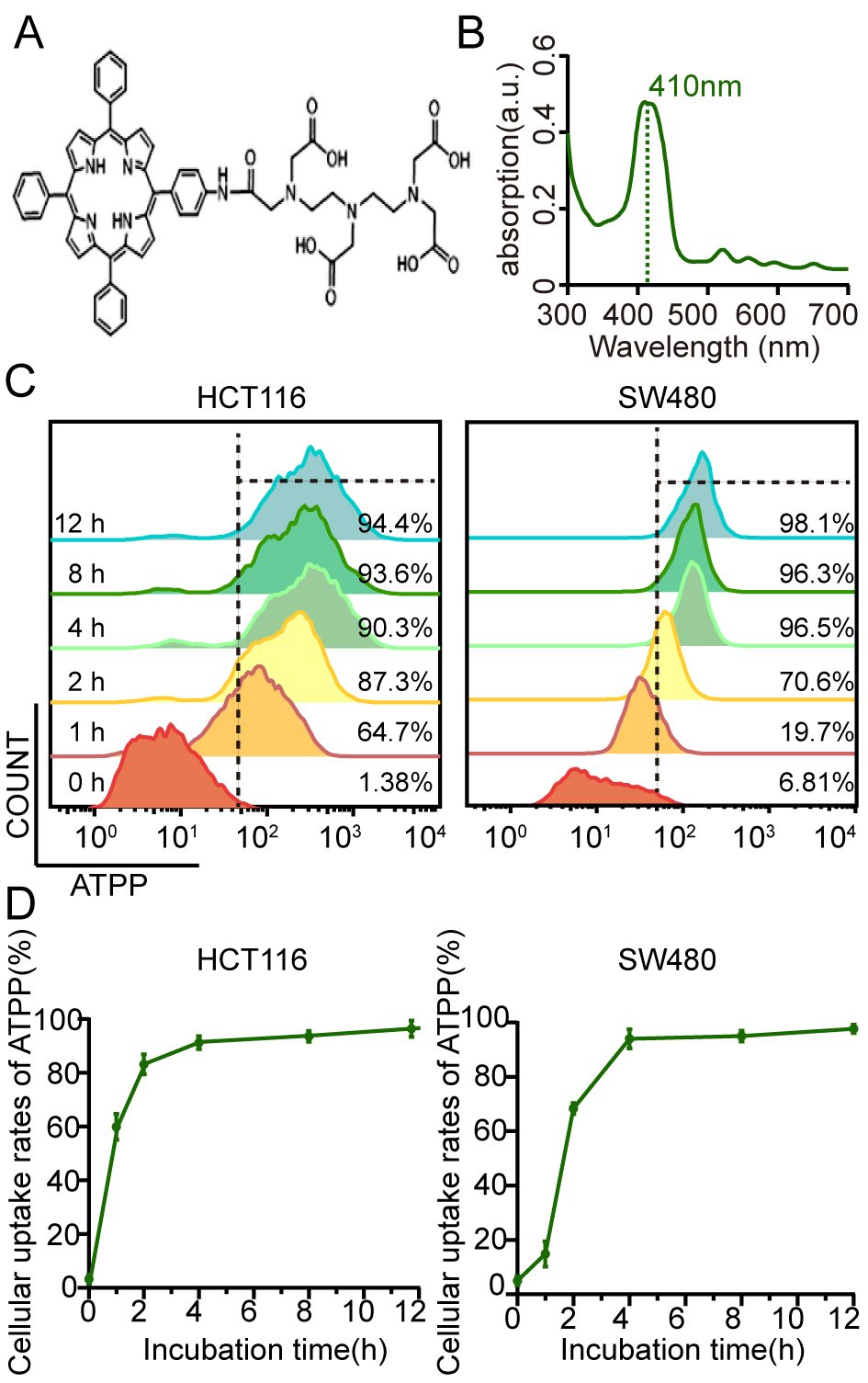 Fig. 1.
The properties of 5-(4-aminophenyl)-10,15,20-triphenylporphyrin
with diethylene-triaminopentaacetic acid (ATPP-DTPA). (A) Basic chemical
structural formula of ATPP-DTPA. (B) The absorption spectrum of ATPP-DTPA in PBS.
(C,D) The intracellular uptake of ATPP-DTPA in HCT116 and SW480 cells were
analyzed by flow cytometry at different periods (n = 3, mean
The CCK-8 assay was utilized to explore whether ATPP-PDT could affect the
viability of CRC cells. As indicated in Fig. 2A, PDT substantially decreased the
proliferation of CRC cells in both human (HCT116 and SW480) and mouse (MC38 and
CT26) CRC cell lines. It’s worth noting that the reduction in CRC cell
proliferation was observed in an ATPP-DTPA dose-dependent manner. The
corresponding concentration of ATPP alone without irradiation did not show
obvious cytotoxic effect on the cells. According to the data of cell survival
rate, we calculated the IC
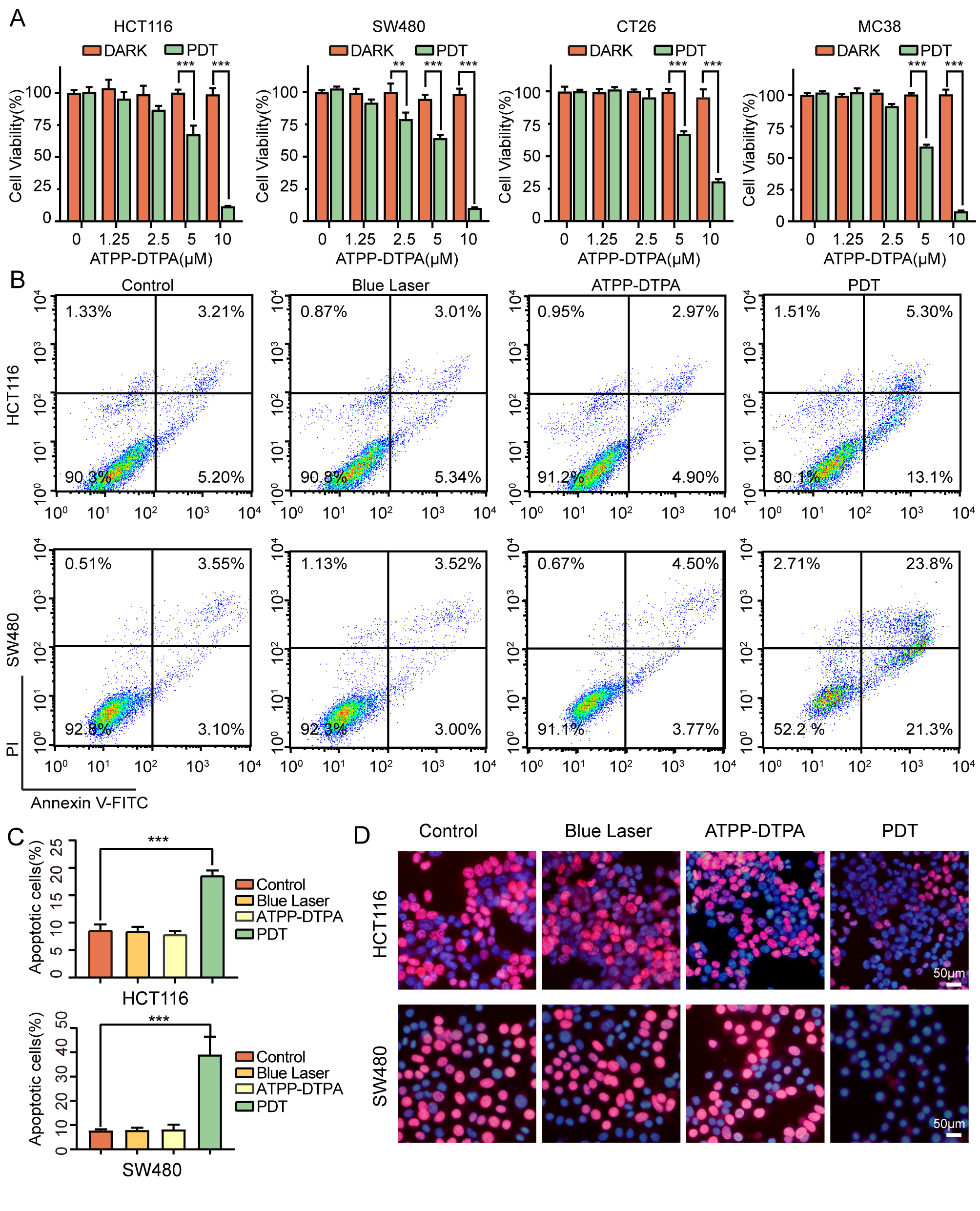 Fig. 2.
Anti-proliferative effects of ATPP-photodynamic therapy (PDT)
against colorectal cancer (CRC) cells. (A) Photodynamic effects and cytotoxic of
ATPP-DTPA on CRC cells (n = 3, mean
Numerous studies have demonstrated direct correlations between the high levels of intracellular ROS generation and the cell damage caused by PDT. Therefore, we detected the ROS generation during the process of ATPP-PDT, fluorescence microscopy suggesting that considerably increased ROS was observed in PDT group, whereas the amount of ROS decreased significantly after adding NAC, a classical ROS scavenger (Fig. 3A,C). Meanwhile, results of flow cytometry (Fig. 3B) further confirmed the same conclusions. Functionally, as shown in Fig. 3D and Supplementary Fig. 3, NAC could significantly mitigate the inhibitory impact of PDT on cell survival and proliferation rate, indicating that ROS plays a critical role in regulating the inhibition of cell growth during PDT.
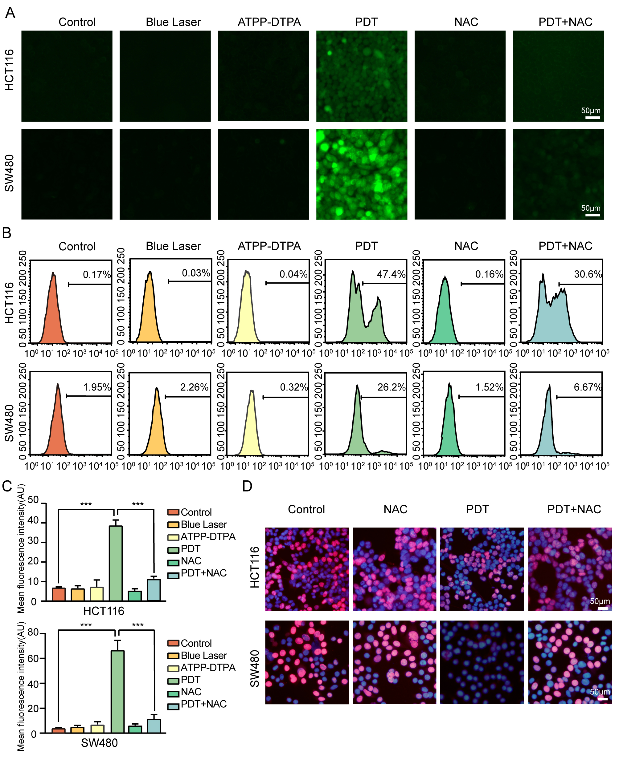 Fig. 3.
Production of reactive oxygen species (ROS) in ATPP-PDT and its
effect on CRC cells. (A) Representative fluorescence images of intracellular ROS
generation in HCT116 and SW480 cells. Scale bar = 50 µm (n = 3, mean
We have observed in Fig. 2B that ATPP-PDT-mediated growth inhibition of CRC cells is related to apoptosis by flow cytometry. To further verify our conclusion, we carried out western blotting experiment (Fig. 4A and Supplementary Fig. 4A). Compared with other groups, ATPP-PDT elevated the expression of cleaved-PARP and cleaved-caspase9, while downregulated the expression of pro-caspase3, and pro-caspase9 in CRC cells. The index of cleaved-PARP could be reversed by using Z-VAD-FMK, a classical apoptosis inhibitor in both SW480 and HCT116 cells, and we could also observe that the pro-caspase3 and pro-caspase9 were reversed in HCT116 cell (Fig. 4B and Supplementary Fig. 4B). Flow cytometry analysis also showed that with the addition of Z-VAD-FMK, the proportion of apoptotic cells significantly decreased (Fig. 4C,D). All the results suggested the occurrence of apoptosis in CRC cells during ATPP-PDT.
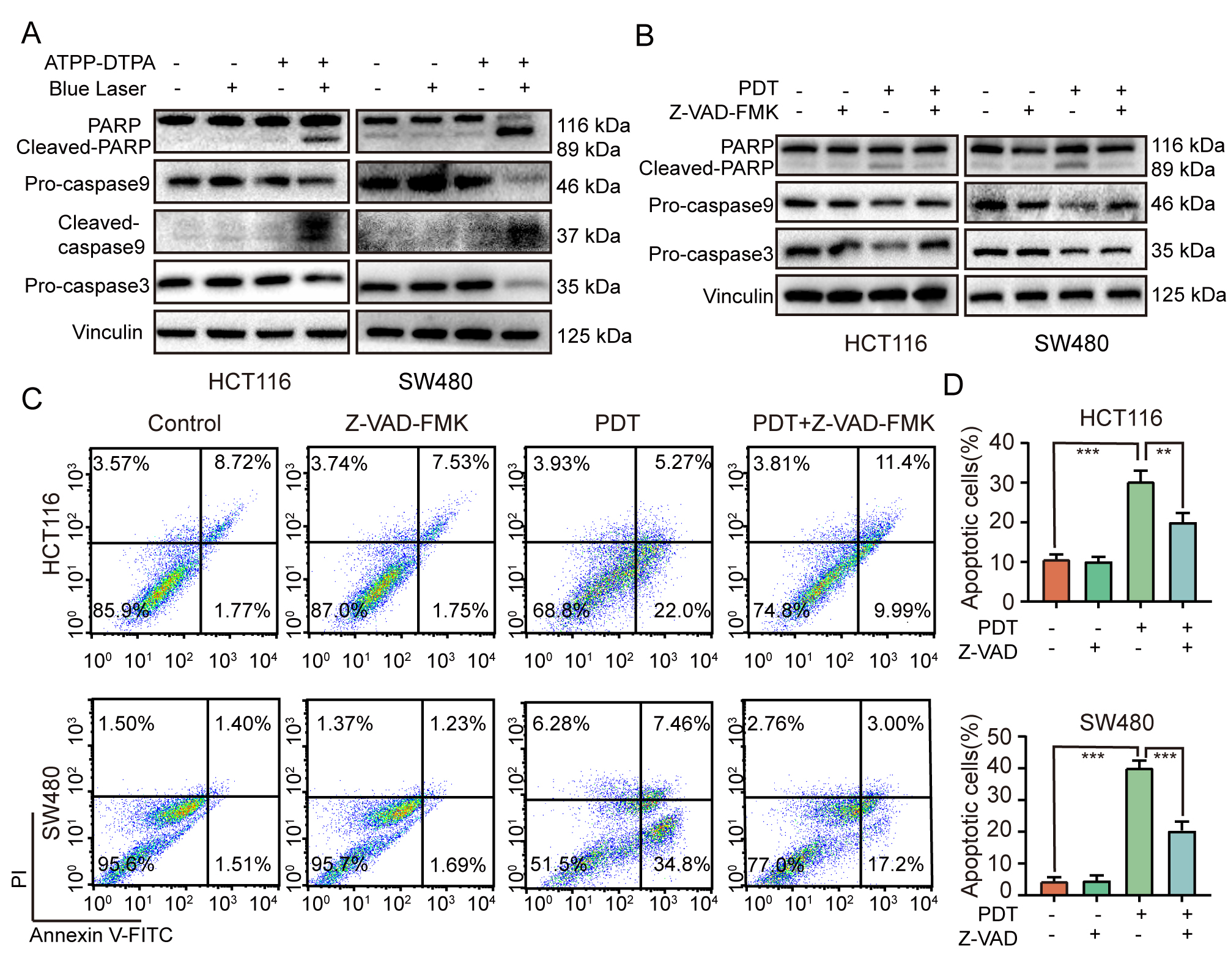 Fig. 4.
450-nm blue laser-mediated ATPP-PDT induces apoptosis in CRC
cells. (A) Expression of apoptosis-related proteins in HCT116 and SW480 cells
after 24 hours treatment in different treatment groups. (B) The impact of the
apoptosis inhibitor Z-VAD-FMK on PDT-induced apoptosis was examined using western
blotting. (C) Flow cytometry analysis of the effect of apoptosis inhibitor
Z-VAD-FMK on ATPP-PDT induced apoptosis. (D) Quantitative analysis of (C) (n = 3,
mean
In order to further explore the potential mechanism of ATPP-PDT inducing cell apoptosis, we further made transcriptome RNA sequencing. As demonstrated in Fig. 5B, there were 3138 differentially expressed genes (DEGs), including 1531 upregulated genes and 1607 downregulated genes. Kyoto Encyclopedia of Genes and Genomes (KEGG) enrichment analysis revealed that the MAPK signal pathway, a classic pathway of apoptosis, was engaged in ATPP-PDT induced apoptosis (Fig. 5A). Phospho-p38 MAPK increased significantly in ATPP-PDT group in both HCT116 and SW480 cells (Fig. 5C and Supplementary Fig. 5A), with the addition of SB202190 (a selective p38 MAPK inhibitor), we could not only observe that the expression of phospho-p38 MAPK was down-regulated again, but found the process of cell apoptosis was inhibited (Fig. 5D and Supplementary Fig. 5B). All these results confirmed the vital role of p38 MAPK signal pathway in ATPP-PDT. What’s more, after adding ROS scavenger NAC, the p38 MAPK pathway was inhibited and the expression of apoptosis-related proteins was reversed (Fig. 5E and Supplementary Fig. 5C). Then, flow cytometry observed that both SB202190 and NAC could significantly decrease the proportion of apoptotic cells in HCT116 and SW480 cells (Fig. 5F–I and Supplementary Fig. 5D–G). These above results suggested that blue laser mediated ATPP-PDT induces apoptosis by ROS activating p38 MAPK signal pathway.
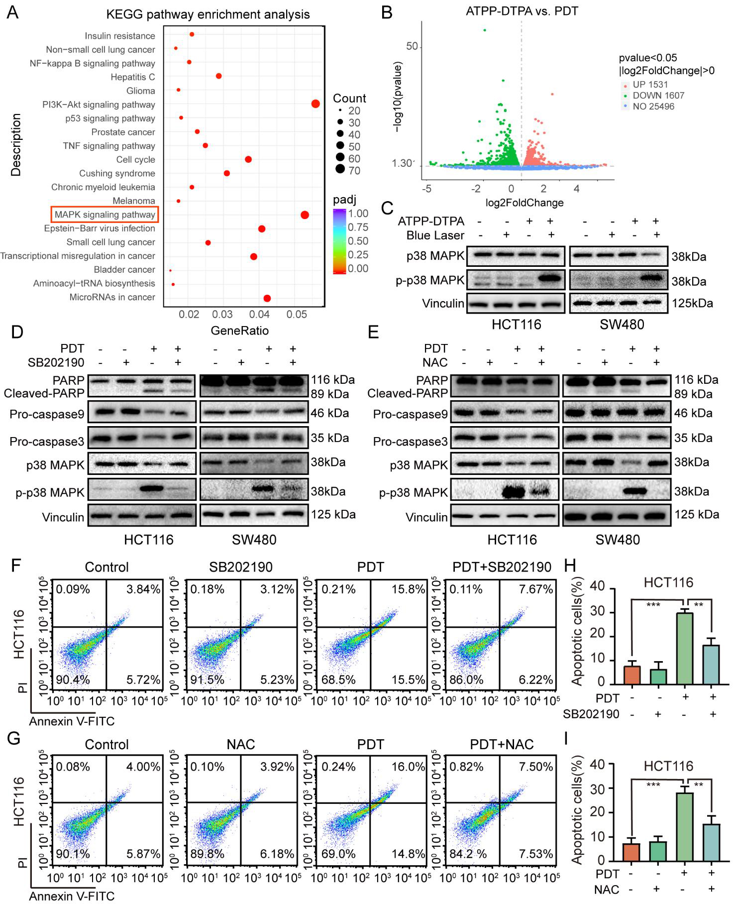 Fig. 5.
ATPP-PDT induces apoptosis by up-regulating ROS/p38 MAPK signal
pathway. (A) Kyoto Encyclopedia of Genes and Genomes (KEGG) pathway analysis of
differential gene expression (DEGs) in PDT group and ATPP-DTPA group in HCT116
cell. (B) Volcano plot of the DEGs in HCT116 cells between PDT group and ATPP-DTPA
group. Western blotting analysis of the expression levels of p38 MAPK
pathway-related proteins after the treatment of 24 h (C), and the effect of
SB202190 (D) and NAC (E) on ATPP-PDT-induced apoptosis and the activation of p38
MAPK pathway. (F,G) Flow cytometry was used to analyze the changes of apoptosis
of HCT116 cells after adding inhibitors SB202190 and NAC, respectively.
Quantitative statistics were shown in the (H,I) respectively. (n = 3, mean
To examined the in vivo therapeutic potential of ATPP-PDT, HCT116 cell subcutaneous xenograft models were constructed in BALB/c nude mice (Fig. 6A). On the 10th day after subcutaneous tumor implantation, the mice received caudal vein injection of ATPP-DTPA and irradiated the tumor site with 450-nm blue laser next day. Compared to other control groups, the tumor development of ATPP-PDT group was strongly inhibited, as seen by the significantly lower tumor volumes and weights (Fig. 6C–E) and the lack of significant changes in body weight (Fig. 6B). Next, we performed H&E and IHC of the tumor tissue and important organs from the mice (Fig. 6F and Supplementary Fig. 6B). In PDT group, substantially lower level of Ki-67 and higher expression of phospho-p38 MAPK and cleaved-PARP were discovered. Meanwhile, we didn’t observe any evident pathological alterations in the major organs, such as the heart, liver, spleen, lung and kidney (Supplementary Fig. 6A). All of the findings suggested that ATPP-PDT could successfully and safely stop CRC tumor development in vivo.
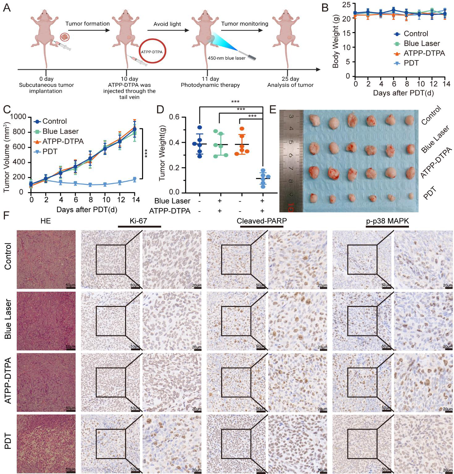 Fig. 6.
Effect of ATPP-PDT mediated by 450-nm blue laser in
vivo. (A) The general schematic illustration of the in vivo
experiment. (B,C) The body weight and tumor volume changes in mice across several
treatment groups throughout the treatment. (D) Comparison of tumor weight after
different treatment. (E) Visuals of the tumor tissues following various
therapies. (F) Tumors from mice were stained with H&E and IHC analysis following
various treatments. Scale bar = 50 µm and 20 µm. (n = 6, mean
As a common malignant tumor, CRC is still posing a huge menace to global health today. Epidemiological data in recent years showed that although the overall incidence of CRC has declined, the increased incidence in young people under the age of 50 is an alarming trend [3, 33, 34]. Surgery is still the cornerstone of radical treatment, and the progress of chemotherapy and radiotherapy has also improved the survival rate of metastatic CRC to a considerable extent, reduced recurrence and prolonged the lives of patients [35]. Because of its advantages such as high efficiency and minimally invasive, PDT has been used as an alternative diagnosis and treatment of CRC in many countries, regardless of whether the patient is in the early or late stage [36, 37]. In our experiment, 450-nm blue laser-mediated ATPP-PDT has significant anticancer effects against CRC in vivo and in vitro, and its biosafety was verified in nude mice. In addition, we observed that ATPP-PDT induced significant apoptosis of colorectal cancer cells by regulating ROS/p38 MAPK signal pathway (Fig. 5). Therefore, 450-nm blue laser with ATPP-DTPA mediated PDT may have a broad clinical application prospect in the treatment of CRC in the future.
Studies have shown that ROS can mediate lipid peroxidation and then induce damage to cancer cells, including apoptosis, autophagy and iron death [38]. Substantial ROS could be produced in the process of PDT [39], while excessive production of ROS may lead to oxidative stress and metabolic disorders, resulting in organelle damage, cell death and inflammation [40, 41]. In our research, we can observe that the intracellular ROS level increased significantly after ATPP-PDT treatment, and this process can be reversed by ROS scavenger NAC. At the same time, we also observed that the decrease of cell viability and proliferation seemed to be related to the production of ROS. Excessive ROS can promote a series of complex molecular signal pathways such as mitochondrial apoptosis to regulate cell apoptosis [42]. As a mechanism of cell death and defense, apoptosis precisely regulates the number of cells to remove unwanted and potentially dangerous cells [43]. In this study, we simply verified the apoptosis of colorectal cancer cells induced by ATPP-PDT, including the increase of apoptosis rate and some changes of apoptosis index, as well as the recovery experiment of inhibitors further confirmed the occurrence of tumor apoptosis.
ATPP-DTPA is partially modified based on the structure of tetraphenyl porphyrin (TPP), which is a symmetrical synthetic porphyrin with numerous benefits over natural porphyrin, including ease of modification [44]. As a result, many nanomaterials have been modified based on TPP for more adaptive PDT. Ming Wu et al. [45] designed the self-luminous nano-system POCL based on TPP to enhance the imaging and therapeutic effect of tumors in vivo and in vitro. In the works of Islam Zmerli et al. [26], bimodal photothermal (PTT)/PDT polydopamine nanoparticles (TPPS3) were successfully synthesized, which have a synergistic impact between PDT and PTT when treating human esophageal squamous cell lines. Using amphiphilic block copolymers of 4-vinylbenzyl-terminated tetraphenyl-porphyrin (VBTP) and maleimide isobutyl polyhedral oligomeric silsesquioxane (MIPOSS), Jianqiu Jin’s group [46] achieved effective PDT treatment on A549 cells. ATPP-DTPA is an amino derivative of tetraphenyl porphyrin modified by DTPA. Actually, other substances like EDTA and tetraphenylethene (TPE) were also utilized for modification. R.R. Valiev et al. [47, 48] calculated and analyzed that the absorption peak of the photosensitizer did not alter considerably and was less susceptible to fluorescence quenching than TPP following EDTA and DTPA modification. Yinpan Zhang et al. [49] used TPE with aggregation-induced emission (AIE) activity to overcome the aggregation quenching (ACQ) effect of ATPP in aqueous phase and enhanced the luminescence intensity in the aggregated state. Jing-Jing Chen’s group [50] verified the apoptosis of gastric cancer cells induced by red light PDT mediated by photosensitizer ATPP-EDTA in vivo and in vitro. However, the application and mechanism exploration of ATPP-DTPA photosensitizer in tumor cells is still lacking at present. In our experiments, we compared the anti-tumor efficacy of ATPP-DTPA with the traditional photosensitizer 5-ALA. The new photosensitizer ATPP-DTPA was significantly more lethal to cancer cells, which means that a lower concentration of photosensitizer can be used in clinical application to reduce its phototoxicity. We also found that ATPP-PDT promoted the apoptosis of CRC cells by up-regulating the expression of p38 MAPK signal pathway, and the addition of SB202190 reduced the apoptosis caused by ATPP-PDT.
Nevertheless, our research still has some limitations. Compared with the red laser commonly used at present, the absorption of porphyrin photosensitizer in the blue region is obviously superior. However, the comparison of the effect of red laser PDT and blue laser PDT is not involved in our experiment. Although in clinical application, the low penetration of blue laser will lead to the reduction of actual absorption, we believe that blue light has its unique indication in superficial cavity tumors, which is worthy of further study. In addition, PDT not only produces ROS to directly destroy the tumor, but also induces inflammatory response and activates the immune response to tumor cells [51]. Whether the ability of blue laser to mediate tumor immunogenic cell death (ICD) is stronger than that of red laser, and the performance of ATPP-DTPA in ICD are both worthy of further study. Finally, the porphyrin ring structure of ATPP can be modified by other groups to make it more suitable for clinical PDT or synergistic effect with other treatments, which is also worth considering.
In this work, we selected a novel photosensitizer ATPP-DTPA, and used a novel 450-nm blue laser for PDT. In the process of ATPP-PDT, a large amount of ROS induced severe apoptosis of CRC cells through p38 MAPK signal pathway, meanwhile ATPP-PDT also effectively inhibited HCT116 subcutaneously tumor in nude mice, providing a potential strategy for CRC treatment in the future.
CRC, Colorectal cancer; PDT, Photodynamic therapy; TPP, tetraphenylporphyrin;
ATPP, Amino derivative of tetraphenyl porphyrin; DTPA,
Diethylenetriaminepentaacetic acid; 5-ALA, 5-Aminolevulinic acid; ROS, Reactive
oxygen species; DMSO, Dimethyl sulfoxide; PBS, phosphate-buffered saline; CCK-8,
Cell Counting Kit-8; EdU, 5-Ethynyl-2
All analyzed data supporting the study are included in this published article and its supplementary information files. And the raw data will be made available from the corresponding author on request.
Conceptualization: YBM, JZ and DLH; methodology: YBM, LJG, YHC, PZ, YFC, RFY, XL, XYW, PG and JZ; data curation: YBM, LJG, YHC, PZ and YFC; formal analysis: YBM, LJG, YHC, PZ and JZ; investigation and project administration: YBM, LJG, YHC, PZ, XYW, PG, JZ and DLH; resources: JZ and DLH; software: YBM, LJG and YHC; supervision: JZ and DLH; writing—original draft: YBM, LJG, YHC, PZ, YFC, RFY, JZ and DLH; writing—review and editing: YBM, LJG, YHC, PZ, YFC, RFY, XYW, PG, JZ, and DLH. All authors contributed to editorial changes in the manuscript. All authors read and approved the final manuscript. All authors have participated sufficiently in the work and agreed to be accountable for all aspects of the work.
The Xi’an Jiaotong University Ethics Committee (Xi’an, China) gave its approval to all in vivo tests. The ethical approval number is XJTU1AF2022LSK-316.
We sincerely appreciate the time and effort of all the researchers involved. We acknowledge Tianjun Liu (from Institute of Biomedical Engineering, Chinese Academy of Medical Sciences) for providing the photosensitizer ATPP-DTPA.
This research was funded by Natural Science Foundation of China (NSFC No. 82073304 to JZ and 82172684 to DLH).
The authors declare no conflict of interest.
References
Publisher’s Note: IMR Press stays neutral with regard to jurisdictional claims in published maps and institutional affiliations.