


1 Department of Operating Room, Shanghai East Hospital, School of Medicine, Tongji University, 200120 Shanghai, China
2 Department of Anesthesiology, Shanghai East Hospital, School of Medicine, Tongji University, 200120 Shanghai, China
3 Department of Nursing, Renji Hospital, School of Medicine, Shanghai Jiaotong University, 200127 Shanghai, China
4 Department of Gastrointestinal Surgery, Shanghai East Hospital, School of Medicine, Tongji University, 200120 Shanghai, China
5 Department of Nursing, Shanghai East Hospital, School of Medicine, Tongji University, 200120 Shanghai, China
†These authors contributed equally.
Abstract
Background: Aberrant splicing has been closely associated with human cancer, though the precise underlying mechanisms linking the two remain not fully understood. Investigating the role of splicing factors in cancer progression may aid in the development of targeted therapies for dysregulated splicing, thereby opening up new avenues for cancer treatment. RNA-binding motif 4 (RBM4) has been identified as a critical participant in the condensin II complex, which is involved in chromosome condensation and stabilization during mitosis. Its significance in tumors is currently gaining attention. The genetic characteristics of RBM4 suggest its potential to elucidate the malignant progression of tumors in a broader context, encompassing various types of cancer, known as pan-cancer. Methods: This study aims to comprehensively explore the potential function of RBM4 in pan-cancer by leveraging existing databases such as The Cancer Genome Atlas (TCGA) and Genotype-Tissue Expression (GTEx). Results: RBM4 is found to be overexpressed in almost all tumors and exhibits significant prognostic and diagnostic efficacy. The correlation between RBM4 and immune signatures, including immune cell infiltration and immune checkpoint genes, indicates that RBM4 could serve as a guiding factor for immunotherapy. Conclusions: As a member of the pan-oncogene, RBM4 has the potential to become a biomarker and therapeutic target for various malignant tumors, offering novel possibilities for precision medicine.
Keywords
- RBM4
- prognosis
- immune infiltration
- pan-cancer
In eukaryotic cells, the process involving the transcription of mRNA to translation involves several steps, including splicing, transport, and stability. These steps are primarily facilitated by RNA-binding proteins (RBPs), which are present in various subcellular organelles and participate in the growth of every cell [1, 2]. Alternative splicing (AS) is a regulatory gene mechanism that regulates protein functions and is related to human cancer. Several studies have stated that abnormal AS functions can lead to cell apoptosis and epithelial-mesenchymal transition, and are involved in metastasis and invasion of malignant cells [3, 4, 5]. Additional research needs to be conducted to determine the role played by dysregulated splicing in cancer. RNA-binding motif 4 (RBM4), which was first identified in Drosophila and regulates circadian rhythms, is expressed in multiple organs in the body, where higher RBM4 levels were found in the testis, ovary, heart, skeletal muscle, and pancreas [6, 7, 8]. At the cellular level, RBM4 is expressed in the nucleoplasm and is found in both nucleoli and puncta. It can also co-localize with SR proteins [1, 9]. RBM4 is an RNA-binding protein that regulates the splicing and stability of a variety of Rnas by binding to specific sequences of RNAs. In cancer, overexpression of RBM4 is generally associated with fewer alternative splicing events and lower levels of cell differentiation. In addition, RBM4 can also through the interaction with E2F1 transcription factors, regulating cell cycle and cancer progression [10, 11]. However, further studies are needed to clarify the specific molecular mechanisms of RBM4 in cancer. Recent studies have indicated that RBM4 was abnormally expressed in several types of cancers, including liver, kidney, gastric, esophageal, and pancreatic cancer, and is involved in the progression of these diseases [10, 12, 13, 14, 15]. RBM4 was also found to be associated with multiple signaling pathways. RBM4 in liver cancer may be affected by the Wnt/beta - catenin and PI3K/Akt signaling pathway and regulation, affect the growth of tumor cells and attack [16, 17]. These effects may be closely related to the progression of Hepatocellular Carcinoma (HCC). RBM4 may also participate in kidney cancer cell apoptosis regulation. It has been found that downregulation of RBM4 may lead to inhibition of tumor cell apoptosis, thereby promoting the growth and spread of renal cancer [16, 18, 19]. The pan-cancer analysis utilizes various public databases to comprehend the molecular mechanisms and the predictive value of genes in tumor biology. This approach facilitates the accurate selection of clinical diagnostic markers and targeted therapy for effective treatment [20, 21, 22]. The exact role of RBM4 in tumors is not yet fully understood and its significance and role in different cancers must be investigated.
This study aims to determine the association between RBM4 and the immune microenvironment by means of pan-cancer analysis, as well as investigate the mechanism used by RBM4 in various tumors. The results of the differential expression analysis revealed that RBM4 expression levels varied significantly in a majority of tumors, whereas the findings of survival analysis demonstrated that RBM4 significantly impacted Adrenocortical carcinoma (ACC), Kidney Chromophobe (KICH), Acute Myeloid Leukemia (LAML), and Liver hepatocellular carcinoma (LIHC). The immune infiltration analysis revealed that RBM4 expression levels were linked to multiple immune cells, and alterations in the immune microenvironment could impact tumor prognosis. Therefore, RBM4 was observed to play a diverse role in pan-cancer analysis, potentially influencing tumor-specific prognosis and immune microenvironment. As a result, RBM4 acts as a novel biomarker and could be used as a probable therapeutic target.
Herein, clinical data and expression profiles of 33 forms of cancers were obtained from The Cancer Genome Atlas (TCGA) (https://portal.gdc.cancer.gov/). Additionally, we collected the TCGA and healthy sample data from the TCGA_ Genotype-Tissue Expression (GTEx) dataset, available at the University of California, Santa Cruz (UCSC) Xena (https://xenabrowser.net/datapages/). Furthermore, the healthy and malignant tissue samples were retrieved based on the immunohistochemical image data derived from Human Protein Atlas (HPA) (https://www.proteinatlas.org/). Furthermore, RBM4 and Gene Expression Profiling Interactive Analysis Database (GEPIA2) (http://gepia2.cancer-pku.cn/#index) were searched for extracting the Top 100 relevant genes from the TCGA dataset. All experiments in the study were based on the standard guidelines issued by TCGA and UCSC. Therefore, we did not acquire ethical approval or informed consent from the patients.
The RBM4 mRNA expression levels in the normal and malignant tissues acquired from the TCGA, TCGA_GTEx, and TCGA paired samples were compared in this study. Furthermore, the RBM4 protein expression levels measured in the data acquired from the healthy and cancerous tissues in the HPA database were also investigated.
Kaplan-Meier (KM) analysis and the log-rank test were conducted to determine the
relationship between RBM4 expression levels and different clinical outcomes, such
as progression-free interval (PFI), overall survival (OS), and disease-specific
survival (DSS) in pan-cancer using TCGA. We displayed survival curves with a
significant p-value
This study has examined the correlation between RBM4 expression levels and other clinical factors, like T stage, gender, N stage, and pathologic stage, in the case of cancers that are affected by RBM4 and can influence prognosis.
Herein, a univariate Cox regression analysis technique was utilized to evaluate
the influence of RBM4 on disease prognosis, with a specific focus on OS, PFI, and
DSS. The sample selection process in this study involved the selection of tumors
with a p-value
The TCGA dataset was assessed with the Tumor Immune Estimation Resource 2.0
(https://cistrome.shinyapps.io/timer/) to determine the link between the RBM4
expression levels and the immune cells like B cells, CD4
To identify the genes that exhibited an RBM4-like expression pattern, a list of the top 100 RBM4-related genes was acquired from the GEPIA2 database. Thereafter, the gene ontology (GO) analysis was carried out, which included biological pathways (BP), cellular components (CC), and molecular functions (MF). These genes were further examined with the Kyoto Encyclopedia of Genes and Genomes (KEGG) analysis to gain further insights into the potential functions of RBM4. For constructing the protein-protein interaction (PPI) network, 100 RBM4-linked genes were used in the Search Tool for the Retrieval of Interacting Genes (STRING) database (https://cn.string-db.org/). The minimum interaction threshold was set at 0.4.
The DESeq R software (Version 4.1.1, R Foundation for Statistical Computing, Vienna, Austria) was used in this study to carry out differential expression analysis of RBM4 in case of cancers that can affect prognosis [23]. Based on the results, we performed GSEA using the clusterProfiler R package (https://bioconductor.org/packages/release/bioc/html/clusterProfiler.html) to study the differential expression of RBM4 in various types of cancer [24].
The HepG2, Huh7, and NCI-H295R cell lines were procured from the Chinese Academy
of Sciences and maintained in Dulbecco’s Modified Eagle Medium (DMEM)
supplemented with 10% heat-inactivated fetal bovine serum (FBS) sourced from
Gemini Bio Products. The cell lines underwent validation through STR profiling
and tested negative for mycoplasma. Cultures were incubated at 37 °C in a humidified
atmosphere with 5% CO
For flow cytometry analysis of the proliferating cells, a Cell-Light EdU Apollo 488 In vitro Flow Cytometry Kit (RiboBio, Guangzhou, China) was used to examine EdU-positive cells according to the manufacturer’s protocol.
As for the cells cultured on plates, they were initially fixed with 4%
paraformaldehyde and subsequently treated with a 1% Triton solution. Following
this, both the sections and cells underwent a blocking step using 10% serum for
30 minutes at room temperature. Next, primary antibodies were applied overnight
at 4 °C. The following day, the sections or cells were thoroughly washed
and then exposed to secondary antibodies labeled with fluorophores (Thermo
Fisher, A48282 and A48287, at a dilution of 1:500, Notch1 Rabbit pAb (A7636),
Cyclin D1 Rabbit mAb (A19038), Phospho-mTOR-S2448 Rabbit pAb (AP0094)) for 1 hour
at room temperature. Finally, an antifade solution containing
4
HepG2 and Huh7 cells were initially placed into 6-well plates. After 24 hours,
the cells were either transfected with siNC, siRBM4, or exposed to a control
adenovirus. Forty-eight hours later, the cells had reached full confluence, and a
cell monolayer was gently scraped using a 200-µL pipette tip. The well was
rinsed twice with serum-free medium (a mixture of 50% MEM and 50% F12) and then
refreshed with a fresh batch of serum-free medium. At the beginning (0 hour) and
subsequently every 12 hours during incubation, images were taken using a light
microscope (LeicaCTR6000 microscope system) at a magnification of
This study includes 32 patients with Liver cancer who were diagnosed and treated at Shanghai East Hospital Affiliated to Tongji University were collected, from September 2019 to 2023. Hepatocellular carcinoma and matched edge or adjacent tissues combined microarray chip, including 32 cases of hepatocellular carcinoma and matched edge or adjacent liver tissues, one case of cancer at 2 points, one case of edge or adjacent cancer at 1 point.
The R (ver. 4.0.2) software was used for statistically analyzing the data. The
Wilcoxon rank-sum test was utilized for comparing the difference between both
groups, while the Spearman rank test was utilized to calculate the link between
these groups. Furthermore, the univariate and multivariate Cox proportional
hazard regression analyses were conducted for screening the factors affecting
disease prognosis. The KM analysis with the log-rank test was utilized for
carrying out survival analysis. A p-value
To investigate the RBM4 expression in pan-cancer, we evaluated the TCGA_GTEx data derived from UCSC. The findings of this analysis suggest that RBM4 exhibited varying expression patterns across a significant number of cancer types. Specifically, RBM4 expression was found to be elevated in ACC, Bladder Urothelial Carcinoma (BLCA), Breast invasive carcinoma (BRCA), Cervical squamous cell carcinoma and endocervical adenocarcinoma (CESC), Cholangiocarcinoma (CHOL), Colon adenocarcinoma (COAD), Lymphoid Neoplasm Diffuse Large B-cell Lymphoma (DLBC), Glioblastoma multiforme (GBM), Head and Neck squamous cell carcinoma (HNSC), Brain Lower Grade Glioma (LGG), Liver hepatocellular carcinoma (LIHC), Lung adenocarcinoma (LUAD), and Lung squamous cell carcinoma (LUSC), while reduced RBM4 expression levels were observed in Esophageal carcinoma (ESCA), Kidney Chromophobe (KICH), Kidney renal clear cell carcinoma (KIRC), Kidney renal papillary cell carcinoma (KIRP), and Ovarian serous cystadenocarcinoma (OV). These outcomes align with the data obtained from the TCGA database (Fig. 1A,B). In addition, the RBM4 expression levels in 18 different types of tumors were analyzed using paired samples from the TCGA dataset (Fig. 1C). Furthermore, the RBM4 protein expression levels in the healthy control and cancer tissue samples derived from different human organs were analyzed using HPA. Furthermore, we extracted representative immunohistochemical (IHC) images from the bladder, lung, liver, pancreas, and stomach tissues to further examine RBM4 expression (Fig. 2). We found that in glioma (GBMLGG), colon adenocarcinoma (COAD), LIHC, ACC and testicular germ cell tumours (TGCT), the expression level of RBM4 in cancer tissues was significantly higher than that in normal control tissues, especially in liver cancer.
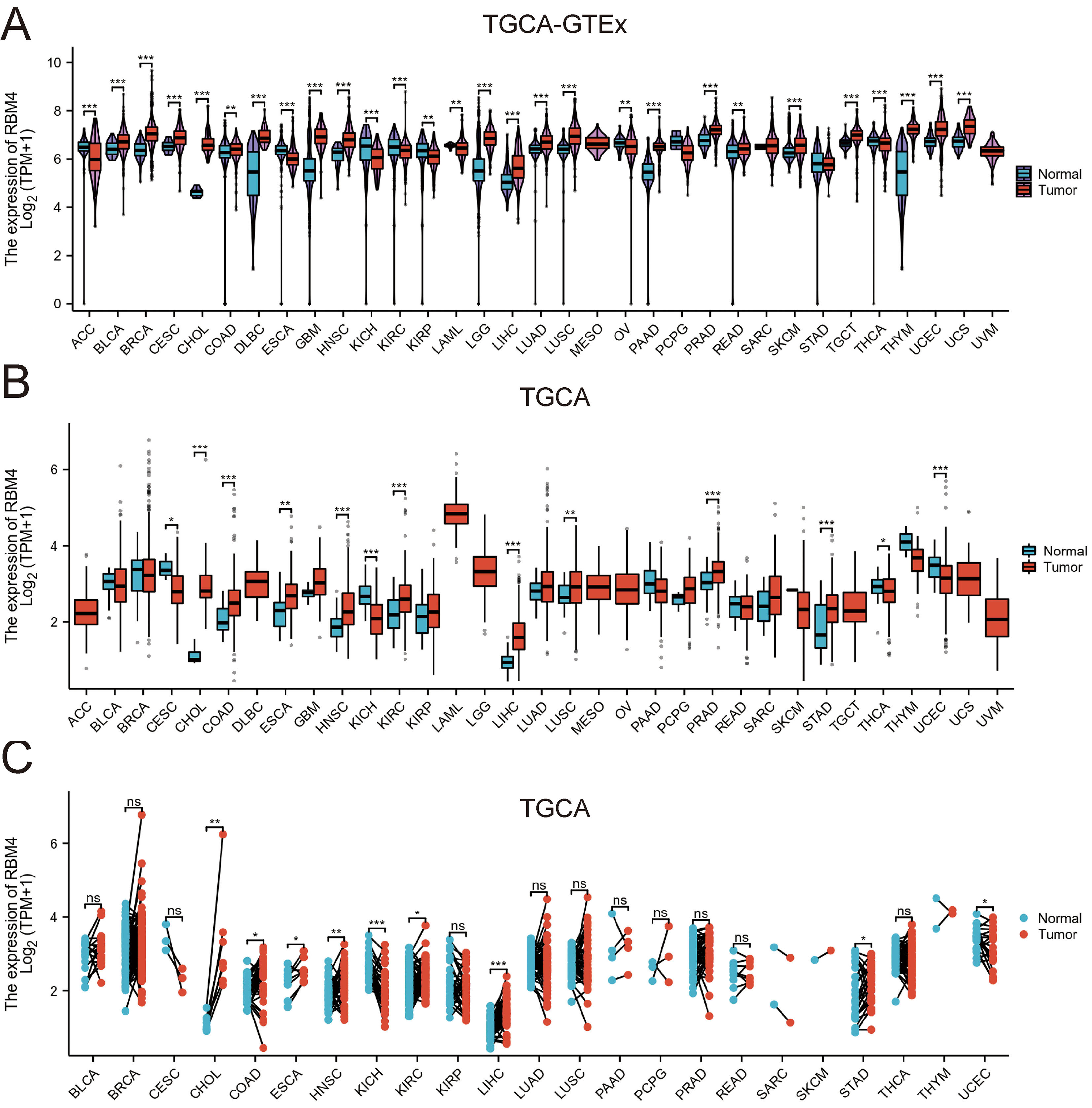 Fig. 1.
The RBM4 mRNA expression in pan-cancer. (A) The RBM4 mRNA
expression levels in 33 types of tumors in TCGA_GTEx samples. (B) The RBM4 mRNA
expression levels in 33 types of tumors in the TCGA database. (C) RBM4 expression
in paired samples of 18 types of cancers in TCGA database. (no significant (ns), p
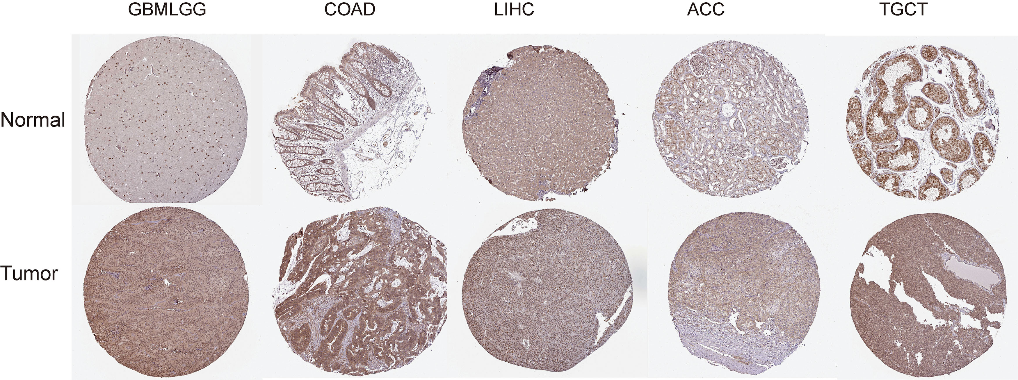 Fig. 2.
IHC images of RBM4 in the healthy and malignant tissue samples extracted from HPA. GBMLGG, glioma; IHC, immunohistochemical; HPA, Human Protein Atlas. Image available from v15.proteinatlas.org.
To assess the prognostic value of RBM4 in pan-cancer, we performed a KM survival analysis to understand the relationship between RBM4 expression levels and clinical outcomes. Initially, the link between RBM4 expression levels and the OS rate in 33 types of cancers was determined (Fig. 3A), and the findings of this analysis showed that the abnormal RBM4 expression was associated with OS in ACC (Fig. 3B), KIRC (Fig. 3C), LUSC (Fig. 3D) and pancreatic adenocarcinoma (PAAD) (Fig. 3E). RBM4 expression upregulation was related to a low OS rate in ACC and LUSC. Finally, elevated RBM4 levels in PAAD and KIRC indicated a long OS. We also determine the correlation between the RBM4 expression levels and DSS (Fig. 4A), and the findings implied that the expression of RBM4 was related to the DSS of ACC (Fig. 4B), KICH (Fig. 4C), STES (Fig. 4D), and LIHC (Fig. 4E). Moreover, the RBM4 expression showed varying associations with DSS based on the type of cancer. Specifically, in ACC and KICH, upregulation of RBM4 was linked to worse DSS. Additionally, when the link between RBM4 expression levels and PFI was investigated (Fig. 5A), the findings implied that RBM4 expression was linked to PFI in ACC (Fig. 5B), LIHC (Fig. 5C), pheochromocytoma and paraganglioma (PCPG) (Fig. 5D), KIRC (Fig. 5E). The results revealed that a high RBM4 expression represented a worse PFI in ACC, LIHC, and PCPG, but a better PFI in KIRC. Furthermore, ROC curves were presented for the 6 types of tumors, wherein RBM4 expression was related to disease prognosis. These curves demonstrate the diagnostic ability of RBM4 in the above tumors (Supplementary Fig. 1A–F).
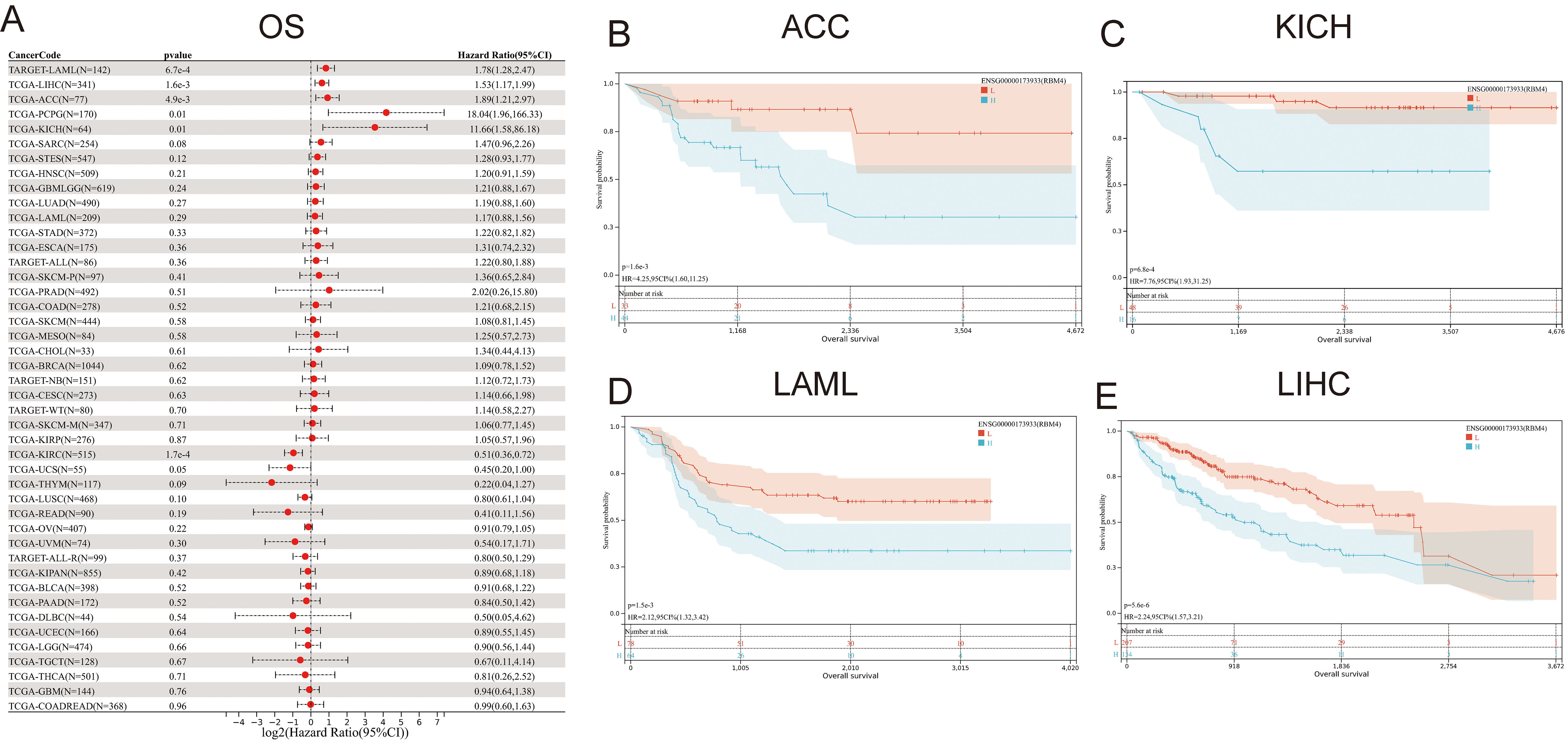 Fig. 3.
Correlation between RBM4 expression and OS in pan-cancer analysis. (A) Forest plots presenting the effects of RBM4 expression levels on OS in pan-cancer. (B–E) Effects of RBM4 expression on OS in different cancers like ACC, KICH, LAML, and LIHC, respectively. OS, Overall Survival.
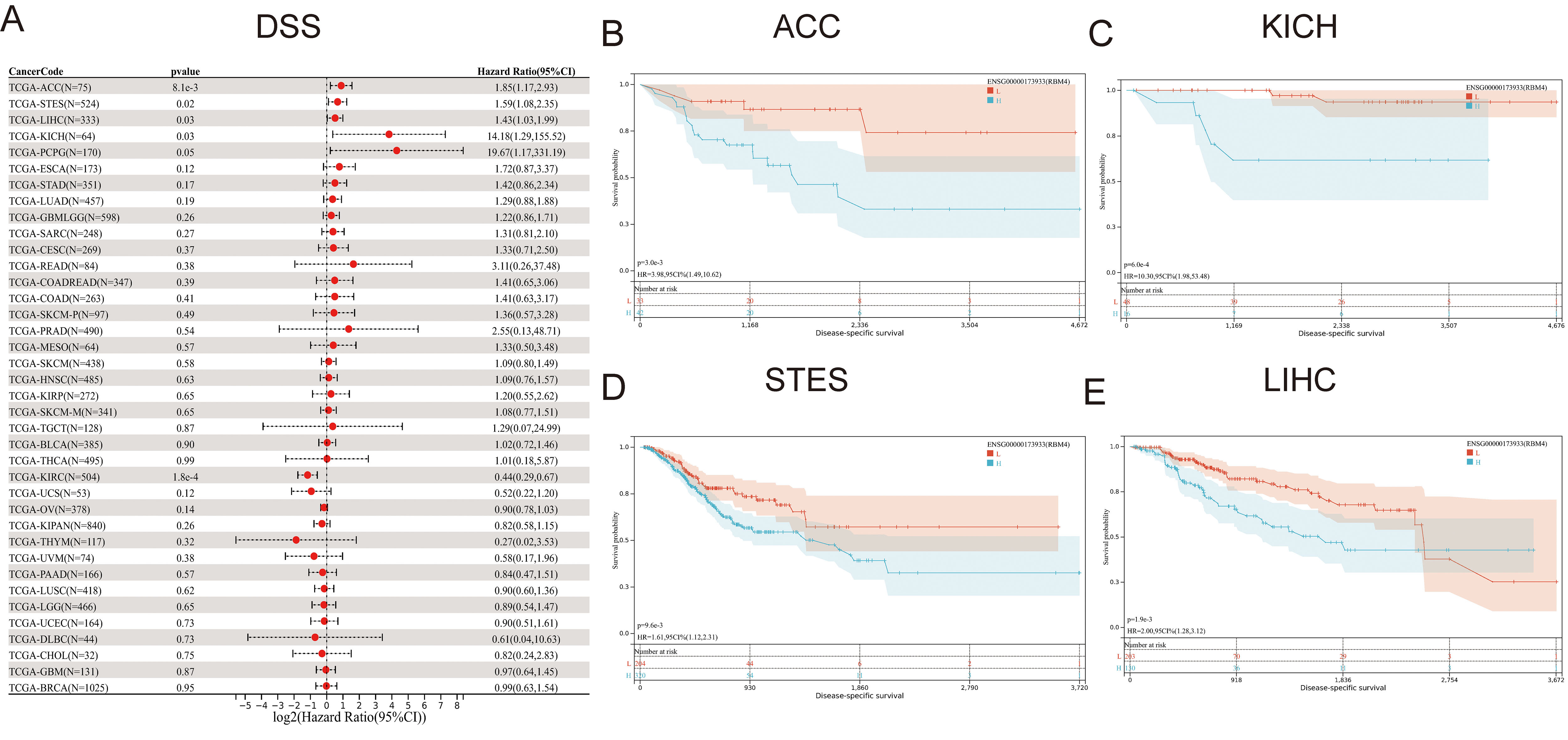 Fig. 4.
Correlation between RBM4 expression and DSS in pan-cancer. (A) Forest Plot depicting the relationship between RBM4 expression levels on DSS in different types of cancers. (B–E) RBM4 survival curves on DSS in ACC, KICH, STES, and LIHC, respectively. DSS, Disease Specific Survival; STES, Stomach and Esophageal carcinoma.
 Fig. 5.
Effect of RBM4 expression levels on PFI in pan-cancer. (A) A forest map describing the relationship between RBM4 expression levels on PFI in TCGA. (B–E) The effect of RBM4 expression levels on PFI in ACC, LIHC, PCPG, and KIRC, respectively. PFI, Progression Free Interval; PCPG, Pheochromocytoma and Paraganglioma.
RBM4 expression levels differed significantly in the I, II, III, and IV pathological stages in several types of cancers such as pan-kidney cohort (KICH + KIRC + KIRP) (KIPAN), KIRC, LIHC, and thyroid carcinoma (THCA) (Fig. 6A). The RBM4 expression varied significantly among the GBMLGG, LGG, KIPAN, KIRC, and LIHC patients, depending on their pathological stage (G1/G2/G3/G4) (Fig. 6B). Based on the M0/M1 pathological stage, we observed significant variations in the RBM4 expression levels in TCGT and Uveal Melanoma (UVM) patients (Fig. 6C). The RBM4 expression varied significantly among prostate adenocarcinoma (PRAD), KIRC, and THCA patients depending on their pathological N stage (N0, N1, N2, or N3) (Fig. 6D). Significant differences were observed in the RBM4 expression levels in the KIPAN, PRAD, KIRC, LIHC, THCA, and UVM patients, based on their pathological T1/T2/T3/T4 stage (Fig. 6E).
 Fig. 6.
Correlation between the RBM4 gene expression and different
clinicopathological stages in pan-cancer. (A) Violin plots showing differential
RBM4 expression levels in the various I/II/III/IV pathological stage. (B) Violin
plots depicting the differential RBM4 expression levels in the G1/G2/G3
pathological stage. (C) Violin plots highlighting the differential RBM4
expression levels in the M0/M1 pathological stage. (D) Violin plots indicate the
differential RBM4 expression levels in the N0/N1/N2/N3 pathological stages. (E)
Violin plots showing the differential RBM4 expression levels in T1/T2/T3/T4
pathological stage. (*p
Three types of cancers including KIRC, LIHC, and COAD with a sample size
 Fig. 7.
Nomogram models were constructed and assessed in KIRC, LIHC, and LUSC. (A) Constructing a nomogram model that involves RBM4 expression in KIRC. (B) Calibration curves were employed to assess the nomogram model in KIRC at 1-year, 3-year, and 5-year. (C) Designing a nomogram model with RBM4 expression in LIHC. (D) The 1-year, 3-year, and 5-year calibration curves were utilized for assessing the prediction accuracy of the nomogram model in LIHC. (E) Developing a nomogram model with RBM4 expression in COAD. (F) To assess the prediction accuracy of the nomogram model in COAD, the 1-, 3-, and 5-year calibration curves were utilized.
Immune cells have a crucial role in preserving the body’s immune equilibrium and
defending against external threats. Over recent years, a mounting body of
research has highlighted the substantial importance of RNA-binding proteins in
modulating the expression and regulation of immune cells within the immune
response regulation [25, 26]. In this study, we analyzed immune infiltration
levels using three established algorithms (xCELL, EPIC, and TIMER) to assess the
link between immune cells and RBM4 expression levels across different cancers.
The results implied that the RBM4 expression significantly promoted immune cell
infiltration, particularly in natural killer cells, macrophages, and neutrophils.
The deconvo_xCell method was used to generate 67 immune cell infiltration scores
for 1080 malignant samples across 44 types of cancers. To determine the
association between genes and the immune cell infiltration scores in every type
of cancer, the corr.test function from the psych tool in R was used to estimate
Pearson’s correlation coefficient. Through this analysis, we identified a
significant link between immune infiltration and RBM4 expression levels in the 44
types of cancers that were studied (Fig. 8A). The Timer method was used to
acquire 6 distinctive immune cell infiltration scores, specifically for
macrophages, CD4
 Fig. 8.
Correlations between RBM4 expression and tumor immune
infiltration. (A) The RBM4 expression is significantly related to the
infiltration levels of different immune cells based on xCELL. (B) The RBM4
expression was seen to be significantly related to the infiltration levels of
different immune cells in the Timer database. (C) The RBM4 expression was
significantly related to the infiltration levels of different immune cells in the
EPIC database. DC, dendritic cell; EPIC, Estimating the Proportion of Immune and Cancer cells. (*p
The fundamental concept behind single-cell sequencing technology involves the isolation of cells on an individual basis, followed by the sequencing of RNA or DNA from each isolated cell. This methodology demands precise cell sorting and the utilization of high-throughput sequencing techniques. In recent years, single-cell RNA sequencing (scRNA-seq) has emerged as the predominant approach in single-cell sequencing technology [27, 28]. Hence, in order to delve deeper into the molecular mechanisms of the RBM4 gene in tumorigenesis, we explored its impact on various types of tumors at the single-cell level. The results of this analysis highlighted a positive relationship between RBM4 expression and hypoxia, angiogenesis, inflammation, and proliferation. Conversely, RBM4 expression was seen to be negatively associated with DNA damage, DNA repair, cell cycle, invasion, and quiescence (Fig. 9A). Herein, the association between RBM4 and functional status in different types of cancer was evaluated. Our findings revealed a positive correlation between RBM4 and various cancer-related processes such as proliferation, metastasis, differentiation, inflammation, quiescence, Epithelial-Mesenchymal Transition (EMT), angiogenesis, hypoxia, and apoptosis in Acute Myeloid Leukemia (AML). Additionally, RBM4 was found to be associated with DNA repair mechanisms in acute lymphoblastic leukemia, angiogenesis in Pheochromocytoma (PC), and angiogenesis, differentiation, and inflammation in RetinoBlastoma (RB). The RBM4 gene expression showed a negative correlation with DNA repair in AML and RB, DNA damage and cell cycle in RB, invasion in Uveal Melanoma (UM) and Ovarian serous cystadenocarcinoma (OV), and EMT in PC. Additionally, RBM4 was also seen to be negatively associated with apoptosis and metastasis in AML (Fig. 9B–F). To determine the biological function of RBM4 in different tumors, 100 RBM4-related genes were acquired from the GEPIA2 database. The findings of the GO analysis (Fig. 10A) revealed that RBM4-related genes were involved in “organelle fission”, “mitotic nuclear division”, “nuclear division”, “mitotic sister chromatid segregation”, and other biological processes. These genes could play a role in “chromosomal region”, “spindle”, and other cellular components. It is involved in “microtubule binding”, “tubulin binding”, “microtubule motor activity”, “chemokine activity”, and other molecular functions. Furthermore, the findings of the KEGG pathway analysis (Fig. 10B) implied that RBM4-linked genes may be linked to “Cell cycle”, “Cellular senescence”, “Oocyte meiosis”, “cytokine and cytokine receptor”, “Viral protein interaction”, and “PPAR signaling pathway”. To determine the function of RBM4, we carried out a GSEA depending on the differential expression analysis of RBM4. This helped in elucidating the biological function of RBM4 in three tumors, namely ACC, LIHC, and BRCA, wherein the RBM4 expression was related to the disease prognosis as presented in Fig. 10C. The findings indicate that RBM4 is mainly associated with DNA Replication, Mitotic G1 Phase and G1S Transition, and Cell Cycle Checkpoints.
 Fig. 9.
Relationship between RBM4 and functional state in different
types of cancers. (A) The interactive bubble chart depicts the correlation
between the RBM4 and functional state in 16 cancers. The correlation between the
RBM4 and functional state in (B) Acute Myeloid Leukemia (AML), (C) Uveal Melanoma
(UM), (D) Pheochromocytoma (PC), (E) RetinoBlastoma (RB), and (F) Ovarian serous
cystadenocarcinoma (OV). X-axis depicts different gene sets; ***p
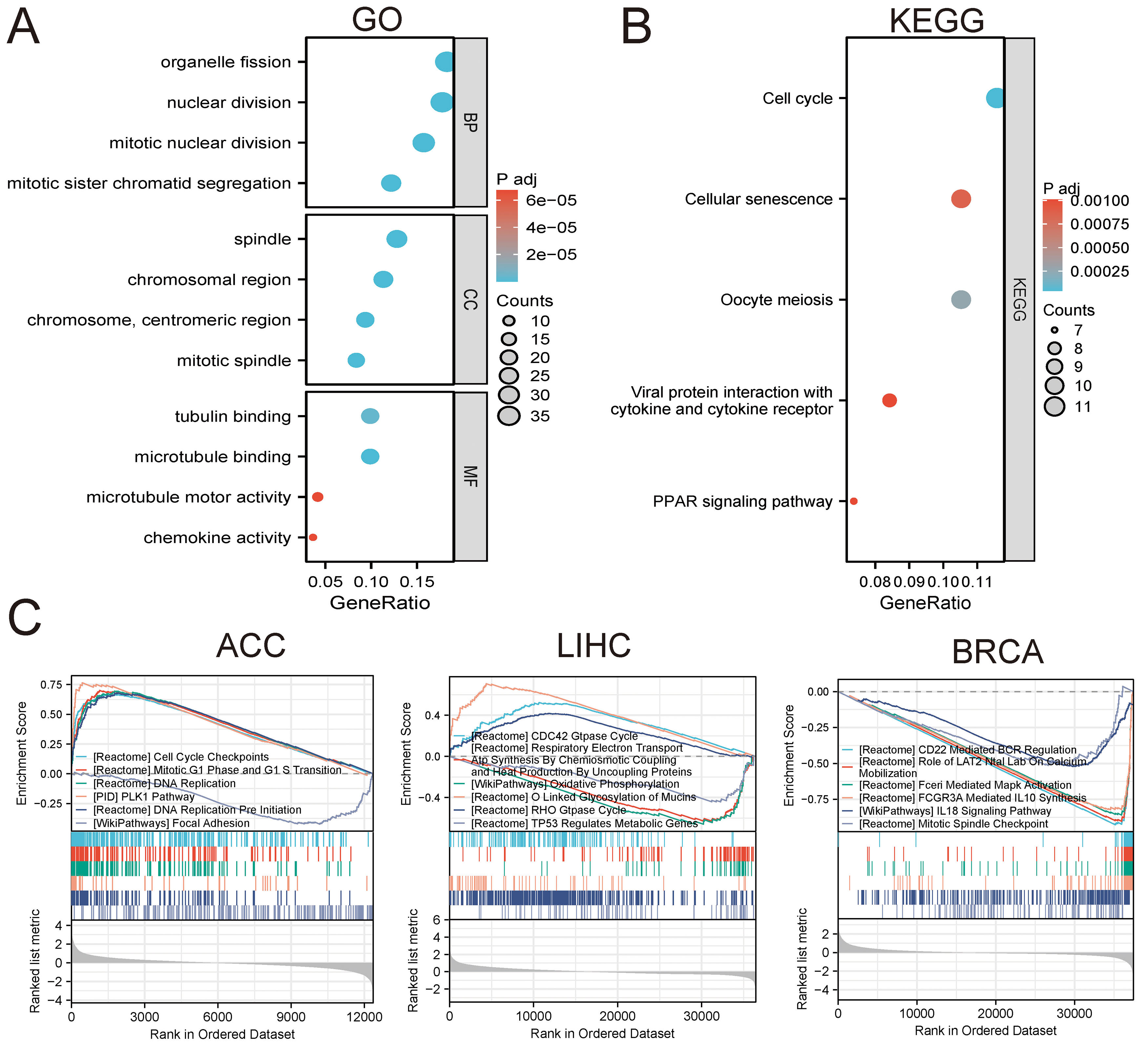 Fig. 10.
Functional enrichment analysis of RBM4-linked genes. (A) GO enrichment analysis of 100 RBM4-related genes, which includes BP, CC, and MF. (B) KEGG pathways analysis using the 100 RBM4-related genes. (C) GSEA based on differential expression analysis in ACC, LIHC, and BRCA, respectively. GO, gene ontology; KEGG, Kyoto Encyclopedia of Genes and Genomes.
To further investigate the role of RBM4 in liver cancer, we chose to study the liver cancer cell line HepG2 and Huh7. We collected a total of 32 paired samples of hepatocellular carcinoma (HCC) and adjacent tissues, which were then assembled into a tissue microarray for analysis. Immunohistochemistry revealed that the staining intensity of RBM4 in the tumor group was significantly higher than that in the adjacent tissue group (Supplementary Fig. 2A,B). We utilized short hairpin RNA (shRNA) to knock down RBM4, confirming the effectiveness of interference through validation experiments (Supplementary Fig. 2C,E). Following RBM4 knockdown in the HepG2 and Huh7 cell line, we conducted EdU experiments, which showed a notable decrease in the percentage of cells displaying positive EdU signals (Supplementary Fig. 2D,F). This suggests a reduction in DNA replication activity and weakened cell proliferation ability upon RBM4 downregulation. To assess the impact of RBM4 on cell migration, we examined whether silencing RBM4 influenced the migration of HepG2 cells. As anticipated, the results showed impaired migration abilities in cells with downregulated RBM4 (Supplementary Fig. 2G,H). After knockdown of RBM4 in the NCI-H295R cell line of ACC, the EdU assay showed a significant decrease in the percentage of cells with edu-positive signals, indicating that DNA replication activity was reduced and ACC cancer cell proliferation was weakened after downregulation of RBM4 expression (Supplementary Fig. 2I,J). GSEA enrichment analysis showed that genes related to differential expression of RBM4 in ACC were mainly enriched in the cell cycle. Knockdown of RBM4 in the NCI-H295R cell line of ACC can inhibit the cell cycle G1-S phase transposition, thereby inhibiting the proliferation (Supplementary Fig. 2K). After knocking out RBM4, ClinD1, P-mTOR and NOTCH1 proteins were significantly decreased by immunofluorescence, indicating that RBM4 could affect the development of ACC through PI3K-Akt and NOTCH signaling pathways (Supplementary Fig. 2L-N). Together, these findings provide further evidence supporting the role of RBM4 as a potential target LIHC and ACC.
Cancer is a life-threatening disease that lacks effective diagnostic and therapeutic targets despite the discovery of biomarkers for targeted therapy in some tumors through the continuous development of science and technology [29, 30]. Mining effective biomarkers for various tumors can lead to the development of targeted and effective treatments for cancers that lack clear targets [31, 32, 33, 34]. This study revealed that RBM4 may impact the prognosis of certain types of cancers and is associated with multiple immune cells. It is worth noting that alterations in immune cell infiltration levels can affect the prognosis of different tumors [35, 36, 37]. The findings implied that the role of immune cells could be influenced by RBM4 expression. The enrichment analysis results indicated that RBM4 could impact tumor progression by implementing various mechanisms such as DNA repair, DNA damage, cell cycle, invasion, and quiescence. Zhang et al. [19] observed that RBM4 could improve the stability of p53 mRNA, promoting the p53 signaling pathway activity, and inhibiting the progression of clear cell renal cell carcinoma (ccRCC). This finding suggests that RBM4 could be employed as a promising target for treating ccRCC patients [10]. The study suggests that miR-504 helps in promoting cell proliferation and inhibiting apoptosis in gastric cancer cells by targeting RBM4 [19]. Wang et al. [38] found that the RBM4 splicing factor regulates cell proliferation, migration, and apoptosis, thereby inhibiting tumor progression. However, very few researchers have investigated the role played by RBM4 in pan-cancer, and its expression status in different types of cancers remains unclear. Here, we have attempted to determine the link between RBM4 expression levels and various types of cancers in pan-cancer by assessing the RBM4 differential expression levels in healthy and malignant tissues in various organs using TCGA, TCGA_GTEx, and TCGA paired samples. Overall, 3 datasets yielded consistent findings, although some inconsistencies and opposing results were observed. For instance, CESC showed contradictory results using the TCGA_GTEx and TCGA datasets owing to the sample size variations in the healthy control patients. Therefore, the sample size of the control group needs to be increased for drawing accurate conclusions.
Recent reports have demonstrated the effect of immune cell infiltration levels
on tumor prognosis. Variations in gene expression levels can alter the immune
microenvironment, which could be used as an effective strategy for tumor
immunotherapy [39, 40, 41]. Initially categorized as an RNA-binding protein primarily
involved in transcription and splicing, RBM4’s role has recently extended into
the realm of immune cells. Emerging studies have unveiled RBM4’s
significance in the expression and regulatory mechanisms of immune cells. It has
come to light that RBM4 plays a pivotal role in processes such as RNA splicing
and post-translational modifications of gene expression when it comes to immune
cell regulation [38, 42]. Notably, in immune cells like lymphocytes, macrophages,
dendritic cells, and others, RBM4’s protein expression has close associations
with immune cell differentiation, activation, and the overall immune regulatory
processes. Moreover, RBM4 protein has been shown to exert influence over critical
biological processes within immune cells, including the regulation of the cell
cycle and apoptosis, further impacting immune cell functionality. Various
research studies have made significant progress in this field [43, 44]. This
particular study highlights the role played by RBM4 in regulating the immune
microenvironment. Our research unveiled substantial connections between RBM4
expression and immune infiltration across 33 distinct cancer types. In numerous
tumor types, RBM4 expression exhibited positive correlations with Th1 cells, NK
cells, Th2 cells, and CD4
This study offers several advantages. Firstly, the mechanism and role of RBM4 were determined in pan-cancer analysis, including expression differences, survival analysis, clinical correlation analysis, immune infiltration analysis, and enrichment analysis. This has expanded our perceptions regarding RBM4. Secondly, we investigated a few selected representative cancers such as LIHC, KIRC, and COAD with a large sample size to determine whether RBM4 affected the tumor prognosis, which increased the reliability of the results. Analyzing the RBM4 expression and immune cells revealed a potential dependence of immune cell function on RBM4 expression, providing further insights into the correlation between RBM4 and tumor immune cells. Finally, the findings in this study implied that RBM4 could play a vital role in tumor development via the PPAR signaling and PLK1 pathways, thus offering directions for mechanistic studies. Despite the care taken in this study, a few limitations were unavoidable. Some of the cancers have smaller sample sizes, while the sample sizes in the control group were sometimes insufficient, leading to ambiguous conclusions. Additionally, this study primarily focused on bioinformatics analysis, and additional in vivo and in vitro experiments must be conducted to confirm the findings.
In conclusion, the findings revealed that RBM4 was related to the prognosis and functions in a variety of cancer types by regulating the immune microenvironment. It may influence the onset and progression of different cancers via the PI3K-AKT signaling pathway, NOTCH signaling pathway, and immune-linked pathways. Furthermore, the particular mechanisms and roles of the molecules still need to be experimentally verified.
ACC, adrenocortical carcinoma; BLCA, bladder urothelial carcinoma; BRCA, breast invasive carcinoma; CESC, cervical squamous cell carcinoma and endocervical adenocarcinoma; CHOL, cholangiocarcinoma; COAD, colon adenocarcinoma; COADREAD, colon adenocarcinoma/rectum adenocarcinoma oesophageal carcinoma; DLBC, lymphoid neoplasm diffuse large B-cell lymphoma; ESCA, oesophageal carcinoma; FPPP, FFPE Pilot Phase II; GBM, glioblastoma multiforme; GBMLGG, glioma; HNSC, head and neck squamous cell carcinoma; KICH, kidney chromophobe; KIPAN, pan-kidney cohort (KICH + KIRC + KIRP); KIRC, kidney renal clear cell carcinoma; KIRP, kidney renal papillary cell carcinoma; LAML, acute myeloid leukaemia; LGG, brain low-grade glioma; LIHC, liver hepatocellular carcinoma; LUAD, lung adenocarcinoma; LUSC, lung squamous cell carcinoma; MESO, mesothelioma; OVO, Varian serous cystadenocarcinoma; PAAD, pancreatic adenocarcinoma; PCPG, pheochromocytoma and paraganglioma; PRAD, prostate adenocarcinoma; READ, rectum adenocarcinoma; SARC, sarcoma; STAD, stomach adenocarcinoma; SKCM, skin cutaneous melanoma; STES, stomach and oesophageal carcinoma; TGCT, testicular germ cell tumours; THCA, thyroid carcinoma; THYM, thymoma; UCEC, uterine corpus endometrial carcinoma; UCS, uterine carcinosarcoma; UVM, uveal melanoma; OS, osteosarcoma; ALL, acute lymphoblastic leukaemia; NB, neuroblastoma; WT, high-risk Wilms tumour.
All raw data can be provided upon request.
JD and JW designed the study, wrote the assay, and drafted the manuscript. HB and YZ contributed to gathering the data and extracted tissue samples. BL carried out data analyses. LN designed the study and supervised and reviewed the manuscript. All authors read and approved the final manuscript. All authors have participated sufficiently in the work to take public responsibility for appropriate portions of the content and agreed to be accountable for all aspects of the work in ensuring that questions related to its accuracy or integrity. All authors contributed to editorial changes in the manuscript.
This study was approved by the Ethics Committees of Shanghai East Hospital, Tongji University School of Medicine (No.2021-221) and was conducted in accordance with the Declaration of Helsinki of the World Medical Association. The requirement for written informed consent from each participating patient was waived in the retrospective analysis.
We thank Bullet Edits Limited for the linguistic editing and proofreading of the manuscript.
This research received no external funding.
The authors declare no conflict of interest.
References
Publisher’s Note: IMR Press stays neutral with regard to jurisdictional claims in published maps and institutional affiliations.