


1 Department of Internal Medicine II, University Hospital Tübingen, 72072 Tübingen, Germany
2 Current Affiliation: Dr. Margarete Fischer-Bosch Institute of Clinical Pharmacology, 70376 Stuttgart, Germany
3 Department of Dermatology, University Hospital Tübingen, 72076 Tübingen, Germany
4 Department of Immunology, University of Tübingen, 72074 Tübingen, Germany
5 Health Sciences North Research Institute, Sudbury, ON P3E 2H3, Canada
Abstract
Background: Currently, there remains an incomplete view of cancer stem cells (CSCs) in solid tumours. Methods: We studied a panel of putative CSC surface markers (ALDH1A1, ABCG2, CD44v7/8, CD44v10, CD133, CD271, and Nestin) in 40 established melanoma cell lines and four early-passage melanoma strains by flow cytometry. We additionally examined 40 formalin-fixed paraffin-embedded melanoma tissues using immunofluorescence microscopy. This was compared with their expression in healthy skin, normal differentiated melanocytes and fibroblasts. Results: Most of the putative CSC markers were expressed by both melanoma cell lines and tissues. When present, these proteins were expressed by the majority of cells in the population. However, the expression of these markers by cells in healthy skin sections, normal differentiated melanocytes, and fibroblasts revealed that differentiated non-malignant cells also expressed CSC markers indicating that they lack of specificity for CSCs. Culturing cell lines under conditions more characteristic of the tumour microenvironment upregulated CSC marker expressions in a proportion of cell lines, which correlated with improved cell growth and viability. Conclusions: The testing of melanoma cell lines (n = 40), early-passage cell strains (n = 4), and melanoma tissues (n = 40) showed that several putative CSC markers (ALDH1A1, ABCG2, CD44v7/8, CD44v10, CD133, CD271, and Nestin) are commonly present in a large proportion of melanoma cells in vitro and in situ. Further, we showed that these putative markers lack specificity for CSCs because they are also expressed in differentiated non-malignant cell types (melanocytes, fibroblasts, and skin), which could limit their use as therapeutic targets. These data are consistent with the emerging notion of CSC plasticity and phenotype switching within cancer cell populations.
Keywords
- melanoma
- cancer stem cells
- cell lines
- tissue
There has been a paradigm shift in our understanding of cancer over the last several decades. Thus, it is now appreciated that instead of consisting entirely of clonally expanded cancer cells, tumours comprise different cell types that are heterogeneous in phenotype and function and that interact in complex ways. Some non-cancerous cell types in the stroma support tumour growth through multiple mechanisms and may even represent the majority of cells in the tumour mass. Moreover, some cancer cells lie dormant, while others retain the capacity for self-renewal and maintain the heterogeneous lineages of cancer cells, which constitute the tumour [1]. The latter have been designated cancer stem cells (CSCs) because they share common features with tissue stem cells, such as a capacity for self-renewal and the ability to produce progeny that can grow and differentiate [2]. They may also express embryonic stem cell markers [3] and have been proposed as possible therapeutic targets [4]. Therefore, these cells have been proposed as the driving force behind tumorigenesis, the “seeds” of metastases, and a factor associated with the failure of cancer treatment due to their resistance to current therapies [5, 6, 7, 8, 9]. For a detailed discussion of CSC plasticity and the challenges of establishing specific biomarkers that may contribute to this problem due to phenotypic switching, for example, see references [10, 11]. However, presently the existence, identification, and roles of CSCs remain incompletely understood, particularly in melanoma, despite the plethora of previous publications in this area [12, 13, 14, 15, 16, 17, 18, 19].
Melanoma is the most aggressive form of skin cancer and one of the deadliest cancers in its metastatic form, yet despite a number of remarkable therapeutic advances, many metastatic patients still face a poor long-term prognosis [20, 21, 22]. An accurate description of CSCs in melanoma may provide a basis for more successful therapies by targeting tumorigenic CSCs. Several studies have attempted to better understand the nature of CSCs in melanoma; however, as noted above, differences regarding their functional properties and expression patterns have been reported. Part of the difficulty in comparing the results concerning CSCs across studies is that different studies have examined different CSC markers but often not in combination [16, 23, 24, 25]. Here, we assessed the expression of multiple putative melanoma CSC markers based on those candidates most likely to be informative, according to the published literature noted above and shown in Supplementary Material 1. This panel of markers was studied in a large number of samples: 40 established melanoma cell lines, 40 melanoma tissue specimens, 4 early-passage melanoma cell strains, and 3 types of differentiated non-malignant cells (melanocytes, fibroblasts, and skin sections). Since our understanding of the function and mechanism of CSC markers is primarily derived from in vivo animal studies or human in vitro studies, the comparison between cell lines and tissue is an important novelty, while the inclusion of non-malignant samples in this study is a further factor, which has not always been addressed in earlier studies.
The diversity of cancer (stem) cells at the genetic and functional levels has also been proposed to be governed by environmental factors [11]. To address this, we also studied melanoma cell lines in an experimental culture model consisting of low oxygen tension and an acidic pH. Hypoxic and acidic conditions are widespread physical features of tumours in vivo [26, 27] but they are usually not included as part of the in vitro modelling of tumours. Therefore, the aim was to improve the accuracy of in vitro culture models and to investigate whether the expression of the CSC markers in melanoma could be influenced by physical features more typical of their environment in vivo. The panel of markers was selected based on our earlier work, which indicated that melanoma tissues expressing higher levels of ABCG2 and CD133 in tumours promoted a significant survival disadvantage to patients, independent of treatment [28].
Established melanoma cell lines (Supplementary Table 1): Forty cell lines were selected from the European Searchable Tumour Line Database (ESTDAB; http://www.ebi.ac.uk/ipd/estdab) [29]. Of the 40 cell lines, 38 were metastasis-derived, and 2 were derived from primary-derived melanomas (EST-66 and EST-83). These cell lines have been certified by DNA fingerprinting and tested for mycoplasma contamination. They are currently also available from the European Collection of Animal Cell Cultures (ECACC, see https://www.culturecollections.org.uk/products/celllines/generalcell/search.jsp?searchtext=estdab&dosearch=true).
Early-passage melanoma cell strains: Four early-passage cell strains were derived from metastatic lesions from patients treated at the Tübingen University Hospital, according to the following protocol [30, 31]: “TüMel 39” (4th passage), “TüMel 49” (3rd passage), “PDX 25” (P2, P1) and “PDX 35” (P0, P1). The PDX early-passage cell strains were initially passaged in mice before being cultured in vitro.
Human epidermal melanocytes: Two sources of primary melanocytes were used: (1) Cells in their third passage derived from the circumcision of a healthy individual treated at the Tübingen University Hospital. (2) Human adult primary epidermal melanocytes purchased from ATCC (Manassas, VA, USA).
Fibroblasts: Neonatal fibroblasts NuFF1 were obtained commercially (Globalstem, Gaithersburg, MD, USA).
Skin sections: Five µm thick sections of formalin-fixed paraffin-embedded normal human skin were sourced commercially (Abcam, Cambridge, UK).
Melanoma tissue samples: Forty formalin-fixed paraffin-embedded metastatic lesions from patients treated at the Tübingen University Hospital Dermatology Department were used. Patients provided their written informed consent for the storage and scientific analysis of tissue samples. The use of these samples was approved by the University of Tübingen Ethics Committee (ethics approval number: 017/2016BO2).
Established melanoma cell lines were cultured in 40 mL RPMI 1640 medium (Life
Technologies, Darmstadt, Germany) supplemented with 10% foetal bovine serum
(FBS) (Sigma-Aldrich, Munich, Germany) in either 20% O
BD LSR II, multicolour flow cytometry was used to measure protein expression, as previously described [32], although with the following modifications. Automatic software compensation was performed to minimise spectral overlap between different fluorochromes, and CST beads were run prior to each sample measurement to control for consistency in machine performance. The following antibodies were used: ALDH1A1-PE (Lot: HG09MY1304, Clone: 03) (Sino Biological Inc., North Wales, PA, USA), Nestin-PE (Lot: 2524561, Clone: 10C2) (Merck Millipore, Temecula, CA, USA), ABCG2-PE (Lot: B143287, Clone: 5D3) (Biolegend, San Diego, CA, USA), CD44v7/8-FITC (Lot: 150715, Clone: VFF-17) (Acris Antibodies, San Diego, CA, USA), CD44v10-FITC (Lot: 9E08V1) (Bioss, Woburn, MA, USA), CD133-APC (Lot: 5150611303, Clone: AC133) and CD271-FITC (Lot: 5150609183, Clone: ME20.4-1.H4) (both from Miltenyi Biotec, Teterow, Germany). The DNA-binding dye ethidium monoazide bromide (Biotium, Hayward, CA, USA) was used to exclude dead cells (incubated on ice under bright light for 20 min) before antibody staining. Data were analysed using FlowJo software version 10.0.7 (Tree Star, Ashland, OR, USA). Cell viability was determined by a commercial viability kit, according to the manufacturer’s instructions (BD Biosciences, Heidelberg, Germany).
Melanoma tissue sections (5 µm thick) were prepared and stained
with antibodies, as previously described [33], but with the following
modifications: an EDTA- and SDS-based antigen retrieval solution containing 25 mM
Tris–HCl (pH 8.5) (Sigma-Aldrich), 1 mM EDTA and 0.05% SDS (both from SERVA
Electrophoresis, Heidelberg, Germany) were used to unmask antigens. The following
antibodies were used: ALDH1A1 rabbit monoclonal (Lot: GR41450-6, Clone: EP1933Y)
(Abcam), CD133 rabbit polyclonal (Lot: X13030523) (Fitzgerald, Acton, MA, USA),
ABCG2 mouse monoclonal (Lot: D15KF02234, Clone: BXP-21) (Biolegend, San Diego,
CA, USA), CD44v7/8 mouse monoclonal (Lot: 051114, Clone: VFF-17) (Bio-Rad,
Hercules, CA, USA), Alexa Fluor 488 donkey anti-rabbit IgG (H+L), Cy3 donkey
anti-mouse IgG (H+L) (both from Jackson ImmunoResearch Laboratories, West Grove,
PA, USA). Fluorescence intensity for each antibody-stained tissue section was
compared by fluorescence from a control tissue (secondary antibody only) mounted
on the same slide. The software PixelStats (designed in-house by the University
Hospital Tübingen, Tübingen, Germany) recorded the mean fluorescence
intensity of microscopy images and was used to create a ratio between the stained
and control tissue sections. A tissue was considered positive if it showed at
least a 50% increase in fluorescence over the control tissue. An average of 12
images per tissue (i.e., 12 for stained and 12 for control) covering the entire
tumour (including all regions of the tumour centre and at the invasive front)
were captured at 20
Statistical analyses were performed using Prism software version 6 (GraphPad, La
Jolla, CA, USA). Changes of less than 10% between different culture conditions
for the same cell line were not considered to be different. Cell line
tumour-associated antigen and CD44 expression data were obtained from ESTDAB
[29]. Correlations were assessed by a non-parametric two-tailed (Spearman)
correlation test. Significance between the two groups was assessed by a
two-tailed non-parametric (Mann–Whitney U) test. Trends across four grouping
variables were assessed by a two-tailed Fisher’s exact contingency test.
Significant relationships were considered as p
We examined 40 different established melanoma cell lines to screen for the
expression of seven putative CSC markers (ALDH1A1, ABCG2, CD44v7/8, CD44v10,
CD133, CD271, and Nestin) and document similarities and differences between
extensively proliferated cells, and those from short-term cultures or freshly
isolated tumour tissue. Cell lines are commonly used in cancer research, and it
is important to document their similarities in ex vivo situations.
First, we identified the number of cell lines from the 40 that expressed each of
these CSC markers. This was achieved by examining the average protein expression
of the entire population for each cell line to determine a fluorescence index
(FI). This approach showed that all 40 cell lines were positive for ALDH1A1,
CD271, and Nestin, while around half expressed ABCG2, and only three were
positive for CD133. Although all 40 cell lines expressed the CD44 molecule [29],
we could only identify four (10%) which expressed the CD44 splice variant
isoforms 7/8 and none that expressed the splice variant isoform 10 (Fig. 1A). In
addition, we examined how many cells within each melanoma cell line were positive
for each marker. The results, considering the frequency of positive cells,
generally agreed with the results obtained from determining the average protein
expression in the entire population (FI), described above: ALDH1A1, CD271, and
Nestin were found to be expressed in the majority of cells in the population for
all 40 cell lines. ABCG2 and CD44v7/8 were found to be present in 0 to 70% and
in 0 to 40% of cells in the population, respectively, while CD133 and CD44v10
were found at a lower maximal frequency (Fig. 1B). We observed that when these
proteins were expressed, they were usually not present in the discrete
sub-populations of positive cells, i.e., a single population of expressing cells
was observed rather than separate populations of positive and negative cells
within a cell line (Fig. 1C). This was the case for both established cell lines
and early-passage cell strains. Additionally, we noted the absence of discrete
positive and negative populations for proteins that showed expression in less
than 100% of the cells—for example, ABCG2 was expressed in between 0 and 70%
of these melanoma cell lines; however, the majority of cells in the population
expressed this protein similarly, with no discrete positive and negative
populations (Fig. 1C, lower panel). However, we also occasionally observed
heterogeneity in the expression of these proteins–for example, Nestin and CD133
were sometimes found to be expressed at different levels in a fraction of cells
in the population (Supplementary Material 2). Interestingly, when
comparing primary- and metastatic-derived cell lines, we observed no marked
differences in the expression of the seven CSC markers (Fig. 1D), while we also
obtained similar results when comparing early-passage cell strains with
established cell lines, i.e., a common expression of CD271, ALDH1A1, Nestin, and
ABCG2 (all 4/4), a rare expression of CD44v7/8 (1/4), and a lack of expression of
CD133 and CD44v10 (data not shown). Finally, we investigated the potential
relationships between the expression of the seven CSC markers. This analysis
revealed correlations between Nestin and ALDH1A1 (p
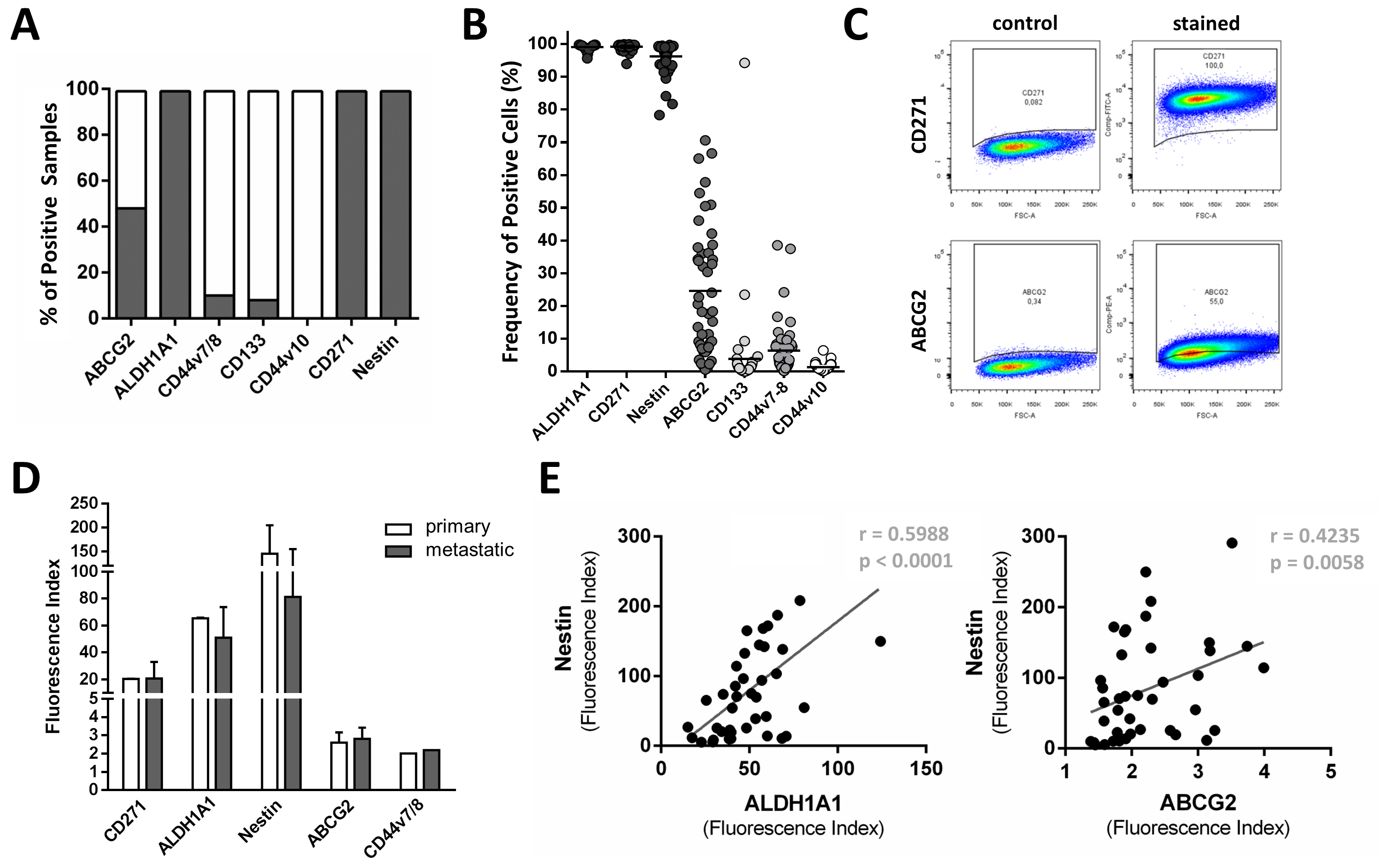 Fig. 1.
Expression patterns and correlations of putative cancer
stem cell (CSC) markers in established melanoma cell lines. Forty melanoma cell
lines were assessed for their expression of ABCG2, ALDH1A1, CD44v7/8, CD44v10,
CD133, CD271, and Nestin by flow cytometry. (A) Average protein expression in the
entire population shows Nestin, CD271, ALDH1A1, and ABCG2 to be commonly
expressed in established cell lines, while CD133 and CD44 variants were less
commonly found. Grey shading indicates positivity for the respective markers. (B)
Assessing the frequency of positive cells within each cell line showed Nestin,
CD271, ALDH1A1, and ABCG2-positive cells to be common in this panel of 40
established cell lines and in the four early-passage cell strains. Lower
frequencies in cells were found for CD133 and CD44 variants. (C) Examples of flow
cytometry plots showing similar expressions by most melanoma cells for CD271 and
ABCG2. Although less than 100% of cells are positive for ABCG2, most cells in
the population express the protein at similar levels. (D) Expression of CSC
markers is similar between primary- and metastatic-derived cell lines. (E)
Expression levels correlated between ALDH1A1 and Nestin (r = 0.5988; p
Since standard in vitro cell culture conditions (20% O
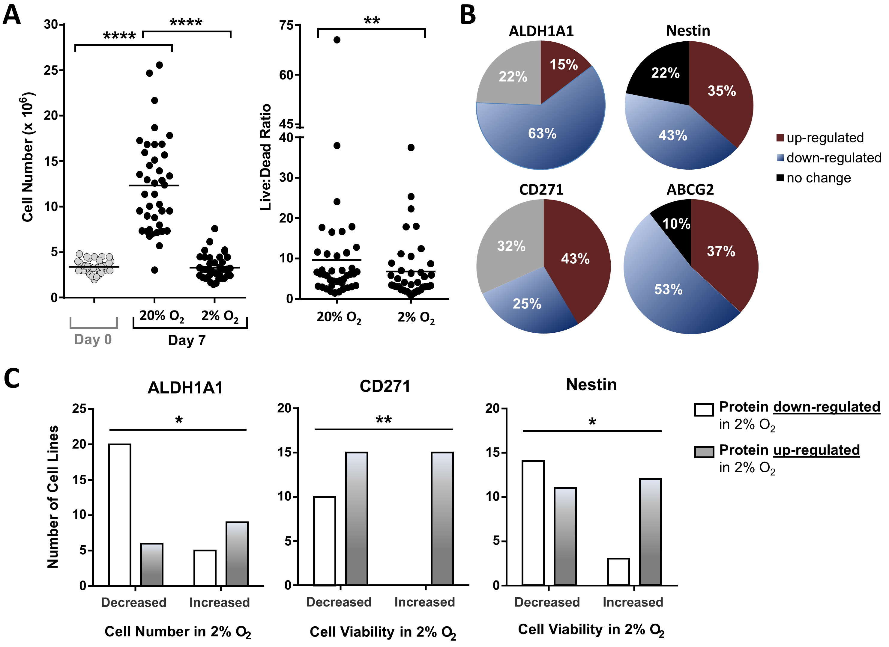 Fig. 2.
Effects of hypoxic and acidic culture conditions on melanoma
cell lines and the expression of putative CSC markers. Forty melanoma cell lines
were cultured under conventional (20% O
The expressions of the CSC markers were also altered by hypoxic and acidic conditions. In the 40 cell lines, ALDH1A1 and ABCG2 were downregulated in most, while CD271 was upregulated in the majority, and Nestin was up- or downregulated in roughly equal numbers (Fig. 2B). Each cell within all cell lines showed similar changes in expression under the test conditions. The cell lines that were negative for any protein under conventional conditions also remained negative in the oxy low/pH low model. The correlations between the expression of Nestin and ALDH1A1 and Nestin and ABCG2 (Fig. 1E) were observed under conventional conditions and remained retained when these cells were cultured in oxy low/pH low (p = 0.0002 for ALDH1A1 vs. Nestin and p = 0.0439 for ABCG2 vs. Nestin) (data not shown).
Changes in the expression of CD271 and Nestin between the conventional and oxy low/pH low culture models were found to correlate with the viability of the melanoma cell lines (p = 0.0063 and p = 0.0258, respectively) (Fig. 2C). Further, we observed that an improvement in viability was associated with the upregulation of these proteins under oxy low/pH low culture conditions, as indicated by the higher live/dead cell ratios. Despite the majority of cell lines tending to downregulate ALDH1A1 in the experimental model, we found improved cell growth in the cell lines where the expression of ALDH1A1 was upregulated (p = 0.0168) (Fig. 2C). No such associations were found for ABCG2. Since the CD44 variants and CD133 were rarely, or not at all, expressed by these cell lines, this analysis could not be performed for these proteins.
To validate the results obtained from the melanoma cell lines in vitro, we examined the expression of the selected CSC markers in situ, using an equal number of melanoma tissue deposits. Since CD133 and CD44v7/8 were rarely present in the melanoma cell lines, we considered it important to compare the results for these proteins obtained in vitro with those in situ. We additionally tested ALDH1A1 and ABCG2 because we found a number of interesting correlations in vitro and these proteins have rarely been studied in melanoma tissue, while CD271, Nestin, and CD44v10 have all been previously examined in situ by other investigators [34, 35, 36]. Consistent with the cell line results, all the CSC markers were commonly expressed in the majority of the melanoma cells in situ, although we did also observe a degree of heterogeneity, which revealed areas in the negative cells or more highly/weakly expressing cells within the tumour regions. In particular, CD133 was expressed more highly in a proportion of melanoma cells in some tissue samples, although in these cases most cells still exhibited positive staining. A common observation was that lower cell expressions resembled the tumour stroma. Notably, ALDH1A1 was an exception to our observation, whereby the common CSC markers were expressed by the majority of cells since we observed it to be either commonly expressed by all cells (17.5% of samples) or to be selectively expressed by a subset of cells (45% of samples) in situ (Supplementary Material 3).
When comparing tissue with established melanoma cell lines, the four putative CSC markers showed different expression patterns in situ (Fig. 3A,B). CD44v7/8 expression was more commonly observed in tissues (37.5% of samples were positive) compared to cell lines (10% of samples). The same was true for ABCG2, which was much more frequently expressed in the tissues (77.5%) rather than in the cell lines (46.34%), whereas ALDH1A1 was expressed only in 62.5% of the melanoma deposits compared to 100% of the cell lines. Interestingly, we found that CD133 was the most commonly expressed putative CSC marker in melanoma tissues, with 95% of the deposits expressing it. This stands in stark contrast to the established cell lines, whereby only a small proportion (7.5%) of cell lines expressed CD133, according to the FI. Representative immunofluorescence images stained for ALDH1A1, ABCG2, CD44v7/8, and CD133 are shown in Fig. 3C. Supplementary Material 3 shows a single representative stained and control image for all four proteins in two tissue samples (in the case of ALDH1A1, it shows one example, which demonstrates the expression exhibited in most cells, with the second example showing the expression in a subset of cells) and Supplementary Material 4 shows one example of a complete set of images comprising all fluorescence images captured throughout one tumour sample for all four proteins.
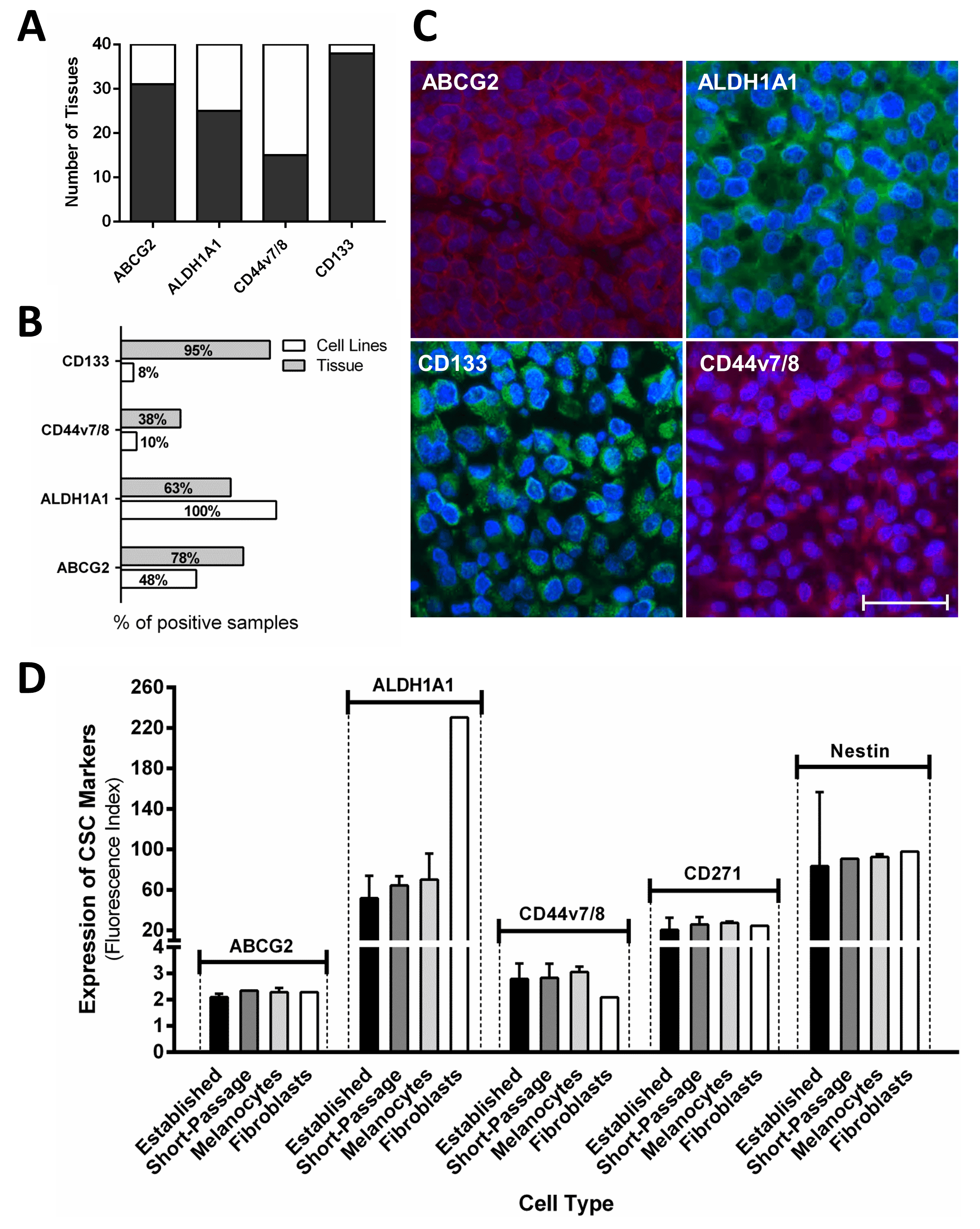 Fig. 3.
Expression of putative CSC markers in melanoma tissue, cell lines, and differentiated non-malignant cells. Immunofluorescence (melanoma tissue) and flow cytometry (cells and cell lines) were used to assess expression levels of putative CSC markers. (A) Almost all 40 melanoma tissues express CD133, and less frequently express ABCG2, ALDH1A1, and CD44v7/8. Grey shading indicates positivity for the respective markers. (B) Forty melanoma cell lines and an equal number of melanoma tissues were compared for their expression of CSC markers; tissues more frequently express CD133, CD44v7/8, and ABCG2, although ALDH1A1 is more common in cell lines. The FI was used to determine positive samples. (C) Representative images from melanoma deposits stained for ALDH1A1, ABCG2, CD44v7/8, and CD133. Scale bar indicates 100 µm. (D) Putative CSC markers are expressed at similar levels by established (n = 40) and early-passage (n = 4) melanoma cell lines, primary epidermal melanocytes (n = 2), and dermal fibroblasts (n = 1).
Since we observed that both melanoma tissues and cell lines expressed these putative CSC markers, next, we examined their expression in benign differentiated cells to test their specificity as CSC markers and to examine their potential use as therapeutic targets in melanoma. Thus, using flow cytometry, we investigated the expression of these molecules in control cells; human dermal fibroblasts and primary human epidermal melanocytes, the latter from two different sources. We additionally tested normal human skin sections for CD133 and ABCG2, using immunofluorescence. Our results demonstrate that these seven putative CSC markers are not specific for cancer or normal stem cells since they were also detected in all the tested benign differentiated cell types examined in this study. Interestingly, the benign samples were found to express these proteins at comparable or occasionally even higher levels than in malignant cell types. A comparison between the expression levels of all proteins tested in the benign and malignant cell types was conducted by flow cytometry and is shown in Fig. 3D. The test results for human skin using immunofluorescence can be found in Supplementary Material 5.
This study surveyed a panel of putative CSC surface markers in large sample numbers in vitro, in situ, and by including multiple differentiated non-malignant cell types, thereby investigating marker- and sample-dependent differences. Since mechanistic studies are either performed in vitro or in vivo with animals, we considered it important to compare melanoma samples in vitro with those in situ, exclusively in human samples. Therefore, we studied the putative CSC markers of CD271, ALDH1A1, Nestin, ABCG2, CD133, CD44v7/8, and CD44v10 in 40 established melanoma cell lines, with four (ABCG2, ALDH1A1, CD44v7/8, and CD133) additionally investigated markers in an equal number of melanoma tissues. We showed that four of the seven putative CSC markers (CD271, ALDH1A1, Nestin, and ABCG2) are commonly expressed in melanoma cell lines, while the remaining three markers were either found very rarely (CD133 and CD44v7/8) or not at all (CD44v10). Except for ALDH1A1, we observed that the remaining CSC markers were expressed more frequently in the tissues than in the cell lines. Substantial differences were seen for CD44v7/8 and CD133, which were rarely found in the cell lines but were more common in the tissues, especially CD133. This infrequent expression of CD133 and CD44v10 in melanoma cell lines is consistent with the results of previous studies [37, 38]. The differences observed between cell lines and tissue may be associated with the selection of melanoma tumours or cells, which are able to grow in vitro as an established cell line. The growth requirements in this artificial environment are likely to differ substantially from those in vivo; thus, the fraction of melanoma tumours, or individual cells within a tumour, which is able to survive surgical excision, processing, and subsequent growth as a monolayer appears to select for melanoma cells or for tumours with a particular profile of CSC marker expression. It is perhaps less likely that these changes occurred during in vitro culturing unless they occur very early because we observed similar results for early passage and established cell lines.
Collectively, our results suggest that the bulk of melanoma cells express similar levels of CSC markers. It was surprising to us that in the majority of cases, the seven tested markers did not show distinguishable sub-populations of positive and negative cells, which also prevented us from isolating these fractions to investigate whether they possess stem-like properties. To strengthen these observations in established cell lines in vitro, we confirmed the expression by most cells in situ, using excised melanoma tissues and in four early-passage cell strains. Indeed, the four early-passage cell strains were less likely to have been altered by in vitro culturing than the established cell lines, while the excised melanoma tissues showed no alterations. A number of prior studies that have shown CSCs to be expressed in only a small proportion of all melanoma cells utilised freshly resected tumour cells, which had undergone enzymatic digestion. This treatment has been shown to reduce the frequency of detected tumour cells expressing CSC markers [13]. In contrast, the present study examined formalin-fixed tissue samples or used cell lines that had undergone brief treatment with a gentler detachment method than trypsin, thereby potentially explaining the observation in our study, where the CSC markers are commonly expressed in melanoma.
Our findings show that the panel of CSC markers investigated here were expressed at a similar level by the majority of melanoma cells. However, these markers are putative and therefore could be non-specific or possibly irrelevant in the identification of CSCs [39]. To address this, we investigated the specificity of these markers for CSCs by testing their expression in benign differentiated cell types of related origin (primary human melanocytes, human dermal fibroblasts, and normal human skin). We found that benign differentiated cells, as well as cancer cells, express these proteins, in line with previous reports on ALDH1, CD44 variants, Nestin, and CD133 [37, 40, 41]. This finding weakens the proposition that the markers examined here are specific for CSCs and leaves open the possibility that more accurate CSC markers in melanoma may still await discovery. Moreover, we examined primary- and metastatic-derived cell lines, meaning it is unlikely that the observed results are due to the dissemination of phenotypic monoclonal metastatic CSCs from a heterogeneous primary tumour. Notably, our results may have consequences for studies that use these markers to isolate CSCs for functional testing, or for studies attempting to target them therapeutically. Noteworthy differences were seen for ALDH1A1; we observed some melanoma tissues where expression occurred in the majority of cells, while other tissues showed the presence of distinct individually positive cells. Perhaps due to genetic heterogeneity, this protein may not be representative in the same population of cells in every melanoma, i.e., ALDH1A1 may be a marker of CSCs in some melanomas but not in others.
We additionally cultured established melanoma cell lines in an experimental oxy low/pH low culture model aimed at more closely mimicking their native environment. We hypothesised that this approach may lead to the selection of cells better adapted to the environment, which may be associated with cells possessing a cancer stem-like phenotype due to the environmental influence on CSCs, which has been previously reported to exist. A previous study in melanoma demonstrated that hypoxia regulated the expression of the molecular CSC marker Oct-4 [42]. This finding hinted that features of the tumour microenvironment are involved in the regulation of CSCs. In line with this finding, we observed a considerable proportion of cell lines that upregulated CSC surface markers in a culture model of hypoxia and acidity. However, we also found that a similar or greater proportion of cell lines downregulated these markers, thereby highlighting the heterogeneous nature of cancer, even those of the same histological origin. The varied results seen in response to our oxy low/pH low culture model suggest that the role of these proteins may not be the same in all melanomas, whether they represent CSCs or not.
In this study we observed that the expression of CSC markers correlated with certain cellular features, suggesting that there are as yet undiscovered roles for these proteins in melanoma. We observed that cells, where certain CSC markers were upregulated under hypoxia and acidity, showed improved viability or cell growth. The importance of these proteins is underlined by the finding that they were expressed in melanoma cell lines as well as in melanoma tissues, indicating that some of them may be essential for tumour maintenance in vivo.
In the present study, we have shown that the expression of CSC surface markers can differ depending on the nature of the sample type examined and the culture environment employed, which may provide some explanation for the large numbers of conflicting previous studies reporting on putative markers of CSCs in melanoma, reconciled by the phenotypic plasticity model [43, 44]. We found that these proteins are commonly expressed in both melanoma cell lines and tissue and that they are associated with important features of melanoma cells. Unlike many studies, the inclusion of differentiated non-malignant samples alongside malignant samples in this work allowed us to investigate their specificity for CSCs, phenotypically modulated or not. This study revealed widespread expression of these proteins in non-malignant cells, which questions their usefulness as CSC markers and may limit their use as therapeutic targets.
CSCs, cancer stem cells; ALDH1A1, Aldehyde Dehydrogenase 1 family member A1; ABCG2, ATP Binding Cassette transporter G2.
The datasets used and/or analyzed during the current study are available from the corresponding author on reasonable request.
LS and NJ performed the experiments. BW and TS provided patients’ samples and data. GP and CS designed the study. LS, GP and CS wrote the paper. All authors contributed to editorial changes in the manuscript. All authors read and approved the final manuscript. All authors have participated sufficiently in the work to take public responsibility for appropriate portions of the content and agreed to be accountable for all aspects of the work in ensuring that questions related to its accuracy or integrity.
Ethical approval was obtained from the University Hospital Tübingen ethics committee (approval number 017/2016BO2).
We are grateful to Christof Zanke (University Hospital Tübingen) for producing software used to assess fluorescence microscopy images. We would also like to express our gratitude to Dr. Heike Niessner and Corinna Kosnopfel of the University Hospital Tübingen for their contribution in the culture of early-passage melanoma cell strains and primary melanocytes, and to Dr. Martin Schlegel for assistance with H&E stainings.
This work was supported by a grant from the German Research Foundation (DFG Pa 361/22-1).
The authors declare no conflict of interest. Given the role as Editor in Chief, Graham Pawelec had no involvement in the peer-review of this article and has no access to information regarding its peer-review. Full responsibility for the editorial process for this article was delegated to Alfredo Budillon.
References
Publisher’s Note: IMR Press stays neutral with regard to jurisdictional claims in published maps and institutional affiliations.