IMR Press / FBL / Volume 28 / Issue 2 / DOI: 10.31083/j.fbl2802038
Open Access Review
Lycium barbarum Ameliorates Neural Damage Induced by Experimental Ischemic Stroke and Radiation Exposure
Yan Huang1,†Xing Zhang1,2,†Ling Chen3,†Bo Xu Ren1,*Feng Ru Tang4,*
Show Less
1 Department of Medical Imaging, Medical School of Yangtze University, 434023 Jingzhou, Hubei, China
2 Medical Imaging Department, Wuhan Hospital of Traditional Chinese and Western Medicine, 430022 Wuhan, Hubei, China
3 Department of General Practice, Wuhan Fourth Hospital, 430033 Wuhan, Hubei, China
4 Radiation Physiology Lab, Singapore Nuclear Research and Safety Initiative, National University of Singapore, 138602 Singapore, Singapore
*Correspondence: boxuren188@163.com (Bo Xu Ren); snrtfr@nus.edu.sg (Feng Ru Tang)
These authors contributed equally.
Front. Biosci. (Landmark Ed) 2023, 28(2), 38; https://doi.org/10.31083/j.fbl2802038
Submitted: 27 December 2022 | Revised: 7 February 2023 | Accepted: 10 February 2023 | Published: 24 February 2023
Copyright: © 2023 The Author(s). Published by IMR Press.
This is an open access article under the CC BY 4.0 license.
Abstract

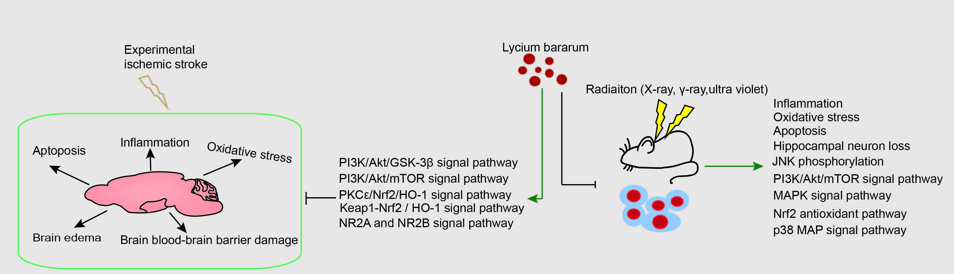
Ischemic stroke and cranial radiotherapy may induce brain inflammatory response, oxidative stress, apoptosis and neuronal loss, and impairment of neurogenesis. Lycium barbarum has anti-oxidation, anti-inflammatory, anti-tumor and anti-aging properties, may produce both neuroprotective and radioprotective effects. In this narrative review paper, we described the neuroprotective effect of Lycium barbarum in different animal models of experimental ischemic stroke and limited studies in irradiated animal models. Relevant molecular mechanisms are also summarized. It has been shown that in experimental ischemic stroke models, Lycium barbarum produces neuroprotective effects by modulating neuroinflammatory factors such as cytokines and chemokines, reactive oxygen species, and neurotransmitter and receptor systems. In irradiation animal models, Lycium barbarum prevents radiation-induced loss of hippocampal interneurons. Given its minimal side-effects, these preclinical studies suggest that Lycium barbarum may be a promising radio-neuro-protective drug that can be used as an adjunct treatment to radiotherapy for brain tumor and in the treatment of ischemic stroke. At molecular levels, Lycium barbarum may regulate PI3K/Akt/GSK-3β, PI3K/Akt/mTOR, PKCε/Nrf2/HO-1, keap1-Nrf2/HO-1, and NR2A and NR2B receptor- related signal transduction pathways to produce neuroprotective effects.

Keywords
ischemic stroke
radiation
radiotherapy
apoptosis
inflammation
oxidative stress
Lycium barbarum
neuroprotective
molecular mechanisms
Lycium barbarum polysaccharides
1. Introduction

Stroke, the leading cause of neurologic disease, is the most common serious manifestation of cerebrovascular disease [1], resulting in a global annual economic burden. It has been reported that ischemic stroke accounts for 70–80% of total stroke events [2]. China is facing the most serious threat of ischemic stroke in the world [3], which is now the second major cause of death worldwide [4], and affects both middle-aged and elderly populations [5, 6]. Ischemic stroke refers to brain tissue and neuronal damage caused by insufficient blood supply [1], and is caused by an interruption in cerebral blood flow induced by thrombosis or embolism [7], leading to neuronal loss, brain atrophy and cognitive decline, which may also occur in patients with Alzheimer’s disease [8], motor functional deficits [9], and multiple cognitive functional deficits [10]. Early and rapid restoration of blood supply within a strict time window is still considered to be the first choice for the treatment of acute ischemic stroke [11, 12]. However, if the recovery of cerebral blood flow exceeds a certain time window, it can result in further neurological damage, resulting in an ischemia-reperfusion injury [13]. Tissue plasminogen activator has been used for the treatment of cerebral infarction for over 20 years, but its side effects limits the prognosis of ischemic stroke [14]. Therefore, there is an urgent need to find new therapies for treating ischemic stroke. It has been shown that drugs that possess anti-apoptosis, anti-inflammatory, and anti-oxidative stress properties, and promote neurogenesis are effective in the treatment of cerebral ischemia [15, 16, 17, 18]. Studies suggest that some Traditional Chinese Medicine (TCM) and natural compounds have neuroprotective properties that can be used for treating ischemic stroke [19, 20].

With the increasing use of radiation for medical diagnosis and therapeutic approaches such as computed tomography (CT), positron emission tomography (PET), radiotherapy and radiopharmaceutical therapy (RPT) of cancers, the risk of exposure to radiation is increasing. Radiotherapy has become one of the most common therapeutic methods for cancer treatment, especially in patients with head and neck tumors, and brain metastases, these patients are exposed to the risk of radiation-induced brain damage [21]. The brain is very sensitive to radiation exposure. Radiation can affect the central nervous system, leading to mental retardation, behavioral changes, cognitive impairment, and neoplastic diseases [22, 23]. Although it is well known that high dose rate radiation causes damage to the brain, skin and eye [24, 25, 26, 27], the effect of low dose radiation on the human brain is still unknown. The anti-radiation drug amifostine is limited in clinical practice due to its side effects [28]. Therefore, it is vital to find drugs with low toxicity, and effective neuroprotection to limit brain damage.

Traditional Chinese Medicines (TCMs) have a long history of application in the treatment of human diseases [29]. Because of their multi-component synergistic effects, multi-targeted therapeutic benefits, low toxicity and side effects, and low price, TCMs may have an advantage when compared to western medicine. Lycium barbarum, a TCM and food supplement [30], has been used for centuries in many countries. Lycium barbarum is also known as Wolfberry or Goji [31]. The fruit, root bark and leaves of Lycium barbarum are widely used as food and pharmaceutical additives [32], and contains rich chemical components [33]. A large number of studies have shown that it has a variety of favorable biological effects in patients with diabetes [32, 34], impaired reproductive systems [35, 36], eye diseases [37, 38], cardiovascular diseases [39, 40] and cancers [41, 42]. Lycium barbarum polysaccharide (LBP), the main active component of Lycium barbarum, has been shown to possess anti-oxidation, anti-inflammatory, anti-tumor and anti-aging properties [43, 44, 45, 46, 47]. Numerous studies have suggested the potential use of LBP or Lycium barbarum in protecting against damage induced by experimental ischemic stroke and radiation exposure [48, 49]. In this study, we review the neuroprotective effects and molecular mechanisms of Lycium barbarum in experimental ischemic strokes and radiation exposure.

2. Neuroprotective Effects of Lycium barbarum on Ischemic Stroke
2.1 Lycium barbarum Inhibits Apoptosis

Mitochondria, the “energy factory” of cells, play an important role in cellular homeostasis [50]. The brain is an energy dependent organ and performs its functions via aerobic metabolism [51]. In the mitochondrial apoptotic pathway, cerebral ischemia leads to altered mitochondrial membrane potential and increased permeability [52], which then causes the release of cytochrome C (Cyt-C) and apoptosis inducing factor (AIF) and the formation of apoptotic bodies [53], promoting the activation of pro caspase-9. Activated caspase-9 can induce the caspase cascade, resulting in the cleavage and activation of caspase-3 [54]. As a key link in the mitochondrial apoptotic pathway, caspase-3 specifically cleaves substrate proteins, such as poly (ADP-ribose) polymerase (PARP), which in turn leads to PARP hyper-activation, DNA damage and apoptosis [55, 56]. In a mouse model of middle cerebral artery occlusion (MCAO), prophylactic gavage with LBP for 7 days significantly reduced neurological deficit scores, the area of cerebral infarction in the ischemic side, and the apoptosis of neurons. In addition, LBP pretreatment reversed the increase of caspase-3 protein activity, the decrease of B-cell lymphoma-2 (Bcl-2) protein expression, and the increase of Bcl-2-associated X (Bax) protein expression [57, 58]. Furthermore, LBP treatment reduced the expression of Cyt-C, caspase-9 and cleaved PARP-1 [59]. Caspase-12 precursors are located in the endoplasmic reticulum [60]. Endoplasmic reticulum stress specifically activates caspase-12, which then cleaves downstream proteins such as caspase-3, causing apoptosis [61]. Cerebral ischemia-reperfusion can induce an increase of caspase-12 protein and mRNA expression, suggesting that the endoplasmic reticulum pathway is involved in the regulation of neuronal apoptosis [62]. In a rat model, gavage of LBP twice a day for 3 days improved neurological function and reduced the water content of brain tissue, and reduced the expression of caspase-12 protein and mRNA [63]. This demonstrated that LBP reduced neuronal apoptosis by inhibiting caspase-12 in the rat model. Similarly, a recent study indicated that LBP played a neuroprotective role by increasing the expression of cyt-C, cleaved caspase-3, and Bcl-2-associated death promoter, through the NR2B signal pathway in experimental ischemic stroke [64] (Fig. 1, Table 1 (Ref. [57, 58, 59, 63, 64, 65, 66, 67, 68, 69, 70, 71, 72, 73, 74])).
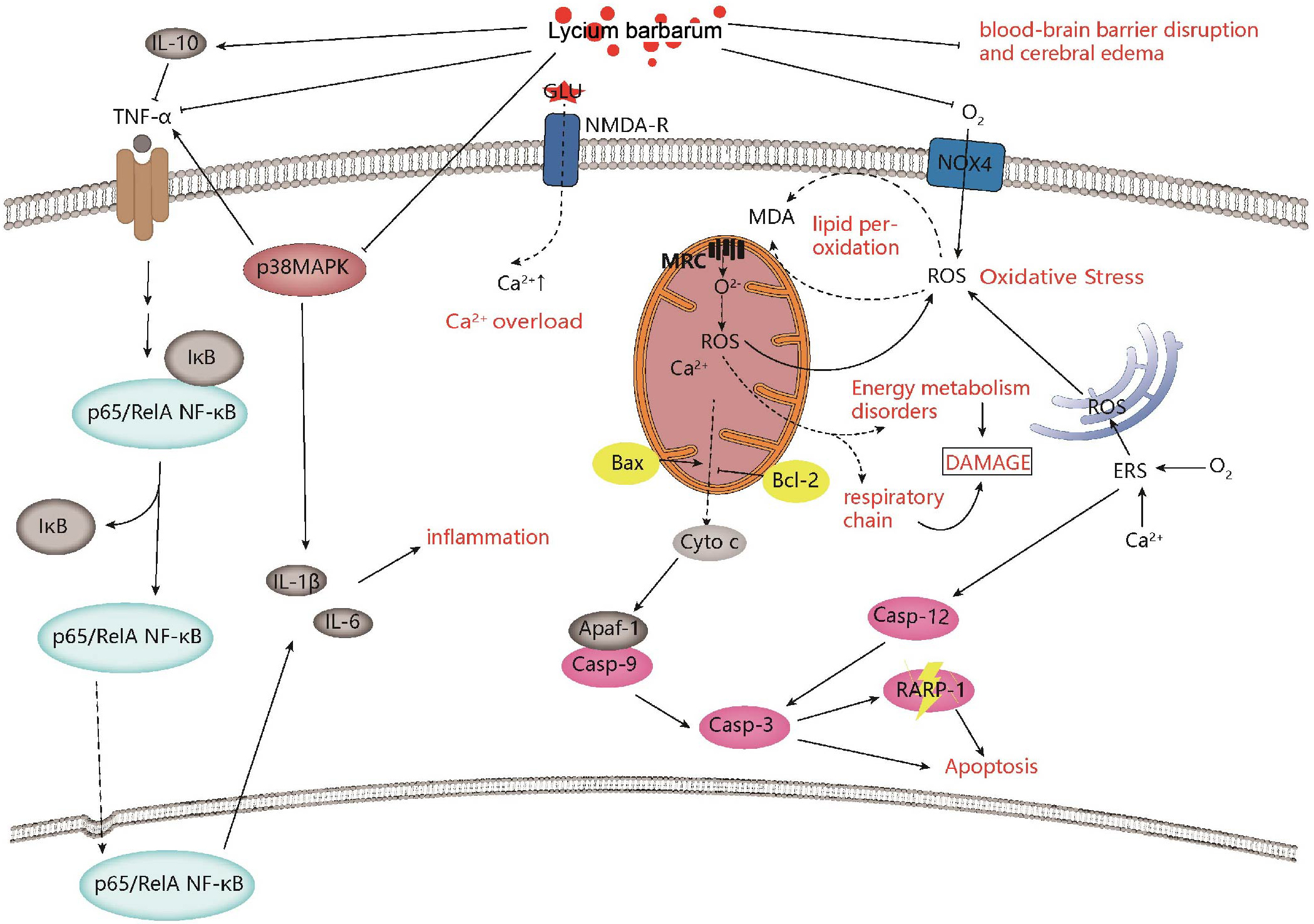
Fig. 1.

Neuroprotective effects and mechanisms of Lycium barbarum on ischemic stroke. Lycium barbarum shows neuroprotective effects against ischemic stroke by improving inflammation, regulating calcium levels, improving mitochondrial function, inhibiting apoptosis, and improving oxidative stress.

Table 1.Neuroprotective effects and molecular mechanisms of Lycium barbarum on ischemic stroke.
Compounds Models Strain, sex, age, and weight Dosage and time Administration and End point Behavioral change Brain effect at cellular level Molecular mechanism References
LBP MCAO model Institute of Cancer Research (ICR) mice, male, 27–32 g 10, 20, 40 mg/kg, before insults Once daily, intragastric administration for 7 days Neurological deficit scores↓, Time spent on the rotating-stick↑, tail flick latency↓, number of autonomous activities↑ p-Akt↑, p-GSK3β (ser9)↑, p-PDK-1↑, Mcl-1↑, PP2A↓, GSK3β mRNA↓ PI3K/AKT-GSK3β pathway [72]
LBP MCAO model ICR mice, male, adult, 20–25 g 10, 20, 40 mg/kg, before insults Once daily, intragastric administration for 7 days Neurological deficit scores↓ caspase-3↓, Bax↓, Bcl-2↑ Inhibit apoptosis [57]
LBP MCAO model ICR mice, male, 5–6 weeks, 23–28 g 10, 20, 40 mg/kg, before insults Once daily, intragastric administration for 7 days Neurological deficit scores↓ Bax↓, Bcl-2↑ Inhibit apoptosis [58]
LBP MCAO model ICR mice, male, 6 weeks, 20–25 g 20, 50, 100 mg/kg, before insults Once daily, intragastric administration for 7 days Neurological deficit scores↓ p65 NF-κB↓, TNF-α↓, IL-6↓, IL-1β Inhibit the activation of P65NF-κB, anti-inflammatory [65]
LBP MCAO model ICR mice, male, adult, 20–25 g 10, 20, 40 mg/kg, before insults Once daily, intragastric administration for 7 days Behavioral test performance↑ p65 NF-κB↓, p38 MAPK↓, TNF-α↓, IL- 1β↓, IL-6↓, IL-10↑, IL-1β mRNA↓, TNF-α mRNA↓ Anti-inflammatory, partly through inhibiting the activation of p65 NF-κB and p38 MAPK [66]
LBP MCAO model SD rats, half male and half female, 220–240 g 60, 30, 15 mg/kg, 2 days before insults Twice daily, intragastric administration for 3 days Neurological deficit scores↓ caspase-12 protein↓, caspase-12 mRNA↓ Inhibit apoptosis [63]
LBP Four-vessel occlusion model Wister rats, male, 220–300 g 20 mg/kg, 7 days before or/and after modeling Once daily, intragastic administration for 7days Memory deficits↑ Neuronal survival↑ Inhibit apoptosis [64]
Oxygen-glucose deprivation (OGD) model Primary cortial neuron from Wister male rats 100mg/L, after oxygen-glucose deprivation Incubate, 24 h Viability of cortical neurons↑ ROS↓, Bcl-2-associated death promoter protein ↓, CytC protein ↓, NR2A protein ↑, pAkt protein ↑, pCREB protein ↑ Activation of NR2A and inhibition of NR2B signialing pathway
LBP MCAO model Kunming mice, male, 25–30 g 10, 20, 40 mg/kg, before insults Once daily, intragastric administration for 7 days - MDA↓, SOD↑, GSH-Px↑, CAT↑, LDH leakage↓, ATP↑ Antioxidant, Improve energy metabolism [69]
LBP MCAO model Kunming mice, male, 25–30 g 10, 20, 40 mg/kg, before insults Once daily, intragastric administration for 5 days - MDA↓, SOD↑, GSH-Px↑, CAT↑, LDH leakage↓, ATP↑ Antioxidant, Improve energy metabolism [68]
LBP MCAO model ICR mice, male, 6 weeks, 20–25 g 20, 50, 100 mg/kg, before insults Once daily, intragastric administration for 7 days Neurological deficit scores↓ Nox4↓, ROS↓, MDA↓, SOD↑, GSH-Px↑ Antioxidant [67]
LBP OGD/RP model primary hippocampal neurons 15, 30, 60 μg/mL, after oxygen and glucose deprivation, before reperfusion Incubate - ROS↓, MDA↓, cleaved-caspase3/caspase3↓, Bcl-2/Bax↑, Beclin 1↓, LC3II/LC3I↓, p62↑, p-Akt↑, p-mTOR↑ Antioxidant, Inhibit autophagy, Inhibit apoptosis, PI3K/Akt/mTOR signal pathway [58]
LBP MCAO model C57BL/6N mice, male, adult, 10–20 weeks 1, 10 mg/kg, before insults Once daily, intragastric administration for 7 days Neurological deficit scores↑, AQP-4↑, glial fibrillary acidic protein↑, EB extravasation↓, IgG-leaky↓, occludin↑ Improve injury of cerebral BBB [70]
LBP MCAO model SD rats, male, 200–220 g, 8 weeks old 25 mg/kg, before MCAO Once daily, injected intraperitoneally for 4 weeks Neurological deficits scores↓ Brain edema ↓, apoptosis↓, IgG leakage↓, occluding protein↑, claudin-5↑, ZO-1↑ Ameliorate ischemia injury via protecting BBB [71]
LBP MCAO model ICR mice, male, 20–25 g 10, 20, 40 mg/kg, before insults Once daily, intragastric administration for 7 days Neurological deficit scores↓ Bcl-2↑, Bax↓, Cyt-C↓, caspase-3↓, caspase -9↓, cleaved PARP-1↓ Attenuate the mitochondrial apoptosis pathway [59]
Lyciumamide A (LyA) MCAO model SPF grade, SD rats, male, 280 ± 20 g 20, 40, 80 mg/kg, immediately after the surgery Peritoneal injection Neurological deficit scores↓ SOD↑, GPx↑, MDA↓, nuclear Nrf2↑, cytoplasmic HO-1↑, LDH leakage↓, Bax↓, Bcl-2↑, cleaved caspase-3↓, p-PKCε↑, Nrf2↑, HO-1↑ Antioxidant via PKCε/Nrf2/HO-1 pathway [73]
OGD/RP model SH-SY5Y cells 0, 5, 10, 20, 40, 80 μΜ Incubate for 8 h before OGD
LBP MCAO model ICR mice, male, adult, 28–30 g 40 mg/kg, 7 days before insults Once daily, intragastric administration for 7 days Neurological deficit scores↓ Nrf2 mRNA↑, HO-1 mRNA↑, LC3 mRNA↓, Ca2+↓, MMP level↑, ROS↓, Nrf2↑, HO-1↑, Keap-1↑, LC3-II↓, Beclin-1↓ Antioxidant via Keap1-Nrf2/HO-1 pathway [74]
OGD/RP model PC12 cells 40 μg/mL Incubate for 24 h before reoxygenation
LBP Transient global ischemia injury rats model Adult male Wister rats, 220–300 g 20 mg/kg Once daily, intragastric administration for 7 days Memory deficits↓ LDH leakage↓, NR2B↓, nNOS↓, Bad↓, Cyt-C↓, cleaved caspase-3↓, ROS↓, Ca2+↓, NR2A↑, p-Akt↑, p-CREB↑ Regulate NR2A and NR2B signal pathway containing NMDA receptors [64]
OGD/RP model Embryos of female Wistar rats at E18 gestation 100 mg/L Incubate for 24 h before reoxygenation
2.2 Lycium barbarum Improves the Inflammatory Response

The inflammatory response is an important target following an acute ischemic stroke. In the classical nuclear factor-κB (NF-κB) pathway, inhibitors of NF-κB (IκB) can be phosphorylated by the IκB kinase (IKK) complex [75], ubiquitinated by ubiquitin ligases and finally degraded by the proteasome, leading to NF-κB nuclear translocation and gene transcription [76]. Many factors, including tumor necrosis factor (TNF) and interleukin 1β (IL-1β), can activate NF-κB [76], which in turn activates inflammatory cells in brain tissue after cerebral ischemia, and mediates the expression of inflammatory cytokines such as tumor necrosis factor-α (TNF-α), interleukin 6 (IL-6), interleukin-8 (IL-8) and IL-1β, which exacerbate the ischemia-reperfusion injury [77]. Notably, crosstalk between different interleukins (ILs) in different immune cells can also affect the outcome of ischemic stroke [78]. In addition, the mitogen-activated protein kinase (MAPK) cascade is also highly activated after ischemic injury. The expression of p38 MAPK is known to be positively correlated with the expression of the pro-inflammatory molecules IL-1β, TNF-α, and IL-6 [79]. Therefore, blocking the excessive activation of the MAPK cascade and the increased production of inflammatory cytokines and is essential to reduce cerebral ischemic injury.

Experiments in mice suggested that LBP significantly improved the neurological symptoms of the brain and reduces the water content and infarct size of brain tissue [65]. LBP reversed the elevated expression of NF-κB p65, TNF-α, IL-6 and IL-1β in the ischemic cerebral cortex [65, 66]. The anti-inflammatory effects of LBP were further supported by reduced TNF-α and IL-1β mRNA expression in astrocytes and microglia in the LBP-treated group, and inhibition of P38 MAPK activation [66]. In addition, Liu et al. [80] found that LBP pretreatment significantly decreased the secretion of inflammatory factors in lipopolysaccharide-induced peritonitis mice models (Fig. 1, Table 2 (Ref. [21, 24, 81, 82, 83, 84, 85, 86, 87, 88, 89]).

Table 2.Radioprotective effects and molecular mechanisms of Lycium barbarum against radiation-induced non-neuronal cell and tissue damage.
Compounds Radiation source and dose Strain, sex, age, and weight Dosage and Time Administration and end point Behavioral change Brain effect at cellular level Molecular mechanism References
The fruits extract of L. barbarum (LBE) γ-ray (whole body), 8.5 and 6.0 Gy C57BL/6 mice, male, 6–8-week-old 1.0, 3.0, 6.0, and 9.0 g/kg, from 7 days before irradiation to 21 days post irradiation Once daily, oral administration, 28 days - TNF-α↓, IL-1β↑, IL-6↑ Immunomodulation and the synergistically modulating effect on the gut microbiota and related metabolites [81]
X-ray (whole body), 5.5 Gy BALB/c mice 3.0 g/kg, 7 days before irradiation to 21 days post irradiation Once daily, oral administration, 28 days -
γ-ray, 4.0 Gy Rat small intestinal epithelial cell line 6 (IEC-6) 100, 250, 500, 2000 μg/mL, 24 h before irradiation Incubate, 24 h -
polysaccharide fraction (LBPF) Ultraviolet (the dorsal region skin) HRS/J mice, female, approximately 8 weeks, - 5% LBPF gel, after irradiation Three times per week, apply, 4 weeks - MMP-1↑, MMP-2↑, MMP-9↑ - [85]
LBP Ultraviolet Immortalized human keratinocytes (HaCaT cells) 300 μg/mL, 24 h berfore irradiation Incubate, 24 h - Phosphorylated p38 protein↓, p38 protein↑, cleaved caspase-3↓, caspase-3↑, MMP-9↓ Nrf2/ARE pathway, p38 MAP pathway [86]
LBP X-rays, 4.0 Gy Kunming mice, male and female, 18–22 g 50, 100, 200 mg/kg, 2 h after irradiation Intraperitoneal injection, 14 days - SOD activity↑, MDA Content↓, CD44↓, CD49d↓ Inhibit apoptosis, antioxidant, and alter the expression of adhesion molecule [83]
LBP Ultraviolet Human skin fibroblast cell line (HSF) 300 μg/mL 0.5, 1, 2, 3 and 4 h before irradiation Incubate, 0.5, 1, 2, 3 and 4 h - Nuclear p-Nrf2↑, ROS↓, lipid peroxide (LPO)↓, SOD↑, glutathione peroxidase (GSP-PX)↑ Nrf2 antioxidant pathway [21]
Aqueous and ethanol extracts of the L. barbarum fruit Ultraviolet Arising retinal pigment epithelia cell line-19 (ARPE-19) 0–200 μg/mL, 2 h before irradiation Incubate, 2 h - ROS↓, γH2AX↓ antioxidant and prevent DNA damage and cell apoptosis [24]
LBP Ultraviolet Rat corneal epithelial (RCE) cells 0, 0.05, 0.1, 0.5, 1, 5, or 10 mg/mL, 24 h before irradiation and 0–6 h after irradiation Incubate, 24 h before irradiation and 0–6 h after irradiation - Bcl-2 mRNA↑, Bax mRNA↓, caspase-3 mRNA↓, caspase-3 protein↓, p-JNK/JNK↓ Attenuate the mitochondrial pathway and inhibit JNK phosphorylation [89]
Lycium ruthenicum Murr X-ray (whole body), 5 Gy Kunming mice, male, 4–6 week old, 25 ± 2 g 2, 4, 8 g/kg, 3, 7, 14 days after irradiation Oral administration, 14 days - caspase-3↓, caspase-6↓, P53↓ Inhibit apoptosis [82]
LBP 60Co-γ (local irradiation), 2.3 Gy Wistar rats, male, 160–200 g 10 mg/kg, 6 h before irradiation Once daily, intragastric administration, 4 weeks - Bcl-2↑, Bax↓ Inhibit apoptosis [84]
Lycium barbarum polysaccharide-rich hydrogel formulation Ultraviolet HRS/J hairless mice, female, 6-weeks -old 5%, 6 weeks after irradiation Three times a week, apply, 3 weeks - c-Fos↓, c-Jun↓, MMP-1↓, MMP-2↓, MMP-9↓, collagen I↑, collagen III↑, fibroblast growth factor-2 (FGF2)↑ MAPK signal pathway [87]
Black wolfberry water extracts Ultraviolet HaCaT cells 2 mg/mL, 12 h before irradiation Incubate, 12 h - P38 MAPK↓, P53↓, caspase-8↓, caspase-3↓, Bcl-2↑ Promote cell proliferation and prevent cell apoptosis [88]
2.3 Lycium barbarum Reduces Oxidative Stress

Brain tissue has a higher oxidative metabolism and fewer antioxidases, and is more sensitive to oxidative stress than any other organ [90]. Oxidative stress is very important in the development of many diseases, especially in ischemic strokes [91, 92, 93]. During normal conditions, the body can produce reactive oxygen species (ROS) during aerobic metabolism and there is a balance between the production and elimination of ROS. As a subtype of nicotinamide adenine dinucleotide phosphate hydride (NADPH), the expression of NADPH oxidase 4 (Nox4) significantly increases during cerebral ischemia, leading to increased production of ROS [94], and reduced activity of endogenous antioxidant enzymes, such as superoxide dismutase (SOD), glutathione peroxidase (GSH-Px), and catalase (CAT) [95], which results in decreased cellular structure and enzymatic activity [96, 97]. Excessive ROS-induced lipid peroxidation can produce large amounts of malondialdehyde (MDA), which further increases brain damage [48, 67]. LBP treatment reduced ROS and MDA, but increased SOD, GSH-Px and CAT production in the mouse brain with ischemic stroke [67, 68, 69]. Western blotting (WB) results showed that LBP reduced Nox4 protein levels in the ischemic side of the cerebral cortex [67], and decreased cerebral ischemia-reperfusion injury (Fig. 1, Table 2).

2.4 Lycium barbarum Alleviates Brain Blood-Brain Barrier (BBB) Damage and Brain Edema

As an important barrier between the brain and peripheral circulation, BBB maintains the homeostasis of the central nervous system. It has been reported that the BBB is destroyed during ischemic stroke resulting in edema formation and hemorrhagic transformation [98, 99], which further exacerbates the ischemic injury [100]. As a key protein responsible for water transport in brain, aquaporin-4 (AQP4) mRNA expression is increased after middle cerebral artery occlusion, which is consistent with the exacerbation of brain edema monitored by magnetic resonance imaging, indicating that the up regulation of AQP4 expression is involved in the formation of brain edema [101, 102]. Matrix metalloproteinases (MMPs) are mainly expressed in the ipsilateral ischemic penumbra vascular endothelial cells, and activated by brain ischemia. The upregulation of matrix metalloproteinase-9 (MMP-9) exacerbates BBB damage [103]. In the mouse MCAO model, LBP treatment reduced brain edema and Evans Blue (EB) extravasation in the ipsilateral hemisphere, and vascular luminal leakage of immunoglobulin G (IgG). The down-regulation of AQP4 and MMP-9 further supports the protective effects of LBP [70]. A recent study showed that pretreatment with LBP protected the BBB by significantly reducing the cerebral infarct volume, cell apoptosis, and IgG leakage, which resulted in decreased hyperglycemia-exacerbated cerebral ischemia/reperfusion injury [71] (Fig. 1, Table 2).

3. The Molecular Mechanisms of Lycium barbarum in Improving Neuronal Damage after Ischemic Stroke
3.1 PI3K/Akt/GSK-3β Signal Pathway

Glycogen synthase kinase-3 (GSK-3) is a multifunctional serine/threonine protein kinase, belonging to the glycogen synthase kinase family [104]. Its dysfunction induces a variety of diseases. GSK-3
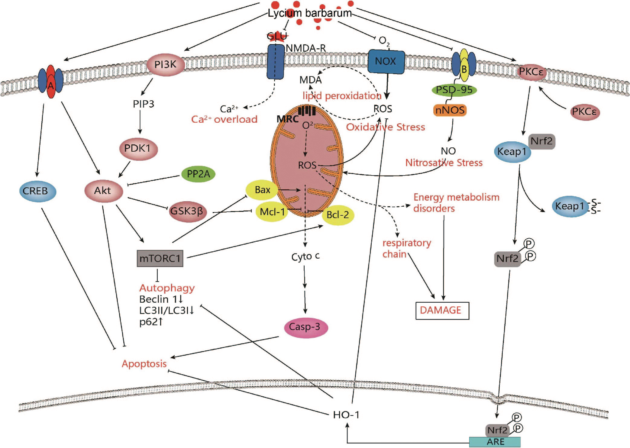
β can be inhibited by photostatidylinositol-3-kinase (PI3K)-activated protein kinase B (Akt), which is involved in the survival signal pathway [105, 106]. Akt inhibits apoptosis by phosphorylating GSK-3β [107, 108]. There have been an increasing number of studies involving the drug inhibition of GSK-3 in the treatment of neurodegeneration and mental diseases [109]. A recent study found that ginsenoside Rd, one of the main active ingredients in Panax ginseng, could improve cognitive function and reduce tau protein phosphorylation via the PI3K/Akt/GSK-3β pathway [110]. LBP pretreatment decreased the incidence of apoptosis in the neurons in the ischemic brain and increased the expression of myeloid cell leukemia-1 (Mcl-1). WB and qRT-PCR showed that the expression of p-Akt, p-GSK-3β (ser9), phosphorylated 3-phosphoinositide-dependent protein kinase 1 (p-PDK-1), and protein phosphatase 2A (PP2A) increased and GSK-3β mRNA decreased in the LBP pretreatment group, suggesting that LBP may protect the brain against cerebral ischemia-reperfusion injury through the PI3K/Akt/GSK-3β pathway [72] (Fig. 2).

Fig. 2.

Intracellular mechanisms of Lycium barbarum in improving ischemic stroke. Lycium barbarum shows neuroprotective effects against ischemic stroke through modulation of PI3K/AKT-GSK3β signaling pathway, PI3K/Akt/mTOR signaling pathway, PKCε/Nrf2/HO-1 signaling pathway, Keap1-Nrf2/HO-1 signaling pathway and NR2B and NR2A-containing NMDA receptor signaling pathway.

3.2 PI3K/Akt/mTOR Signal Pathway

Activation and modulation of the PI3K/Akt/mammalian target of rapamycin (mTOR) signal pathway has been shown to regulate apoptosis and autophagy [111]. The PI3K/Akt/mTOR signal pathway has been found to play an important role in angiogenesis, including endothelial cell survival, migration and tube formation [112, 113], which suggests that it may become a promising therapeutic target in ischemic stroke [114, 115]. It has been reported that ginsenoside Rg1 treatment activated the PI3K/Akt/mTOR signal pathway in cerebral cortical ischemia after strokes [116]. LBP inhibited neuronal apoptosis by reversing cleaved-caspase 3/caspase 3 and Bax/Bcl-2. In addition, Beclin 1 expression and microtubule-associated protein 1 light chain 3 II (LC3-II)/microtubule-associated protein 1 light chain 3 I (LC3-I) were decreased in the LBP-treated group, but p62 expression was increased, indicating that LBP suppressed neuronal autophagy, which was further supported by transmission electron microscopy. A mechanistic study revealed that LBP treatment increased the expression of p-Akt and p-mTOR proteins, confirming that LBP may exert neuroprotective effects through the PI3K/Akt/mTOR signal pathway [62] (Fig. 2).

3.3 PKCε/Nrf2/HO-1 Signal Pathway

Protein kinase C (PKC) belongs to the serine/threonine kinase family and mediates the phosphorylation of nuclear factor E2-related factor 2 (Nrf2) at ser-40 site and its antioxidant response [117, 118]. Nrf2 is one of the key regulators of endogenous antioxidant molecules, its nuclear translocation induces the expression of the cytoprotective gene heme oxygenase 1 (HO-1) [119]. It has been reported that HO-1 is an effective target for the protection of cerebral ischemia [120]. The cytoplasmic activity of PKC was decreased and membrane activity of PKC was increased during cerebral ischemia [121]. Reperfusion injury consistently reduced the levels of Nrf2 and HO-1 [122].

In the Sprague-Dawley (SD) rat model, Lycium barbarum treatment improved the neurological deficit score and brain infarct volume. Furthermore, it reduced oxidative stress, partly and significantly increased the expression of nuclear Nrf2 and cytoplasmic HO-1 in ischemic cerebral cortex; as knocking out either Nrf2 or HO-1 reduces the protective effect of lyciumamide A (lyA). In addition, lyA-induced Nrf2 nuclear translocation and the up regulation of HO-1 expression were inhibited by the knockout of PKCε, suggesting that the neuroprotective effect of lyA is mediated by activating the PKCε/Nrf2/HO-1 signal pathway [73].

An in-vitro experiment showed that LyA treatment reduced lactate dehydrogenase (LDH) leakage, cleaved caspase-3 levels, Bax/Bcl-2 levels, and the number of apoptotic cells [73] (Fig. 2).

3.4 keap1-Nrf2/HO-1 Signal Pathway

Kelch-like ECH-associated protein 1 (Keap1) is localized to the actin cytoskeleton [123]. The Neh2 structural domain of Nrf2 can be bound to Keap1 in the cytoplasm under physiological conditions and subsequently degraded by ubiquitination, to maintain dynamic stability [124]. Pathological conditions can induce Nrf2 phosphorylation, dissociation from Keap1 and nuclear translocation, and binds to the nuclear antioxidant response element (ARE) to promote the expression of antioxidant genes such as HO-1 [125, 126], enhancing the antioxidant capacity of the organism.

Experiments in mice revealed that ischemic injury disrupted neurological function and cortical brain electrical activity and reduced cortical blood perfusion on the ischemic side, while LBP treatment reversed these abnormal changes. The expression of Nrf2, HO-1 and Keap-1 were increased, while the expression of LC3-II and Beclin-1 was decreased after LBP treatment. Cell experiments revealed that LBP increased cell survival, reduced intracellular ROS levels and intracellular free calcium ion content, and stabilized mitochondrial membrane potential. Nrf2 inhibitor significantly reduced the Nrf2 and HO-1 protein content and increased the expression of Keap-1, LC3-II and Beclin-1 protein on the ischemic side compared to the LBP group, which was consistent in mouse and cell experiments. These studies suggest that LBP may protect ischemic brain injury by regulating the Keap1-Nrf2/HO-1 signal pathway [74] (Fig. 2).

3.5 NR2A and NR2B Signal Pathway

The activation of different subunits of the glutamate receptor/N-methyl-D-aspartate receptor (NMDAR) effects several pathways. For instance, activation of intrasynaptic NMDAR 2A (NR2A) stimulates the survival signaling pathway, while the activation of the extrasynaptic NMDAR 2B (NR2B) triggers the apoptotic pathway. In models of cerebral ischemia and other neurodegenerative diseases, the inhibition of NR2B-containing NMDARs are neuroprotective, while the inhibition of NR2A-containing NMDARs results in neural death [127, 128]. The inhibition of postsynaptic density-95 (PSD-95) expression selectively attenuates excitotoxicity triggered by NMDAR, demonstrating the importance of PSD-95 in effectively coupling NMDAR activity to NO toxicity [129]. Once ischemic injury occurs, neuronal nitric oxide synthase (nNOS) can be transferred to the cell membrane to form the NR2B-PSD-95-nNOS complex which subsequently produces a large amount of nitric oxide (NO) resulting in neuronal damage.

In the early ischemic phase, LBP attenuates neuronal damage by preventing the upregulation of NR2B and nNOS. In the late ischemic phase, LBP reduces calcium influx and mitochondrial permeability, preventing the overexpression of nNOS, the Bcl-2 associated agonist of cell death (Bad), Cyt-C, and cleaved-caspase-3 in the NR2B signal pathway. LBP increased the expression of NR2A, p-Akt, and the phosphorylated cAMP response to element binding (p-CREB). These results suggest that LBP may attenuate ischemic damage to hippocampal neurons by the NR2A and NR2B signal pathway containing NMDA receptors [64] (Fig. 2).

4. Radioprotective Effects and Molecular Mechanisms of Lycium barbarum against Radiation-Induced Damage to the Brain and Other Organs

Several studies have shown that Lycium barbarum or Lycium barbarum extract play a vital role against radiation-induced damage [26, 49, 81, 82]. In the mouse X-ray radiation model, Lycium ruthenicum Murr reversed radiation-induced decrease of the body weight, alterations in hematology, thymus and spleen indexes, and reduced the expression of caspase-3, caspase-6 and P53 [82]. LBP inhibited X-ray-induced apoptosis in bone marrow mononuclear cells (BMNC), reduced oxidative damage and decreased the expression of adhesion molecules CD44 and CD49d [83]. In the γ-ray radiation model, the extract of L. barbarum reduced radiation damage to C57BL/6 mice, BALB/c mice and the rat small intestinal epithelial cell line 6 (IEC-6) through immunomodulation and its synergistic effects on intestinal flora and related metabolites. At molecular level, it reduced TNF-α, IL-1β and IL-6 expression [81]. In addition, LBP pretreatment significantly improved the reproductive function of Wistar rats, significantly upregulated the expression of Bcl-2, while downregulating the expression of Bax, and inhibited the apoptosis of spermatogenic cells [84]. Lycium barbarum also promoted cell proliferation and prevented apoptosis by regulating the expression of metalloproteinases [85] and modulating the Nrf2/ARE pathway, and the p38 MAP, MAPK pathway [26, 86, 87, 88]. It prevented ultra violet (UV)-induced skin and eye damage due to its antioxidant and antiapoptotic effects [27], the inhibition of the mitochondrial pathway, and the phosphorylation of c-Jun NH2-terminal kinase (JNK) [89] (Table 2).

In the brain, Lycium barbarum berry extraction prevented radiation-induced hippocampal neuron loss, and alleviated radiation-induced spatial memory and emotional impairment, and improved the behavioral performance of BALB/c mice exposed to acute 5.5 Gy X-ray [49]. LBP pretreatment for 2 weeks reduced brain MDA, but enhanced SOD and GSH-Px expression. It shortened the escape latency period and space exploration time of SD rats exposed to a single 20 Gy X-ray during the Morris water maze test. LBP pretreatment for 1 hour significantly inhibited the apoptosis of hippocampal neurons, increased the expression of Bcl-2 protein, and decreased the expression of Bax protein and capsase-3 protein. Furthermore, the protein expression of PI3K, Akt, and mTOR increased, indicating that LBP may prevent the radiation-induced apoptosis of hippocampal neurons through the PI3K/Akt/mTOR signal pathway [130]. Furthermore, in in-vitro studies, LBP improved cell viability, and had a neuroprotective role in spinal cord neurons exposed to 10Gy X-ray radiation by up-regulating the expression of LC3II/I and Beclin-1 [131, 132] (Table 3, Ref. [49, 130, 131, 132]).

Table 3.Neuroprotective effects and molecular mechanisms of Lycium barbarum against radiation-induced brain damage.
Compounds Radiation source and dose Strain, sex, age, and weight Dosage and time Administration and end point Behavioral change Brain effect at cellular level Molecular mechanism References
Lycium barbarum berry extract X-ray (whole body), 5.5 Gy BALB/c mice, male, 8-week-old, 22 ± 2 g 10 g/kg, 2 h after irradiation Once daily, oral administration, 4 weeks Tail suspension immobility times↓, forced swimming immobility times↓, the average total travelling distance↑, the average central area staying time↓, the average escape latency↓, the average platform crossing time↑, platform quadrant resident time↑ NeuN immunopositive neurons in the hilus↑, CB immunopositive interneurons in the the strata radiatum lacunosum, moleculare (SRLM) and stratum oriens (SO)↑, PV positive interneurons in the CA1 stratum pyramidum (CA1-SP), and the stratum granulosum of the dentate gyrus (DG-SG)↑ Improves Radiation-Induced hippocampal neuron loss, improvement of spatial memory and depression [49]
LBP X-ray (Head), 20 Gy SD rat, male, -, 180–200 g 50 mg/kg, before irradiation Once daily, intragastric administration, 2 weeks The escape latency period↓, space exploration time↓ MDA↓, SOD activity↑, GSH-Px activity↑ PI3K/Akt/mTOR signaling pathway and oxidative stress [130]
X-ray, 30 Gy Primary hippocampal neurons from fetus of SD rats 50 μg/mL, 1 h before irradiation Incubate, 1 h Phosphatidylinositol-3-kinase (PI3K)↑, protein kinase B (Akt)↑, mammalian target of rapamycin (mTOR)↑, B-cell lymphoma/leukemia 2 (Bcl-2)↑, Bcl-2 associated X protein (Bax)↓, capsase-3↓
LBP X-ray, 10 Gy Primary spinal cord neurons from rats 10, 25, 40 mg/L Incubate, 24 h Cell viability↑ LC3II/LC3I↑ Promote autophagy [131]
LBP X-ray, 10 Gy Spinal cord nerve cells 10, 25, 40 mg/L Incubate, 24 h Cell viability↑ LC3II/LC3I↑, Beclin-1↑, Number of autophagy lysosomes ↑ Promote autophagy [132]

Based on the molecular mechanism of radiation-induced brain damage seen in these studies [133, 134, 135, 136, 137], the neuroprotective effect of Lycium barbarum in the ischemic stroke model and other diseases may also apply to radiation-induce brain damage (Fig. 3).
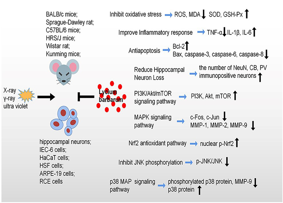
Fig. 3.

Radioprotective effects and molecular mechanisms of Lycium barbarum against radiation-induce damage. Lycium barbarum shows protective effects against radiation through inhibiting oxidative stress, cell apoptosis, reduce hippocampal neuron loss and improve inflammatory response, specially, modulation of PI3K/Akt/mTOR signaling pathway, MAPK signaling pathway, Nrf2 signaling pathway, JNK signaling pathway and p38 MAP signaling pathway.

5. Discussion and Conclusions

Ischemic stroke, caused by a decrease of blood supply to a certain region of the brain due to obstruction of a blood vessel, is the second leading cause of death worldwide [11]. In addition, there is a shift in the ischemic stroke burden to younger individuals from elderly groups [137]. Despite progress in the understanding of the pathophysiological mechanisms in stroke over the past 30 years, cognitive impairment and depression, the common complications of stroke [138, 139], remains difficult for treatment and rehabilitation. In this paper, we reviewed the molecular mechanism of ischemic stroke, including apoptosis, inflammation, oxidative stress, and various signal pathways. A recent study found that the RANKL genetic variation played an important role in ischemic stroke [140]. Recombinant tissue plasminogen activator, the only Food and Drug Administration approved therapy for ischemic stroke, has important limitations in the treatment of ischemic stroke because of the narrow therapeutic time window time of 4.5 h and the potential risk of hemorrhagic transformation [141, 142]. In recent decades, many studies have focused on the pathophysiology and mechanisms of stroke and found that neuroprotection is a promising strategy for the treatment of stroke [143]. Unfortunately, most neuroprotective drugs have failed to translate to clinical practice [144, 145]. However, the potential impact of sulfonylures in the outcome of type 2 diabetic patients with ischemic stroke has suggested that it can decrease ischemic stroke induced damage [146]. Recently, Traditional Chinese Medicines have attracted more attention due to its effective neuroprotective roles. Unlike Western medicine, the Traditional Chinese Medicines have unique advantages due to their regulatory effects at multiple targets and organs. LBP, as the main active ingredient of Lycium barbarum, has been used for traditional herbal and food supplements for many years in Asiatic countries. Many studies indicated that LBP play an anti-oxidative role in oxidative liver injury [147], and it can protect ganglion cells against retinal ischemic/reperfusion injury [148]. Song et al. [149] found that LBP could reduce glucose deprivation-induced injury in PC-12 cells. Similarly, several studies have suggested that it can limit ischemic stroke injury [64, 149]. Considering the neuroprotective effect of Lycium barbarum on ischemia stroke injury, Lycium barbarum may be used as a potential therapeutic agent for ischemic injury in the future clinical trials.

Radiotherapy kills the tumor cells, but also affects the adjacent normal cells [150], which greatly increases the risk of radiation-induced brain damage. In addition, radiodiagnosis, accidental or environmental high dose/dose rate radiation contamination can cause biological effects, such as behavioral disorders, apoptosis, inflammation, neuronal injury, and oxidative stress [151, 152, 153, 154]. Amifostine, a radioprotective drug used with radiotherapy for patients with head and neck cancer, is limited in its wide application due to its relatively large side effects. Therefore, it is urgent to develop radioprotective drugs with low side effects. Many Traditional Chinese Medicine can improve radiation-induced damage [155]. The neuroprotective effect of LBP has been reported in both in vivo and in-vitro models [43]. LBP improves microglia damage induced by bipolar pulse current by regulating autophagy [156], and Lycium barbarum water extract improves brain trauma induced cognitive impairment by prevention of neuronal apoptosis and promotion of regeneration of hippocampal neurons [157].

Due to many shared neuropathological changes between ischemic stroke and radiation exposure such as neuro-inflammation, neuronal damage, apoptosis, oxidative stress, BBB damage, and impaired neurogenesis, the neuroprotective effect of Lycium barbarum in ischemic stroke may be applied to radiation-induced brain damage. Additional extensive research on the radio-neuroprotective effect of Lycium barbarum will be necessary to determine its role in clinical practice.

Although more and more attention has been paid to ischemic stroke, therapeutic treatments are limited to intravenous thrombolysis with recombinant tissue plasminogen activator [158, 159]. The combined complexity of the brain structure and the timeliness with which patients receive treatment compromise clinical treatment of ischemic stroke [11]. Similarly, the mechanism of radiation-induced brain damage is still unclear and there is no ideal radioprotective drug in clinical practice. Many experimental studies have strongly suggested that Lycium barbarum produces neuroprotective effects on neurological and neuropsychiatric diseases, ischemic stroke and injury induced by radiation exposure. These protective effects are mainly achieved by regulating brain oxidative stress, inflammation and neuronal apoptosis. By preventing neuronal loss, less neural pathway will be destroyed, and more newly generated neurons are integrated into the dentate gyrus-related afferent and efferent pathways which will reduce ischemic stroke- and radiation-induced impairment of learning and memory and patients’ symptoms.

The molecular mechanisms and neuroprotective effect of Lyceum barbarum in lacunar versus non-lacunar acute ischemic stroke will need further study, since the pathophysiology, prognosis and clinical features of acute small-vessel ischemic strokes are different from other types of cerebral infarcts [160]. This review provides evidence that Lycium barbarum treatment might be a promising treatment for the protection of the brain against acute ischemic stroke injury in humans. The radioprotective effect of Lycium barbarum on other organs such as skin, eye, reproductive systems and the intestine may also apply to the brain. Further studies on the radio-neuroprotective effect of Lycium barbarum on other neurodegenerative disorders may provide the evidence needed to use Lycium barbarum as a supplementary treatment after radiotherapy to prevent acute radiation exposure-induced chronic brain damage.

In this review paper, only limited numbers of studies on the radio-neuroprotective role of Lycium barbarum were available. Since ischemic stroke and radiation exposure share many similar neuropathological changes, it is predicted that further study will provide promising results to confirm the radio-neuro-protective role of Lycium barbarum to translate it for clinical use.

Abbreviations

CT, computed tomography; PET, positron emission tomography; RPT, radiopharmaceutical therapy; TCMs, Traditional Chinese Medicines; LBP, Lycium barbarum polysaccharide; Cyt-C, cytochrome C; AIF,apoptosis inducing factor; PARP, poly (ADP-ribose) polymerase; MCAO, middle cerebral artery occlusion; LBP, Lycium barbarum polysaccharide; Bcl-2, B-cell lymphoma-2; Bax, Bcl-2-associated X; NF-κB, nuclear factor-κB; IκB, inhibitor of NF-κB; IKK, IκB kinase; TNF, tumour necrosis factor; IL-1β, interleukin-1β; TNF-α, tumour necrosis factor-α; IL-6, interleukin-6; IL-8, interleukin-8; MAPK, mitogen-activated protein kinase; NADPH, nicotinamide adenine dinucleotide phosphate; Nox4, NADPH oxidase 4; ROS, reactive oxygen species; SOD, superoxide dismutase; GSH-Px, glutathione peroxidase; CAT, catalase; MDA, malondialdehyde; BBB, blood-brain barrier; WB, western blotting; AQP4, aquaporin protein-4; MMPs, matrix metalloproteinases; MMP-9, matrix metalloproteinase-9; EB, Evans Blue; IgG, immunoglobulin G; GSK-3, glycogen synthase kinase-3; PI3K, photostatidylinositol-3-kinase; Akt, protein kinase B; Mcl-1, myeloid cell leukemia-1; qRT-PCR, real-time quantitative reverse transcription polymerase chain reaction; p-, phosphorylated; p-PDK-1, phosphorylated 3-phosphoinositide-dependent protein kinase 1; PP2A, protein phosphatase 2A; mTOR, mammalian target of rapamycin; LC3-II, microtubule-associated protein 1 light chain 3 II; LC3- I, microtubule-associated protein 1 light chain 3 I; PKC, protein kinase C; Nrf2, nuclear factor erythroid 2-related factor 2; HO-1, heme oxygenase-1; SD, Sprague-Dawley; lyA, lyciumamide; LDH, lactate dehydrogenase; Keap1, Kelch-like ECH-associated protein 1; ARE, antioxidant response elements; NR2A, NMDAR 2A; NR2B, NMDAR 2B; PSD-95, postsynaptic density-95; nNOS, neuronal nitric oxide synthase; NO, nitric oxide; Bad, Bcl-2 associated agonist of cell death; CREB, cAMP response element binding protein; BMNC, bone marrow mononuclear cells; IEC-6, intestinal epithelial cell line 6; UV, ultra violet; JNK, c-Jun NH2-terminal kinase; OGD/RP, oxygen and glucose deprivation/reperfusion; SRLM, strata radiatum lacunosum moleculare; SO, stratum oriens; CA1-SP, CA1 stratum pyramidum; DG-SG, stratum granulosum of the dentate gyrus; LBE, the fruits extract of L. barbarum; IEC-6, intestinal epithelial cell line 6; LBPF, Lycium barbarum polysaccharide fraction; HaCaT cells, Immortalized human keratinocytes; HSF, Human skin fibroblast cell line; LPO, lipid peroxide; GSP-PX, glutathione peroxidase; ARPE-19, arising retinal pigment epithelia cell line-19; γH2AX, phosphated histone family 2 A variant; RCE, rat corneal epithelial; FGF2, fibroblast growth factor-2.

Author Contributions

YH, XZ and LC contributed to the acquisition and interpretation of data, they contributed equally. BXR and FRT contributed to the conception of the research and the revision of key elements.

Ethics Approval and Consent to Participate

Not applicable.

Acknowledgment

Not applicable.

Funding

This research was funded by the following research grants from the National Natural Science Foundation of China (Grant no. 81772223) to BXR. Grants from National Research Foundation of Singapore to Singapore Nuclear Research and Safety Initiative (FRT).

Conflict of Interest

The authors declare no conflict of interest.

References
[1]
Feske SK. Ischemic Stroke. The American Journal of Medicine. 2021; 134: 1457–1464.
[2]
Zhou Y, Liao J, Mei Z, Liu X, Ge J. Insight into Crosstalk between Ferroptosis and Necroptosis: Novel Therapeutics in Ischemic Stroke. Oxidative Medicine and Cellular Longevity. 2021; 2021: 9991001.
[3]
Wang YJ, Li ZX, Gu HQ, Zhai Y, Zhou Q, Jiang Y, et al. China Stroke Statistics: an update on the 2019 report from the National Center for Healthcare Quality Management in Neurological Diseases, China National Clinical Research Center for Neurological Diseases, the Chinese Stroke Association, National Center for Chronic and Non-communicable Disease Control and Prevention, Chinese Center for Disease Control and Prevention and Institute for Global Neuroscience and Stroke Collaborations. Stroke and Vascular Neurology. 2022; 7: 415–450.
[4]
Paul S, Candelario-Jalil E. Emerging neuroprotective strategies for the treatment of ischemic stroke: An overview of clinical and preclinical studies. Experimental Neurology. 2021; 335: 113518.
[5]
Broussalis E, Killer M, McCoy M, Harrer A, Trinka E, Kraus J. Current therapies in ischemic stroke. Part A. Recent developments in acute stroke treatment and in stroke prevention. Drug Discovery Today. 2012; 17: 296–309.
[6]
Paul SL, Srikanth VK, Thrift AG. The large and growing burden of stroke. Current Drug Targets. 2007; 8: 786–793.
[7]
Qin C, Yang S, Chu YH, Zhang H, Pang XW, Chen L, et al. Signaling pathways involved in ischemic stroke: molecular mechanisms and therapeutic interventions. Signal Transduction and Targeted Therapy. 2022; 7: 215.
[8]
Boyle PA, Yang J, Yu L, Leurgans SE, Capuano AW, Schneider JA, et al. Varied effects of age-related neuropathologies on the trajectory of late life cognitive decline. Brain. 2017; 140: 804–812.
[9]
Buchman AS, Yu L, Boyle PA, Levine SR, Nag S, Schneider JA, et al. Microvascular brain pathology and late-life motor impairment. Neurology. 2013; 80: 712–718.
[10]
Hogan AM, Pit-ten Cate IM, Vargha-Khadem F, Prengler M, Kirkham FJ. Physiological correlates of intellectual function in children with sickle cell disease: hypoxaemia, hyperaemia and brain infarction. Developmental Science. 2006; 9: 379–387.
[11]
Woodruff TM, Thundyil J, Tang SC, Sobey CG, Taylor SM, Arumugam TV. Pathophysiology, treatment, and animal and cellular models of human ischemic stroke. Molecular Neurodegeneration. 2011; 6: 11.
[12]
Zamanian JL, Xu L, Foo LC, Nouri N, Zhou L, Giffard RG, et al. Genomic analysis of reactive astrogliosis. The Journal of Neuroscience. 2012; 32: 6391–6410.
[13]
Eltzschig HK, Eckle T. Ischemia and reperfusion–from mechanism to translation. Nature Medicine. 2011; 17: 1391–1401.
[14]
Barthels D, Das H. Current advances in ischemic stroke research and therapies. Biochimica et Biophysica Acta. Molecular Basis of Disease. 2020; 1866: 165260.
[15]
Li Y, Li S, Li D. Breviscapine Alleviates Cognitive Impairments Induced by Transient Cerebral Ischemia/Reperfusion through Its Anti-Inflammatory and Anti-Oxidant Properties in a Rat Model. ACS Chemical Neuroscience. 2020; 11: 4489–4498.
[16]
Liu B, Li F, Shi J, Yang D, Deng Y, Gong Q. Gastrodin ameliorates subacute phase cerebral ischemia reperfusion injury by inhibiting inflammation and apoptosis in rats. Molecular Medicine Reports. 2016; 14: 4144–4152.
[17]
Zhu T, Meng XB, Dong DX, Zhao LY, Qu MW, Sun GB, et al. Xuesaitong injection (lyophilized) combined with aspirin and clopidogrel protect against focal cerebral ischemic/reperfusion injury in rats by suppressing oxidative stress and inflammation and regulating the NOX2/IL-6/STAT3 pathway. Annals of Palliative Medicine. 2021; 10: 1650–1667.
[18]
Zhu T, Wang L, Xie W, Meng X, Feng Y, Sun G, et al. Notoginsenoside R1 Improves Cerebral Ischemia/Reperfusion Injury by Promoting Neurogenesis via the BDNF/Akt/CREB Pathway. Frontiers in Pharmacology. 2021; 12: 615998.
[19]
Zhu T, Fang BY, Meng XB, Zhang SX, Wang H, Gao G, et al. Folium Ginkgo extract and tetramethylpyrazine sodium chloride injection (Xingxiong injection) protects against focal cerebral ischaemia/reperfusion injury via activating the Akt/Nrf2 pathway and inhibiting NLRP3 inflammasome activation. Pharmaceutical Biology. 2022; 60: 195–205.
[20]
Zhu T, Wang L, Feng Y, Sun G, Sun X. Classical Active Ingredients and Extracts of Chinese Herbal Medicines: Pharmacokinetics, Pharmacodynamics, and Molecular Mechanisms for Ischemic Stroke. Oxidative Medicine and Cellular Longevity. 2021; 2021: 8868941.
[21]
Wilke C, Grosshans D, Duman J, Brown P, Li J. Radiation-induced cognitive toxicity: pathophysiology and interventions to reduce toxicity in adults. Neuro-Oncology. 2018; 20: 597–607.
[22]
Marazziti D, Baroni S, Catena-Dell’Osso M, Schiavi E, Ceresoli D, Conversano C, et al. Cognitive, psychological and psychiatric effects of ionizing radiation exposure. Current Medicinal Chemistry. 2012; 19: 1864–1869.
[23]
Loganovsky KN, Fedirko PA, Kuts KV, Marazziti D, Antypchuk KY, Perchuk IV, et al. Brain and eye as potential targets for ionizing radiation impact. Part І. the consequences of irradiation of the participants of the liquidation of the chornobyl accident. Problemy Radiatsiinoi Medytsyny Ta Radiobiolohii. 2020; 25: 90–129.
[24]
Tang FR, Loke WK, Wong P, Khoo BC. Radioprotective effect of ursolic acid in radiation-induced impairment of neurogenesis, learning and memory in adolescent BALB/c mouse. Physiology & Behavior. 2017; 175: 37–46.
[25]
Wang SW, Ren BX, Qian F, Luo XZ, Tang X, Peng XC, et al. Radioprotective effect of epimedium on neurogenesis and cognition after acute radiation exposure. Neuroscience Research. 2019; 145: 46–53.
[26]
Liang B, Peng L, Li R, Li H, Mo Z, Dai X, et al. Lycium barbarum polysaccharide protects HSF cells against ultraviolet-induced damage through the activation of Nrf2. Cellular & Molecular Biology Letters. 2018; 23: 18.
[27]
Hsieh FC, Hung CT, Cheng KC, Wu CY, Chen YC, Wu YJ, et al. Protective Effects of Lycium barbarum Extracts on UVB-Induced Damage in Human Retinal Pigment Epithelial Cells Accompanied by Attenuating ROS and DNA Damage. Oxidative Medicine and Cellular Longevity. 2018; 2018: 4814928.
[28]
Bhat SA, Sood A, Shukla R, Hanif K. AT2R Activation Prevents Microglia Pro-inflammatory Activation in a NOX-Dependent Manner: Inhibition of PKC Activation and p47phox Phosphorylation by PP2A. Molecular Neurobiology. 2019; 56: 3005–3023.
[29]
Corson TW, Crews CM. Molecular understanding and modern application of traditional medicines: triumphs and trials. Cell. 2007; 130: 769–774.
[30]
Zhang C, Teng F, Tu J, Zhang D. Ultrasound-enhanced protective effect of tetramethylpyrazine against cerebral ischemia/reperfusion injury. PLoS ONE. 2014; 9: e113673.
[31]
Chan HHL, Lam HI, Choi KY, Li SZC, Lakshmanan Y, Yu WY, et al. Delay of cone degeneration in retinitis pigmentosa using a 12-month treatment with Lycium barbarum supplement. Journal of Ethnopharmacology. 2019; 236: 336–344.
[32]
Yao X, Peng Y, Xu LJ, Li L, Wu QL, Xiao PG. Phytochemical and biological studies of Lycium medicinal plants. Chemistry & Biodiversity. 2011; 8: 976–1010.
[33]
Duan W, Zhang Z, Zhu J, Zhang D, Qian D, Teng F, et al. Comparative Analysis of the Phenolic Profile of Lycium barbarum L. Fruits from Different Regions in China. Molecules. 2022; 27: 5842.
[34]
Zhou W, Yang T, Xu W, Huang Y, Ran L, Yan Y, et al. The polysaccharides from the fruits of Lycium barbarum L. confer anti-diabetic effect by regulating gut microbiota and intestinal barrier. Carbohydrate Polymers. 2022; 291: 119626.
[35]
Liu CL, Zhang Q, Zhang SH, Mu CL, Yao P, Jiao HY, et al. Lycium barbarum polysaccharide reduces testicular spermatogenic injury in Immp2l-/-mice through GPX4 and AIF pathways. Zhonghua Nan Ke Xue. 2021; 27: 387–393. (In Chinese)
[36]
Andoni E, Curone G, Agradi S, Barbato O, Menchetti L, Vigo D, et al. Effect of Goji Berry (Lycium barbarum) Supplementation on Reproductive Performance of Rabbit Does. Animals. 2021; 11: 1672.
[37]
Yang C, Zhao Q, Li S, Pu L, Yu L, Liu Y, et al. Effects of Lycium barbarum L. Polysaccharides on Vascular Retinopathy: An Insight Review. Molecules. 2022; 27: 5628.
[38]
Au NPB, Kumar G, Asthana P, Gao F, Kawaguchi R, Chang RCC, et al. Clinically relevant small-molecule promotes nerve repair and visual function recovery. NPJ Regenerative Medicine. 2022; 7: 50.
[39]
Toh DWK, Xia X, Sutanto CN, Low JHM, Poh KK, Wang JW, et al. Enhancing the cardiovascular protective effects of a healthy dietary pattern with wolfberry (Lycium barbarum): A randomized controlled trial. The American Journal of Clinical Nutrition. 2021; 114: 80–89.
[40]
Toh DWK, Low JHM, Kim JE. Cardiovascular disease risk reduction with wolfberry consumption: a systematic review and meta-analysis of randomized controlled trials. European Journal of Nutrition. 2022; 61: 1177–1186.
[41]
Han L, Yan Y, Fan M, Gao S, Zhang L, Xiong X, et al. Pt3R5G inhibits colon cancer cell proliferation through inducing ferroptosis by down-regulating SLC7A11. Life Sciences. 2022; 306: 120859.
[42]
Sun L, Zuo C, Liu X, Guo Y, Wang X, Dong Z, et al. Combined Photothermal Therapy and Lycium barbarum Polysaccharide for Topical Administration to Improve the Efficacy of Doxorubicin in the Treatment of Breast Cancer. Pharmaceutics. 2022; 14: 2677.
[43]
Peng XC, Huang JR, Wang SW, Liu L, Liu ZZ, Sethi G, et al. Traditional Chinese Medicine in Neuroprotection after Brain Insults with Special Reference to Radioprotection. Evidence-Based Complementary and Alternative Medicine. 2018; 2018: 2767208.
[44]
Tian X, Liang T, Liu Y, Ding G, Zhang F, Ma Z. Extraction, Structural Characterization, and Biological Functions of Lycium Barbarum Polysaccharides: A Review. Biomolecules. 2019; 9: 389.
[45]
Cao S, Du J, Hei Q. Lycium barbarum polysaccharide protects against neurotoxicity via the Nrf2-HO-1 pathway. Experimental and Therapeutic Medicine. 2017; 14: 4919–4927.
[46]
Zheng G, Ren H, Li H, Li X, Dong T, Xu S, et al. Lycium barbarum polysaccharide reduces hyperoxic acute lung injury in mice through Nrf2 pathway. Biomedicine & Pharmacotherapy. 2019; 111: 733–739.
[47]
Kwok SS, Bu Y, Lo ACY, Chan TCY, So KF, Lai JSM, et al. A Systematic Review of Potential Therapeutic Use of Lycium Barbarum Polysaccharides in Disease. BioMed Research International. 2019; 2019: 4615745.
[48]
Shi Z, Wu D, Yao JP, Yao X, Huang Z, Li P, et al. Protection against Oxygen-Glucose Deprivation/Reperfusion Injury in Cortical Neurons by Combining Omega-3 Polyunsaturated Acid with Lyciumbarbarum Polysaccharide. Nutrients. 2016; 8: 41.
[49]
Guo L, Du QQ, Cheng PQ, Yang TT, Xing CQ, Luo XZ, et al. Neuroprotective Effects of Lycium barbarum Berry on Neurobehavioral Changes and Neuronal Loss in the Hippocampus of Mice Exposed to Acute Ionizing Radiation. Dose-response. 2021; 19: 15593258211057768.
[50]
Cagalinec M, Safiulina D, Liiv M, Liiv J, Choubey V, Wareski P, et al. Principles of the mitochondrial fusion and fission cycle in neurons. Journal of Cell Science. 2013; 126: 2187–2197.
[51]
Lv J, Guan W, You Q, Deng L, Zhu Y, Guo K, et al. RIPC provides neuroprotection against ischemic stroke by suppressing apoptosis via the mitochondrial pathway. Scientific Reports. 2020; 10: 5361.
[52]
Kuwana T, Mackey MR, Perkins G, Ellisman MH, Latterich M, Schneiter R, et al. Bid, Bax, and lipids cooperate to form supramolecular openings in the outer mitochondrial membrane. Cell. 2002; 111: 331–342.
[53]
Love S. Apoptosis and brain ischaemia. Progress in Neuro-psychopharmacology & Biological Psychiatry. 2003; 27: 267–282.
[54]
Sugawara T, Fujimura M, Noshita N, Kim GW, Saito A, Hayashi T, et al. Neuronal death/survival signaling pathways in cerebral ischemia. NeuroRx. 2004; 1: 17–25.
[55]
Endres M, Wang ZQ, Namura S, Waeber C, Moskowitz MA. Ischemic brain injury is mediated by the activation of poly(ADP-ribose)polymerase. Journal of Cerebral Blood Flow and Metabolism. 1997; 17: 1143–1151.
[56]
Boulares AH, Yakovlev AG, Ivanova V, Stoica BA, Wang G, Iyer S, et al. Role of poly(ADP-ribose) polymerase (PARP) cleavage in apoptosis. Caspase 3-resistant PARP mutant increases rates of apoptosis in transfected cells. The Journal of Biological Chemistry. 1999; 274: 22932–22940.
[57]
Wu CX, Wang TF, Yu JQ. Lycium barbarum Polysaccharide Pretreatment Attenuates Cerebral Ischemic Reperfusion Injury by Inhibiting Apoptosis in Mice. Journal of Chinese Medicinal Materials. 2015; 38: 1454–1459. (In Chinese)
[58]
Li XY, Pan YT, Li YH. Neuroprotective effect and its mechanism of lycium barbarum polysaccharide on cerebral ischemia-reperfusion injury in mice. The Chinese Journal of Clinical Pharmacology. 2017; 33: 2054–2057. (In Chinese).
[59]
Wang T, Li Y, Wang Y, Zhou R, Ma L, Hao Y, et al. Lycium barbarum polysaccharide prevents focal cerebral ischemic injury by inhibiting neuronal apoptosis in mice. PLoS ONE. 2014; 9: e90780.
[60]
Zhang JH, Zhao ZY, Zhang H. Role of caspase-12 in cerebral neuron apoptosis mediated by ER stress after fetal rat ischemia/reperfusion injury. Chongqing Medicine. 2007; 834–835. (In Chinese)
[61]
Shen Y, Lv B. Study of glycyrrhizin regulating caspase-12 apoptosis signaling pathway of endoplasmic reticulum stress to alleviate the colonic inflammatory response in ulcerative colitis. China Journal of Traditional Chinese Medicine and Pharmacy. 2020; 35: 3872–3877. (In Chinese)
[62]
Zhang H, Song LC, Jia CH, Liu YY, Lv YL. Neuronal apoptosis and expression of caspase-12 mRNA and protein following focal cerebral ischemia-reperfusion in rats. Chinese Pharmacological Bulletin. 2008; 24: 1069–1072.
[63]
Wang X, Chen LF, Tan CH, Xie DX. Protective Effect of Lycium barbarum Polysaccharides on Cerebral Ischemia-reperfusion Injury Model Rats. China Pharmacy. 2014; 25: 1365–1367. (In Chinese)
[64]
Shi Z, Zhu L, Li T, Tang X, Xiang Y, Han X, et al. Neuroprotective Mechanisms of Lycium barbarum Polysaccharides Against Ischemic Insults by Regulating NR2B and NR2A Containing NMDA Receptor Signaling Pathways. Frontiers in Cellular Neuroscience. 2017; 11: 288.
[65]
Ge JB, Lu HJ, Song XJ, Li M, Chen DD, Wu F. Protective effects of LBP on cerebral ischemia reperfusion injury in mice and mechanism of inhibiting NF-κB, TNF-α, IL-6 and IL-1β. China Journal of Chinese Materia Medica. 2017; 42: 326–331. (In Chinese)
[66]
Zhao P, Zhou R, Zhu XY, Liu G, Zhao YP, Ma PS, et al. Neuroprotective Effects of Lycium barbarum Polysaccharide on Focal Cerebral Ischemic Injury in Mice. Neurochemical Research. 2017; 42: 2798–2813.
[67]
Ge J, Lu H, Song X. Neuroprotection of LBP on a mouse model of transient focal cerebral ischemia and its protective mechanisms of inhibiting oxidative stress. Journal of Apoplexy and Nervous Diseases. 2016; 33: 790–794. (In Chinese)
[68]
Wang HB, Li YX, Hao YJ, Wang TF, Lei Z, Wu Y, et al. Neuroprotective effects of LBP on brain ischemic reperfusion neurodegeneration. European Review for Medical and Pharmacological Sciences. 2013; 17: 2760–2765.
[69]
Hao YJ. Effects of lycium barbarum polysaccharide on oxidative stress after cerebral ischemic and reperfusion injury in mouse. Ningxia Medical Journal. 2012; 34: 1069–1071. (In Chinese)
[70]
Yang D, Li SY, Yeung CM, Chang RCC, So KF, Wong D, et al. Lycium barbarum extracts protect the brain from blood-brain barrier disruption and cerebral edema in experimental stroke. PLoS ONE. 2012; 7: e33596.
[71]
Zhao Q, Jing YM, He MT, Jing L, Xi YF, Zhang JZ. Lycium Barbarum polysaccharides ameliorates hyperglycemia-exacerbated cerebral ischemia/reperfusion injury via protecting blood-brain barrier. Transplant Immunology. 2023; 76: 101757.
[72]
Zhu XY. The research on Lycium barbarum Polysaccharides and its Mechanism involved in PI3K/Akt-GSK3β pathway on mice brain after focal cerebral ischemia. Ningxia Medical University Thesis for Application of Master’s Degree. 2017. (In Chinese)
[73]
Gao K, Liu M, Ding Y, Yao M, Zhu Y, Zhao J, et al. A phenolic amide (LyA) isolated from the fruits of Lycium barbarum protects against cerebral ischemia-reperfusion injury via PKCε/Nrf2/HO-1 pathway. Aging. 2019; 11: 12361–12374.
[74]
Wu W. Effects of Lycium barbarum Polysaccharides on Keap1-Nrf2/HO-1 pathway after Cerebral Ischemic Injury. Ningxia Medical University. 2018. (In Chinese)
[75]
Harari OA, Liao JK. NF-κB and innate immunity in ischemic stroke. Annals of the New York Academy of Sciences. 2010; 1207: 32–40.
[76]
Ridder DA, Schwaninger M. NF-kappaB signaling in cerebral ischemia. Neuroscience. 2009; 158: 995–1006.
[77]
Tu XK, Yang WZ, Chen JP, Chen Y, Ouyang LQ, Xu YC, et al. Curcumin inhibits TLR2/4-NF-κB signaling pathway and attenuates brain damage in permanent focal cerebral ischemia in rats. Inflammation. 2014; 37: 1544–1551.
[78]
Zhu H, Hu S, Li Y, Sun Y, Xiong X, Hu X, et al. Interleukins and Ischemic Stroke. Frontiers in Immunology. 2022; 13: 828447.
[79]
Murata Y, Fujiwara N, Seo JH, Yan F, Liu X, Terasaki Y, et al. Delayed inhibition of c-Jun N-terminal kinase worsens outcomes after focal cerebral ischemia. The Journal of Neuroscience. 2012; 32: 8112–8115.
[80]
Liu ZC, Yu WW, Zhou HC, Lan ZC, Wu T, Xiong SM, et al. Lycium barbarum polysaccharides ameliorate LPS-induced inflammation of RAW264.7 cells and modify the behavioral score of peritonitis mice. Journal of Food Biochemistry. 2021; 45: e13889.
[81]
Zheng Y, Pang X, Zhu X, Meng Z, Chen X, Zhang J, et al. Lycium barbarum mitigates radiation injury via regulation of the immune function, gut microbiota, and related metabolites. Biomedicine & Pharmacotherapy. 2021; 139: 111654.
[82]
Duan Y, Chen F, Yao X, Zhu J, Wang C, Zhang J, et al. Protective Effect of Lycium ruthenicum Murr. Against Radiation Injury in Mice. International Journal of Environmental Research and Public Health. 2015; 12: 8332–8347.
[83]
Zhou J, Pang H, Li W, Liu Q, Xu L, Liu Q, et al. Effects of Lycium barbarum Polysaccharides on Apoptosis, Cellular Adhesion, and Oxidative Damage in Bone Marrow Mononuclear Cells of Mice Exposed to Ionizing Radiation Injury. BioMed Research International. 2016; 2016: 4147879.
[84]
Luo Q, Li J, Cui X, Yan J, Zhao Q, Xiang C. The effect of Lycium barbarum polysaccharides on the male rats׳ reproductive system and spermatogenic cell apoptosis exposed to low-dose ionizing irradiation. Journal of Ethnopharmacology. 2014; 154: 249–258.
[85]
Neves LMG, Tim CR, Floriano EM, da Silva de Avó LR, Fernandes JB, Parizotto NA, et al. Lycium barbarum polysaccharide fraction associated with photobiomodulation protects from epithelium thickness and collagen fragmentation in a model of cutaneous photodamage. Lasers in Medical Science. 2021; 36: 863–870.
[86]
Li H, Li Z, Peng L, Jiang N, Liu Q, Zhang E, et al. Lycium barbarum polysaccharide protects human keratinocytes against UVB-induced photo-damage. Free Radical Research. 2017; 51: 200–210.
[87]
Neves LMG, Parizotto NA, Tim CR, Floriano EM, Lopez RFV, Venâncio T, et al. Polysaccharide-rich hydrogel formulation combined with photobiomodulation repairs UV-induced photodamage in mice skin. Wound Repair and Regeneration. 2020; 28: 645–655.
[88]
Jia YE, Ren LC, Yan HL, Dermatology DO. Effects of Black Wolfberry Water Extracts on Proliferation and Apoptosis and Apoptosis-related-protein ExpressionLevel of HaCaT Cells after UVB Radiation. Chinese General Practice. 2017; 20: 3400–3404. (In Chinese)
[89]
Du S, Han B, Li K, Zhang X, Sha X, Gao L. Lycium barbarum Polysaccharides Protect Rat Corneal Epithelial Cells against Ultraviolet B-Induced Apoptosis by Attenuating the Mitochondrial Pathway and Inhibiting JNK Phosphorylation. BioMed Research International. 2017; 2017: 5806832.
[90]
Pacher P, Beckman JS, Liaudet L. Nitric oxide and peroxynitrite in health and disease. Physiological Reviews. 2007; 87: 315–424.
[91]
Moloney JN, Cotter TG. ROS signalling in the biology of cancer. Seminars in Cell & Developmental Biology. 2018; 80: 50–64.
[92]
Huang D, Siaw-Debrah F, Wang H, Ye S, Wang K, Wu K, et al. Transplanting Rac1-silenced bone marrow mesenchymal stem cells promote neurological function recovery in TBI mice. Aging. 2020; 13: 2822–2850.
[93]
Zhang L, Wu J, Duan X, Tian X, Shen H, Sun Q, et al. NADPH Oxidase: A Potential Target for Treatment of Stroke. Oxidative Medicine and Cellular Longevity. 2016; 2016: 5026984.
[94]
Chen H, Song YS, Chan PH. Inhibition of NADPH oxidase is neuroprotective after ischemia-reperfusion. Journal of Cerebral Blood Flow and Metabolism. 2009; 29: 1262–1272.
[95]
Liu Y, Zhang L, Liang J. Activation of the Nrf2 defense pathway contributes to neuroprotective effects of phloretin on oxidative stress injury after cerebral ischemia/reperfusion in rats. Journal of the Neurological Sciences. 2015; 351: 88–92.
[96]
Omidifar N, Nili-Ahmadabadi A, Nakhostin-Ansari A, Lankarani KB, Moghadami M, Mousavi SM, et al. The modulatory potential of herbal antioxidants against oxidative stress and heavy metal pollution: plants against environmental oxidative stress. Environmental Science and Pollution Research International. 2021; 28: 61908–61918.
[97]
Li G, Ye C, Zhu Y, Zhang T, Gu J, Pan J, et al. Oxidative Injury in Ischemic Stroke: A Focus on NADPH Oxidase 4. Oxidative Medicine and Cellular Longevity. 2022; 2022: 1148874.
[98]
Turner RJ, Sharp FR. Implications of MMP9 for Blood Brain Barrier Disruption and Hemorrhagic Transformation Following Ischemic Stroke. Frontiers in Cellular Neuroscience. 2016; 10: 56.
[99]
Abdullahi W, Tripathi D, Ronaldson PT. Blood-brain barrier dysfunction in ischemic stroke: targeting tight junctions and transporters for vascular protection. American Journal of Physiology. Cell Physiology. 2018; 315: C343–C356.
[100]
LI Wen qian LHq, PENG Yong jun, XU Shu ying, WU Xu. Currents situation of blood - brain barrier dysfunction after ischemic stroke. The Chinese Journal of Clinical Pharmacology. 2020; 36: 3533–3637. (In Chinese)
[101]
Manley GT, Fujimura M, Ma T, Noshita N, Filiz F, Bollen AW, et al. Aquaporin-4 deletion in mice reduces brain edema after acute water intoxication and ischemic stroke. Nature Medicine. 2000; 6: 159–163.
[102]
Taniguchi M, Yamashita T, Kumura E, Tamatani M, Kobayashi A, Yokawa T, et al. Induction of aquaporin-4 water channel mRNA after focal cerebral ischemia in rat. Brain Research. Molecular Brain Research. 2000; 78: 131–137.
[103]
Chaturvedi M, Kaczmarek L. Mmp-9 inhibition: a therapeutic strategy in ischemic stroke. Molecular Neurobiology. 2014; 49: 563–573.
[104]
Embi N, Rylatt DB, Cohen P. Glycogen synthase kinase-3 from rabbit skeletal muscle. Separation from cyclic-AMP-dependent protein kinase and phosphorylase kinase. European Journal of Biochemistry. 1980; 107: 519–527.
[105]
Kumar P, Miller AI, Polverini PJ. p38 MAPK mediates gamma-irradiation-induced endothelial cell apoptosis, and vascular endothelial growth factor protects endothelial cells through the phosphoinositide 3-kinase-Akt-Bcl-2 pathway. The Journal of Biological Chemistry. 2004; 279: 43352–43360.
[106]
Liu RL, Xiong QJ, Shu Q, Wu WN, Cheng J, Fu H, et al. Hyperoside protects cortical neurons from oxygen-glucose deprivation-reperfusion induced injury via nitric oxide signal pathway. Brain Research. 2012; 1469: 164–173.
[107]
Tao RR, Ji YL, Lu YM, Fukunaga K, Han F. Targeting nitrosative stress for neurovascular protection: new implications in brain diseases. Current Drug Targets. 2012; 13: 272–284.
[108]
Ding X. Research on the mechanism of the inhibitory effects of TMP on rat focal cerebral ischemia-reperfusion injury. Nanjing University of Chinese Medicine Doctoral Thesis. 2007. (In Chinese)
[109]
Eldar-Finkelman H, Martinez A. GSK-3 Inhibitors: Preclinical and Clinical Focus on CNS. Frontiers in Molecular Neuroscience. 2011; 4: 32.
[110]
Zhang X, Shi M, Ye R, Wang W, Liu X, Zhang G, et al. Ginsenoside Rd attenuates tau protein phosphorylation via the PI3K/AKT/GSK-3β pathway after transient forebrain ischemia. Neurochemical Research. 2014; 39: 1363–1373.
[111]
Wang Z, Zhou L, Zheng X, Chen G, Pan R, Li J, et al. Autophagy protects against PI3K/Akt/mTOR-mediated apoptosis of spinal cord neurons after mechanical injury. Neuroscience Letters. 2017; 656: 158–164.
[112]
Lee JH, Kim C, Um JY, Sethi G, Ahn KS. Casticin-Induced Inhibition of Cell Growth and Survival Are Mediated through the Dual Modulation of Akt/mTOR Signaling Cascade. Cancers. 2019; 11: 254.
[113]
Li S, Haigh K, Haigh JJ, Vasudevan A. Endothelial VEGF sculpts cortical cytoarchitecture. The Journal of Neuroscience. 2013; 33: 14809–14815.
[114]
Chong ZZ, Yao Q, Li HH. The rationale of targeting mammalian target of rapamycin for ischemic stroke. Cellular Signalling. 2013; 25: 1598–1607.
[115]
Choudhury GR, Ding S. Reactive astrocytes and therapeutic potential in focal ischemic stroke. Neurobiology of Disease. 2016; 85: 234–244.
[116]
Chen J, Zhang X, Liu X, Zhang C, Shang W, Xue J, et al. Ginsenoside Rg1 promotes cerebral angiogenesis via the PI3K/Akt/mTOR signaling pathway in ischemic mice. European Journal of Pharmacology. 2019; 856: 172418.
[117]
Huang HC, Nguyen T, Pickett CB. Regulation of the antioxidant response element by protein kinase C-mediated phosphorylation of NF-E2-related factor 2. Proceedings of the National Academy of Sciences of the United States of America. 2000; 97: 12475–12480.
[118]
Huang HC, Nguyen T, Pickett CB. Phosphorylation of Nrf2 at Ser-40 by protein kinase C regulates antioxidant response element-mediated transcription. The Journal of Biological Chemistry. 2002; 277: 42769–42774.
[119]
Ge M, Yao W, Yuan D, Zhou S, Chen X, Zhang Y, et al. Brg1-mediated Nrf2/HO-1 pathway activation alleviates hepatic ischemia-reperfusion injury. Cell Death & Disease. 2017; 8: e2841.
[120]
Ding Y, Chen M, Wang M, Li Y, Wen A. Posttreatment with 11-Keto-β-Boswellic Acid Ameliorates Cerebral Ischemia-Reperfusion Injury: Nrf2/HO-1 Pathway as a Potential Mechanism. Molecular Neurobiology. 2015; 52: 1430–1439.
[121]
Chen KL, Dong WW. Effect of Dengzhanhua on Neuronal Apoptosis Induced by Cerebral Ischemia. Chinese Journal of Clinical Neurosciences. 2000; 5–7. (In Chinese)
[122]
Zhi SM, Fang GX, Xie XM, Liu LH, Yan J, Liu DB, et al. Melatonin reduces OGD/R-induced neuron injury by regulating redox/inflammation/apoptosis signaling. European Review for Medical and Pharmacological Sciences. 2020; 24: 1524–1536.
[123]
Kang MI, Kobayashi A, Wakabayashi N, Kim SG, Yamamoto M. Scaffolding of Keap1 to the actin cytoskeleton controls the function of Nrf2 as key regulator of cytoprotective phase 2 genes. Proceedings of the National Academy of Sciences of the United States of America. 2004; 101: 2046–2051.
[124]
Yang Y, Jiang S, Yan J, Li Y, Xin Z, Lin Y, et al. An overview of the molecular mechanisms and novel roles of Nrf2 in neurodegenerative disorders. Cytokine & Growth Factor Reviews. 2015; 26: 47–57.
[125]
Itoh K, Wakabayashi N, Katoh Y, Ishii T, Igarashi K, Engel JD, et al. Keap1 represses nuclear activation of antioxidant responsive elements by Nrf2 through binding to the amino-terminal Neh2 domain. Genes & Development. 1999; 13: 76–86.
[126]
Baird L, Dinkova-Kostova AT. The cytoprotective role of the Keap1-Nrf2 pathway. Archives of Toxicology. 2011; 85: 241–272.
[127]
Liu Y, Wong TP, Aarts M, Rooyakkers A, Liu L, Lai TW, et al. NMDA receptor subunits have differential roles in mediating excitotoxic neuronal death both in vitro and in vivo. The Journal of Neuroscience. 2007; 27: 2846–2857.
[128]
Chen M, Lu TJ, Chen XJ, Zhou Y, Chen Q, Feng XY, et al. Differential roles of NMDA receptor subtypes in ischemic neuronal cell death and ischemic tolerance. Stroke. 2008; 39: 3042–3048.
[129]
Sattler R, Xiong Z, Lu WY, Hafner M, MacDonald JF, Tymianski M. Specific coupling of NMDA receptor activation to nitric oxide neurotoxicity by PSD-95 protein. Science. 1999; 284: 1845–1848.
[130]
Wang YL, Liu QF. Effect of Lycium barbarum polysaccharide protecting against apoptosis of hippocampal neurons and on PI3K/Akt/mTOR signaling pathway in rats with radiation-induced brain injury. Chinese Journal of Public Health. 2019; 35: 1043–1045. (In Chinese)
[131]
Guan SZH, De XM, Pang KH, Yang HF. Protective effects of lycium barbarum polysaccharides on spinal cord neurons after radiation injury. Carcinomatous Aberration Mutation. 2019; 31: 45–48. (In Chinese)
[132]
Pang KH. Effect of Lycium Barbarum Polysaccharides on autophagy induced by radiation injury of spinal nerve cells in vitro. Ningxia Medical University. 2018. (In Chinese)
[133]
Tang FR, Loke WK, Khoo BC. Postnatal irradiation-induced hippocampal neuropathology, cognitive impairment and aging. Brain & Development. 2017; 39: 277–293.
[134]
Yang B, Ren BX, Tang FR. Prenatal irradiation-induced brain neuropathology and cognitive impairment. Brain & Development. 2017; 39: 10–22.
[135]
Wang QQ, Yin G, Huang JR, Xi SJ, Qian F, Lee RX, et al. Ionizing Radiation-Induced Brain Cell Aging and the Potential Underlying Molecular Mechanisms. Cells. 2021; 10: 3570.
[136]
Lee RX, Tang FR. Radiation-induced neuropathological changes in the oligodendrocyte lineage with relevant clinical manifestations and therapeutic strategies. International Journal of Radiation Biology. 2022; 98: 1519–1531.
[137]
Stack CA, Cole JW. Ischemic stroke in young adults. Current Opinion in Cardiology. 2018; 33: 594–604.
[138]
Makin SDJ, Turpin S, Dennis MS, Wardlaw JM. Cognitive impairment after lacunar stroke: systematic review and meta-analysis of incidence, prevalence and comparison with other stroke subtypes. Journal of Neurology, Neurosurgery, and Psychiatry. 2013; 84: 893–900.
[139]
Nabavi SF, Habtemariam S, Di Lorenzo A, Sureda A, Khanjani S, Nabavi SM, et al. Post-Stroke Depression Modulation and in Vivo Antioxidant Activity of Gallic Acid and Its Synthetic Derivatives in a Murine Model System. Nutrients. 2016; 8: 248.
[140]
Biscetti F, Giovannini S, Straface G, Bertucci F, Angelini F, Porreca C, et al. RANK/RANKL/OPG pathway: genetic association with history of ischemic stroke in Italian population. European Review for Medical and Pharmacological Sciences. 2016; 20: 4574–4580.
[141]
Xiong XY, Liu L, Yang QW. Refocusing Neuroprotection in Cerebral Reperfusion Era: New Challenges and Strategies. Frontiers in Neurology. 2018; 9: 249.
[142]
Tao T, Liu M, Chen M, Luo Y, Wang C, Xu T, et al. Natural medicine in neuroprotection for ischemic stroke: Challenges and prospective. Pharmacology & Therapeutics. 2020; 216: 107695.
[143]
Li C, Zhao Z, Luo Y, Ning T, Liu P, Chen Q, et al. Macrophage-Disguised Manganese Dioxide Nanoparticles for Neuroprotection by Reducing Oxidative Stress and Modulating Inflammatory Microenvironment in Acute Ischemic Stroke. Advanced Science. 2021; 8: e2101526.
[144]
Minnerup J, Sutherland BA, Buchan AM, Kleinschnitz C. Neuroprotection for stroke: current status and future perspectives. International Journal of Molecular Sciences. 2012; 13: 11753–11772.
[145]
Dávalos A, Alvarez-Sabín J, Castillo J, Díez-Tejedor E, Ferro J, Martínez-Vila E, et al. Citicoline in the treatment of acute ischaemic stroke: an international, randomised, multicentre, placebo-controlled study (ICTUS trial). Lancet. 2012; 380: 349–357.
[146]
Arboix A. Potential impact of sulfonylureas in the outcome of type 2 diabetic patients with ischemic stroke. Stroke. 2007; 38: 2413–2414.
[147]
Wu HT, He XJ, Hong YK, Ma T, Xu YP, Li HH. Chemical characterization of Lycium barbarum polysaccharides and its inhibition against liver oxidative injury of high-fat mice. International Journal of Biological Macromolecules. 2010; 46: 540–543.
[148]
He M, Pan H, Chang RCC, So KF, Brecha NC, Pu M. Activation of the Nrf2/HO-1 antioxidant pathway contributes to the protective effects of Lycium barbarum polysaccharides in the rodent retina after ischemia-reperfusion-induced damage. PLoS ONE. 2014; 9: e84800.
[149]
Song S, Lin F, Zhu P, Wu C, Zhao S, Han Q, et al. Lycium barbarum polysaccharide alleviates oxygen glucose deprivation-induced PC-12 cells damage by up-regulating miR-24. Artificial Cells, Nanomedicine, and Biotechnology. 2019; 47: 3994–4000.
[150]
Liu X, Cotrim A, Teos L, Zheng C, Swaim W, Mitchell J, et al. Loss of TRPM2 function protects against irradiation-induced salivary gland dysfunction. Nature Communications. 2013; 4: 1515.
[151]
Xie LW, Cai S, Zhao TS, Li M, Tian Y. Green tea derivative (-)-epigallocatechin-3-gallate (EGCG) confers protection against ionizing radiation-induced intestinal epithelial cell death both in vitro and in vivo. Free Radical Biology & Medicine. 2020; 161: 175–186.
[152]
Turnquist C, Harris BT, Harris CC. Radiation-induced brain injury: current concepts and therapeutic strategies targeting neuroinflammation. Neuro-Oncology Advances. 2020; 2: vdaa057.
[153]
Gürich HG. New ideas on the pathogenesis and pathologic in anatomy of pancreatitis. Deutsches Medizinisches Journal. 1971; 22: 698–701. (In German)
[154]
Pipová Kokošová N, Kisková T, Vilhanová K, Štafuriková A, Jendželovský R, Račeková E, et al. Melatonin mitigates hippocampal and cognitive impairments caused by prenatal irradiation. The European Journal of Neuroscience. 2020; 52: 3575–3594.
[155]
Zhang X, Chen X, Wang L, He C, Shi Z, Fu Q, et al. Review of the Efficacy and Mechanisms of Traditional Chinese Medicines as a Therapeutic Option for Ionizing Radiation Induced Damage. Frontiers in Pharmacology. 2021; 12: 617559.
[156]
Bie M, Lv Y, Ren C, Xing F, Cui Q, Xiao J, et al. Lycium barbarum polysaccharide improves bipolar pulse current-induced microglia cell injury through modulating autophagy. Cell Transplantation. 2015; 24: 419–428.
[157]
Hu X, Qu Y, Chu Q, Li W, He J. Investigation of the neuroprotective effects of Lycium barbarum water extract in apoptotic cells and Alzheimer’s disease mice. Molecular Medicine Reports. 2018; 17: 3599–3606.
[158]
Macrez R, Ali C, Toutirais O, Le Mauff B, Defer G, Dirnagl U, et al. Stroke and the immune system: from pathophysiology to new therapeutic strategies. The Lancet. Neurology. 2011; 10: 471–480.
[159]
Farina M, Vieira LE, Buttari B, Profumo E, Saso L. The Nrf2 Pathway in Ischemic Stroke: A Review. Molecules. 2021; 26: 5001.
[160]
Rudilosso S, Rodríguez-Vázquez A, Urra X, Arboix A. The Potential Impact of Neuroimaging and Translational Research on the Clinical Management of Lacunar Stroke. International Journal of Molecular Sciences. 2022; 23: 1497.

Publisher’s Note: IMR Press stays neutral with regard to jurisdictional claims in published maps and institutional affiliations.

Share
Back to top