


 , Sara Vitalini 8, Marcello Iriti 9,10,*
1 Department of Forensic Medicine and Toxicology, School of Medicine, Urmia University of Medical Sciences, 5714783734 Urmia, Iran
2 Center for Molecular Biosciences (CMBI), Institute of Pharmacy/Pharmacognosy, University of Innsbruck, 6020 Innsbruck, Austria
3 Department of Biology, Payame Noor University, 19395-4697 Tehran, Iran
4 Department of Pharmaceutical Nanotechnology, School of Pharmacy, Mashhad University of Medical Sciences, 13944-91388 Mashhad, Iran
5 Industrial Biotechnology Research Group, Institute of Biotechnology, Ferdowsi University of Mashhad, 9177948974 Mashhad, Iran
6 Medical Toxicology Research Centre, Mashhad University of Medical Sciences, 917794-8564 Mashhad, Iran
7 Applied Biomedical Research Center, Mashhad University of Medical Sciences, 917794-8564 Mashhad, Iran
8 Department of Food, Environmental and Nutritional Sciences, Università degli Studi di Milano, 20133 Milan, Italy
9 Department of Biomedical, Surgical and Dental Sciences, Università degli Studi di Milano, 20133 Milan, Italy
10 National Interuniversity Consortium of Materials Science and Technology (INSTM), 50121 Firenze, Italy
Academic Editor: Graham Pawelec
Abstract
Background: In the present study, resveratrol was used to prepare
complexes of cerium and nanoceria, also coated with gold (CeO
Keywords
- resveratrol
- nanogold
- cerium oxide
- green synthesis
- cell toxicity
- core-shells
- nanocomposite
Phytoalexins are diverse plant secondary metabolites possessing supportive roles
against biotic and abiotic stressors, whereas they have represented potent
biological properties [1, 2]. Resveratrol (3,4
The cerium oxide (nanoceria, NCs) have demonstrated anticancer activities, due
to its interesting changes in oxidation states, while it can act similar to the
antioxidant enzymes (catalase and superoxide dismutase mimetic activities)
[14, 15, 16], subsequently, showing protective attribute as an active scavenger of
reactive oxygen/nitrogen species (ROS/RNS) with unlimited cycles that involves
changes in oxidation states of Ce
The combination of NCs with gold could enhance the anticancer effects of the NCs
along with the biocompatibility of the particles [27]. Due to the potential
application of gold in different nanoplatforms for the treatment of cancer, as an
active agent or a nanocarrier, it can be a good choice to form nanocomposites
with NCs [28, 29, 30]. The gold inertness and its nontoxic nature are of great use in
the design of new active nanomaterials and nanocomposites for biomedical
applications [31, 32, 33, 34]. The factors that can be crucial in determining the final
anticancer properties can be the size, shape, and ratio of the components [35, 36]. Gold nanoparticles’ toxicity is directly proportional to the size of
nanoparticles. The larger particles are less toxic, and the smaller ones have
substantial toxicity towards cancer cells [37, 38, 39, 40]. Therefore, the NC seeds’ size
influences the bioactivity of CeO
Herein, the complexation of a phytochemical resveratrol and Ce
The following analyses were performed for characterization of the nanoparticles:
powder X-ray diffraction (PXRD), Fourier transforms infrared spectroscopy (FTIR),
transmission electron microscopy (TEM), Dynamic light scattering (DLS), and
To prepare the nanoceria, 0.012 mol resveratrol and 0.004 mol cerium nitrate
hexahydrate (Ce(NO
Liver cancer cell line (HepG2) and human foreskin fibroblasts (HFF) were
attained from Iran’s Pasteur Institute. Cell developmental was performed in RPMI
and DMEM media, supplemented with 100
The PXRD confirmed the formation of gold coated nanoceria (CeO
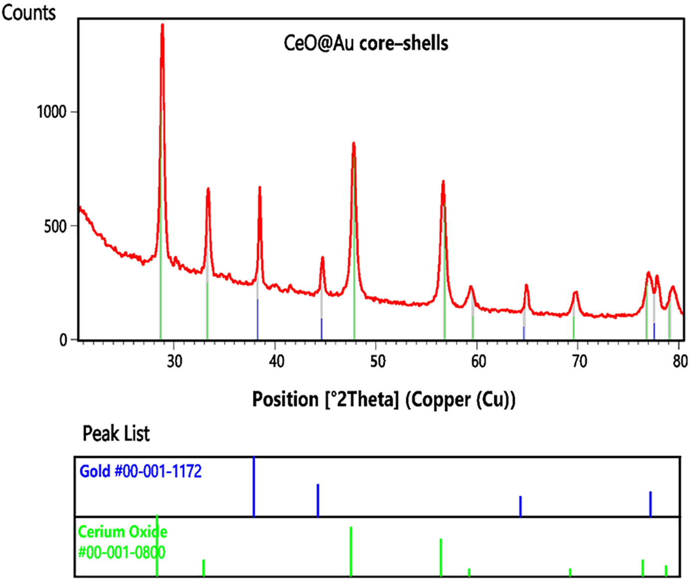 Fig. 1.
The PXRD pattern of CeO
The calculated 2theta value (d space, intensity, HKL) of cerium oxide were 28.68
˚ (3.11 Å, 100.0%, 111), 33.28 ˚ (2.69 Å, 25.0%, 200), 47.83 ˚
(1.90 Å, 80.0%, 220), 56.78 ˚ (1.62 Å, 60.0%, 311), 59.60 ˚ (1.55
Å, 10.0%, 222), 69.58 ˚ (1.35 Å, 10.0%, 400), 76.81 ˚ (1.24
Å, 25.0%, 331), and 79.08 (1.21 Å, 16.0%, 420) and the ones for gold
layer were 38.27 ˚ (2.35 Å, 42.6%, 111), 44.60 ˚ (2.03 Å, 22.6%,
200), 64.68 ˚ (1.44 Å, 14.1%, 220), and 77.55 ˚ (1.23 Å, 17.0%,
311). The comparison of the calculated and experimental data indicated the
successful synthesis of cerium oxide and the reduction of Au
The absorption bands of FTIR between
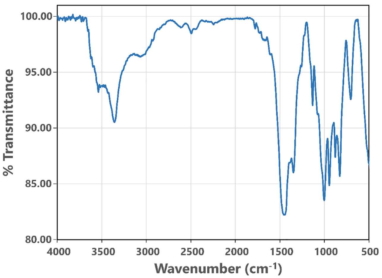 Fig. 2.
The FTIR spectrum of CeO
The TEM analysis was performed to determine the size of CeO
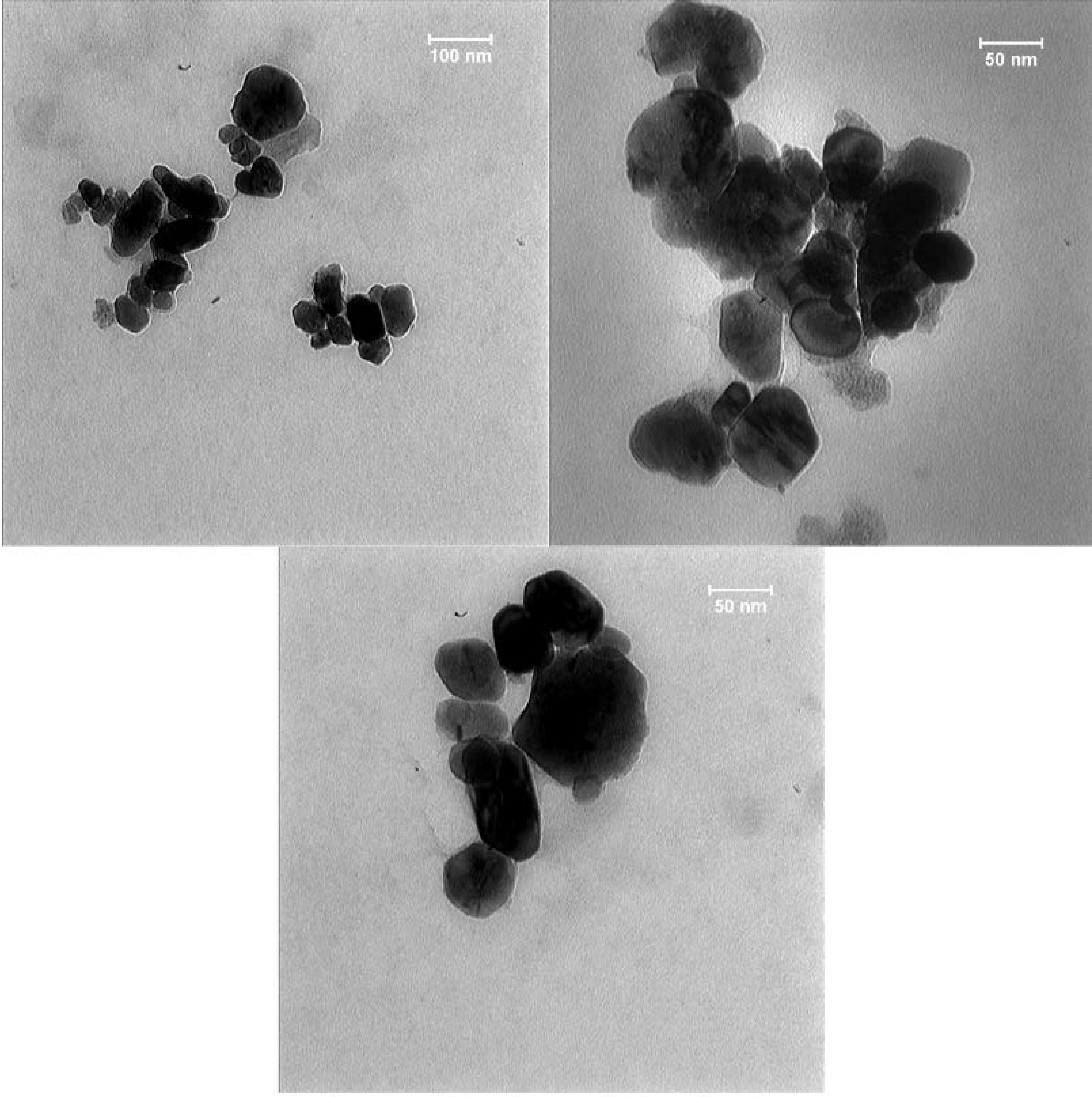 Fig. 3.
The TEM images of CeO
The hydrodynamic size and the partial surface charge of the particles were
measured using the DLS and
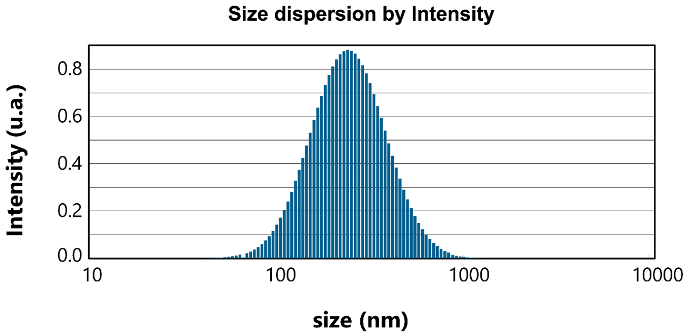 Fig. 4.
The hydrodynamic size of CeO
Nanotechnology-mediated delivery can be employed as a beneficial device in increasing the bioavailability of resveratrol. Nanoscience is an emerging part of investigation that operates at the crossways of biology, physico-chemicals, engineering, pharmacology, and medicine [22, 46, 47, 48]. Designing of NPs (nano particles) for an effective and controlled delivery of anticancer factors is being considered as one of the most important applications of nanoscience [24, 49, 50]. Few experiments have attempted to study potency of resveratrol for the synthesis of nanoparticles. It has been shown that the solid lipid NPs loaded with resveratrol can cross the cell membrane, while it can be enhanced via increasing the exposure time of cells to resveratrol [51]. Another study utilizing bovine serum albumin-bound resveratrol NPs in primary ovarian cancer of mice showed that not only the NP-bound resveratrol was easier to work with because of the improved solubility, it had also a greater influence on prevent of tumor growth, compared to pure resveratrol [52]. The anti-cancer effect of gold nanoparticles against breast, testicular, liver, and lung cancer cells has been proven in a concentration-dependent manner [30, 51, 52, 53, 54, 55]. In another study, the produced gold nanoparticles provided a safe and great system for the delivery of gapmers in cancerous cells, which meaningfully down-regulated mutant p53 proteins and changed molecular markers related to cell growth and apoptosis [56].
The anticancer activity of the produced NC was remarkable, whereas it possessed lower toxic effect on normal cells. The results clearly exhibited the toxicity of NC against cancer cells in a concentration and time dependent manner (Fig. 5). Although resveratrol alone was effective against cancer cells, higher efficacy in toxicity against cancer cell lines was observed in combination with nanoparticles.
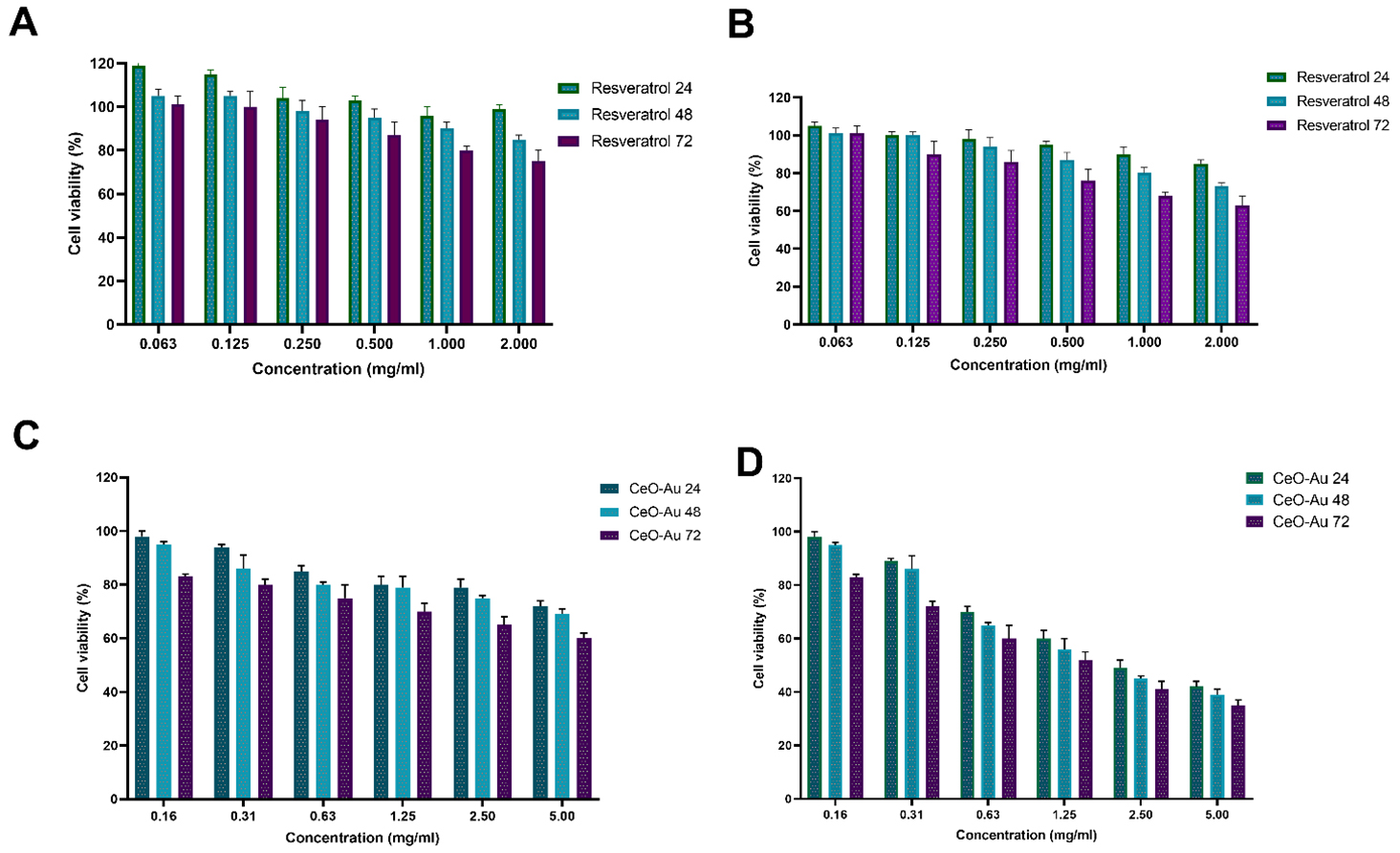 Fig. 5.
Cellular toxicity effect of biosynthesized NCs and resveratrol against HepG2 as a cancerous cell lines (B and D) and HFF as a normal cell lines (A and C).
The nanoceria particles were successfully synthesized using resveratrol to form
complexes as a precursor for further calcination procedure. The synthesized
nanoceria was also coated with a gold layer to increase their anticancer
efficiency. The prepared nanoceria were fully characterized and analyzed through
conventional methods. The experiment has shown the particle size of
Conceptualization, AGA, JM, FN, METY; methodology, MM, AH, AHM, SV; investigation, AGA, JM, MSA, MM, AH, AHM, FN, MN, MQ, METY, MI; project administration, METY, MI.
Not applicable.
The authors are warmly grateful to the Mashhad University of Medical Sciences for support and kindly providing facilities to perform the experiments.
This research received no external funding.
The authors declare no conflict of interest. MI is serving as one of the Editorial Board and Guest Editor of this journal. We declare that MI had no involvement in the peer review of this article and has no access to information regarding its peer review.Full responsibility for the editorial process for this article was delegated to GP.
References
Publisher’s Note: IMR Press stays neutral with regard to jurisdictional claims in published maps and institutional affiliations.