IMR Press / FBL / Volume 27 / Issue 5 / DOI: 10.31083/j.fbl2705146
Open Access Review
Impact of New Drugs for Therapeutic Intervention in Alzheimer’s Disease
Show Less
1 Department of Biochemistry and Physiology, Physiology Section, Faculty of Pharmacy and Food Sciences, University of Barcelona, 08028 Barcelona, Spain
2 Laboratory of Cellular and Molecular Pathology, Institute of Biomedical Sciences, Faculty of Health Sciences, Universidad Autónoma de Chile, 3460000 Talca, Chile
3 Department of Pharmacology, Toxicology and Therapeutic Chemistry, Faculty of Pharmacy and Food Sciences, University of Barcelona, 08028 Barcelona, Spain
4 Institut de Neurociences (INUB), University of Barcelona, 08028 Barcelona, Spain
5 Biomedical Research Networking Centre in Neurodegenerative Diseases (CIBERNED), 28001 Madrid, Spain
6 Department of Pharmacy, Pharmaceutical Technology and Physical Chemistry, Faculty of Pharmacy and Food Sciences, University of Barcelona, 08028 Barcelona, Spain
7 ACE, Alzheimer Center Barcelona, Universitat Internacional de Catalunya (UIC), 08017 Barcelona, Spain
8 Unit of Biochemistry and Pharmacology, Faculty of Medicine and Health Sciences, University of Rovira i Virgili, 43003 Reus (Tarragona), Spain
9 Departamento de Biología Celular y Molecular, Centro Universitario de Ciencias Biológicas y Agropecuarias (CUCBA), Universidad de Guadalajara, 44100 Zapopan, Mexico
10 Department of Cellular Biology, Physiology and Immunology, Faculty of Biology, University of Barcelona, 08028 Barcelona, Spain
*Correspondence: camins@ub.edu (Antoni Camins)
Academic Editor: Graham Pawelec
Front. Biosci. (Landmark Ed) 2022, 27(5), 146; https://doi.org/10.31083/j.fbl2705146
Submitted: 11 December 2021 | Revised: 2 February 2022 | Accepted: 12 February 2022 | Published: 6 May 2022
Copyright: © 2022 The Author(s). Published by IMR Press.
This is an open access article under the CC BY 4.0 license.
Abstract

The increases in population ageing and growth are leading to a boosting in the number of people living with dementia, Alzheimer’s disease (AD) being the most common cause. In spite of decades of intensive research, no cure for AD has been found yet. However, some treatments that may change disease progression and help control symptoms have been proposed. Beyond the classical hypotheses of AD etiopathogenesis, i.e., amyloid beta peptide (Aβ) accumulation and tau hyperphosphorylation, a trend in attributing a key role to other molecular mechanisms is prompting the study of different therapeutic targets. Hence, drugs designed to modulate inflammation, insulin resistance, synapses, neurogenesis, cardiovascular factors and dysbiosis are shaping a new horizon in AD treatment. Within this frame, an increase in the number of candidate drugs for disease modification treatments is expected, as well as a focus on potential combinatory multidrug strategies.The present review summarizes the latest advances in drugs targeting Aβ and tau as major contributors to AD pathophysiology. In addition, it introduces the most important drugs in clinical studies targeting alternative mechanisms thought to be involved in AD’s neurodegenerative process.

Keywords
dementia
clinical studies
novel therapies
neuroinflammation
synapses
1. Introduction

Alzheimer’s disease (AD) is the most important neurodegenerative disorder. It is closely related to the aging process, and it is associated with a functional cognitive decline that ultimately causes patients’ death. It was first described in 1906 by Alois Alzheimer, a German psychiatrist who reported the case of a 51-year-old woman with cognitive impairment, disorientation, delirium, and other behavioral changes [1]. Although qualitatively described at the beginning of the twentieth century, the molecular identities of the two main neuropathological hallmarks of AD, i.e., the amyloid beta peptide (Aβ) found in plaques and the hyperphosphorylated tau protein found in neurofibrillary tangles (NFTs), were not identified until some years later [2, 3, 4, 5]. Since these first findings, the neuropathological evaluation of AD has evolved even further in recent years and currently it also recognizes multiple comorbidities that contribute to the process of clinical dementia [6, 7, 8]. The clinical criteria for AD diagnosis were modernized in 1984 and refined in 2011 and 2018 [9]. Nowadays, the criteria have progressed and point to the use of potential biomarkers to characterize the preclinical stages of the disease [9]. Hopefully, this will allow to carry out more effective treatments to slow or delay the development of the disease.

Despite the existence of several descriptive hypotheses trying to explain the causes of AD, its etiology remains unknown. Beyond the microscopic finding of senile Aβ plaques and NFTs in AD patients, there are other processes that appear to be related with AD’s neuropathology, including neuroinflammation (inflammatory hypothesis), ATP and glucose levels disturbances (metabolic hypothesis) and dysfunction of acetylcholine-containing neurons (cholinergic hypothesis), the last one introducing an interesting therapeutic approach [10, 11] that we discuss below. In addition, genetic predisposition or mutations have also been found to promote the onset of the disease, the apolipoprotein E (APOE) gene being the strongest genetic risk factor known for AD development [12]. Interestingly, in recent years, it has been proposed that synaptic alterations may be a key initial process involved in cognitive loss [13]. Within this frame, alterations in the signaling pathway of brain-derived neurotrophic factor (BDNF) and its receptor tropomyosin-related kinase B (TrkB), which are essential in maintaining synaptic functions, may be behind the memory impairment in AD [14]. In this sense, the mechanisms involved in synaptic pathophysiology constitute suitable therapeutic targets to be considered when developing new effective drugs in AD.

On another front, the potential pathologies that coexist with AD during aging inflict additional damage to the brain, hence increasing the risk of dementia [15]. If the risk of suffering more than one disease increases with age, mixed pathologies may be a predominant cause of dementia in the elderly. In this sense, comorbidities such as type 2 diabetes mellitus (T2DM) may explain the higher risk of dementia in the elderly [16]. Likewise, the specific combinations of mixed pathologies and their relative contribution to the loss of cognition can vary widely for each individual. Clearly, a better understanding of the role of these mixed pathologies in AD may allow to unveil the brain alterations involved in age-related synaptic and neuronal loss leading to cognitive impairment. Moreover, from a therapeutic point of view, a combined therapy could be necessary to stop AD progression, as well as different pathologies associated with aging [17].

In the present review, we focus on therapeutic strategies designed to mitigate the risk and modify the evolution of AD. Specifically, we describe drugs under clinical study targeting Aβ, tau, neuroinflammation and other risk factors, which may serve in future therapeutic approaches (Table 1, Ref. [18, 19, 20, 21, 22, 23, 24, 25, 26, 27, 28, 29, 30, 31, 32, 33, 34, 35, 36, 37]).

Table 1.Summary of reported therapies and clinical outcomes.
Row Ref. Phase clinical trials Biopharma name Main findings
1 [18] Aducanumab FDA Biogen Immunotherapy. Causes reduction in amyloid plaques, which is very likely to cause a reduction in clinical decline due to AD.
Approval
2 [19] Donanemab (Phase 3) Eli Lilly Immunotherapy. Humanized IgG1 antibody directed at an N-terminal pyroglutamate Aβ epitope that is present only in established plaques. Under research.
NCT04437511
NCT04640077
3 [20] Lecanemab (Phase 3) BioArctic Neuroscience Immunotherapy. Humanized IgG1 antibody which selectively binds strongly to soluble Aβ protofibrils. Under research.
4 [21] Gamunex (Phase 2/3 trial) Grifols Biologicals Inc. Immune Globulin Intravenous (IGIV) infusion administered in participants with mild to moderate AD. Decreases Aβ from the central nervous system. Under research.
5 [22] Azeliragon (Discontinued) Pfizer, TransTech Pharma, Inc., RAGE Inhibitor. RAGE is involved in amyloid transport into the brain.
NCT03980730 vTv Therapeutics LLC
6 [24] Tideglusib (Phase 2) Zeltia Group Non-ATP-competitive glycogen synthase kinase 3 (GSK-3) inhibition.
7 [25] Lithium (Phase 2) Unknown Reduces cognitive and functional decline in amnestic MCI and modifies Alzheimer’s disease-related CSF biomarkers. Under research.
NCT01055392
8 [26] TRx0237 (Phase 3) TauRx Therapeutics Ltd Prevents tau aggregation and attenuates downstream pathological consequences of aberrant tau.
NCT03446001
9 [27] Tilavonemab or ABBV-8E12 (Discontinued) AbbVie, C2N Diagnostics, LLC It binds to tau’s N-terminus. Under research.
NCT02880956
11 [27] Gosuranemab or BIIB092 (Discontinued) Biogen Humanized IgG4 monoclonal anti-tau antibody. Neutralizes toxicity of tau at the preclinical level.
NCT03352557
12 [23] Masitinib (Phase 3) AB Science Inhibits the protein tyrosine kinase c-kit, PDGF and FGF receptors, and fyn and lyn kinases. Under research.
NCT01872598
13 [28] ALZT-OP1 AZTherapies, Inc. Combination regimen of cromolyn (designated ALZT OP1a) and ibuprofen (designated ALZT OP1b). Ibuprofen is a nonsteroidal anti-inflammatory drug and cromolyn acts as an anti-inflammatory compound by suppressing cytokine release and decreased soluble Aβ levels in the brains of APPswe/PS1ΔE9 a familial AD mice model. Under research.
NCT02547818
14 [29] GV-971 Shanghai Green Valley Pharmaceuticals Mixture of acidic linear oligosaccharides derived from brown algae. Restores the gut microbial profile to normal and decreases microglial activation, brain Aβ burden, tau hyperphosphorylation, and cognitive deficits in mice models of AD. Under research.
15 [30] Atuzaginstat (Phase 2/3) Cortexyme, Inc. Protease inhibitor targeting the lysine proteases of the periodontal pathogen Porphyromonas gingivalis. which is involved in AD. Under research.
NCT03823404
16 Liraglutide (N/A) Novo Nordisk A/S Analog of glucagon-like peptide 1. GLP-1 prevented the decline of cerebral glucose metabolism in AD patients. Under research.
NCT01469351
17 [31] Metformin (Phase 2/3) A drug widely used to treat Type 2 diabetes. Previous studies reported positive results in preclinical models of AD. Under research.
NCT04098666
18 [32] NE3107 (Phase 3) BioVie Inc Decreases inflammation and improves insulin function in human and preclinical models. Under research.
NCT04669028
19 [33] Losartan (Phase 2/3) Merck The study aims to evaluate the effects of aerobic exercise training and intensive vascular risk reduction (Losartan + amlodipine) on cognitive performance in older adults who have high risk for AD. Under research.
NCT02913664
20 [34] Troriluzole (Phase 2) (Discontinued) Biohaven Pharmaceuticals Prodrug formulation of riluzole. Inhibits voltage-gated sodium channels, therefore decreasing glutamate levels at the synaptic cleft.
NCT03605667
21 [35] Blarcamesine (Anavex 2-73) (Phase 2/3) Anavex Life Science Corp. Binds sigma-1 receptor in the high nanomolar and the muscarinic receptor in the low micromolar range. Under research.
NCT03790709
22 [36] Levetiracetam (Phase 3) UCB S.A. Anti-convulsant drug and a modulator of the synaptic vesicle protein modulator SV2A. Under research.
NCT03486938
23 [37] Icosapent ethyl (IPE), Formulation of omega-3 fat ethyl eicosapentaenoic acid (ethyl-EPA) purified from fish oil. Under research.
Ethyl eicosapentaenoic acid (E-EPA),
AMR101, Miraxion
(Phase 2/3)
NCT02719327
2. Classification of Alzheimer’s Disease

AD can be classified into early-onset familial AD (or autosomal dominant AD, EOAD) which affects 5–10% of patients, and late onset Alzheimer’s disease (sporadic AD, LOAD) affecting 90–95% of AD patients [38].

EOAD is caused by mutations in genes encoding amyloid precursor protein (APP), presenilin (PSEN)1 and PSEN2. Regarding APP, it is a type I membrane protein that can be cleaved through three distinct pathways, α-secretase cleavage, β-secretase cleavage, and γ-secretase cleavage. Under pathophysiological conditions, APP is mostly cleaved sub sequentially by β and γ-secretases. These lead to different peptides, such as Aβ40 and Aβ42, the latter being the most dangerous since it is more prone to form fibrils and neurotoxic Aβ aggregates [39]. About 35 different APP mutations have been associated with the pathogenesis of AD, most of them leading to elevated levels of APP and an increase in β-secretase cleavage, resulting in higher production and aggregation of Aβ [40]. In turn, PSEN1 and PSEN2 are two homologous multi-transmembrane proteins that represent the catalytic nucleus of the γ-secretase complex. Mutations in these two genes are the most known cause of EOAD, and lead to a deterioration in γ-secretase activity, resulting in a Aβ42 overproduction [41]. The fact that excessive accumulation of Aβ protein due to genetic mutations in APP, PSEN1 and PSEN2 genes lead to EOAD constitutes the base of the amyloid cascade hypothesis.

Unlike EOAD, LOAD is considered a multifactorial disease in which over 20 genetic risk sites could be involved, including clusterin, Sortilin-1 related receptor, TREM2 and ApoE genes, among others [42]. The ApoE gene stands as the most important one. It encodes the major apolipoprotein of the central nervous system, playing key roles in lipid transport, growth, repair, reorganization, and maintenance of neurons [43]. Hence, ApoE facilitates cellular uptake of lipoproteins by binding to members of the LDLR family and participates in the activation of signaling pathways involved in the modulation of lipid homeostasis. Two amino acid substitutions at positions 112 and 158 lead to three possible isoforms of ApoE, namely ApoE2, ApoE3 and ApoE4, which are encoded by three common alleles (ε2, ε3, ε4) [44]. Whereas ApoE ε2 allele was associated with a reduced risk of developing AD [45], one copy of the ε4 allele increases LOAD risk by 3~4-fold [46]. Thus, a substitution of amino acids at position 112 (cysteine to arginine), which affects the stability of the N-terminal and C-terminal domain helix package, results in better binding capacity of ApoE4 to lipids and less efficient clearance of Aβ, amyloid plaques, and/or soluble neurofibrillary tangles [47].

3. Current Treatments for Alzheimer’s Disease

The treatments of AD fall into two main categories: symptomatic and disease-modifying. The purpose of symptomatic treatments is cognitive improvement or control of neuropsychiatric symptoms, without having an impact on the biological causes leading to neuronal death. By contrast, disease-modifying treatments are designed to induce neuroprotection through changing the neuropathology of AD, often acting on a variety of intermediate mechanisms. Unfortunately, most therapeutic agents developed in the last 15 years have failed. In the European Union, only four symptomatic treatments have been approved so far, three cholinesterase inhibitors (donepezill, galantamine and rivastigmine) and an n-methyl-D-aspartate receptor antagonist (memantine), which was approved more than a decade ago.

More recently, two new drugs are being administered with the aim of slowing the evolution of AD. These are sodium oligomannate (approved in China) and adacunumab (approved by the FDA). These drugs are indicated for the treatment of a moderate stage of AD, and are mainly based in the cholinergic hypothesis, initially proposed by Davies and Maloney in 1976 [48]. Choline acetyltransferase is a key enzyme for the synthesis of acetylcholine. Davies and Maloney compared the activities of key enzymes involved in the synthesis of different neurotransmitters, including acetylcholine, γ-aminobutyric acid, dopamine, norepinephrine, and 5-hydroxytryptamine, in brains of patients with AD and control brains. They found that choline acetyltransferase activity was significantly reduced in the amygdala, hippocampus, and cortex of AD patients’ brains, whereas the activity of the other enzymes responsible for the synthesis of neurotransmitters in these areas was within the normal range [48]. Since then, inhibitors of the enzyme acetylcholinesterase (the enzyme responsible for hydrolyzing acetylcholine) have been used in order to achieve higher levels of acetylcholine at the synapses of AD patients [49]. Although these treatments can relieve cognitive impairment and improve quality of life in patients with mild to moderate AD, they have no significant effect on the onset or progression of the disease. Hence, the scope of these drugs is merely symptomatic.

3.1 Cholinesterase Inhibitors

Cholinesterase inhibitors inhibit the activity of the enzyme acetylcholinesterase (AChE), which hydrolyzes the neurotransmitter acetylcholine left over in the synaptic space, producing choline and acetic acid. This inhibition decreases acetylcholine elimination, hence leading to an increased availability of the neurotransmitter in the synaptic space. Although this has been shown to be effective in improving the cognitive function of patients with mild to moderate AD [50], no evidence indicates that these drugs slow progression of mild cognitive impairment (MCI) in non-demented patients, and it is clear that they do not prevent the development of dementia.

Currently, three cholinesterase inhibitors (rivistagmine, donepezil and galantamine) are being used for AD treatment. Rivastigmine selectively inhibits both cortical AChE in the central nervous system and butyrylcholinesterase (BuChE), which is documented as the predominant cholinesterase in many key regions affected in AD, including the hippocampus, thalamic nuclei and the amygdala [51]. Donepezil is a reversible and highly selective inhibitor of AChE which has also been found to be effective in treating cognitive impairment in patients with mild to moderate AD [52]. Indeed, Birks and Harvey [53] reported that a 10 mg/day treatment of donepezil during 52 weeks improved cognitive function, daily living activities performance and behavior in AD patients with mild, moderate or severe dementia. Finally, galantamine is characterized by two pharmacological mechanisms that involve the inhibition of AChE and the binding to nicotinic acetylcholine receptors in order to modulate allosterically the actions of ligands [54]. It also has very little activity in inhibiting BuChE. The clinical benefits of galantamine in AD patients have been recently demonstrated by Xu and colleagues [55], who reported a significant beneficial effect on cognitive improvement evaluated with the Mini-Mental State Examination. This improvement on cognitive decline, although modest, was persistent. Since the authors confirmed the usefulness of galantamine in the treatment of AD, studies assessing a combinatory therapy of galantamine with other drugs would be interesting.

3.2 N-methyl D-aspartate [NMDA] Receptor Antagonist: Memantine

Learning and memory processes involve long-term potentiation (LTP), a persistent and rapid increase in synaptic transmission mediated by the neurotransmitter glutamate through the NMDA receptor [56]. Indeed, NMDA receptors are abundant in the pyramidal cells of the hippocampus and cortex (areas involved in cognition, learning, and memory). However, high glutamate levels are associated with neurotoxicity through the activation of the NMDA GluN2B extrasynaptic receptors, leading to long-term depression (LTD), spine shrinkage and synaptic loss through a mechanism known as excitotoxicity [57]. In addition, these receptors also induce an increase in intracellular calcium and mitochondrial alterations, as well as an increase in reactive oxygen species (ROS) and nitric oxide (NO) production, which also contribute to neuronal cell death [58].

Memantine is a non-competitive, moderate-affinity NMDA receptor antagonist thought to decrease glutamate-induced excitotoxicity while allowing the physiological actions of glutamate on learning and memory [59]. Nevertheless, a study conducted by Peters and colleagues [60] reported that a combined treatment of galantamine plus memantine did not offer additional cognitive or functional advantages in patients with mild-to-moderate AD patients as compared with MCI patients who received galantamine alone. However, both drugs should not be discarded in a future disease-modifying strategy for AD.

4. New Strategies in AD Treatment

Despite being such an important disease, the number of drugs in development for AD is much lower than in other diseases with a higher therapeutic arsenal. This reflects the fact that AD’s biology is poorly understood, and the availability of biomarkers is a very limited. Moreover, the duration of clinical trials for assessing AD treatments is very long, which increases the risk of failure.

In any case, we may wonder why the treatments in development are failing or are not effective. Based on numerous trials of failed drugs in patients with AD, a plausible explanation could be that Aβ therapies are being administered too late, when the disease is completely developed and the effectiveness of the treatments is dramatically reduced. Therefore, an earlier (pre-symptomatic) diagnosis should be made, including a rethinking of the AD diagnostic criteria, which should be based primarily on biomarkers. Following this line of thought, drugs in phase III clinical development are being tested primarily in subjects during the early stages of the disease (MCI), in the preclinical phase of AD or even in asymptomatic subjects at high risk of developing AD. An additional explanation could be that the initial hypotheses proposed for β-amyloid and tau as the main responsible neurotoxins for AD, are not able to entirely explain the pathophysiology of the disease. Hence, β-amyloid plaques and NFT would have a secondary role in AD’s origin. Indeed, if we review the clinical trials developed during the last 5 years, we find a progressive emphasis on non-amyloid targets, including candidate treatments for inflammation, synapse and neuronal protection, vascular factors, neurogenesis, and epigenetic interventions. There has also been an increase in the study of “reused drugs”, that is to say, drugs that are used to treat other pathologies but are also thought to be useful for AD treatment. Two clear examples of these are escitalopram and metformin [61, 62]. In any case, the complexity of AD’s etiopathogenesis demands multiple therapeutic strategies that can be proposed according to the molecular and physiological processes involved. Below we describe the targets of the drugs which are currently under study.

4.1 Beta Amyloid as a Therapeutic Target

The “amyloid cascade hypothesis” is, probably, the most accepted pathophysiological hypothesis in AD. As already explained, it proposes that an altered cleavage of APP by β-secretases (BACE1) and γ-secretases generate Aβ soluble neurotoxic oligomers (AβOs) capable of producing fibrils that are deposited in the brain as Aβ plaques. In the past, plaque formation was thought to be the cause of neuronal death, but currently is well known that AβOs are responsible of neurotoxicity through different mechanisms, including excitotoxicity, oxidative stress, mitochondrial dysfunction, and alteration of synapses [63]. Hence, it has been shown that AβOs bind to NMDA receptors, generating excitotoxicity both in preclinical models and AD patients [64, 65]. Likewise, AβOs interact, directly or indirectly, with a wide range of presynaptic and postsynaptic neuronal receptors, including ionotropic (iGluR) and metabotropic (mGluR) glutamate receptors, the cellular prion protein (PrPc), neuroligines, neurexins and insulin receptors, among others [63]. For this reason, the amyloid cascade hypothesis is more or less related to most of the other hypotheses trying to explain the pathophysiology of AD. In this way, the tau hypothesis states that the formation of AβOs leads to the activation of kinases leading to tau hyperphosphorylation and its polymerization into insoluble NFTs [66]. Moreover, the inflammatory hypothesis proposes that the formation of AβOs is followed by the activation of microglia and the neuroinflammatory response, contributing to neurotoxicity [67].

Based on the amyloidogenic hypothesis of AD, the elimination of oligomers (soluble) and plaques (insoluble) with monoclonal antibodies could be able to decrease the progression of the disease [18]. Monoclonal antibodies develop an immune response against these A
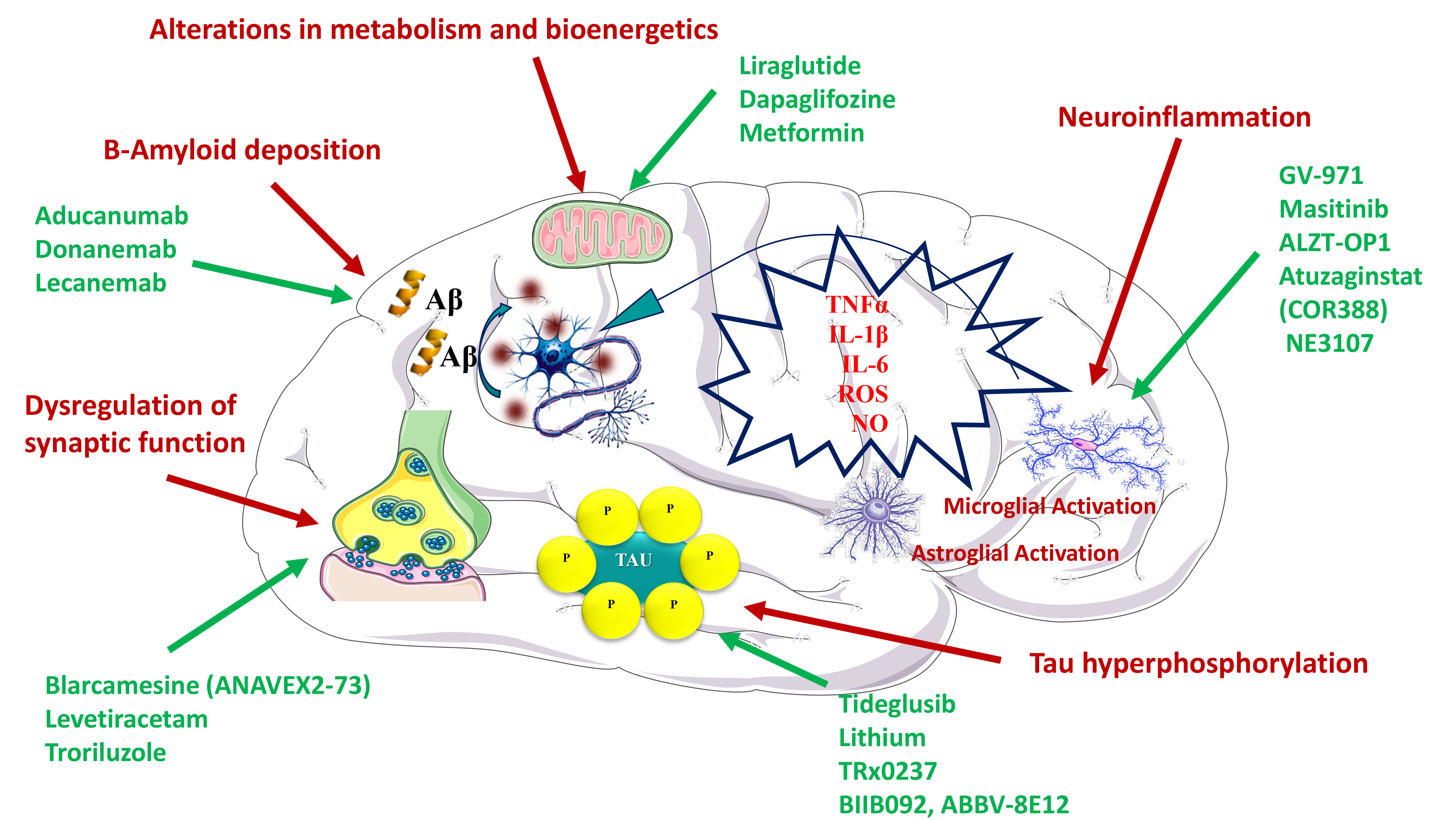
β peptides causing an increase in their clearance. Indeed, Aβ peptides are a common target of phase II and III drug development programs, and some monoclonal antibodies against oligomers, plaques or protofibrils have been widely studied [19]. However, until 2020–2021, most drugs aiming to promote amyloid clearance failed to improve cognitive performance out rightly, irrespective of whether they were monoclonal antibodies or vaccines. In this sense, aducanumab, a monoclonal antibody targeting Aβ protofibrils, was a turning point. Based on the results of two-phase III clinical trials (EMERGE and ENGAGE), the FDA approved its use for AD treatment in 2021. Notwithstanding, these two phase III clinical trials were stopped prematurely in 2019, due to the low efficacy of the drug. While one of the trials showed a positive trend, the other showed no clinical benefit [68, 69]. Subsequently, it was observed that participants who received sufficiently high doses of aducanumab showed a therapeutic improvement in both trials. Accordingly, the researchers suggested that aducanumab was effective in decreasing dementia at a clinical level (Fig. 1). Nevertheless, and despite its FDA approval, much controversy exists about the efficacy of aducanumab in the treatment of AD, as well as its pricing. Indeed, it has been criticized that the approval of aducanumab is based more on speculation than on contrasted clinical data demonstrating a real efficacy. In this sense, the European Medicines Agency (EMA) [70] recommends the denegation of a marketing authorization, due to many doubts about its efficacy and safety. Moreover, it is unclear how aducanumab affects the different types of amyloid oligomers that could be responsible for cognitive loss. In addition, after years of research, all drugs targeting the amyloid cascade have failed in clinical studies, suggesting that amyloid reduction does not lead to a significant clinical benefit in AD. For instance, solanezumab and bapineuzumab showed no clinical benefit in phase III trials, resulting in the end of their development programs.

Fig. 1.

Mechanisms proposed to explain the efficacy of different drugs in the treatment of Alzheimer’s disease. Pathophysiological mechanisms are depicted in red, whereas the specific therapeutic agents are depicted in green.

In turn, donanemab, which targets Aβ plaques, reported both a decrease in Aβ aggregates and a reduction in the rate of cognitive and functional decline in a phase II study [71]; while a larger phase III trial is underway (NCT # 04437511). Another monoclonal antibody, lecanemab (which targets Aβ oligomer protofibrils) is also in late-stage assays (NCT # 03887455). Swanson and colleagues reported that lecanemab treatment resulted in an effective dose-dependent reduction in clinical deterioration when compared to placebo [20]. In addition, after 18 months of treatment, they found a dose-dependent reduction in brain amyloid (assessed by PET). Taken together, the findings of this double-blind trial across multiple cognitive endpoints and biomarkers support a therapeutic effect for targeting specific oligomeric species (protofibrils) in the process of pathophysiologic amyloid generation in AD.

Aβ peptides are able to bind to serum albumin and cross the blood-brain barrier (BBB) [72]. In this sense, it has been proposed that replacing serum albumin in plasma could be a suitable therapy for AD, since it would allow to remove Aβ from the central nervous system (CNS). Moreover, the antioxidant properties of albumin could provide additional therapeutic benefits. Following this line of thought, combining plasma exchange with intravenous immunoglobulin infusion (which also binds Aβ) could further increase the amyloid clearance from the brain. In this regard, Gamunex® is a human intravenous immunoglobulin (IVIG) that is administered concomitantly with human albumin. A phase III multicenter CT (AMBAR) examined the effects of human albumin infusion plasmapheresis combined with IVIG in patients with mild-moderate AD [21]. 347 patients were randomized into three treatment arms and a control group. This study showed that plasma exchange could slow down cognitive and functional decline in AD. However, more studies are needed to clarify the positive effects of this therapy.

On another front, the receptor for advanced glycation end products (RAGE) not only binds and transports Aβ peptides from the blood to the brain, but also induces neuroinflammation [73]. For this reason, RAGE has emerged as a potential target for AD treatment, and drugs blocking the Aβ-RAGE interaction could show numerous therapeutic actions, including the reduction of neuroinflammation and Aβ levels in the brain and the decrease of cognitive impairment. Azeliragon is a RAGE antagonist that decreases Aβ transport into the brain, thus preventing the toxic effects of oligomers and reducing the neuroinflammatory effects of glial cells [22]. In a phase III CT, azeliragon did not improve functional outcomes, so it was terminated [22]. Subsequently, a phase II CT comparing azeliragon with placebo was performed in patients with AD and T2DM during six months. In December 2020, vTv Therapeutics Inc. announced that the study did not meet the primary efficacy endpoint. The company will continue to analyze the data to determine if there are potential benefits or future applications of this drug in AD and/or other diseases [44].

As already mentioned, the fact that most drugs aiming to promote amyloid clearance have failed, irrespective of whether they are monoclonal antibodies or vaccines, challenges the amyloid cascade hypothesis. Moreover, some studies show that patients treated with these drugs eliminate amyloid from the brain but do not improve cognitively [74, 75]. For that matter, a more reconciling Aβ hypothesis suggests that AD has at least two stages, a first one in which Aβ accumulates over time and may be a key factor involved in AD onset, and a second stage, with an ongoing AD where amyloid loses importance and where tau and glia activation are key pathophysiological mechanisms. However, other factors should not be ruled out. Indeed, according to synaptic deficiency hypothesis of AD, Aβ and tau would be secondary to cell injury and the role of neurotrophins in synaptic maintenance and neuronal survival, hence attributing to anti-amyloid treatments a very limited therapeutic efficacy. In any case, several other therapies are in various stages of clinical development, including drugs directed against tau accumulation or dissemination.

4.2 Tau as a Therapeutic Target

Tau is a microtubule-associated protein that can be found both in the CNS and at the peripheral level. In a physiological context, tau exists as a soluble protein that, apart from promoting correct assembly for the stabilization of the microtubular structure, also plays a role in the balance of axonal transport and synapses [76]. Tau is regulated both by normal homeostatic responses and by stress responses, through a series of post-translational modifications such as glycosylation, ubiquitination, glycation, nitration, oxidation and phosphorylation, the last one being the most important one [77]. Tau phosphorylation is regulated by several kinases, including glycogen synthase kinase 3 (GSK3β) and extracellular cyclin 5-dependent kinase (CDK5). As we have explained, tau protein is hyperphosphorylated in AD patients, which favors and leads to fibrillation and aggregation of the protein, forming helical filaments that make up intracellular NFTs that destabilize the neuronal cytoskeleton. In neurodegenerative diseases, tau is a biomarker that migrates to the extracellular compartment and increases its concentration in the cerebrospinal fluid (CSF). The tangles appear initially in the entorhinal cortex of the temporal lobe, spreading to limbic areas such as the hippocampus and finally affecting large areas of the neocortex [78]. The histopathological diagnosis of AD includes the assessment of the quantity and location of NFTs, its number and density being correlated with the intensity of the dementia [23]. Although GSK3β and CDK5 are the most important kinases responsible for tau hyperphosphorylation, other kinases such as Protein Kinase C, Protein Kinase A, the serine/threonine kinase ERK2, caspase 3 and caspase 9, which can be activated by Aβ, also have a prominent role [79]. Beyond tau hyperphosphorylation and formation of NFTs, a “tau propagation hypothesis” has been proposed, stating that poorly folded fibrillar tau aggregates can propagate through cells in a similar way to prions [80].

Given that the severity of tau pathology is more correlated with the progression of cognitive impairment than Aβ [81], multiple studies trying to ascertain tau modulation, (de) phosphorylation and hyperphosphorylation processes in AD have been carried out in order to find suitable treatments. For instance, preclinical studies have reported that sodium selenite, a PP2A activator, restores synaptic plasticity and improves learning and memory in Tg mice with an advanced stage of tauopathy [82]. Focusing on clinical studies, GSK3β inhibitors such as tideglusib (ClinicalTrials.gov Identifier: NCT01350362) and lithium have been tested [24] (Fig. 1). Specifically, Forlenza and colleagues reported that a long-term lithium treatment in subtherapeutic doses (lithium carbonate was prescribed to concentrations 0.25–0.5 mEq/L), can be safe and well tolerated by patients, which supports its potential administration in patients with cognitive deficits (trial registration at clinicaltrials.gov: NCT01055392) [25].

In turn, the second-generation tau aggregation inhibitor LMTX (TRx0237) has been extensively investigated and it is the only drug in this group that reached clinical phase III. Hence, two clinical phase III trials showed that a LMTX monotherapy seems to slow cognitive decline in patients with mild to moderate AD (NCT01689246, NCT01689233). Consequently, additional clinical phase III studies are underway (NCT03446001) [26].

In addition to these strategies, relatively new approaches based on tau have also been investigated; however, some of them did not show results in clinical trials and have been discontinued. These include inhibition of acetylation and deglucosylation of tau (MK-8719 and ASN120290, respectively), monoclonal antibodies against tau (Gosuranemab or BIIB092, Tilavonemab or ABBV-8E12) and reduction of tau levels by an antisense oligonucleotide (ASO) (IONIS-MAPTRx) [27].

4.3 Immunity and Inflammation as Therapeutic Targets

The notion that inflammation and the immune responses play a fundamental role in the pathophysiology of AD is supported by many studies [83]. Much research has been focused on the involvement of astrocytes, microglia and CNS mast cells, but also on the impact of systemic inflammation caused by intestinal and gut microbiota. These have led to the development of different drugs targeting the inflammatory process that could result in neurodegeneration, including tyrosine kinase inhibitors that modulate mast cells (masitinib), monoclonal antibodies that regulate microglial activity as daratumumab (against CD38), AL002 (against TREM2) or AL003 (against SIGLEC-3), and curcumin, which also has antioxidant properties. The use of montelukast (a leukotriene receptor antagonist) and intestinal microbiota transplants, among others, are also being studied.

Microglia are CNS-resident phagocytes that play a vital role in maintaining neuronal plasticity and synapse remodeling [84, 85]. They have been shown to be involved in the maintenance of neural networks by releasing neurotrophic factors such as BDNF, but also contribute to brain homeostasis by exerting synaptic elimination [86]. Microglia can be activated by the accumulation of proteins that act as a pathological trigger, producing the migration of the microglial cells and the onset of an innate immune response. Indeed, microglia actively participate as supportive cells by engulfing Aβ in the AD brain, which may favor a cognitive improvement [87]. The Aβ peptide can trigger the neuroinflammatory process through different microglial receptors, including toll-like receptors (TLRs), receptors for advanced glycation end products, pentraxins, and the complement cascade, among others [88]. One of the most studied receptors involved in this process are TLRs, the TLR2 and TLR4 subtypes being considered critical in the microglial recognition of Aβ [89, 90]. Downstream TLR signaling through NF-κB pathways, activator protein 1, and interferon regulatory factor (IRF) lead to transcription of pro-inflammatory genes and secretion of pro-inflammatory cytokines and chemokines. After binding to receptors, microglia cells also endocyte Aβ oligomers and destroy them by endolysosomal degradation, where microglial proteases such as neprilysin and insulin degrading enzyme (IDE) play an important role [91]. However, in severe AD, microglial clearance of Aβ is inefficient due to the increased concentrations of cytokines downregulating the expression of Aβ phagocytosis receptors, such as Myeloid Cell 2 Activation Receptor (TREM2). TREM2 is surface receptor of the Ig superfamily, widely expressed in microglia and involved in the mediation of phagocytic clearance of neuronal remains [92]. It has been shown that a rare mutation in TREM2 (R47H) leads to the inability of the receptors to remove Aβ from the CNS, hence increasing the risk of developing AD [93]. In light of all these findings, microglia constitute an interesting therapeutic target against AD, either because a microglial overstimulation can increase synaptic elimination and lead to cognitive loss, or because increasing TREM2 activity could prevent Aβ accumulation.

On another front, mast cells (MCs) have an important physiological function and are involved in immunity and inflammation, especially in allergic inflammation. Upon activation, MCs increase the synthesis and secretion of many inflammatory mediators, such as TNF-α, serotonin, histamine and heparin, among others [94]. As key components of the immune system, the pathophysiological changes of these cells affect multiple organs. In the CNS, the contribution of MCs to neuroinflammation is beyond doubt, but less is known about their role in neurodegenerative diseases such as AD. In this sense, it has been suggested that MCs could favor alterations in the BBB and interact with microglia. Regarding the latter, some studies have shown that mast cell-glia interaction can occur in different ways [95, 96, 97]. Hence, MCs degranulation and the release of histamine and TNF-α activate microglia [96]. Moreover, during inflammation, ATP derived from MCs activates purinergic P2 receptors in the microglia leading to IL-33 secretion [98]. In turn, IL-33 causes MCs to release IL-6, IL-13 and the monocyte chemoattractant protein-1 (MCP-1), which also regulates microglial activity. In addition, the secretion of tryptase from MCs stimulates microglial release of TNF-α, IL-6 and reactive oxygen species (ROS) through the activation of the protease-activated receptor-2 (PAR-2) [99]. Interestingly, PAR-2 also upregulates the expression of microglial ATP-sensitive ionotropic P2X4 receptors which, upon ATP binding, lead to the secretion of BDNF, a key mediator in synaptic plasticity [100].

Masitinib is currently one of the most promising research drugs targeting inflammatory diseases such as rheumatoid arthritis, intestinal disease, asthma and mastocytosis. However, a potential new application of masitinib in neurodegenerative diseases, such as AD and multiple sclerosis (MS), has emerged. Masitinib inhibits c-kit tyrosine kinase, a surface receptor expressed by MCs and many other cells, which plays a prominent role as a regulator of the migration of neuronal stem and progenitor cells to areas of brain injury [101]. Hence, masitinib is thought to exert a neuroprotective effect through its activity on MCs and other non-neuronal cells of the CNS, with a subsequent modulation of inflammatory and neurodegenerative processes. It is currently in the phase 3 of a clinical trial in patients with mild to moderate AD (ClinicalTrials.gov Identifier: NCT01872598). Masitinib is administered as adjunctive therapy to patients who have been treated for a minimum of 6 months with a stable dose of a cholinesterase inhibitor (donepezil, rivastigmine or galantamine) and/or memantine. So far, the company has reported some positive results from this study. Masitinib is also capable of blocking the Src TK Fyn family in a nanomolar range. Fyn is a cytoplasmic tyrosine kinase (TK) belonging to the Src kinase (SFK) family involved in multiple CNS transduction pathways, including synaptic transmission, myelination, axon guidance, and formation of oligodendrocytes [102]. Shirazi and Wood reported that that Fyn is upregulated in the brain of AD patients and demonstrated the presence of a Fyn phosphorylation site in the tau-matched helical filament, supporting a role of Fyn in the neuropathogenesis of AD [103]. Therefore, treatment with masitinib may provide two benefits in the pathology of AD: (i) a reduction in neuroinflammation by modulating the mast cell-glia axis and (ii) a cognitive enhancement through a Fyn inhibition.

After many failures with monotreatments, the combination of cromolyn and ibuprofen (ALZT-OP1) has been proposed as a suitable treatment for LOAD [28]. Cromolyn acts as a modulator of MCs and microglia, as well as an Aβoligomerization inhibitor [104]. In turn, ibuprofen is an anti-inflammatory drug with interesting effects such as α-secretase activation -thus modulating Aβ production-, microglia inhibition, activation of PPAR γ and modulation of synapses through the inhibition of Rho family GTPases [105, 106, 107].

XPro1595 is a second-generation inhibitor of tumor necrosis factor (TNFα), an inflammatory factor implicated in AD pathology [108]. XPro1595 selectively neutralizes soluble TNF (sTNF) and inactivates it. Previous preclinical studies reported that targeting of sTNF/TNFR1 signaling with XPro1595 decreased brain alterations in immune cell populations associated with the neuroinflammatory process in 5xFAD (Tg) mouse model [109]. In addition, XPro1595 treatment rescued impaired LTP and also decreased the production or accumulation of amyloid. Furthermore, XPro1595 has a potential application in inflammatory states associated to obesogenic diets and T2DM. Hence, XPro1595 prevents the development of insulin resistance and risk for AD [110].

From a different point of view, some studies have proposed an association between intestinal microbiota and AD, in which dysbiosis cause neuroinflammation induced by activation of the microglia [111]. In this regard, one of the latest interesting drugs is sodium oligomannate (GV-971), a derivative of brown marine algae based on a mixture of acidic linear oligosaccharides ranging from dimers to decamers [112]. It was developed by Green Valley for reconditioning the intestinal microbiota and treating mild-moderate AD. A preclinical study showed that oral administration of GV-971 for one month markedly altered the composition of the microbiota and reduced the concentrations of phenylalanine and isoleucine in a murine model of AD [113]. These was paralleled by a reduction in microglial activation, as well as in Th1 responses and cytokines in the brain. Simultaneously, oligomannate treatment reduced Aβ deposition and tau phosphorylation. These findings led to a study of GV-971 in patients with mild-moderate AD in a randomized, multicenter, double blind, placebo-controlled phase II clinical trial in China [29, 114]. The objective of this trial was to establish the optimal dose, safety and efficacy of GV-971 capsules. The authors found a cognitive improvement assessed by changes in the ADAS-Cog-12 (Alzheimer’s Disease Assessment Scale) score from baseline to week 24 in patients treated with the drug. GV-971 also demonstrated significant and sustained efficacy in enhancing cognition in his first phase III trial [29]. The effects were more pronounced at the end point and in those patients with the most severe cognitive impairment. In 2019, GV-971 obtained conditional approval in China to treat AD patients; however, additional trials are needed to investigate the full mechanism of action of sodium oligomannate. In 2020, the FDA approved the request for a global phase III trial (Green Memory) to evaluate the safety and efficacy of GV-971 in additional populations (United States, Europe, and Asia). If the results of this study are in the same line that those seen in phase III in China, GV-971 probably could receive a worldwide regulatory approval by the FDA and the EMA.

Beyond the relation between intestinal microbiota and AD, recent studies also support an association between periodontitis, a chronic inflammatory oral disease, and neurodegeneration [115]. It is known that there is a relationship between host immune responses and pathogenic burden of microbial biofilm [116, 117], which is made up of several microorganisms, such as Porphyromonas gingivalis, and their toxic products, such as fimbrins, gingipain and lipopolysaccharides (LPS). Both periodontopathogenic bacteria and their products can enter the bloodstream, promoting the expression of inflammatory mediators that can damage other organs, including the brain. Interestingly, gingipains are proteases that generate pathogenicity factors such as Arg-gingipain (Rgp) and Lys-gingipain (Kgp), which can damage tau [30, 118]. In this regard, atuzaginstat (COR388) is an inhibitor of gingipains, which is being tested in a phase II/III clinical study for AD treatment (NCT03823404) (Fig. 1).

4.4 Metabolism and Bioenergetics as Therapeutic Targets

It is widely known that glucose is the main source of energy of the mammalian brain, and several pathologies of the CNS are a consequence of disturbed central or peripheral glucose energy metabolism [119]. In this sense, impaired glucose metabolism has been shown to be related with AD pathophysiology. Hence, alterations in brain glucose uptake have been described in patients with initial symptoms that precede the development of AD [120]. Moreover, it seems that insulin improves glucose uptake in the brain by increasing the activation of hippocampal insulin receptor, which plays a key role in synaptic plasticity, learning and memory [121]. Therefore, the rescue of cerebral insulin signaling and glucose metabolism constitutes a tempting goal to treat neurodegeneration.

A priori, many drugs used to treat T2DM could be recycled to also treat AD, including 116 (glucagon-like peptide 1 receptor agonist), dapaglifozine (SGLT2 inhibitor) and metformin, among others. Regarding the latter, metformin is currently the first-line treatment for T2DM, widely prescribed due to its safeness and virtual absence of side effects. Preclinical results in murine models of AD showed that metformin delayed the progression of cognitive impairment [122]. In turn, Luchsinger and colleagues reported that metformin treatment significantly improves the total recall in the selective reminding test, but not the Alzheimer Disease Assessment Scale-Cognition (ADAS-Cog), in a study with patients with amnestic MCI (ClinicalTrials.gov Identifier: NCT00620191). The authors concluded that additional clinical studies with a larger number of patients are necessary to demonstrate a greater efficacy of the drug. Other studies evaluating the efficacy of metformin on AD biomarkers and cognitive ability (ClinicalTrials.gov Identifier: NCT01965756) and for dementia prevention (NCT04098666) have been conducted. The results of the study NCT01965756 showed that metformin improved executive function and there was a trend in improving the learning process, memory and attention [31]. Regarding the study NCT04098666, no results have yet been published.

NE3107 is another drug which also reached the phase III (Clinicaltrials.gov: NCT04669028). Previous studies have shown that this drug has a dual therapeutic effect, presenting both neuroinflammatory and antidiabetic properties [32]. Hence, NE3107 has been reported to inhibit the inflammatory ERK pathway and to the improve insulin signaling. This dual effect makes this drug interesting in the treatment of AD (Fig. 1).

On a different note, intranasal administration of insulin glulisine is also being evaluated with the aim to increase insulin signaling in the brain [123]. Promising results have been published regarding intranasal insulin administration in patients with amnestic MCI and AD (ClinicalTrials.gov Identifier: NCT01767909). However, more studies are needed to better characterize the neuroprotective effect of insulin and brain insulin receptor stimulation in neurodegeneration.

4.5 Cardiovascular Risk Factors as Therapeutic Targets

An association between cognitive impairment in non-demented individuals and cardiovascular risk factors has been reported [124]. Hence, the inflammatory process associated with aging, atherosclerotic cardiovascular diseases and dementia could share common molecular mechanisms. In this sense, the control of cardiovascular risk factors could be an appropriate strategy to reduce or prevent the incidence of dementia [125].

Following this line of thought, drugs acting on cardiovascular and cerebrovascular dysfunctions, BBB and neurovascular unit, hypertension, atherosclerosis, amyloid cerebral angiopathy and lymphatic/glyphic system dysfunction could be useful for AD treatment. For instance, losartan, an antihypertensive drug, reduced plasma and brain Aβ1-42 levels in a murine model of AD [126, 127]. In this sense, different antihypertensive drugs that could be used against neurodegeneration are currently under study, including ARAII drugs (candesartan, telmisartan) and combination treatments such as telmisartan + perindopril (ARAII + ACE inhibitors), and losartan + amlodipine + atorvastatin + exercise (ARAII + Ca2+ blocker + an anti-cholesterol agent) (NCT02913664) [33, 128, 129, 130]. These drugs can modulate APP/Aβ metabolism, hence preserving cognitive function in addition to improve cerebrovascular function.

4.6 Synapses as Therapeutic Targets

Previous studies have reported a dysregulation of synaptic functions in AD [131, 132]. In fact, synaptic loss and dysfunction is strongly correlated with the cognitive decline observed in AD patients [133]. Indeed, surviving neurons in AD’s neurodegenerative process have been shown to lose synapses, and synaptic dysfunction has been shown to precede amyloid plaque deposition, as LTP impairment is present in early stages in the hippocampus of AD mice [134, 135, 136]. Hence, the loss of synaptic homeostasis or the integrity of the neural network would precede neuronal death and be key to AD development, an idea that fits with the proposed theories and clinical manifestations. Consequently, memory deficits in AD may even begin two decades before the first symptoms appear. Synapse degeneration is thought to begin in dendritic spines and, specifically, with a decrease in the number of molecules that regulate spinal signaling.

It has been shown that glutamate is involved in the development of dendritic spines [137]. On this basis, some drugs targeting neurotransmitters and mechanisms of neurogenesis are being studied (Fig. 1). An example is troriluzole, also known as BHV4157 (proriluzole), which is a prodrug of riluzole. Riluzole inhibits voltage-gated sodium channels and reduces synaptic glutamate by increasing its uptake and inhibiting its release [138]. Indeed, riluzole reduces synaptic glutamate levels by increasing the expression and function of glial glutamate transporters responsible for synaptic glutamate clearance [34]. Its efficacy in the treatment of AD is currently being evaluated in phase 2–3 clinical trials (Clinicaltrials.gov NCT03605667), although so far, the results do not seem to be encouraging.

In turn, blarcamesine (ANAVEX2-73) is an agonist of the Sigma-1 receptor (S1R) and modulates cholinergic muscarinic receptors in mice [135, 139, 140]. It has been reported that an oral dose (30 or 50 mg) of blarcamesine, followed by an intravenous (IV) dose (3 or 5 mg) in a second period have suitable safety and tolerability in patients with mild-to-moderate AD in a Phase IIa clinical study [35]. Furthermore, phase IIb/III clinical studies consisting in 48 weeks of daily treatment with blarcamesine or placebo, and primary outcomes of ADAS-Cog and ADCS-ADL (Activities of Daily Living) evaluations are being conducted (ClinicalTrials.gov Identifier: NCT03790709). Additionally, in preclinical models of Rett syndrome (RTT), a neurodevelopmental disorder associated with increased risk of cognitive impairment, blarcamesin improved calcium homeostasis, which favors an improvement in mitochondrial and synaptic functions in all brain regions [135]. Besides that, the anticonvulsant levetiracetam has also shown potential as synapse modulator against neurodegeneration. Levetiracetam is an antiepileptic drug that binds to SV glycoprotein 2A (SV2A), a constituent of synaptic vesicle membranes at presynaptic terminals, involved in vesicle trafficking and exocytosis [141]. SV2A is expressed in excitatory and inhibitory synapses in the brain, including the hippocampus, and alterations in this protein have been associated with AD [142, 143]. Interestingly, Rao and Savas [36] reported that levetiracetam lowers Aβ42 levels in APP knock-in mice models of amyloid pathology by normalizing the abundance of presynaptic endocytosis machinery, hence favoring a change in the processing of amyloid precursor protein towards the non-amyloidogenic pathway. Other study showed that a treatment with a low dose of levetiracetam (125 mg twice daily), significantly improved memory performance [36].

Currently, a phase II-III study is evaluating the efficacy of AGB101, a proprietary extended-release formulation of levetiracetam, on slowing cognitive and functional impairment in patients with MCI (ClinicalTrials.gov Identifier: NCT03486938). No results have been reported yet.

4.7 Dietary and Supplemental Strategies

The fact that diet quality and composition play a role in virtually all health conditions, affecting incidence, complications, management, recovery and quality of life is well supported by mounting evidence [144]. AD is not an exception, and currently several supplements and dietary strategies claim to promote cognitive enhancement. However, to date, no evidence-based product on the market has demonstrated a clear capacity to prevent or slow AD’s progression.

Omega-3 polyunsaturated Fatty Acids (ω3 PUFAs) are among the nutritional and dietary factors that have shown most consistent positive research findings to prevent cognitive decline in older adults [145]. Linoleic and α-linolenic acid are examples of ω3 PUFAs which are obtained directly through diet or supplementation. Long-chain ω3 PUFAs (eicosapentaenoic acid and docosahexaenoic acid), have been investigated in different studies as a therapeutic strategy to mitigate cognitive loss, and progression to AD [146, 147]. These compounds can play an important role in improving cognitive impairment through multiple mechanisms such as modulation of the inflammatory process, or acting on genes that regulate the retinoic acid receptor and peroxisome proliferator-activated receptor, as well as the rigidity of the cell membrane [148]. In this context, icosapent ethyl (IPE), a purified form of the omega-3 fatty acid eicosapentaenoic acid (EPA), is under investigation in a clinical study (ClinicalTrials.gov Identifier: NCT02719327). The aim of the research is to evaluate the efficacy of 18 months of IPE therapy in 150 cognitively healthy veterans aged 50 to 75 with increased risk of developing AD due to a parental history of the disease and increased prevalence of the APOE4. The evaluation will be carried out through magnetic resonance imaging (MRI) and the evaluation of cognitive biomarkers for AD in CSF. The study is projected to be finished in early 2023. In the same line, a phase III clinical study (NCT00440050) determined whether chronic supplementation with docosahexaenoic acid (DHA) was capable of slowing the progression of cognitive and functional decline in mild to moderate AD. However, the results showed no differences between DHA supplementation and placebo groups [37].

5. Conclusions

Despite the numerous failures in the development of therapies that modify the evolution of AD, current technological progress in the fields of biomarkers assays and genetics presents an unprecedented opportunity to reshape AD therapeutic strategies towards the medicine of precision. As with other diseases, an early detection of AD is essential. Hence, assessing the blood levels of AD biomarkers such as tau and amyloid in asymptomatic patients could be beneficial as an early approximation [149]. In symptomatic patients, positron emission tomography and magnetic resonance imaging approaches to assess Aβ and tau can be performed, along with CSF tests, if necessary, for accurate identification of the disease. Once we have an early diagnosis, the patient should be treated with the most appropriate drug cocktail, specifically designed for their individual etiology and stage of AD. In this regard, the recognition of multiple causal and protective genes (genetic resilience) and the positive effects of lifestyle interventions (such as diet) highlight the possibility of developing different alternatives for the future. Hence, the combination of pharmacological and non-pharmacological strategies could be an interesting therapeutic approach.

Undoubtedly, the trends in therapeutic strategies for AD will involve an increase in the diversity of non-amyloid or tau targets, including inflammation, insulin resistance, synapse and neuronal protection, cardiovascular factors, neurogenesis and epigenetic interventions. Indeed, some authors consider that AD should no longer be considered a brain disease, since its development is also attributed to peripheral factors as, for instance, intestinal dysbiosis. Hence, the increasing knowledge of the mechanisms involved in AD may favor the development of novel therapies based, for example, on the reconstitution of the intestinal microbiota. In this sense, sodium oligomannate opens a promising new therapeutic line of disease-modifying therapies worthy of future research. In passive immunotherapy, the FDA-approved aducanumab stands out, but anti-tau treatments still need to demonstrate its clinical efficacy. Also, an increase in the number of candidate drugs for disease modification treatments is expected, as well as a focus on potential combinatory multidrug strategies.

Author Contributions

JO, ME, AMAC, ESL, assisted in the preparation of the figures, designed the concept of the manuscript, provided supervision. JO and AC wrote the manuscript. MC, TE, CBZ, GGC, MEUG, EV, JF and CA critically reviewed the manuscript and substantially contributed to the writing and content of the manuscript. All authors provided their contribution in writing the manuscript, critically reviewing the completed manuscript, and approved the submitted version of the manuscript.

Ethics Approval and Consent to Participate

Not applicable.

Acknowledgment

The authors would like to thank Anton Kabanen for English checking and the Reviewers for their opinions and suggestions.

Funding

Jordi Olloquequi and Miren Ettcheto are Serra Húnter fellows. Amanda Cano acknowledges the support of the Spanish Ministry of Science, Innovation and Universities under the grant Juan de la Cierva (FJC2018-036012-I). Authors acknowledge the support of Spanish Ministry of Economy and Competitiveness under project SAF2017-84283-R, and Biomedical Research Networking Centre in Neurodegenerative Diseases (CIBERNED) under the project CB06/05/0024. The authors have no other relevant affiliations or financial involvement with any organization or entity with a financial interest in or financial conflict with the subject matter or materials discussed in the manuscript apart from those disclosed.

Conflict of Interest

The authors declare no conflict of interest. AC is serving as the guest editor and the editorial board member of this journal. We declare that AC had no involvement in the peer review of this article and has no access to information regarding its peer review. Full responsibility for the editorial process for this article was delegated to GP.

References
[1]
Möller HJ, Graeber MB. The case described by Alois Alzheimer in 1911. European Archives of Psychiatry and Clinical Neuroscience. 1998; 248: 111–122.
[2]
Brion JP, Couck AM, Passareiro E, Flament-Durand J. Neurofibrillary tangles of Alzheimer’s disease: an immunohistochemical study. Journal of Submicroscopic Cytology. 1985; 17: 89–96.
[3]
Grundke-Iqbal I, Iqbal K, Tung YC, Quinlan M, Wisniewski HM, Binder LI. Abnormal phosphorylation of the microtubule-associated protein tau (tau) in Alzheimer cytoskeletal pathology. Proceedings of the National Academy of Sciences of the United States of America. 1986; 83: 4913–4917.
[4]
Mirra SS, Heyman A, McKeel D, Sumi SM, Crain BJ, Brownlee LM, et al. The Consortium to Establish a Registry for Alzheimer’s Disease (CERAD). Part II. Standardization of the neuropathologic assessment of Alzheimer’s disease. Neurology. 1991; 41: 479–486.
[5]
Hyman BT, Phelps CH, Beach TG, Bigio EH, Cairns NJ, Carrillo MC, et al. National Institute on Aging-Alzheimer’s Association guidelines for the neuropathologic assessment of Alzheimer’s disease. Alzheimer’s & Dementia. 2012; 8: 1–13.
[6]
2021 Alzheimer’s disease facts and Figures. Alzheimers Dementia. 2021; 17: 327–406.
[7]
Neuman KM, Molina-Campos E, Musial TF, Price AL, Oh K, Wolke ML, et al. Evidence for Alzheimer’s disease-linked synapse loss and compensation in mouse and human hippocampal CA_1 pyramidal neurons. Brain Structure & Function. 2015; 220: 3143–3165.
[8]
Scheff SW, Neltner JH, Nelson PT. Is synaptic loss a unique hallmark of Alzheimer’s disease? Biochemical Pharmacology. 2014; 88: 517–528.
[9]
Ausó E, Gómez-Vicente V, Esquiva G. Biomarkers for Alzheimer’s Disease Early Diagnosis. Journal of Personalized Medicine. 2020; 10: 114.
[10]
Du X, Wang X, Geng M. Alzheimer’s disease hypothesis and related therapies. Translational Neurodegeneration. 2018; 7: 1–7.
[11]
Ferreira-Vieira TH, Guimaraes IM, Silva FR, Ribeiro FM. Alzheimer’s disease: Targeting the Cholinergic System. Current Neuropharmacology. 2016; 14: 101–115.
[12]
Stocker H, Möllers T, Perna L, Brenner H. The genetic risk of Alzheimer’s disease beyond APOE ε4: systematic review of Alzheimer’s genetic risk scores. Translational Psychiatry. 2018; 8: 166.
[13]
Dietrich K, Bouter Y, Müller M, Bayer TA. Synaptic Alterations in Mouse Models for Alzheimer Disease-a Special Focus on N-Truncated Abeta 4-42. Molecules. 2018; 23: 718.
[14]
Jackson J, Jambrina E, Li J, Marston H, Menzies F, Phillips K, et al. Targeting the Synapse in Alzheimer’s Disease. Frontiers in Neuroscience. 2019; 13: 735.
[15]
Duthie A, Chew D, Soiza RL. Non-psychiatric comorbidity associated with Alzheimer’s disease. QJM. 2011; 104: 913–920.
[16]
Cholerton B, Baker LD, Montine TJ, Craft S. Type 2 Diabetes, Cognition, and Dementia in Older Adults: toward a Precision Health Approach. Diabetes Spectrum. 2016; 29: 210–219.
[17]
Salloway SP, Sevingy J, Budur K, Pederson JT, DeMattos RB, Von Rosenstiel P, et al. Advancing combination therapy for Alzheimer’s disease. Alzheimer’s & Dementia. 2020; 6: e12073.
[18]
Prins ND, Scheltens P. Treating Alzheimer’s disease with monoclonal antibodies: current status and outlook for the future. Alzheimer’s Research & Therapy. 2013; 5: 56.
[19]
van Dyck CH. Anti-Amyloid-β Monoclonal Antibodies for Alzheimer’s Disease: Pitfalls and Promise. Biological Psychiatry. 2018; 83: 311–319.
[20]
Swanson CJ, Zhang Y, Dhadda S, Wang J, Kaplow J, Lai RYK, et al. A randomized, double-blind, phase 2b proof-of-concept clinical trial in early Alzheimer’s disease with lecanemab, an anti-Aβ protofibril antibody. Alzheimer’s Research & Therapy. 2021; 13: 80.
[21]
M. Boada, López OL, Olazarán J, Núñez L, Pfeffer M, Puente O, et al. Neuropsychological, neuropsychiatric, and quality-of-life assessments in Alzheimer’s disease patients treated with plasma exchange with albumin replacement from the randomized AMBAR study. Alzheimer’s & Dementia. 2021. (in press)
[22]
Burstein AH, Sabbagh M, Andrews R, Valcarce C, Dunn RI, Altstiel L. Development of Azeliragon, an Oral Small Molecule Antagonist of the Receptor for Advanced Glycation Endproducts, for the Potential Slowing of Loss of Cognition in Mild Alzheimer’s Disease. The Journal of Prevention of Alzheimer’s Disease. 2018; 5: 149–154.
[23]
Scheltens P, De Strooper B, Kivipelto M, Holstege H, Chételat G, Teunissen CE, et al. Alzheimer’s disease. The Lancet. 2021; 397: 1577–1590.
[24]
Matsunaga S, Fujishiro H, Takechi H. Efficacy and Safety of Glycogen Synthase Kinase 3 Inhibitors for Alzheimer’s Disease: a Systematic Review and Meta-Analysis. Journal of Alzheimer’s Disease. 2019; 69: 1031–1039.
[25]
Forlenza OV, Radanovic M, Talib LL, Gattaz WF. Clinical and biological effects of long-term lithium treatment in older adults with amnestic mild cognitive impairment: randomised clinical trial. The British Journal of Psychiatry. 2019; 215: 668–674.
[26]
Wilcock GK, Gauthier S, Frisoni GB, Jia J, Hardlund JH, Moebius HJ, et al. Potential of Low Dose Leuco-Methylthioninium Bis(Hydromethanesulphonate) (LMTM) Monotherapy for Treatment of Mild Alzheimer’s Disease: Cohort Analysis as Modified Primary Outcome in a Phase III Clinical Trial. Journal of Alzheimer’s Disease. 2018; 61: 435–457.
[27]
Sexton C, Snyder H, Beher D, Boxer AL, Brannelly P, Brion JP, et al. Carrillo: Current directions in tau research: Highlights from Tau 2020. Alzheimers Dement. 2021. (in press)
[28]
Lozupone M, Solfrizzi V, D’Urso F, Di Gioia I, Sardone R, Dibello V, et al. Anti-amyloid-β protein agents for the treatment of Alzheimer’s disease: an update on emerging drugs. Expert Opinion on Emerging Drugs. 2020; 25: 319–335.
[29]
Xiao S, Chan P, Wang T, Hong Z, Wang S, Kuang W, et al. A 36-week multicenter, randomized, double-blind, placebo-controlled, parallel-group, phase 3 clinical trial of sodium oligomannate for mild-to-moderate Alzheimer’s dementia. Alzheimer’s research & therapy. 2021; 13: 62.
[30]
Olsen I. Porphyromonas gingivalis-Induced Neuroinflammation in Alzheimer’s Disease. Frontiers in Neuroscience. 2021; 15: 691016.
[31]
Koenig AM, Mechanic-Hamilton D, Xie SX, Combs MF, Cappola AR, Xie L, et al. Effects of the Insulin Sensitizer Metformin in Alzheimer Disease: Pilot Data from a Randomized Placebo-controlled Crossover Study. Alzheimer Disease and Associated Disorders. 2017; 31: 107–113.
[32]
Reading CL, Ahlem CN, Murphy MF. NM101 Phase III study of NE3107 in Alzheimer’s disease: rationale, design and therapeutic modulation of neuroinflammation and insulin resistance. Neurodegenerative Disease Management. 2021; 11: 289–298.
[33]
Kehoe PG, Turner N, Howden B, Jarutyte L, Clegg SL, Malone IB, et al. Safety and efficacy of losartan for the reduction of brain atrophy in clinically diagnosed Alzheimer’s disease (the RADAR trial): a double-blind, randomised, placebo-controlled, phase 2 trial. The Lancet Neurology. 2021; 20: 895–906.
[34]
Doble A. The pharmacology and mechanism of action of riluzole. Neurology. 1996; 47: S233–S241.
[35]
Hampel H, Williams C, Etcheto A, Goodsaid F, Parmentier F, Sallantin J, et al. A precision medicine framework using artificial intelligence for the identification and confirmation of genomic biomarkers of response to an Alzheimer’s disease therapy: Analysis of the blarcamesine (ANAVEX2‐73) Phase 2a clinical study. Alzheimer’s & Dementia. 2020; 6: e12013.
[36]
Rao NR, Savas JN. Levetiracetam Treatment Normalizes Levels of Presynaptic Endocytosis Machinery and Restores Nonamyloidogenic APP Processing in App Knock-in Mice. Journal of Proteome Research. 2021; 20: 3580–3589.
[37]
Quinn JF, Raman R, Thomas RG, Yurko-Mauro K, Nelson EB, Van Dyck C, et al. Docosahexaenoic Acid Supplementation and Cognitive Decline in Alzheimer Disease. JAMA. 2010; 304: 1903–1911.
[38]
Reitz C, Rogaeva E, Beecham GW. Late-onset vs nonmendelian early-onset Alzheimer disease: a distinction without a difference? Neurology Genetics. 2020; 6: e512.
[39]
Hardy J, Selkoe DJ. The amyloid hypothesis of Alzheimer’s disease: progress and problems on the road to therapeutics. Science (New York, N.Y.). 2002; 297: 353–356.
[40]
Tcw J, Goate AM. Genetics of β-Amyloid Precursor Protein in Alzheimer’s Disease. Cold Spring Harbor Perspectives in Medicine. 2017; 7: a024539.
[41]
Xiao X, Liu H, Liu X, Zhang W, Zhang S, Jiao B. APP, PSEN1, and PSEN2 Variants in Alzheimer’s Disease: Systematic Re-evaluation According to ACMG Guidelines. Frontiers in Aging Neuroscience. 2021; 13: 695808.
[42]
Bellenguez C, Grenier-Boley B, Lambert J. Genetics of Alzheimer’s disease: where we are, and where we are going. Current Opinion in Neurobiology. 2020; 61: 40–48.
[43]
Husain MA, Laurent B, Plourde M. APOE and Alzheimer’s Disease: From Lipid Transport to Physiopathology and Therapeutics. Frontiers in Neuroscience. 2021; 15: 630502.
[44]
Phillips MC. Apolipoprotein E isoforms and lipoprotein metabolism. IUBMB Life. 2014; 66: 616–623.
[45]
Reiman EM, Arboleda-Velasquez JF, Quiroz YT, Huentelman MJ, Beach TG, Caselli RJ, et al. Exceptionally low likelihood of Alzheimer’s dementia in APOE_2 homozygotes from a 5,000-person neuropathological study. Nature Communications. 2020; 11: 667.
[46]
Karch CM, Goate AM. Alzheimer’s Disease Risk Genes and Mechanisms of Disease Pathogenesis. Biological Psychiatry. 2015; 77: 43–51.
[47]
Hunsberger H, Pinky P, Smith W, Suppiramaniam V, Reed M. The role of APOE_4 in Alzheimer’s disease: strategies for future therapeutic interventions. Neuronal Signaling. 2019; 3: NS20180203.
[48]
Davies P, Maloney AJ. Selective loss of central cholinergic neurons in Alzheimer’s disease. The Lancet. 1976; 2: 1403.
[49]
Santos TCD, Gomes TM, Pinto BAS, Camara AL, Paes AMDA. Naturally Occurring Acetylcholinesterase Inhibitors and Their Potential Use for Alzheimer’s Disease Therapy. Frontiers in Pharmacology. 2018; 9: 1192.
[50]
Anand P, Singh B. A review on cholinesterase inhibitors for Alzheimer’s disease. Archives of Pharmacal Research. 2013; 36: 375–399.
[51]
Alcolea-Palafox M, Posada-Moreno P, Ortuño-Soriano I, Pacheco-del-Cerro JL, Martínez-Rincón C, Rodríguez-Martínez D, et al. Chapter 8 - Research Strategies Developed for the Treatment of Alzheimer’s Disease. Reversible and Pseudo-Irreversible Inhibitors of Acetylcholinesterase: Structure-Activity Relationships and Drug Design. Drug Design and Discovery in Alzheimer’s Disease. 2014; 426–477.
[52]
Dooley M, Lamb HM. Donepezil: a review of its use in Alzheimer’s disease. Drugs & Aging. 2000; 16: 199–226.
[53]
Birks JS, Harvey RJ. Donepezil for dementia due to Alzheimer’s disease. Cochrane Database of Systematic Reviews. 2018; 6: Cd001190.
[54]
Lilienfeld S. Galantamine–a novel cholinergic drug with a unique dual mode of action for the treatment of patients with Alzheimer’s disease. CNS Drug Reviews. 2002; 8: 159–176.
[55]
Xu H, Garcia-Ptacek S, Jönsson L, Wimo A, Nordström P, Eriksdotter M. Long-term Effects of Cholinesterase Inhibitors on Cognitive Decline and Mortality. Neurology. 2021; 96: e2220–e2230.
[56]
Huh S, Baek S, Lee K, Whitcomb DJ, Jo J, Choi S, et al. The reemergence of long-term potentiation in aged Alzheimer’s disease mouse model. Scientific Reports. 2016; 6: 29152.
[57]
Babaei P. NMDA and AMPA receptors dysregulation in Alzheimer’s disease. European Journal of Pharmacology. 2021; 908: 174310.
[58]
Kabir MT, Sufian MA, Uddin MS, Begum MM, Akhter S, Islam A, et al. NMDA Receptor Antagonists: Repositioning of Memantine as a Multitargeting Agent for Alzheimer’s Therapy. Current Pharmaceutical Design. 2019; 25: 3506–3518.
[59]
Folch J, Busquets O, Ettcheto M, Sánchez-López E, Castro-Torres RD, Verdaguer E, et al. Memantine for the Treatment of Dementia: a Review on its Current and Future Applications. Journal of Alzheimer’s Disease. 2018; 62: 1223–1240.
[60]
Peters O, Fuentes M, Joachim LK, Jessen F, Luckhaus C, Kornhuber J, et al. Combined treatment with memantine and galantamine-CR compared with galantamine-CR only in antidementia drug naïve patients with mild-to-moderate Alzheimer’s disease. Alzheimers & Dementia. 2015; 1: 198–204.
[61]
Wood H. Could antidepressant drugs aid Alzheimer disease prevention? Nature Reviews Neurology. 2020; 16: 599–599.
[62]
Poor SR, Ettcheto M, Cano A, Sanchez-Lopez E, Manzine PRM, Olloquequi J, et al. Metformin a Potential Pharmacological Strategy in Late Onset Alzheimer’s Disease Treatment. Pharmaceuticals. 2021; 14: 890.
[63]
Fontana IC, Zimmer AR, Rocha AS, Gosmann G, Souza DO, Lourenco MV, et al. Amyloid‐β oligomers in cellular models of Alzheimer’s disease. Journal of Neurochemistry. 2020; 155: 348–369.
[64]
Viola KL, Klein WL. Amyloid β oligomers in Alzheimer’s disease pathogenesis, treatment, and diagnosis. Acta Neuropathologica. 2015; 129: 183–206.
[65]
Cline EN, Bicca MA, Viola KL, Klein WL. The Amyloid-β Oligomer Hypothesis: Beginning of the third Decade. Journal of Alzheimer’s Disease. 2018; 64: S567–S610.
[66]
Bloom GS. Amyloid-β and tau: the trigger and bullet in Alzheimer disease pathogenesis. JAMA Neurology. 2014; 71: 505–508.
[67]
Forloni G, Balducci C. Alzheimer’s Disease, Oligomers, and Inflammation. Journal of Alzheimer’s Disease. 2018; 62: 1261–1276.
[68]
Kuller LH, Lopez OL. ENGAGE and EMERGE: Truth and consequences? Alzheimer’s & Dementia. 2021; 17: 692–695.
[69]
Salloway S, Chalkias S, Barkhof F, Burkett P, Barakos J, Purcell D, et al. Amyloid-Related Imaging Abnormalities in 2 Phase 3 Studies Evaluating Aducanumab in Patients with Early Alzheimer Disease. JAMA Neurology. 2022; 79: 13.
[70]
Mahase E. Aducanumab: European agency rejects Alzheimer’s drug over efficacy and safety concerns. The BMJ. 2021; 375: n3127
[71]
Mintun MA, Lo AC, Duggan Evans C, Wessels AM, Ardayfio PA, Andersen SW, et al. Donanemab in Early Alzheimer’s Disease. New England Journal of Medicine. 2021; 384: 1691–1704.
[72]
Costa M, Páez A. Emerging insights into the role of albumin with plasma exchange in Alzheimer’s disease management. Transfusion and Apheresis Science. 2021; 60: 103164.
[73]
Cai Z, Liu N, Wang C, Qin B, Zhou Y, Xiao M, et al. Role of RAGE in Alzheimer’s Disease. Cellular and Molecular Neurobiology. 2016; 36: 483–495.
[74]
Jeremic D, Jiménez-Díaz L, Navarro-López JD. Past, present and future of therapeutic strategies against amyloid-β peptides in Alzheimer’s disease: a systematic review. Ageing Research Reviews. 2021; 72: 101496.
[75]
Decourt B, Boumelhem F, Pope ED, Shi J, Mari Z, Sabbagh MN. Critical Appraisal of Amyloid Lowering Agents in AD. Current Neurology and Neuroscience Reports. 2021; 21: 39.
[76]
Barbier P, Zejneli O, Martinho M, Lasorsa A, Belle V, Smet-Nocca C, et al. Role of Tau as a Microtubule-Associated Protein: Structural and Functional Aspects. Frontiers in Aging Neuroscience. 2019; 11: 204.
[77]
Buée L, Delacourte A. Tau Phosphorylation. Functional Neurobiology of Aging. 2001; 64: 315–332.
[78]
DeTure MA, Dickson DW. The neuropathological diagnosis of Alzheimer’s disease. Molecular Neurodegeneration. 2019; 14: 32.
[79]
Sayas CL, Ávila J. GSK-3 and Tau: A Key Duet in Alzheimer’s Disease. Cells. 2021; 10: 721.
[80]
Lewis J, Dickson DW. Propagation of tau pathology: hypotheses, discoveries, and yet unresolved questions from experimental and human brain studies. Acta Neuropathologica. 2016; 131: 27–48.
[81]
Kent SA, Spires-Jones TL, Durrant CS. The physiological roles of tau and Aβ: implications for Alzheimer’s disease pathology and therapeutics. Acta Neuropathologica. 2020; 140: 417–447.
[82]
Van der Jeugd A, Parra-Damas A, Baeta-Corral R, Soto-Faguás CM, Ahmed T, LaFerla FM, et al. Reversal of memory and neuropsychiatric symptoms and reduced tau pathology by selenium in 3xTg-AD mice. Scientific Reports. 2018; 8: 6431.
[83]
Kinney JW, Bemiller SM, Murtishaw AS, Leisgang AM, Salazar AM, Lamb BT. Inflammation as a central mechanism in Alzheimer’s disease. Alzheimer’s & Dementia. 2018; 4: 575–590.
[84]
Yao K, Zu H. Microglial polarization: novel therapeutic mechanism against Alzheimer’s disease. Inflammopharmacology. 2020; 28: 95–110.
[85]
Hong S, Beja-Glasser VF, Nfonoyim BM, Frouin A, Li S, Ramakrishnan S, et al. Complement and microglia mediate early synapse loss in Alzheimer mouse models. Science. 2016; 352: 712–716.
[86]
Miyanishi K, Sato A, Kihara N, Utsunomiya R, Tanaka J. Synaptic elimination by microglia and disturbed higher brain functions. Neurochemistry International. 2021; 142: 104901.
[87]
Heneka MT, Carson MJ, Khoury JE, Landreth GE, Brosseron F, Feinstein DL, et al. Neuroinflammation in Alzheimer’s disease. The Lancet Neurology. 2015; 14: 388–405.
[88]
Salminen A, Ojala J, Kauppinen A, Kaarniranta K, Suuronen T. Inflammation in Alzheimer’s disease: amyloid-beta oligomers trigger innate immunity defence via pattern recognition receptors. Progress in Neurobiology. 2009; 87: 181–194.
[89]
Walter S, Letiembre M, Liu Y, Heine H, Penke B, Hao W, et al. Role of the Toll-Like Receptor 4 in Neuroinflammation in Alzheimer’s Disease. Cellular Physiology and Biochemistry. 2007; 20: 947–956.
[90]
Liu S, Liu Y, Hao W, Wolf L, Kiliaan AJ, Penke B, et al. TLR2 is a Primary Receptor for Alzheimer’s Amyloid β Peptide to Trigger Neuroinflammatory Activation. the Journal of Immunology. 2012; 188: 1098–1107.
[91]
Ries M, Sastre M. Mechanisms of Aβ Clearance and Degradation by Glial Cells. Frontiers in Aging Neuroscience. 2016; 8: 160.
[92]
Ulland TK, Colonna M. TREM2 — a key player in microglial biology and Alzheimer disease. Nature Reviews Neurology. 2018; 14: 667–675.
[93]
Cosker K, Mallach A, Limaye J, Piers TM, Staddon J, Neame SJ, et al. Microglial signalling pathway deficits associated with the patient derived R47H TREM2 variants linked to AD indicate inability to activate inflammasome. Scientific Reports. 2021; 11: 13316.
[94]
Krystel-Whittemore M, Dileepan KN, Wood JG. Mast Cell: a Multi-Functional Master Cell. Frontiers in Immunology. 2016; 6: 620.
[95]
Skaper SD, Facci L, Giusti P. Mast cells, glia and neuroinflammation: partners in crime? Immunology. 2014; 141: 314–327.
[96]
Sandhu JK, Kulka M. Decoding Mast Cell-Microglia Communication in Neurodegenerative Diseases. International Journal of Molecular Sciences. 2021; 22: 1093.
[97]
Skaper SD, Facci L, Zusso M, Giusti P. Neuroinflammation, Mast Cells, and Glia: Dangerous Liaisons. The Neuroscientist. 2017; 23: 478–498.
[98]
Salcman B, Affleck K, Bulfone-Paus S. P2X Receptor-Dependent Modulation of Mast Cell and Glial Cell Activities in Neuroinflammation. Cells. 2021; 10: 2282.
[99]
Zhang X, Wang Y, Dong H, Xu Y, Zhang S. Induction of Microglial Activation by Mediators Released from Mast Cells. Cellular Physiology and Biochemistry. 2016; 38: 1520–1531.
[100]
Yuan H, Zhu X, Zhou S, Chen Q, Zhu X, Ma X, et al. Role of mast cell activation in inducing microglial cells to release neurotrophin. Journal of Neuroscience Research. 2010; 88: 1348–1354.
[101]
Sun L, Lee J, Fine HA. Neuronally expressed stem cell factor induces neural stem cell migration to areas of brain injury. The Journal of Clinical Investigation. 2004; 113: 1364–1374.
[102]
C. Matrone, F. Petrillo, R. Nasso and G. Ferretti: Fyn Tyrosine Kinase as Harmonizing Factor in Neuronal Functions and Dysfunctions. International Journal of Molecular Sciences. 2020; 21: 4444.
[103]
Shirazi SK, Wood JG. The protein tyrosine kinase, fyn, in Alzheimer’s disease pathology. Neuroreport. 1993; 4: 435–437.
[104]
C. Albertini, M. Naldi, S. Petralla, S. Strocchi, D. Grifoni, B. Monti, et al. From Combinations to Single-Molecule Polypharmacology-Cromolyn-Ibuprofen Conjugates for Alzheimer’s Disease. Molecules. 2021; 26: 1112.
[105]
Jaturapatporn D, Isaac MGEKN, McCleery J, Tabet N. Aspirin, steroidal and non-steroidal anti-inflammatory drugs for the treatment of Alzheimer’s disease. The Cochrane Database of Systematic Reviews. 2012; CD006378.
[106]
Fu Q, Hue J, Li S. Nonsteroidal anti-inflammatory drugs promote axon regeneration via RhoA inhibition. The Journal of Neuroscience. 2007; 27: 4154–4164.
[107]
Zhou Y, Su Y, Li B, Liu F, Ryder JW, Wu X, et al. Nonsteroidal anti-inflammatory drugs can lower amyloidogenic Abeta42 by inhibiting Rho. Science. 2003; 302: 1215–1217.
[108]
Perry RT, Collins JS, Wiener H, Acton R, Go RC. The role of TNF and its receptors in Alzheimer’s disease. Neurobiology of Aging. 2001; 22: 873–883.
[109]
MacPherson KP, Sompol P, Kannarkat GT, Chang J, Sniffen L, Wildner ME, et al. Peripheral administration of the soluble TNF inhibitor XPro1595 modifies brain immune cell profiles, decreases beta-amyloid plaque load, and rescues impaired long-term potentiation in 5xFAD mice. Neurobiology of Disease. 2017; 102: 81–95.
[110]
De Sousa Rodrigues ME, Houser MC, Walker DI, Jones DP, Chang J, Barnum CJ, et al. Targeting soluble tumor necrosis factor as a potential intervention to lower risk for late-onset Alzheimer’s disease associated with obesity, metabolic syndrome, and type 2 diabetes. Alzheimer’s Research & Therapy. 2019; 12: 1.
[111]
Kowalski K, Mulak A. Brain-Gut-Microbiota Axis in Alzheimer’s Disease. Journal of Neurogastroenterology and Motility. 2019; 25: 48–60.
[112]
Syed YY. Sodium Oligomannate: first Approval. Drugs. 2020; 80: 441–444.
[113]
Wang X, Sun G, Feng T, Zhang J, Huang X, Wang T, et al. Sodium oligomannate therapeutically remodels gut microbiota and suppresses gut bacterial amino acids-shaped neuroinflammation to inhibit Alzheimer’s disease progression. Cell Research. 2019; 29: 787–803.
[114]
Wang T, Kuang W, Chen W, Xu W, Zhang L, Li Y, et al. A phase II randomized trial of sodium oligomannate in Alzheimer’s dementia. Alzheimer’s Research & Therapy. 2020; 12: 110.
[115]
Liccardo D, Marzano F, Carraturo F, Guida M, Femminella GD, Bencivenga L, et al. Potential Bidirectional Relationship Between Periodontitis and Alzheimer’s Disease. Frontiers in Physiology. 2020; 11: 683.
[116]
Elwishahy A, Antia K, Bhusari S, Ilechukwu NC, Horstick O, Winkler V. Porphyromonas Gingivalis as a Risk Factor to Alzheimer’s Disease: A Systematic Review. Journal of Alzheimer’s Disease Reports. 2021; 5: 721–732.
[117]
Yu W, Lu L, Ji X, Qian Q, Lin X, Wang H. Recent Advances on Possible Association between the Periodontal Infection of Porphyromonas gingivalis and Central Nervous System Injury. Journal of Alzheimer’s Disease. 2021; 84: 51–59.
[118]
Haditsch U, Roth T, Rodriguez L, Hancock S, Cecere T, Nguyen M, et al. Alzheimer’s Disease-Like Neurodegeneration in Porphyromonas gingivalis Infected Neurons with Persistent Expression of Active Gingipains. Journal of Alzheimer’s Disease. 2020; 75: 1361–1376.
[119]
Mergenthaler P, Lindauer U, Dienel GA, Meisel A. Sugar for the brain: the role of glucose in physiological and pathological brain function. Trends in Neurosciences. 2013; 36: 587–597.
[120]
Mosconi L, Andrews RD, Matthews DC. Comparing brain amyloid deposition, glucose metabolism, and atrophy in mild cognitive impairment with and without a family history of dementia. Journal of Alzheimer’s Disease. 2013; 35: 509–524.
[121]
Spinelli M, Fusco S, Grassi C. Brain insulin resistance impairs hippocampal plasticity. Vitamins and Hormones. 2020; 114: 281–306.
[122]
Chen Y, Zhao S, Fan Z, Li Z, Zhu Y, Shen T, et al. Metformin attenuates plaque-associated tau pathology and reduces amyloid-β burden in APP/PS1 mice. Alzheimer’s Research & Therapy. 2021; 13: 40.
[123]
Avgerinos KI, Kalaitzidis G, Malli A, Kalaitzoglou D, Myserlis PG, Lioutas V. Intranasal insulin in Alzheimer’s dementia or mild cognitive impairment: a systematic review. Journal of Neurology. 2018; 265: 1497–1510.
[124]
Stakos DA, Stamatelopoulos K, Bampatsias D, Sachse M, Zormpas E, Vlachogiannis NI, et al. The Alzheimer’s Disease Amyloid-Beta Hypothesis in Cardiovascular Aging and Disease. Journal of the American College of Cardiology. 2020; 75: 952–967.
[125]
Santos CY, Snyder PJ, Wu W, Zhang M, Echeverria A, Alber J. Pathophysiologic relationship between Alzheimer’s disease, cerebrovascular disease, and cardiovascular risk: a review and synthesis. Alzheimer’s & Dementia. 2017; 7: 69–87.
[126]
Drews HJ, Klein R. Losartan Improves Memory, Neurogenesis and Cell Motility in Transgenic Alzheimer’s Mice. Pharmaceuticals. 2021; 14: 166.
[127]
Ongali B, Nicolakakis N, Tong X, Aboulkassim T, Papadopoulos P, Rosa-Neto P, et al. Angiotensin II type 1 receptor blocker losartan prevents and rescues cerebrovascular, neuropathological and cognitive deficits in an Alzheimer’s disease model. Neurobiology of Disease. 2014; 68: 126–136.
[128]
Liu J, Liu S, Tanabe C, Maeda T, Zou K, Komano H. Differential effects of angiotensin II receptor blockers on Aβ generation. Neuroscience Letters. 2014; 567: 51–56.
[129]
Ihara M, Saito S. Drug Repositioning for Alzheimer’s Disease: Finding Hidden Clues in Old Drugs. Journal of Alzheimer’s Disease. 2020; 74: 1013–1028.
[130]
Liu CH, Sung PS. Telmisartan use and risk of dementia in type 2 diabetes patients with hypertension: A population-based cohort study. 2021; 18: e1003707.
[131]
Terry RD, Masliah E, Salmon DP, Butters N, DeTeresa R, Hill R, et al. Physical basis of cognitive alterations in alzheimer’s disease: Synapse loss is the major correlate of cognitive impairment. Annals of Neurology. 1991; 30: 572–580.
[132]
Scheff SW, Price DA, Schmitt FA, Mufson EJ. Hippocampal synaptic loss in early Alzheimer’s disease and mild cognitive impairment. Neurobiology of Aging. 2006; 27: 1372–1384.
[133]
Walker CK, Herskowitz JH. Dendritic Spines: Mediators of Cognitive Resilience in Aging and Alzheimer’s Disease. The Neuroscientist. 2021; 27: 487–505.
[134]
Chidambaram SB, Rathipriya AG, Bolla SR, Bhat A, Ray B, Mahalakshmi AM, et al. Dendritic spines: Revisiting the physiological role. Progress in Neuro-Psychopharmacology & Biological Psychiatry. 2019; 92: 161–193.
[135]
Kaufmann WE, Sprouse J, Rebowe N, Hanania T, Klamer D, Missling CU. ANAVEX®2-73 (blarcamesine), a Sigma-1 receptor agonist, ameliorates neurologic impairments in a mouse model of Rett syndrome. Pharmacology Biochemistry and Behavior. 2019; 187: 172796.
[136]
Mango D, Saidi A, Cisale GY, Feligioni M, Corbo M, Nisticò R. Targeting Synaptic Plasticity in Experimental Models of Alzheimer’s Disease. Frontiers in Pharmacology. 2019; 10: 778.
[137]
Mattison HA, Popovkina D, Kao JPY, Thompson SM. The role of glutamate in the morphological and physiological development of dendritic spines. The European Journal of Neuroscience. 2014; 39: 1761–1770.
[138]
Binvignat O, Olloquequi J. Excitotoxicity as a Target against Neurodegenerative Processes. Current Pharmaceutical Design. 2020; 26: 1251–1262.
[139]
Ruscher K, Wieloch T. The involvement of the sigma-1 receptor in neurodegeneration and neurorestoration. Journal of Pharmacological Sciences. 2015; 127: 30–35.
[140]
Reyes ST, Deacon RMJ, Guo SG, Altimiras FJ, Castillo JB, van der Wildt B, et al. Effects of the sigma-1 receptor agonist blarcamesine in a murine model of fragile X syndrome: neurobehavioral phenotypes and receptor occupancy. Scientific Reports. 2021; 11: 17150.
[141]
Alrabiah H. Levetiracetam. Profiles of Drug Substances, Excipients, and Related Methodology. 167–204. Ed Brittain HG. Elsevier. The Netherlands. 2019.
[142]
Chen M, Mecca AP, Naganawa M, Finnema SJ, Toyonaga T, Lin S, et al. Assessing Synaptic Density in Alzheimer Disease with Synaptic Vesicle Glycoprotein 2a Positron Emission Tomographic Imaging. JAMA Neurology. 2018; 75: 1215–1224.
[143]
Bastin C, Bahri MA, Meyer F, Manard M, Delhaye E, Plenevaux A, et al. In vivo imaging of synaptic loss in Alzheimer’s disease with [18F]UCB-H positron emission tomography. European Journal of Nuclear Medicine and Molecular Imaging. 2020; 47: 390–402.
[144]
Segal L, Opie RS. A nutrition strategy to reduce the burden of diet related disease: access to dietician services must complement population health approaches. Frontiers in Pharmacology. 2015; 6: 160.
[145]
Tomata Y, Larsson SC, Hägg S. Polyunsaturated fatty acids and risk of Alzheimer’s disease: a Mendelian randomization study. European Journal of Nutrition. 2020; 59: 1763–1766.
[146]
Zhu R, Chen M, Zhang Z, Wu T, Zhao W. Dietary fatty acids and risk for Alzheimer’s disease, dementia, and mild cognitive impairment: a prospective cohort meta-analysis. Nutrition. 2021; 90: 111355.
[147]
Wood AHR, Chappell HF. Dietary and supplemental long-chain omega-3 fatty acids as moderators of cognitive impairment and Alzheimer’s disease. 2021. (in press)
[148]
Janssen CIF, Kiliaan AJ. Long-chain polyunsaturated fatty acids (LCPUFA) from genesis to senescence: the influence of LCPUFA on neural development, aging, and neurodegeneration. Progress in Lipid Research. 2014; 53: 1–17.
[149]
Altuna-Azkargorta M, Mendioroz-Iriarte M. Blood biomarkers in Alzheimer’s disease. Neurología. 2021; 36: 704–710.
Share
Back to top