IMR Press / FBL / Volume 27 / Issue 3 / DOI: 10.31083/j.fbl2703102
Open Access Review
Role of cytokine storm in coronavirus infections: culprit or accomplice?
Show Less
1 Department of Critical Care Medicine, The Third Affiliated Hospital of Soochow University, 213004 Changzhou, Jiangsu, China
2 Department of Anesthesiology and Perioperative Medicine, The First Affiliated Hospital of Nanjing Medical University, 210029 Nanjing, Jiangsu, China
3 Department of Anesthesiology, Tongji Hospital, Tongji Medical College, Huazhong University of Science and Technology, 430030 Wuhan, Hubei, China
4 Department of Anesthesiology, The Second Affiliated Changzhou People’s Hospital of Nanjing Medical University, 213004 Changzhou, Jiangsu, China
*Correspondence: drzhubin@126.com (Bin Zhu); chunyang@njmu.edu.cn (Chun Yang)
Academic Editor: Graham Pawelec
Front. Biosci. (Landmark Ed) 2022, 27(3), 102; https://doi.org/10.31083/j.fbl2703102
Submitted: 30 November 2021 | Revised: 19 January 2022 | Accepted: 27 January 2022 | Published: 17 March 2022
Copyright: © 2022 The Author(s). Published by IMR Press.
This is an open access article under the CC BY 4.0 license.
Abstract

At present, there are seven known types of human coronaviruses (HCoVs), which can be further divided into two categories: low pathogenic and highly pathogenic. The low pathogenic HCoVs infect the upper respiratory tract, mainly causing mild, cold-like respiratory diseases. By contrast, highly pathogenic HCoVs mainly infect the lower respiratory tract and cause fatal types of pneumonia, which include severe acute respiratory syndrome (SARS) and Middle East respiratory syndrome (MERS), as well as the recent outbreak of coronavirus disease 2019 (COVID-19). Highly pathogenic HCoV infection has a high morbidity and mortality, which is usually related to the strong immune response induced by highly proinflammatory cytokines, which is also known as “cytokine storm”. Therefore, it is particularly important to explore the role of cytokine storm in the process of highly pathogenic HCoV infection. We review the epidemiological and clinical manifestations of highly pathogenic HCoV infection, and reveal the pathology of cytokine storm and its role in the process of highly pathogenic HCoV infection.

Keywords
coronavirus
cytokine storm
SARS
MERS
COVID-19
review
1. Introduction

Coronavirus is an enveloped virus; with a single-strand, positive-sense RNA genome size of about 26–32 kb, it is the largest known RNA virus [1]. At present, there are seven known types of human coronaviruses (HCoVs): severe acute respiratory syndrome coronavirus (SARS-CoV); Middle East respiratory syndrome CoV (MERS-CoV); SARS-CoV2; and HCoV-229E, HCoV-OC43, HCoV-NL63, and HCoV-HKU1, which cause only mild upper respiratory diseases [2, 3, 4, 5].

In recent years, novel coronaviruses have been detected periodically around the world. For example, SARS was caused by a SARS-CoV outbreak in Guangdong Province in South China from 2002 to 2003 [6]. A sustainable severe respiratory disease with MERS-CoV as the pathogen broke out in the Middle East in 2012 [7], and more recently, an outbreak of COVID-19 occurred in Wuhan, China, from the end of 2019 to the beginning of 2020 [5]. All three highly pathogenic HCoV infections caused severe pneumonia, which can further develop into fatal acute lung injury (ALI) and acute respiratory distress syndrome (ARDS) with high morbidity and mortality [6, 8, 9, 10]. However, the specific mechanism of serious diseases caused by highly pathogenic HCoV infection remains unclear. It is noteworthy that excessive inflammatory response—that is, cytokine storm—is considered to play a vital role in fatal pneumonia caused by highly pathogenic HCoV infection [11]. Therefore, in this review, we summarize the epidemiology and clinical manifestations of highly pathogenic HCoV infections, and describe the pathology of cytokine storms and their role in the course of highly pathogenic HCoV infection.

2. Epidemiology and clinical manifestations of highly pathogenic HCoVs infection
2.1 SARS

SARS was first diagnosed in Guangdong, China, and quickly spread around the world in 2002–2003 [12, 13]. In March 2003, a new HCoV was identified as the pathogen of SARS and was promptly named SARS-CoV [14, 15]. By the end of the epidemic in July 2003, SARS-CoV had spread to 29 countries and regions; a total of 8098 patients were infected and 744 died (case fatality rate, 9.6%) [16]. At the beginning of the SARS epidemic, almost all diagnosed patients had been exposed to animals before the onset of the disease. After the SARS pathogen had been identified, SARS-CoV was found in palm civets on the market [17, 18]. However, new studies have shown that palm civets are only intermediate hosts, and the SARS-CoV found in them is actually transmitted by other animals [18, 19]. Moreover, multiple studies have reported the discovery of a novel coronavirus associated with human SARS-CoV in horseshoe bats [20, 21]. These lines of evidence suggest that horseshoe bats may be the natural hosts of SARS-CoV, whereas palm civets are the intermediate hosts.

SARS-CoV infection may exhibit a variety of clinical characteristics, ranging from asymptomatic or mild fever to acute respiratory tract infections, and even ARDS or death [12]. In addition to respiratory manifestations, such as fever and cough, SARS can also present nonpulmonary features such as myalgia and diarrhea [22]. In general, SARS-CoV replicates quickly after infecting the host. At this stage, patients with SARS will develop fever and cough, then develop high fever, hypoxemia, and pneumonia-like symptoms; eventually, about 20% of the patients will develop ARDS, and some of them may even die [11].

2.2 MERS

Since MERS-CoV was first isolated from the sputum of a 60-year-old man in Saudi Arabia in September 2012 [9], the number of MERS cases reported to the World Health Organization (WHO) has increased steadily. By December 2019, there have been a total of 2468 laboratory-confirmed cases of MERS-CoV infection, of whom 851 died, which translates to a mortality rate of about 34.5%. Of these, approximately 82% occurred in Saudi Arabia, and most of these cases resulted from outbreaks in hospitals and families [23]. In addition, similar to SARS-CoV, MERS-CoV spreads to humans across species. Increasing evidence has shown that the main intermediate hosts of MERS-CoV are camels, and its natural hosts are bats [24, 25, 26].

MERS-CoV can cause acute respiratory infection, but its severity ranges from asymptomatic to mild respiratory symptoms to life-threatening ARDS with high mortality [27]. The most common clinical manifestations are fever, sore throat, cough, myalgia, hard breathing, and other influenza-like symptoms, which rapidly develop into pneumonia [28]. In addition, MERS-CoV can also cause gastrointestinal symptoms, such as abdominal pain, vomiting, and diarrhea [29]. Most healthy individuals show mild to moderate respiratory diseases, but individuals with dysregulated immune response and severe respiratory diseases often develop ARDS [30].

2.3 COVID-19

In December 2019, a highly transmissible pneumonia, caused by a novel coronavirus SARS-CoV2, broke out in Wuhan, China [31, 32]. On February 11, 2020, the WHO named this novel coronavirus disease as COVID-19. Thus far, the virus has spread rapidly more than 200 countries and regions around the world. As of October 31, 2021, there have been a total of over 246 million confirmed cases of COVID-19, of whom nearly 5 million have died, and the number continues to rise [33]. Studies have shown that SARS-CoV2 can spread mainly from person-to-person through respiratory droplets or aerosol particles produced when infected people cough, sneeze, or talk [34]. Based on the analysis of the current epidemiological data, the basic reproductive number of SARS-CoV2 was estimated at 2.19 (95% confidence interval, 2.08 to 2.36), and median incubation period was estimated to be 5.1 days (95% confidence interval, 4.5 to 5.8 days), and it was confirmed that case and close contact isolation are effective in reducing transmission of SARS-CoV2 [35, 36]. It is worth noting that the genetic similarity between SARS-CoV2 and SARS-CoV is 79.6%, and the similarity with bat coronavirus is as high as 96% [37, 38]. Although the origin of SARS-CoV2 is unclear, current evidence points to bats as the likely natural host of SARS-CoV2 [39].

Available evidence suggests that the clinical setting of COVID-19 is complex. A series of studies have found that the most common symptoms of COVID-19 are fever, fatigue and dry cough [10, 40, 41, 42]. Meanwhile, the digestive symptoms, such as diarrhea, vomiting and abdominal pain, and neurological symptoms, such as olfactory and taste disorders, language disturbances, cerebellar ataxia and delirium, are also common in COVID-19 patients [43, 44, 45, 46]. In addition, ground-glass opacity and subpleural lesions were found to be common CT signs of SARS-COV2 infection [47]. Of note, a study has shown that more than half of the patients with severe COVID-19 have chronic diseases such as cardiovascular diseases, endocrine system disease and respiratory system disease, among whom 17% developed ARDS, and 11% deteriorated with multiple organ failure within a short period [40]. In a summary of a report of 72,314 cases from China, 81% were mild and 19% were severe and critical requiring mechanical ventilation [48]. Similar results were seen in a multicenter study in Germany, in which 17% of patients received mechanical ventilation and found that common comorbidities of COVID-19 were hypertension, diabetic, arrhythmias and kidney failure [49]. A cohort study conducted in the US showed that 20.3% of hospitalized COVID-19 patients died and 19.4% were admitted to ICU [50]. In addition, COVID-19 can produce a series of mental health challenges such as anxiety, depression, traumatic stress reactions and suicide [51]. Furthermore, a study has confirmed a significantly higher incidence of heart failure, stroke, chronic kidney disease stages 3–5 and chronic liver disease in COVID-19 patients after discharge from hospital than in the general population [52].

2.4 Characteristics of highly pathogenic HCoV infection

Here’s what we know, similar to influenza and respiratory syncytial virus (RSV), fever and cough are the most common symptoms of highly pathogenic HCoVs infection. In addition, studies have shown that the epidemiology of COVID-19 is related to temperature, radiation/sunlight and latitude [53, 54, 55]. Meanwhile, a recent study further confirmed that the COVID-19 incidence is negatively correlated with temperature and humidity [56]. These evidences demonstrated the seasonality of COVID-19.

However, highly pathogenic HCoVs are more likely to cause severe respiratory tract infection, ARDS and even multiple organ failure compared with low pathogenic HCoVs and seasonal influenza [57]. In addition, patients with COVID-19 frequently present with bilateral pneumonia and multiple ground-glass shadows on lung CT imaging, which provides a reliable detection method for early diagnosis of COVID-19 [58]. It is worth noting that some patients with COVID-19 are prone to fatal complications such as pulmonary embolism, septic shock or hemorrhagic stroke [59]. Furthermore, Idilman et al. [60] recruited 31 COVID-19 patients with suspected of having pulmonary thromboembolism who underwent pulmonary dual-energy computed tomography angiography and found that perfusion defects may be a sign of systemic microangiopathy with micro-thrombosis. These findings suggest that the clinical manifestations of patients infected with highly pathogenic HCoVs are not limited to severe respiratory symptoms, but also involve other organs and cause systemic damage. However, the specific mechanism is still unclear. It is important to note that some SARS patients with diminishing viral load still develop ARDS, suggesting that mechanisms other than viral virulence may mediate tissue damage [61]. Moreover, patients infected with highly pathogenic HCoVs showed significant changes in white blood cell count and immune biomarkers such as IL-6 [62]. Furthermore, a cohort study has found that hyperinflammatory syndrome, including secondary haemophagocytic lymphohistiocytosis, cytokine release syndrome, macrophage activation syndrome and macrophage activation-like syndrome of sepsis, was associated with the prognosis of COVID-19 [63]. Taken together, these studies substantiated the concept that immune mechanisms are involved in the occurrence and development of highly pathogenic HCoVs infection.

3. Cytokine storm in highly pathogenic HCoV infection
3.1 Mechanisms of cytokine storm

During virus infection, the innate immune response triggered by the host is the first line of defense against the virus. However, an overactive immune response may lead to immunopathology [64]. The innate immune system of the host recognizes viral pathogen-associated molecular patterns through pattern recognition receptors, thereby stimulating the expression of proinflammatory cytokines [65, 66]. These proinflammatory cytokines lead to the accumulation of inflammatory cells and acute inflammatory response, often leading to tissue or organ damage [67]. In most cases, the repair process after the beginning of the inflammatory response can fully restore the function of the tissue or organ, and some severe inflammatory reactions will lead to persistent tissue or organ dysfunction [68]. In some cases, excessive inflammatory cytokines such as interferons (IFNs), tumor necrosis factors (TNFs), interleukins (ILs), and chemokines (Table 1, Ref. [10, 55, 56, 57, 58, 59, 60, 66, 67, 68, 69, 70, 71]) are produced and accumulated, causing a cytokine storm and multiple organ dysfunction [68].

Table 1.Excessive cytokines and chemokines in highly pathogenic human coronavirus infection.
HCoVs infection Patient characteristics Cytokines and chemokines Ref.
SARS SARS IFN-γ, IL-1, IL-6, IL-12, IL-8, IP-10, MCP-1 Chien et al. [55]
Wong et al. [56]
Zhang et al. [57]
severe SARS IFN-α, IFN-γ, IP10, MCP-1 Cameron et al. [58]
Theron et al. [59]
Cameron et al. [60]
MERS MERS IFN-γ, TNF-α, IL-15, IL-17 Mahallawi et al. [66]
severe or moderate IL-6, IL-1RA, IP10, MCP-1 Shin et al. [67]
MERS
severe MERS IFN-α, IL-6, IP10, IL-8, RANTES Kim et al. [68]
Min et al. [69]
COVID-19 COVID-19 IL-1, IL-6, IL-7, IL-8, IL-9, IL-10, IFN-γ, TNFα, IP10, MCP-1, MIP1α, G-CSF Huang et al. [10]
severe COVID-19 IL-2, IL-6, IL-7, IL-10, IP10, MCP-1, MIP1α, TNFα, G-CSF Huang et al. [10]
Chen et al. [70]
Liu et al. [71]
HCoVs, human coronavirus; SARS, severe acute respiratory syndrome; MERS, Middle East respiratory syndrome; COVID-19, coronavirus disease-19; IFN, interferons; IL, interleukin; IP10, interferon-gamma inducible protein 10; MCP-1, monocyte chemoattractant protein-1; TNF, tumor necrosis factor; RANTES, regulated upon activation normal T-cell expressed and secreted; MIP, macrophage inflammatory protein; G-CSF, granulocyte colony-stimulating factor.
3.2 Cytokine storm in SARS-CoV infection

Innate immune response plays a key role in combating SARS-CoV infection. Studies have shown that macrophages infected with SARS-CoV have low expression of IFN-α/β, but significantly high expression of monocyte chemoattractant protein-1 (MCP-1) and IFN-gamma inducible protein 10 (IP10) [69]. In addition, dendritic cells infected with SARS-CoV had low expression of antiviral cytokines IFN-α/β, moderately up-regulated proinflammatory cytokines TNF-α and IL-6, and significantly up-regulated inflammatory chemokine IP10, macrophage inflammatory protein 1α (MIP-1α), MCP-1, and regulated upon activation of normal T-cell expressed and secreted (RANTES) [70]. In addition, SARS-CoV-infected airway epithelial cells expressed a large number of MCP-1, RANTES, and IP10 [71]. These lines of evidence suggest that SARS-CoV induces the production of inflammatory factors after infecting immune cells, which plays a role in fighting against the virus; conversely, however, excessive production of these cytokines may also lead to the imbalance of host innate immune response.

It is noteworthy that many studies have shown that the levels of proinflammatory cytokines (IFN-γ, IL-1, IL-6, IL-12) and chemokines (IL-8, IP10, MCP-1) are significantly elevated in patients with SARS [72, 73, 74]. In contrast, patients with SARS have very low levels of IL-10 [72]. In addition, compared to patients with mild to moderate SARS, the serum levels of IFN-α, IFN-γ, IP10, and MCP-1 in patients with severe SARS were significantly higher [75, 76, 77]. These results suggest that proinflammatory cytokines and chemokines may play a role in the immunopathological pathogenesis of SARS. These studies suggest that the imbalance or exaggeration of SARS-CoV-infected alveolar epithelial cells, dendritic cells, and macrophages to proinflammatory cytokines and chemokines may play an important role in the pathogenesis of SARS.

3.3 Cytokine storms in MERS-CoV infection

MERS-CoV has a high morbidity and mortality. Although its pathological mechanism remains unknown to date, accumulating evidence indicates that immune response plays an important role [78]. Studies have shown that macrophages infected with MERS-CoV highly express IL-12, IFN-γ, IP10, MCP-1, MIP-1α and IL-8 [79]. In addition, monocytes and plasmacytoid dendritic cells infected with MERS-CoV express a large number of type I and III IFNs [80]. Moreover, studies have shown that airway epithelial cells infected with MERS-CoV induce delayed high expression of inflammatory cytokines (IL-1β, IL-6, IL-8) [81]. These results suggest that MERS-CoV can induce host immune cells to express a variety of cytokines, and these abnormally expressed inflammatory cytokines and chemokines may play an important role in its pathogenesis.

Recent studies have shown that there is a significant proinflammatory cytokine response in the acute phase of human infection with MERS-CoV, and the serum concentrations of IFN-γ, TNF-α, IL-15, and IL-17 in patients with MERS are significantly higher than those in healthy individuals [82]. In addition, the levels of IL-6, IL-1RA, IP10, and MCP-1 in the plasma of patients with severe or moderate MERS increased significantly, and the degree of this increase was related to the severity of the disease, and decreased to the basic level during the recovery period of the disease [83]. In addition, studies have also shown higher serum levels of IFN-α, IL-6, IP10, IL-8, and RANTES in patients with severe MERS compared to patients with mild MERS [84, 85]. Moreover, the increase of serum cytokines was well correlated with the severity of chest infiltration in patients with severe MERS [84]. Although causality is unclear, elevated serum levels of pro-inflammatory cytokines have been associated with the severity of MERS-CoV infection.

Many patients with MERS-CoV infection, therefore, exhibit excessive proinflammatory cytokines and chemokines, which leads to a cytokine storm and plays an important role in the pathogenesis of MERS-CoV.

3.4 COVID-19 and cytokine storm

COVID-19, now considered a worldwide pandemic disease with high morbidity and mortality, is caused by SARS-CoV2. In the latest study, tissue biopsy samples obtained from an autopsy of a dead patient with COVID-19 showed that the pathological features of COVID-19 were very similar to those of SARS-CoV and MERS-CoV infection [86, 87, 88]. Studies have shown that lymphocytes, particularly CD4+ T and CD8+ T cells, are significantly reduced in almost all patients with COVID-19 [89, 90]. In addition, C-reactive protein, neutrophil count and the neutrophil-to-lymphocyte ratio in the peripheral blood of patients with COVID-19 are increased, and that the neutrophil-to-CD8+ T cell ratio in patients with severe COVID-19 was significantly higher than that in patients with mild diseases [91]. Therefore, understanding the complex immune dysregulation is essential to the diagnosis and treatment of COVID-19.

The study found that plasma concentrations of IL-1, IL-7, IL-8, IL-9, IL-10, IFN-γ, TNF-α, IP10, MCP-1, MIP1α, and granulocyte colony-stimulating factor (G-CSF) in patients with COVID-19 were higher [10]. In addition, the plasma levels of IL-2, IL-7, IL-10, IP10, MCP-1, MIP1α, TNF-α, and G-CSF in patients with COVID-19 who were admitted to the ICU were higher than those without ICU admission [10]. After that, a series of studies have found that circulating IL-2, IL-6, IL-10, TNF-α and IFN-γ levels of patients with severe COVID-19 are significantly higher compared to mild to moderate illness [89, 91]. Meanwhile, mounting evidence supports the role of cytokine-mediated neuroinflammation as the underlying mechanism in COVID-19-related encephalopathy [45, 46]. It is noteworthy that studies have shown that IL-6 is not only correlated with the severity of disease, but also associated with mortality rates of COVID-19 infection [92]. Paradoxically, the administration of tocilizumab did not improve survival in hospitalized patients with COVID-19 pneumonia who were not receiving mechanical ventilation [93]. However, another study found that IL-6 antagonists reduced 28-day all-cause mortality in patients hospitalized for COVID-19 compared with usual care or placebo [94]. In addition, tocilizumab is considered a viable treatment option for patients with COVID-19 at risk for cytokine storm [95]. Although a variety of studies have shown that patients with severe SARS-CoV2 have higher pro-inflammatory cytokines, anti-cytokine treatments have shown inconsistent results, with some trials showing improvement and others showing no effect.

4. Pathogenesis of cytokine storm in highly pathogenic HCoV infection

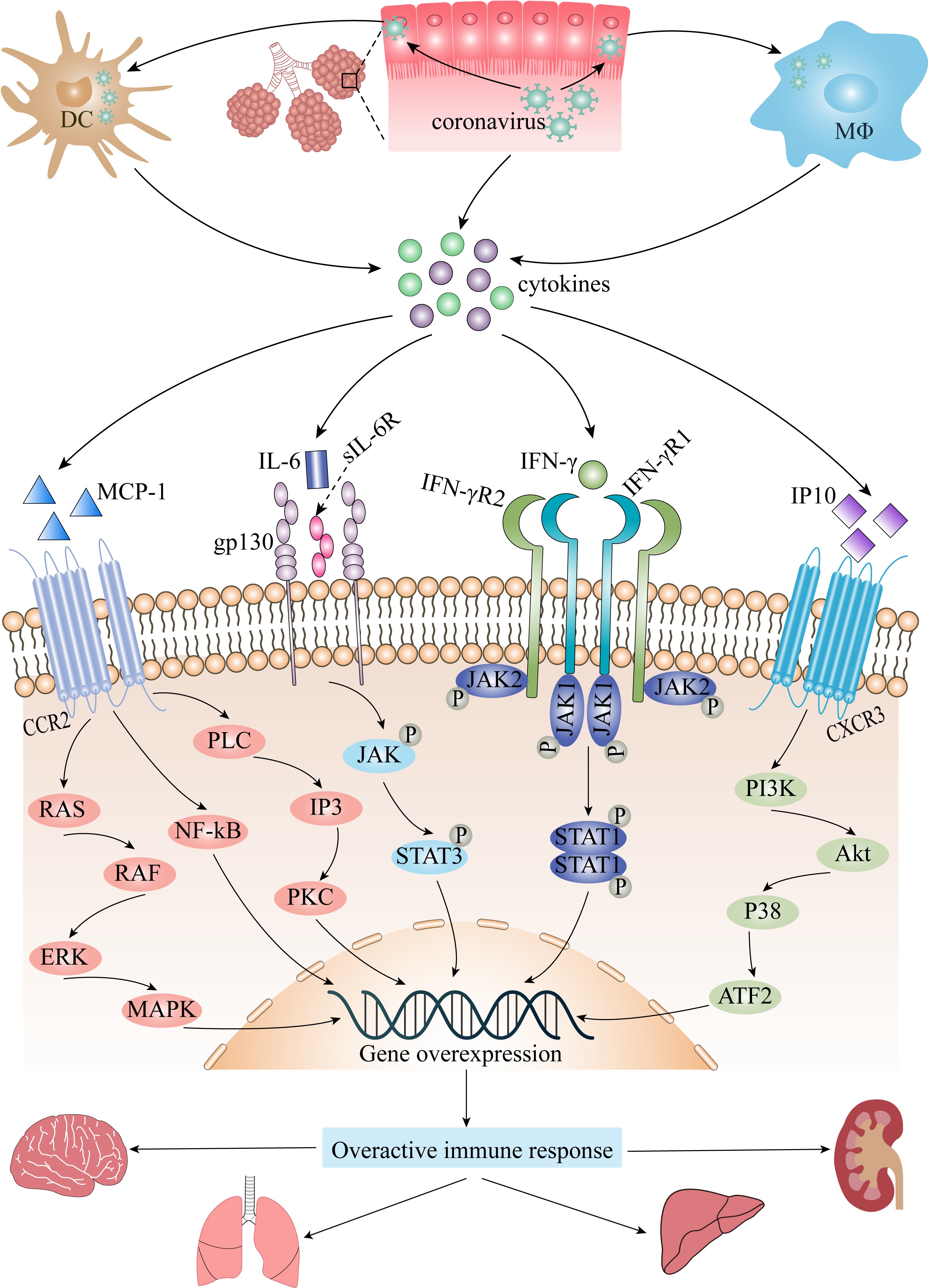
Cytokine storms play an important role in highly pathogenic HCoV infection. Current studies have found that the main elevated cytokines in the plasma of critically ill patients with SARS, MERS, or COVID-19 are IFN-γ, IL-6, IP10, and MCP-1 [10, 77, 83]. Therefore, a cytokine storm may trigger the downstream cascade signal pathway through the binding of these proinflammatory cytokines and chemokines to their respective receptors, then regulate the expression of a series of corresponding genes and participate in severe injury caused by highly pathogenic HCoV infection (Fig. 1).

Fig. 1.

The role of cytokine storm in coronavirus infection. Akt, protein kinase B; ATF2, activating transcription factor 2; CCR2, CC chemokine receptor 2; CXCR3, CXC chemokine receptor 3; DC, dendritic cell; ERK, extracellular signal regulated kinase; gp130, interleukin-6 receptor subunit-beta; IFN-γ, interferon-gamma; IFN-γR, interferon-gamma receptor; IL-6, interleukin-6; IP10, interferon-gamma inducible protein 10; IP3, inositol 1,4,5-trisphosphate; JAK, Janus kinase; MAPK, mitogen-activated protein kinase; MCP-1, monocyte chemoattractant protein-1; MΦ, macrophage; NF-κB, nuclear factor-kappa B; PI3K, phosphatidylinositol 3-kinases; PKC, protein kinase C; PLC, phospholipase C; sIL-6R, soluble forms of interleukin-6 receptor; STAT, signal transducer and activator of transcription.

4.1 IFN-γ

IFNs are a class of cytokines that play an important role in the innate immune response induced by viral infection [96]. According to the specificity of the receptor, they can be divided into three types—type I, type II, and type III. IFN-γ is the only type II IFN, and its receptor is IFN-γR1/IFN-γR2. In addition, IFN-γ is also important for adaptive immunity, especially Th1 cell-mediated immunity, and it can enhance the antiviral activity of type I IFN [97, 98]. After binding to its receptor, IFN-γ activates Janus kinase (JAK) 1 and JAK2, and then recruits and phosphorylates signal transducer and activator of transcription (STAT) 1. Phosphorylated STAT1 homodimers form the IFN-γ activation factor (GAF), then enter the nucleus and bind to IFN-γ-activated sequence (GAS) to activate the expression of IFN-stimulated genes (ISGs) [99]. Normally, the products encoded by ISGs have antiviral or immunomodulatory functions [68]. However, when there is a large amount of IFN-γ in the serum of patients with severe SARS or MERS, this induces an excessive expression of ISGs through the JAK–STAT signaling pathway, which leads to immune disorders and cytokine storms, and aggravates tissue or organ damage caused by highly pathogenic HCoV infection.

The cyclic GMP-AMP synthase (cGAS)-stimulator of interferon genes (STING) signaling pathway is important for the production of IFN. cGAS, a pattern recognition receptor of cytoplasmic double-stranded DNA (dsDNA), catalyzes the reaction between GTP and ATP and generates the small molecule cyclic GMP-AMP (cGAMP) after sensing the dsDNA derived from pathogens or damage of host tissues, and then activates STING to induce the phosphorylation of TANK-binding kinase 1 (TBK1) and interferon regulatory factor 3 (IRF3), which ultimately induces IFN expression and secretion [100, 101]. Previous reports have indicated that both SARS-CoV and SARS-CoV2 enter cells via binding with angiotensin converting enzyme 2 (ACE2) receptor to cause tissue and organ damage [62]. In addition, self-DNA release due to tissue damage can over-activate the cGAS-STING pathway and cause lung injury [102]. Meanwhile, as mentioned previously, IFN-γ level is increased in SARS, MERS and COVID-19 patients [76, 82, 90]. Therefore, it is speculated that the self-DNA released after tissue and organ damage caused by viral virulence after HCoV infection over-activates the cGAS-STING pathway, and then induces the increases in the levels of IFN, thus amplifying the damage of highly pathogenic HCoV to the body.

4.2 IL-6

IL-6 is an important cytokine that participates in the regulation of immune response [103]. Its receptors include IL-6 receptor-α (IL-6R) and the soluble forms of IL-6R (sIL-6R). There are two signaling pathways in which IL-6 plays a role: the classic signaling pathway and the trans-signaling pathway [104]. In the classic signaling pathway, IL-6 binds to IL-6R and then forms a heterohexameric complex with the IL-6 receptor subunit-β (gp130), which in turn activates the JAK–STAT3 pathway and leads to the transcription of STAT3 target genes [104]. Because IL-6R is mainly expressed in the specific subsets of leukocytes, megakaryocytes, hepatocytes, and some epithelial cells, the classical IL-6 signaling pathway is limited in these types of cells [105]. In the trans-signaling pathway, IL-6 binds to sIL-6R and then binds to gp130 to form a polymer, thereby activating the JAK-STAT3 pathway [104]. Although both of them activate the JAK-STAT3 pathway, the current view is that when the concentration of IL-6 is low, the anti-inflammatory effect of IL-6 may be expressed through the classic signaling pathway; however, when the concentration of IL-6 increases, the proinflammatory effect of IL-6 may be expressed by initiating the trans-signaling pathway [106]. Therefore, the high concentration of IL-6 in the serum of patients with highly pathogenic HCoV infection may aggravate tissue or organ damage through the IL-6 trans-signaling pathway.

Although IL-6 is significantly elevated in patients with severe COVID-19 and MERS, as previously noted, and the cytokine storm is strongly associated with patient mortality. However, it should be noted that the cytokine response of patients with severe COVID-19 is distinct from that of patients with bacterial sepsis, in particular, the elevated IL-6 levels are significantly lower [107, 108]. Very recently, a study has confirmed that routine use of tocilizumab in hospitalized COVID-19 patients is not supported [109]. Moreover, studies have shown that anti-IL-6 receptor treatments fail to benefit the survival of hospitalized patients with COVID-19, and even increase the secondary infections in severe patients [110, 111]. These results suggest that IL-6 plays a complex role in the occurrence and development of COVID-19, and the specific mechanism remains unclear. Studies have indicated that plasma plasminogen activator inhibitor-1 (PAI-1) in patients with severe COVID-19 is positively related to IL-6 levels, and the production of PAI-1 can be reduced by inhibiting IL-6 signaling pathway [112]. PAI-1 is a factor associated with vascular endothelial injury, and its increase indicates vascular endothelial lesions and coagulation dysfunction. Interestingly, thromboembolic events be more frequent in COVID-19 associated cytokine storm compared to other cytokine storms [113]. Therefore, the abnormally elevated IL-6 in patients with highly pathogenic HCoV may be involved in the occurrence of thromboembolic events in severe patients by promoting the production of PAI-1. In addition, low 25 (OH) vitamin D levels were associated with disease severity in patients hospitalized with COVID-19 [114]. Furthermore, studies have found that vitamin D3 is associated with the inflammatory response of virus infected patients, especially can reduce the level of IL-6 in patients with RSV infection [115]. Does this imply that the pathogenic mechanism of IL-6 in the occurrence and development of coronavirus infection is regulated by vitamin D3. In conclusion, current studies have shown that IL-6 plays a crucial role in the process of highly pathogenic HCoV infection, and more studies are needed to further clarify the specific mechanism in the future.

4.3 IP10

IP10, a chemokine with a molecular weight of 10 kDa, is induced by IFN-γ, and is mainly expressed by neutrophils, eosinophils, monocytes, epithelial cells, endothelial cells, and stromal cells [116, 117]. Its only specific receptor is CXC chemokine receptor (CXCR) 3, a transmembrane G protein coupled receptor, which is mainly expressed on T cells, B cells, natural killer cells, macrophages, and dendritic cells [118]. After binding to the receptor CXCR3, IP10 activates a variety of downstream signal pathways, resulting in increased chemotactic activity, thereby regulating immune response, cell proliferation, and other biological functions [119, 120]. Study has confirmed that IP10-CXCR3 signaling pathway plays a vital role in the pathological deterioration of ARDS caused by virus or non-virus [121]. In addition, IP10 may be a key chemokine for COVID-19 associated cytokine storm and an independent risk factor for the increase in mechanical ventilation time after COVID-19 patients develop ARDS, and it is expected to become a potential therapeutic target for COVID-19 [122, 123].

It is worth pointing out that IP10 was significantly elevated in severe SARS, MERS and COVID-19 patients, but the specific role of IP10 remains unclear. Study has shown that IP10 can inhibit vascular endothelial repair, which means IP10 can promote thrombosis [124]. Interestingly, autopsy pathological evidences have found that thrombosis is a common clinical manifestation in patients with severe COVID-19 [125]. In addition, a previous study suggested that IP10 has neurotoxic potential [126]. Meanwhile, some scholars believe that IP10 plays a key role in olfactory dysfunction in COVID-19 patients [127]. These lines of evidence suggest that IP10 may be involved in the development of highly pathogenic HCoV infection by damaging vascular endothelia and exerting neurotoxicity. However, the specific mechanisms that cause these pathological processes are still being explored. It is known that macrophages expressing high levels of CXCR3 can be recruited by IP10 to produce large amounts of IL-6. In addition, high level of IL-6 can induce the high expression of IP10 in the cerebrospinal fluid of patients with subarachnoid hemorrhage [128]. Thus, whether IP10 and IL-6 form a positive feedback loop in the occurrence and development of the disease and eventually cause cytokine storm. It has been found that a high concentration of IP10 in the serum of patients with H1N1 can mediate the formation of ALI by activating the phosphatidylinositol 3-kinases (PI3K)-protein kinase B (Akt)-p38-activating transcription factor 2 (ATF2) signaling pathway [129]. Based on this, we speculate that excessive IP10 in the serum of critically ill patients with highly pathogenic HCoV may also mediate tissue or organ injury by activating the PI3K-Akt-p38-ATF2 signaling pathway.

4.4 MCP-1

MCP-1 is a chemokine with a molecular weight of 13 kDa that belongs to the CC subfamily [130]. It can be expressed by a variety of cells, including monocytes, macrophages, endothelial cells, smooth muscle cells, fibroblasts, epithelial cells, mesangial cells, astrocytes and microglial cells [131, 132]. Its specific receptor, CCR2, is a G protein coupled receptor that crosses the membrane seven times [131]. After binding to CCR2, MCP-1 activates a series of signaling pathways, regulates the activation and chemotactic migration of target cells, and then participates in the regulation of immune response [133]. The specific signaling pathways include phospholipase C (PLC)-inositol 1,4,5-trisphosphate (IP3)-rotein kinase C (PKC), nuclear factor-kappa B (NF-κB), and extracellular signal regulated kinases (ERKs), c-Jun N-terminal kinase, and mitogen-activated protein kinase (MAPK) [134, 135]. According to the chemotactic mechanism of MCP-1, it can be known that a large amount of MCP-1 in the serum of patients with SARS, MERS, or COVID-19 excessively activates downstream signaling pathways, which makes inflammatory cells largely chemotactic. Consequently, inflammatory cells potentially contribute to cytokine storms by expressing a large number of proinflammatory cytokines and chemokines, thus resulting in tissue or organ damage.

5. Conclusions

Highly pathogenic HCoV infections can cause respiratory diseases with varying severity, with high morbidity and mortality. Increased levels of proinflammatory cytokines and chemokines were found in the plasma of patients with SARS, MERS, or COVID-19, and this increase was related to the severity of the disease. Therefore, it is considered that cytokine storm is associated with the pathogenic mechanism of highly pathogenic HCoV infection. In addition, the increased cytokines in the plasma of critically ill patients with SARS, of MERS and COVID-19 were mainly IFN-γ, IL-6, IP-10, and MCP-1; in particular, the increased levels of IP-10 or MCP-1 in the plasma of all critically ill has been noted patients. Therefore, it is considered that the cytokine storm in highly pathogenic HCoV infection triggers the downstream cascade signal pathway through the binding of these proinflammatory cytokines and chemokines to their respective receptors, thus aggravating tissue or organ damage. Selective blocking of the IFN-γ, IL-6, IP10, and MCP-1 corresponding signaling pathways is beneficial in mitigating the damage caused by highly pathogenic HCoV infection.

Abbreviations

ACE2, angiotensin converting enzyme 2; Akt, protein kinase B; ALI, acute lung injury; ARDS, acute respiratory distress syndrome; ATF2, activating transcription factor 2; CCR2, CC chemokine receptor 2; cGAMP, cyclic GMP-AMP; cGAS, cyclic GMP-AMP synthase; COVID-19, coronavirus disease-19; CXCR3, CXC chemokine receptor 3; DC, dendritic cell; dsDNA, double-stranded DNA; ERK, extracellular signal regulated kinase; GAF, IFN-γ activation factor; GAS, IFN-γ-activated sequence; G-CSF, granulocyte colony-stimulating factor; gp130, interleukin-6 receptor subunit-beta; HCoVs, human coronavirus; IFN, interferon; IFN-γ, interferon-gamma; IFN-γR, interferon-gamma receptor; IL, interleukin; IL-6, interleukin-6; IL-6R, IL-6 receptor; IP3, inositol 1,4,5-trisphosphate; IP10, interferon-gamma inducible protein 10; IRF3, interferon regulatory factor 3; ISGs, IFN-stimulated genes; JAK, Janus kinase; MAPK, mitogen-activated protein kinase; MCP-1, monocyte chemoattractant protein-1; MERS, Middle East respiratory syndrome; MIP, macrophage inflammatory protein; MΦ, macrophage; NF-κB, nuclear factor-kappa B; PAI-1, plasminogen activator inhibitor-1; PI3K, phosphatidylinositol 3-kinases; PKC, protein kinase C; PLC, phospholipase C; RANTES, regulated upon activation normal T-cell expressed and secreted; RSV, respiratory syncytial virus; SARS, severe acute respiratory syndrome; sIL-6R, soluble forms of interleukin-6 receptor; STAT, signal transducer and activator of transcription; STING, stimulator of interferon genes; TBK1, TANK-binding kinase 1; TNF, tumor necrosis factor; WHO, World Health Organization.

Author contributions

CY and BZ designed the work. RX, CL, XX, YH, BZ and CY collected and reviewed the references. RX wrote the first draft. CY and BZ wrote and reviewed the final version of the manuscript. All authors discussed and contributed to the manuscript.

Ethics approval and consent to participate

Not applicable.

Acknowledgment

Not applicable.

Funding

This research received no external funding.

Conflict of interest

The authors declare no conflict of interest. Chun Yang is serving as one of the Editorial Board members of this journal. We declare that Chun Yang had no involvement in the peer review of this article and has no access to information regarding its peer review. Full responsibility for the editorial process for this article was delegated to Graham Pawelec.

References
[1]
Weiss SR, Navas-Martin S. Coronavirus Pathogenesis and the Emerging Pathogen Severe Acute Respiratory Syndrome Coronavirus. Microbiology and Molecular Biology Reviews. 2005; 69: 635–664.
[2]
Kin N, Miszczak F, Lin W, Gouilh MA, Vabret A. Genomic Analysis of 15 Human Coronaviruses OC43 (HCoV-OC43s) Circulating in France from 2001 to 2013 Reveals a High Intra-Specific Diversity with New Recombinant Genotypes. Viruses. 2016; 7: 2358–2377.
[3]
Forni D, Cagliani R, Clerici M, Sironi M. Molecular Evolution of Human Coronavirus Genomes. Trends in Microbiology. 2017; 25: 35–48.
[4]
Cui J, Li F, Shi Z. Origin and evolution of pathogenic coronaviruses. Nature Reviews Microbiology. 2019; 17: 181–192.
[5]
Thompson R. Pandemic potential of 2019-nCoV. The Lancet Infectious Diseases. 2020; 20: 280.
[6]
Peiris JSM, Lai ST, Poon LLM, Guan Y, Yam LYC, Lim W, et al. Coronavirus as a possible cause of severe acute respiratory syndrome. Lancet. 2003; 361: 1319–1325.
[7]
Raj VS, Osterhaus AD, Fouchier RA, Haagmans BL. MERS: Emergence of a novel human coronavirus. Current Opinion in Virology. 2014; 5: 58–62.
[8]
Drosten C, Günther S, Preiser W, van der Werf S, Brodt H, Becker S, et al. Identification of a novel coronavirus in patients with severe acute respiratory syndrome. The New England Journal of Medicine. 2003; 348: 1967–1976.
[9]
Zaki AM, van Boheemen S, Bestebroer TM, Osterhaus ADME, Fouchier RAM. Isolation of a novel coronavirus from a man with pneumonia in Saudi Arabia. The New England Journal of Medicine. 2012; 367: 1814–1820.
[10]
Huang C, Wang Y, Li X, Ren L, Zhao J, Hu Y, et al. Clinical features of patients infected with 2019 novel coronavirus in Wuhan, China. The Lancet. 2020; 395: 497–506.
[11]
Channappanavar R, Perlman S. Pathogenic human coronavirus infections: causes and consequences of cytokine storm and immunopathology. Seminars in Immunopathology. 2018; 39: 529–539.
[12]
Cheng VCC, Lau SKP, Woo PCY, Yuen KY. Severe acute respiratory syndrome coronavirus as an agent of emerging and reemerging infection. Clinical Microbiology Reviews. 2007; 20: 660–694.
[13]
Lee N, Hui D, Wu A, Chan P, Cameron P, Joynt GM, et al. A major outbreak of severe acute respiratory syndrome in Hong Kong. The New England Journal of Medicine. 2003; 348: 1986–1994.
[14]
Kuiken T, Fouchier RAM, Schutten M, Rimmelzwaan GF, van Amerongen G, van Riel D, et al. Newly discovered coronavirus as the primary cause of severe acute respiratory syndrome. Lancet. 2003; 362: 263–270.
[15]
Ksiazek TG, Erdman D, Goldsmith CS, Zaki SR, Peret T, Emery S, et al. A novel coronavirus associated with severe acute respiratory syndrome. The New England Journal of Medicine. 2003; 348: 1953–1966.
[16]
Smith RD. Responding to global infectious disease outbreaks: lessons from SARS on the role of risk perception, communication and management. Social Science & Medicine. 2007; 63: 3113–3123.
[17]
Guan Y, Zheng BJ, He YQ, Liu XL, Zhuang ZX, Cheung CL, et al. Isolation and characterization of viruses related to the SARS coronavirus from animals in southern China. Science. 2003; 302: 276–278.
[18]
Kan B, Wang M, Jing H, Xu H, Jiang X, Yan M, et al. Molecular evolution analysis and geographic investigation of severe acute respiratory syndrome coronavirus-like virus in palm civets at an animal market and on farms. Journal of Virology. 2005; 79: 11892–11900.
[19]
Tu C, Crameri G, Kong X, Chen J, Sun Y, Yu M, et al. Antibodies to SARS coronavirus in civets. Emerging Infectious Diseases. 2005; 10: 2244–2248.
[20]
Lau SKP, Woo PCY, Li KSM, Huang Y, Tsoi H, Wong BHL, et al. Severe acute respiratory syndrome coronavirus-like virus in Chinese horseshoe bats. Proceedings of the National Academy of Sciences of the United States of America. 2005; 102: 14040–14045.
[21]
Hu B, Zeng L, Yang X, Ge X, Zhang W, Li B, et al. Discovery of a rich gene pool of bat SARS-related coronaviruses provides new insights into the origin of SARS coronavirus. PLoS Pathogens. 2017; 13: e1006698.
[22]
Peiris JSM, Yuen KY, Osterhaus ADME, Stöhr K. The severe acute respiratory syndrome. The New England Journal of Medicine. 2003; 349: 2431–2441.
[23]
Hui DS, Azhar EI, Kim Y, Memish ZA, Oh M, Zumla A. Middle East respiratory syndrome coronavirus: risk factors and determinants of primary, household, and nosocomial transmission. The Lancet Infectious Diseases. 2018; 18: e217–e227.
[24]
Drosten C, Kellam P, Memish ZA. Evidence for camel-to-human transmission of MERS coronavirus. The New England Journal of Medicine. 2014; 371: 1359–1360.
[25]
Conzade R, Grant R, Malik MR, Elkholy A, Elhakim M, Samhouri D, et al. Reported Direct and Indirect Contact with Dromedary Camels among Laboratory-Confirmed MERS-CoV Cases. Viruses. 2019; 10.
[26]
Anthony SJ, Gilardi K, Menachery VD, Goldstein T, Ssebide B, Mbabazi R, et al. Further Evidence for Bats as the Evolutionary Source of Middle East Respiratory Syndrome Coronavirus. MBio. 2018; 8.
[27]
Al-Tawfiq JA, Auwaerter PG. Healthcare-associated infections: the hallmark of Middle East respiratory syndrome coronavirus with review of the literature. Journal of Hospital Infection. 2019; 101: 20–29.
[28]
Al-Tawfiq JA, Hinedi K, Ghandour J, Khairalla H, Musleh S, Ujayli A, et al. Middle East respiratory syndrome coronavirus: a case-control study of hospitalized patients. Clinical Infectious Diseases. 2014; 59: 160–165.
[29]
Assiri A, Al-Tawfiq JA, Al-Rabeeah AA, Al-Rabiah FA, Al-Hajjar S, Al-Barrak A, et al. Epidemiological, demographic, and clinical characteristics of 47 cases of Middle East respiratory syndrome coronavirus disease from Saudi Arabia: a descriptive study. The Lancet. Infectious Diseases. 2013; 13: 752–761.
[30]
Arabi YM, Arifi AA, Balkhy HH, Najm H, Aldawood AS, Ghabashi A, et al. Clinical Course and Outcomes of Critically Ill Patients with Middle East Respiratory Syndrome Coronavirus Infection. Annals of Internal Medicine. 2014; 160: 389–397.
[31]
Lu H, Stratton CW, Tang Y. Outbreak of pneumonia of unknown etiology in Wuhan, China: the mystery and the miracle. Journal of Medical Virology. 2020; 92: 401–402.
[32]
Hui DS, I Azhar E, Madani TA, Ntoumi F, Kock R, Dar O, et al. The continuing 2019-nCoV epidemic threat of novel coronaviruses to global health — the latest 2019 novel coronavirus outbreak in Wuhan, China. International Journal of Infectious Diseases. 2020; 91: 264–266.
[33]
Situation Reports. Available at: https://www.who.int/emergencies/diseases/novel-coronavirus-2019/situation-reports (Accessed: 2 November 2021).
[34]
Shereen MA, Khan S, Kazmi A, Bashir N, Siddique R. COVID-19 infection: Origin, transmission, and characteristics of human coronaviruses. Journal of Advanced Research. 2020; 24: 91–98.
[35]
Sun K, Wang W, Gao L, Wang Y, Luo K, Ren L, et al. Transmission heterogeneities, kinetics, and controllability of SARS-CoV-2. Science. 2021; 371.
[36]
Lauer SA, Grantz KH, Bi Q, Jones FK, Zheng Q, Meredith HR, et al. The Incubation Period of Coronavirus Disease 2019 (COVID-19) from Publicly Reported Confirmed Cases: Estimation and Application. Annals of Internal Medicine. 2020; 172: 577–582.
[37]
Wu A, Peng Y, Huang B, Ding X, Wang X, Niu P, et al. Genome Composition and Divergence of the Novel Coronavirus (2019-nCoV) Originating in China. Cell Host & Microbe. 2020; 27: 325–328.
[38]
Wu F, Zhao S, Yu B, Chen Y, Wang W, Song Z, et al. A new coronavirus associated with human respiratory disease in China. Nature. 2020; 579: 265–269.
[39]
Lau SKP, Luk HKH, Wong ACP, Li KSM, Zhu L, He Z, et al. Possible Bat Origin of Severe Acute Respiratory Syndrome Coronavirus 2. Emerging Infectious Diseases. 2020; 26: 1542–1547.
[40]
Chen N, Zhou M, Dong X, Qu J, Gong F, Han Y, et al. Epidemiological and clinical characteristics of 99 cases of 2019 novel coronavirus pneumonia in Wuhan, China: a descriptive study. The Lancet. 2020; 395: 507–513.
[41]
Wang D, Hu B, Hu C, Zhu F, Liu X, Zhang J, et al. Clinical Characteristics of 138 Hospitalized Patients with 2019 Novel Coronavirus–Infected Pneumonia in Wuhan, China. JAMA. 2020; 323: 1061.
[42]
Guan W, Ni Z, Hu Y, Liang W, Ou C, He J, et al. Clinical Characteristics of Coronavirus Disease 2019 in China. New England Journal of Medicine. 2020; 382: 1708–1720.
[43]
Pan L, Mu M, Yang P, Sun Y, Wang R, Yan J, et al. Clinical Characteristics of COVID-19 Patients with Digestive Symptoms in Hubei, China: a Descriptive, Cross-Sectional, Multicenter Study. American Journal of Gastroenterology. 2020; 115: 766–773.
[44]
Giacomelli A, Pezzati L, Conti F, Bernacchia D, Siano M, Oreni L, et al. Self-reported Olfactory and Taste Disorders in Patients with Severe Acute Respiratory Coronavirus 2 Infection: a Cross-sectional Study. Clinical Infectious Diseases. 2020; 71: 889–890.
[45]
Pensato U, Muccioli L, Cani I, Janigro D, Zinzani PL, Guarino M, et al. Brain dysfunction in COVID‐19 and CAR‐T therapy: cytokine storm‐associated encephalopathy. Annals of Clinical and Translational Neurology. 2021; 8: 968–979.
[46]
Perrin P, Collongues N, Baloglu S, Bedo D, Bassand X, Lavaux T, et al. Cytokine release syndrome‐associated encephalopathy in patients with COVID‐19. European Journal of Neurology. 2021; 28: 248–258.
[47]
Zhang J, Cao Y, Tan G, Dong X, Wang B, Lin J, et al. Clinical, radiological, and laboratory characteristics and risk factors for severity and mortality of 289 hospitalized COVID‐19 patients. Allergy. 2021; 76: 533–550.
[48]
Wu Z, McGoogan JM. Characteristics of and Important Lessons from the Coronavirus Disease 2019 (COVID-19) Outbreak in China. JAMA. 2020; 323: 1239.
[49]
Karagiannidis C, Mostert C, Hentschker C, Voshaar T, Malzahn J, Schillinger G, et al. Case characteristics, resource use, and outcomes of 10 021 patients with COVID-19 admitted to 920 German hospitals: an observational study. The Lancet Respiratory Medicine. 2020; 8: 853–862.
[50]
Rosenthal N, Cao Z, Gundrum J, Sianis J, Safo S. Risk Factors Associated with in-Hospital Mortality in a us National Sample of Patients with COVID-19. JAMA Network Open. 2020; 3: e2029058.
[51]
Gruber J, Prinstein MJ, Clark LA, Rottenberg J, Abramowitz JS, Albano AM, et al. Mental health and clinical psychological science in the time of COVID-19: Challenges, opportunities, and a call to action. American Psychologist Journal. 2021; 76: 409–426.
[52]
Ayoubkhani D, Khunti K, Nafilyan V, Maddox T, Humberstone B, Diamond I, et al. Post-covid syndrome in individuals admitted to hospital with covid-19: retrospective cohort study. The British Medical Journal. 2021; 372: n693.
[53]
Burra P, Soto-Díaz K, Chalen I, Gonzalez-Ricon RJ, Istanto D, Caetano-Anollés G. Temperature and Latitude Correlate with SARS-CoV-2 Epidemiological Variables but not with Genomic Change Worldwide. Evolutionary Bioinformatics. 2021; 17: 117693432198969.
[54]
Sajadi MM, Habibzadeh P, Vintzileos A, Shokouhi S, Miralles-Wilhelm F, Amoroso A. Temperature, Humidity, and Latitude Analysis to Estimate Potential Spread and Seasonality of Coronavirus Disease 2019 (COVID-19). JAMA Network Open. 2020; 3: e2011834.
[55]
Nichols GL, Gillingham EL, Macintyre HL, Vardoulakis S, Hajat S, Sarran CE, et al. Coronavirus seasonality, respiratory infections and weather. BMC Infectious Diseases. 2021; 21: 1101.
[56]
Fontal A, Bouma MJ, San-José A, López L, Pascual M, Rodó X. Climatic signatures in the different COVID-19 pandemic waves across both hemispheres. Nature Computational Science. 2021; 1: 655–665.
[57]
Song P, Li W, Xie J, Hou Y, You C. Cytokine storm induced by SARS-CoV-2. Clinica Chimica Acta. 2020; 509: 280–287.
[58]
Zhao D, Yao F, Wang L, Zheng L, Gao Y, Ye J, et al. A Comparative Study on the Clinical Features of Coronavirus 2019 (COVID-19) Pneumonia With Other Pneumonias. Clinical Infectious Diseases. 2020; 71: 756–761.
[59]
Piroth L, Cottenet J, Mariet A, Bonniaud P, Blot M, Tubert-Bitter P, et al. Comparison of the characteristics, morbidity, and mortality of COVID-19 and seasonal influenza: a nationwide, population-based retrospective cohort study. The Lancet Respiratory Medicine. 2021; 9: 251–259.
[60]
Idilman IS, Telli Dizman G, Ardali Duzgun S, Irmak I, Karcaaltincaba M, Inkaya AC, et al. Lung and kidney perfusion deficits diagnosed by dual-energy computed tomography in patients with COVID-19-related systemic microangiopathy. European Radiology. 2021; 31: 1090–1099.
[61]
Peiris JSM, Chu CM, Cheng VCC, Chan KS, Hung IFN, Poon LLM, et al. Clinical progression and viral load in a community outbreak of coronavirus-associated SARS pneumonia: a prospective study. Lancet. 2003; 361: 1767–1772.
[62]
Moore JB, June CH. Cytokine release syndrome in severe COVID-19. Science. 2020; 368: 473–474.
[63]
Webb BJ, Peltan ID, Jensen P, Hoda D, Hunter B, Silver A, et al. Clinical criteria for COVID-19-associated hyperinflammatory syndrome: a cohort study. The Lancet Rheumatology. 2020; 2: e754–e763.
[64]
Li G, Fan Y, Lai Y, Han T, Li Z, Zhou P, et al. Coronavirus infections and immune responses. Journal of Medical Virology. 2020; 92: 424–432.
[65]
Medzhitov R. Recognition of microorganisms and activation of the immune response. Nature. 2007; 449: 819–826.
[66]
Barton GM. A calculated response: control of inflammation by the innate immune system. The Journal of Clinical Investigation. 2008; 118: 413–420.
[67]
Hussell T, Goulding J. Structured regulation of inflammation during respiratory viral infection. The Lancet. Infectious Diseases. 2010; 10: 360–366.
[68]
Tisoncik JR, Korth MJ, Simmons CP, Farrar J, Martin TR, Katze MG. Into the eye of the cytokine storm. Microbiology and Molecular Biology Reviews. 2012; 76: 16–32.
[69]
Cheung CY, Poon LLM, Ng IHY, Luk W, Sia S, Wu MHS, et al. Cytokine responses in severe acute respiratory syndrome coronavirus-infected macrophages in vitro: possible relevance to pathogenesis. Journal of Virology. 2005; 79: 7819–7826.
[70]
Law HKW, Cheung CY, Ng HY, Sia SF, Chan YO, Luk W, et al. Chemokine up-regulation in SARS-coronavirus-infected, monocyte-derived human dendritic cells. Blood. 2005; 106: 2366–2374.
[71]
Yen Y, Liao F, Hsiao C, Kao C, Chen Y, Wu-Hsieh BA. Modeling the Early Events of Severe Acute Respiratory Syndrome Coronavirus Infection in Vitro. Journal of Virology. 2006; 80: 2684–2693.
[72]
Chien J, Hsueh P, Cheng W, Yu C, Yang P. Temporal changes in cytokine/chemokine profiles and pulmonary involvement in severe acute respiratory syndrome. Respirology. 2007; 11: 715–722.
[73]
Wong CK, Lam CWK, Wu AKL, Ip WK, Lee NLS, Chan IHS, et al. Plasma inflammatory cytokines and chemokines in severe acute respiratory syndrome. Clinical and Experimental Immunology. 2004; 136: 95–103.
[74]
Zhang Y, Li J, Zhan Y, Wu L, Yu X, Zhang W, et al. Analysis of Serum Cytokines in Patients with Severe Acute Respiratory Syndrome. Infection and Immunity. 2004; 72: 4410–4415.
[75]
Cameron MJ, Bermejo-Martin JF, Danesh A, Muller MP, Kelvin DJ. Human immunopathogenesis of severe acute respiratory syndrome (SARS). Virus Research. 2008; 133: 13–19.
[76]
Theron M, Huang K, Chen Y, Liu C, Lei H. A probable role for IFN-gamma in the development of a lung immunopathology in SARS. Cytokine. 2008; 32: 30–38.
[77]
Cameron MJ, Ran L, Xu L, Danesh A, Bermejo-Martin JF, Cameron CM, et al. Interferon-mediated immunopathological events are associated with atypical innate and adaptive immune responses in patients with severe acute respiratory syndrome. Journal of Virology. 2007; 81: 8692–8706.
[78]
Mubarak A, Alturaiki W, Hemida MG. Middle East Respiratory Syndrome Coronavirus (MERS-CoV): Infection, Immunological Response, and Vaccine Development. Journal of Immunology Research. 2019; 2019: 6491738.
[79]
Zhou J, Chu H, Li C, Wong BH, Cheng Z, Poon VK, et al. Active Replication of Middle East Respiratory Syndrome Coronavirus and Aberrant Induction of Inflammatory Cytokines and Chemokines in Human Macrophages: Implications for Pathogenesis. The Journal of Infectious Diseases. 2014; 209: 1331–1342.
[80]
Scheuplein VA, Seifried J, Malczyk AH, Miller L, Höcker L, Vergara-Alert J, et al. High secretion of interferons by human plasmacytoid dendritic cells upon recognition of Middle East respiratory syndrome coronavirus. Journal of Virology. 2015; 89: 3859–3869.
[81]
Lau SKP, Lau CCY, Chan K, Li CPY, Chen H, Jin D, et al. Delayed induction of proinflammatory cytokines and suppression of innate antiviral response by the novel Middle East respiratory syndrome coronavirus: implications for pathogenesis and treatment. The Journal of General Virology. 2014; 94: 2679–2690.
[82]
Mahallawi WH, Khabour OF, Zhang Q, Makhdoum HM, Suliman BA. MERS-CoV infection in humans is associated with a pro-inflammatory Th1 and Th17 cytokine profile. Cytokine. 2018; 104: 8–13.
[83]
Shin H, Kim Y, Kim G, Lee JY, Jeong I, Joh J, et al. Immune Responses to Middle East Respiratory Syndrome Coronavirus during the Acute and Convalescent Phases of Human Infection. Clinical Infectious Diseases. 2019; 68: 984–992.
[84]
Kim ES, Choe PG, Park WB, Oh HS, Kim EJ, Nam EY, et al. Clinical Progression and Cytokine Profiles of Middle East Respiratory Syndrome Coronavirus Infection. Journal of Korean Medical Science. 2017; 31: 1717–1725.
[85]
Min C, Cheon S, Ha N, Sohn KM, Kim Y, Aigerim A, et al. Comparative and kinetic analysis of viral shedding and immunological responses in MERS patients representing a broad spectrum of disease severity. Scientific Reports. 2018; 6: 25359.
[86]
Xu Z, Shi L, Wang Y, Zhang J, Huang L, Zhang C, et al. Pathological findings of COVID-19 associated with acute respiratory distress syndrome. The Lancet Respiratory Medicine. 2020; 8: 420–422.
[87]
Ng DL, Al Hosani F, Keating MK, Gerber SI, Jones TL, Metcalfe MG, et al. Clinicopathologic, Immunohistochemical, and Ultrastructural Findings of a Fatal Case of Middle East Respiratory Syndrome Coronavirus Infection in the United Arab Emirates, April 2014. The American Journal of Pathology. 2016; 186: 652–658.
[88]
Ding Y, Wang H, Shen H, Li Z, Geng J, Han H, et al. The clinical pathology of severe acute respiratory syndrome (SARS): a report from China. The Journal of Pathology. 2003; 200: 282–289.
[89]
Chen G, Wu D, Guo W, Cao Y, Huang D, Wang H, et al. Clinical and immunological features of severe and moderate coronavirus disease 2019. Journal of Clinical Investigation. 2020; 130: 2620–2629.
[90]
Song J, Zhang C, Fan X, Meng F, Xu Z, Xia P, et al. Immunological and inflammatory profiles in mild and severe cases of COVID-19. Nature Communications. 2020; 11: 3410.
[91]
Liu J, Li S, Liu J, Liang B, Wang X, Wang H, et al. Longitudinal characteristics of lymphocyte responses and cytokine profiles in the peripheral blood of SARS-CoV-2 infected patients. EBioMedicine. 2020; 55: 102763.
[92]
Zhou F, Yu T, Du R, Fan G, Liu Y, Liu Z, et al. Clinical course and risk factors for mortality of adult inpatients with COVID-19 in Wuhan, China: a retrospective cohort study. The Lancet. 2020; 395: 1054–1062.
[93]
Salama C, Han J, Yau L, Reiss WG, Kramer B, Neidhart JD, et al. Tocilizumab in Patients Hospitalized with Covid-19 Pneumonia. New England Journal of Medicine. 2021; 384: 20–30.
[94]
Group WHOREAfC-TW, Shankar-Hari M, Vale CL, Godolphin PJ, Fisher D, Higgins JPT, et al. Association Between Administration of IL-6 Antagonists and Mortality Among Patients Hospitalized for COVID-19: A Meta-analysis. The Journal of the American Medical Association. 2021; 326: 499–518.
[95]
Luo P, Liu Y, Qiu L, Liu X, Liu D, Li J. Tocilizumab treatment in COVID‐19: a single center experience. Journal of Medical Virology. 2020; 92: 814–818.
[96]
Fensterl V, Sen GC. Interferons and viral infections. BioFactors. 2009; 35: 14–20.
[97]
Malmgaard L. Induction and Regulation of IFNs during Viral Infections. Journal of Interferon & Cytokine Research. 2004; 24: 439–454.
[98]
Takaoka A, Yanai H. Interferon signalling network in innate defence. Cellular Microbiology. 2006; 8: 907–922.
[99]
Schneider WM, Chevillotte MD, Rice CM. Interferon-stimulated genes: a complex web of host defenses. Annual Review of Immunology. 2014; 32: 513–545.
[100]
Wu J, Sun L, Chen X, Du F, Shi H, Chen C, et al. Cyclic GMP-AMP is an endogenous second messenger in innate immune signaling by cytosolic DNA. Science. 2013; 339: 826–830.
[101]
Ablasser A, Chen ZJ. CGAS in action: Expanding roles in immunity and inflammation. Science. 2019; 363.
[102]
Ma R, Ortiz Serrano TP, Davis J, Prigge AD, Ridge KM. The cGAS‐STING pathway: the role of self‐DNA sensing in inflammatory lung disease. The FASEB Journal. 2020; 34: 13156–13170.
[103]
Hunter CA, Jones SA. IL-6 as a keystone cytokine in health and disease. Nature Immunology. 2015; 16: 448–457.
[104]
Johnson DE, O’Keefe RA, Grandis JR. Targeting the IL-6/JAK/STAT3 signalling axis in cancer. Nature Reviews. Clinical Oncology. 2019; 15: 234–248.
[105]
Wolf J, Rose-John S, Garbers C. Interleukin-6 and its receptors: a highly regulated and dynamic system. Cytokine. 2015; 70: 11–20.
[106]
Yeung YT, Aziz F, Guerrero-Castilla A, Arguelles S. Signaling Pathways in Inflammation and Anti-inflammatory Therapies. Current Pharmaceutical Design. 2019; 24: 1449–1484.
[107]
Del Valle DM, Kim-Schulze S, Huang H, Beckmann ND, Nirenberg S, Wang B, et al. An inflammatory cytokine signature predicts COVID-19 severity and survival. Nature Medicine. 2020; 26: 1636–1643.
[108]
Leisman DE, Ronner L, Pinotti R, Taylor MD, Sinha P, Calfee CS, et al. Cytokine elevation in severe and critical COVID-19: a rapid systematic review, meta-analysis, and comparison with other inflammatory syndromes. The Lancet Respiratory Medicine. 2020; 8: 1233–1244.
[109]
Soin AS, Kumar K, Choudhary NS, Sharma P, Mehta Y, Kataria S, et al. Tocilizumab plus standard care versus standard care in patients in India with moderate to severe COVID-19-associated cytokine release syndrome (COVINTOC): an open-label, multicentre, randomised, controlled, phase 3 trial. The Lancet Respiratory Medicine. 2021; 9: 511–521.
[110]
Hermine O, Mariette X, Tharaux PL, Resche-Rigon M, Porcher R, Ravaud P, et al. Effect of Tocilizumab vs Usual Care in Adults Hospitalized With COVID-19 and Moderate or Severe Pneumonia: A Randomized Clinical Trial. JAMA Internal Medicine. 2021; 181: 32–40.
[111]
Stone JH, Frigault MJ, Serling-Boyd NJ, Fernandes AD, Harvey L, Foulkes AS, et al. Efficacy of Tocilizumab in Patients Hospitalized with Covid-19. New England Journal of Medicine. 2020; 383: 2333–2344.
[112]
Kang S, Tanaka T, Inoue H, Ono C, Hashimoto S, Kioi Y, et al. IL-6 trans-signaling induces plasminogen activator inhibitor-1 from vascular endothelial cells in cytokine release syndrome. Proceedings of the National Academy of Sciences. 2020; 117: 22351–22356.
[113]
Klok FA, Kruip MJHA, van der Meer NJM, Arbous MS, Gommers D, Kant KM, et al. Confirmation of the high cumulative incidence of thrombotic complications in critically ill ICU patients with COVID-19: an updated analysis. Thrombosis Research. 2020; 191: 148–150.
[114]
De Smet D, De Smet K, Herroelen P, Gryspeerdt S, Martens GA. Serum 25(OH)D Level on Hospital Admission Associated with COVID-19 Stage and Mortality. American Journal of Clinical Pathology. 2021; 155: 381–388.
[115]
Anderson J, Do LAH, Toh ZQ, Hoe E, Reitsma A, Mulholland K, et al. Vitamin D Induces Differential Effects on Inflammatory Responses During Bacterial and/or Viral Stimulation of Human Peripheral Blood Mononuclear Cells. Frontiers in Immunology. 2020; 11: 602.
[116]
Luster AD, Ravetch JV. Genomic characterization of a gamma-interferon-inducible gene (IP-10) and identification of an interferon-inducible hypersensitive site. Molecular and Cellular Biology. 1988; 7: 3723–3731.
[117]
Dyer KD, Percopo CM, Fischer ER, Gabryszewski SJ, Rosenberg HF. Pneumoviruses infect eosinophils and elicit MyD88-dependent release of chemoattractant cytokines and interleukin-6. Blood. 2009; 114: 2649–2656.
[118]
Loetscher M, Gerber B, Loetscher P, Jones SA, Piali L, Clark-Lewis I, et al. Chemokine receptor specific for IP10 and mig: structure, function, and expression in activated T-lymphocytes. The Journal of Experimental Medicine. 1997; 184: 963–969.
[119]
Shen Q, Zhang R, Bhat NR. MAP kinase regulation of IP10/CXCL10 chemokine gene expression in microglial cells. Brain Research. 2006; 1086: 9–16.
[120]
Shahabuddin S, Ji R, Wang P, Brailoiu E, Dun N, Yang Y, et al. CXCR3 chemokine receptor-induced chemotaxis in human airway epithelial cells: role of p38 MAPK and PI3K signaling pathways. American Journal of Physiology-Cell Physiology. 2006; 291: C34–C39.
[121]
Ichikawa A, Kuba K, Morita M, Chida S, Tezuka H, Hara H, et al. CXCL10-CXCR3 enhances the development of neutrophil-mediated fulminant lung injury of viral and nonviral origin. American Journal of Respiratory and Critical Care Medicine. 2013; 187: 65–77.
[122]
Zhang N, Zhao YD, Wang XM. CXCL10 an important chemokine associated with cytokine storm in COVID-19 infected patients. European Review for Medical and Pharmacological Sciences. 2020; 24: 7497–7505.
[123]
Blot M, Jacquier M, Aho Glele LS, Beltramo G, Nguyen M, Bonniaud P, et al. CXCL10 could drive longer duration of mechanical ventilation during COVID-19 ARDS. Critical Care. 2020; 24: 632.
[124]
Lupieri A, Smirnova NF, Solinhac R, Malet N, Benamar M, Saoudi A, et al. Smooth muscle cells-derived CXCL10 prevents endothelial healing through PI3Kgamma-dependent T cells response. Cardiovascular Research. 2020; 116: 438–449.
[125]
Wichmann D. Autopsy Findings and Venous Thromboembolism in Patients with COVID-19. Annals of Internal Medicine. 2020; 173: 1030.
[126]
Quint JK, Donaldson GC, Goldring JJP, Baghai-Ravary R, Hurst JR, Wedzicha JA. Serum IP-10 as a Biomarker of Human Rhinovirus Infection at Exacerbation of COPD. Chest. 2010; 137: 812–822.
[127]
Oliviero A, de Castro F, Coperchini F, Chiovato L, Rotondi M. COVID-19 Pulmonary and Olfactory Dysfunctions: is the Chemokine CXCL10 the Common Denominator? The Neuroscientist. 2021; 27: 214–221.
[128]
Niwa A, Osuka K, Nakura T, Matsuo N, Watabe T, Takayasu M. Interleukin-6, MCP-1, IP-10, and MIG are sequentially expressed in cerebrospinal fluid after subarachnoid hemorrhage. Journal of Neuroinflammation. 2017; 13: 217.
[129]
Wang W, Yang P, Zhong Y, Zhao Z, Xing L, Zhao Y, et al. Monoclonal antibody against CXCL-10/IP-10 ameliorates influenza a (H1N1) virus induced acute lung injury. Cell Research. 2013; 23: 577–580.
[130]
Van Coillie E, Van Damme J, Opdenakker G. The MCP/eotaxin subfamily of CC chemokines. Cytokine & Growth Factor Reviews. 1999; 10: 61–86.
[131]
Deshmane SL, Kremlev S, Amini S, Sawaya BE. Monocyte Chemoattractant Protein-1 (MCP-1): an Overview. Journal of Interferon & Cytokine Research. 2009; 29: 313–326.
[132]
França CN, Izar MCO, Hortêncio MNS, do Amaral JB, Ferreira CES, Tuleta ID, et al. Monocyte subtypes and the CCR2 chemokine receptor in cardiovascular disease. Clinical Science. 2017; 131: 1215–1224.
[133]
Daly C, Rollins BJ. Monocyte chemoattractant protein-1 (CCL2) in inflammatory disease and adaptive immunity: therapeutic opportunities and controversies. Microcirculation. 2003; 10: 247–257.
[134]
Yadav A, Saini V, Arora S. MCP-1: chemoattractant with a role beyond immunity: a review. Clinica Chimica Acta. 2010; 411: 1570–1579.
[135]
Haller H, Bertram A, Nadrowitz F, Menne J. Monocyte chemoattractant protein-1 and the kidney. Current Opinion in Nephrology and Hypertension. 2016; 25: 42–49.
Share
Back to top