IMR Press / FBL / Volume 27 / Issue 11 / DOI: 10.31083/j.fbl2711318
Open Access Review
Quo vadis PGRMC? Grand-Scale Biology in Human Health and Disease
Show Less
1 School of Dentistry and Medical Sciences, Charles Sturt University, Wagga Wagga, NSW 2678, Australia
2 ACRF Department of Cancer Biology and Therapeutics, The John Curtin School of Medical Research, Acton, ACT 2601, Australia
*Correspondence: mcahill@csu.edu.au (Michael A. Cahill)
Academic Editors: Giuseppe Ingravallo and Graham Pawelec
Front. Biosci. (Landmark Ed) 2022, 27(11), 318; https://doi.org/10.31083/j.fbl2711318
Submitted: 12 October 2022 | Revised: 8 November 2022 | Accepted: 15 November 2022 | Published: 30 November 2022
Copyright: © 2022 The Author(s). Published by IMR Press.
This is an open access article under the CC BY 4.0 license.
Abstract

The title usage of Latin Quo vadis ‘where are you going’ extends the question Unde venisti from where ‘did you come?’ posed in the accompanying paper and extends consideration of how ancient eukaryotic and eumetazoan functions of progesterone receptor membrane component (PGRMC) proteins (PGRMC1 and PGRMC2 in mammals) could influence modern human health and disease. This paper attempts to extrapolate to modern biology in terms of extensions of hypothetical ancestral functional states from early eukaryotes and the last eumetazoan common ancestor (LEUMCA), to relativize human metabolic physiology and disease. As novel cell types and functional specializations appeared in bilaterian animals, PGRMC functions are hypothesized to have continued to be part of the toolkit used to develop new cell types and manage increasingly complex tasks such as nerve-gut-microbiome neuronal and hormonal communication. A critical role of PGRMC (as one component of a new eumetazoan genetic machinery) is proposed in LEUMCA endocrinology, neurogenesis, and nerve-gut communication with possible involvement in circadian nicotinamide adenine dinucleotide synthesis. This model would explain the contribution of PGRMC to metabolic and differentiation/behavioral changes observed in age-related diseases like diabetes, cancer and perhaps aging itself. Consistent with proposed key regulation of neurogenesis in the LEUMCA, it is argued that Alzheimer’s disease is the modern pathology that most closely reflects the suite of functions related to PGRMC biology, with the ‘usual suspect’ pathologies possibly being downstream of PGRMC1. Hopefully, these thoughts help to signpost directions for future research.

Keywords
steroid biology
eukaryogenesis
membrane-associated progesterone receptor
neurogenesis: neurobiology
synapse
cyP51A1
heme
redox
metabolism
translational control
eumetazoa
eumetazoans
LEUMCA
gastrulation
gastrulation organizer
blastoporal axial organizer
pluripotent stem cells
TMEM97
sigma-2 receptor
cell motility
sleep
epigenetics
aging
aging clock
tyrosine phosphorylation
epithelial-mesenchymal transition
EMT
cancer
diabetes
intestinal microbiome
1. Quo vadis: Where Are You Going PGRMC?
1.1 PGRMC Biology as Fulcrum for Human Disease

This paper is meant to be read after the accompanying paper [1]. Hopefully the reader has been guided to a vantage point perspective that encompasses early eukaryotic roles for membrane-associated progesterone receptor (MAPR) proteins in heme homeostasis, mitochondrial regulation, steroid biology and oxygen responses, as well as recognition of the last eumetazoan common ancestor (LEUMCA) as the evolutionary platform from which our bilaterian body plan evolved, and the extrapolated possible roles of progesterone receptor membrane component (PGRMC) proteins in modern human biology. Most vertebrates have two pgrmc genes, pgrmc1 and pgrmc2, following a gene duplication in the early chordate lineage [2, 3]. Following the conventions of the accompanying paper, this paper will refer to PGRMC1 or PGRMC2 proteins for mammals, or to PGRMC proteins for non-chordates. For common reference, all amino acid numbering refers to the cognate residues of human PGRMC1.

This evolutionary PGRMC1 vantage point provides a conceptual framework from which to assess the role of PGRMC phosphorylation (then and now), where the field currently stands, and where its future trajectory should be oriented: Quo vadis? While this question might have been more accurately asked of the LEUMCA, perhaps from a modern medical perspective we should consider it to incorporate: Unde venis pervenit? (‘Where have you arrived’? But it is meant to mean: “Now that we know this about PGRMC, where should medical science go?”).

The interconnected systems of a modern bilaterian body (e.g., central nervous system mediated coordination of behavior based upon sensory inputs, hormonal and nervous system control of body functions, etc.) all evolved in small sequential steps from the LEUMCA, an organism with a body complexity presumably somewhat similar to that of cnidarians [1]. If there were processes that continuously required PGRMC tyrosine phosphorylation events (at some stage of the life cycle) during that evolutionary pathway (as conservation of Y139 and Y180 among eumetazoans would suggest), then we can expect those processes to be very important in our own biology.

Take, for example, neural coordination of muscle contraction, or of secretory glands. In the LEUMCA, as in cnidarians [1], these cells were adjacent. As more complex bilaterian body plans developed then the basic existing LEUMCA functions had to be adapted to be effective over longer distance via circuitous routes (e.g., nerve axons and hormones). There is a strong case that PGRMC involvement in processes like vasculogenesis, hormonal secretion into the blood, or axon guidance and synaptic function (etc.), represent bilaterian adaptations of pre-existing LEUMCA PGRMC functions. From this vantage point, which is why the narrative of the accompanying paper [1] has circuitously brought us here, stopping to appreciate the lookouts along the way, we can proceed to consider PGRMC functions in modern human biology, as well as to reflectively interpret human biology in terms of PGRMC function.

1.2 PGRMC and the Gut, Its Microbiota, and Metabolic Control

The gastrulation organizer produces the symbiotic microbiota cavity, or gut. The LEUMCA was the first organism in the eumetazoan lineage to possess a specialized gut (excluding ctenophores, which apparently evolved nerves and gut independently: accompanying paper [1]), and so this topic presents an apt starting point to consider human health.

The intestinal microbiota is intimately associated with human life from early fetal stages to adult [4]. Bacteria have possibly been involved in the evolution of animals from the time of the LEUMCA, or before. All animal epithelia are colonized by bacteria. Microbial communities that colonize the gut of cnidarians differ from those of the ectoderm, suggesting gut-specific roles, although their functions are poorly studied (reviewed by [5]). In triploblastic bilaterians the gut forms a microbe-filled tube, where the gut flora ferments digested food into products that can be used by the host animal under anoxic conditions, e.g., from worms [6] to mammals [7]. In nematodes, bacterial metabolites regulate host muscle mitochondria to attenuate age-related mitochondrial fragmentation, which increases worm lifespan [8]. As discussed in the accompanying publication [1], sleep arose in the LEUMCA, with the evolution of a nervous system. In the diploblastic body plan of the LEUMCA, the physical distance between gut bacteria and neurons was small.

The author is unaware of any evidence that the cnidarian sleep-like state [9] involves bacteria at all. However, a historically early and at the time perplexing discovery is highly interesting in this context. Although it has received scant recognition, in the author’s opinion, this is a classic reminder of how international research progress can be retarded due to the sometimes speciously misguided failure of grant review panels to recognize important research directions.

In the late 1980s and early 1990s a team at the University of Newcastle in Australia were working on slow wave sleep and sleep disturbances, where they had generated a series of solid publications [10, 11, 12]. By the early 1990’s they had biochemically fractionated blood to identify a bioactive peptide. It was discovered (and duly ignored for years) that human sleep is regulated by bacterial peptides [13, 14]. The very observation that none of the key involved authors (Brown, Price, King or Husband) was able to generate follow-up publications in that area is informative that medical funding grant panels scorned and rejected the seismically paradigm-stirring hypothesis of what must have been their next research project funding applications. Scientific meetings and the top journals of the day featured talks on transcription factors (Myc, Jun, Fos, AP1, cyclic AMP-response element binding protein (CREB), p53, etc.), transcriptional regulators such as Rb and P300, or an excitingly (or repetitively) growing string of kinases. Fecal bacteria influencing sleep were not on the radar of the tolerable!

The author personally attended a visiting scientific seminar given by Dr. Brown in the early 1990s, while undertaking PhD studies in Hannover, Germany, and so can recall this case well. It is ironic that years later PGRMC own research brings the author back to the same system, to stumble across this work while investigating that sleep originated in the first organism to develop a gut with presumably specialized gut flora. Having experienced the grant funding system in the same country for a dozen years, the author comments on the retardation of knowledge acquisition by 1990s grant panels by providing quotes from two recent reviews which acknowledge that Brown and colleagues were monumentally correct.

(1) “It appears that a crucial role in the production of reactive oxygen species can be attributed to gut microbiota, due to their ability to shape our behavior and neurodevelopment through their maintenance of the central nervous system” [15].

(2) “Gut microbial metabolites influence central and hepatic clock gene expression and sleep duration in the host and regulate body composition through circadian transcription factors” [16].

The gut microbiota may well also influence human ageing [17]. In terms of PGRMC biology, consider this as another potential system where original LEUMCA functional foundations laid down by the gastrulation organizer may have been co-opted and adapted during evolution of the vertebrate body plan and its physiology.

Glycolytic biology is another potential (hypothetical) area where PGRMC gut cell functions may have been inherited from the LEUMCA. In mammals, colonic epithelial cells (colonocytes) consume oxygen to promote gut lumen hypoxia associated with obligate anaerobic healthy gut microbiota, as related in the accompanying paper [1]. The degree of hypoxia is important to the health of the host mammal. Failure to maintain hypoxic levels permits the expansion of facultative anaerobes, leading to dysbiosis that is associated with several pathologies [7].

In the gut epithelium, stem cells buried in hypoxic crypts divide to produce progeny of increasing degrees of differentiation which are continually pushed up out of the crypts and along the surface of tower-like villi. Mature colonocytes on the villi surface absorb nutrients from the gut lumen, with which they are in direct contact. In this process, stem cells in intestinal crypts perform Warburg glycolytic metabolism. Cells change to aerobic metabolism as differentiated progeny cells move from crypts to the epithelium of the intestinal villi. The differentiation process involves activation of fatty acid catabolism and oxidative phosphorylation by mature colonocytes, consuming oxygen to promote hypoxia of the gut lumen. Thereby, colonocyte aerobic metabolism is central in maintaining a healthy gut microbial population [7].

In light of recent recognition that PGRMC1 modulates Warburg/glycolytic metabolism [18, 19, 20], it is likely that PGRMC1 tyrosine phosphorylation (1) was involved with the origin of the gut, as the first structure formed after/by gastrulation, and (2) is associated with the manipulation of oxygen levels associated with Warburg versus oxidative phosphorylation metabolism that affects microbiota composition that has been associated with multiple pathologies, including inflammatory bowel disease [21], cardiovascular disease [22], neurodegenerative disorders [23], and ageing [8, 24], among other diseases. The general field has been reviewed [25], as has the role of gut microbiota in disease [26]. Whereas a direct role for PGRMC1 in most of these processes remains speculative, evidence for its role in Warburg metabolism and the embryogenic origin of animal tissues via the organizer is strong.

1.3 PGRMC1 and Diabetes

The intestinal tract is the site of insulin secretion because the pancreas is topologically connected to the intestinal epithelium via the common bile duct. It has been suggested that altered PGRMC protein activity could be strongly associated with diabetes [27, 28]. Because of the insulin/glucagon-like effects induced, a role of PGRMC1 phosphorylation in diabetes is very likely (e.g., insulin receptor activation of phosphatidylinositol 3-kinase (PI3K)/Akt signaling and induced vesicle fusions to plasma membrane in myocytes [27], as well as insulin-like effects of PGRMC1 over-expression on metabolic enzymes [19]). Indeed, Craven’s group has demonstrated PGRMC1 regulation of the sub-cellular translocation of both the insulin receptor and glucose transporters to the plasma membrane [27], which are accompanied by increased glycolysis. Further to the ‘pan-metabolic’ role of PGRMC1, Sabbir et al. [29] also reported physical association between PGRMC1 and hexokinase, the first glycolytic enzyme. Atif et al. [30] reported that high levels of progesterone (P4) reduce both cytoplasmic glycolysis and mitochondrial oxidative phosphorylation in glioblastoma cells. They did not provide data on the mechanism of mitochondrial regulation, but discussed possible PGRMC1 involvement, reminiscent of the P4-induced and PGRMC1-associated Warburg effect observed in gestational diabetes [31].

Bearing in mind this ‘pan-metabolic’ biology, the Korean group of Lee et al. [32], using PGRMC1 knockout cells and mice, demonstrated in cultured hepatocytes that PGRMC1 is involved in the regulation of phosphoenolpyruvate carboxy kinase (PEPCK), one of the key enzymes of gluconeogenesis (the mutually exclusive inverse pathway to glycolysis). The mechanism involved the PGRMC1-mediated activation of cAMP synthesis by adenyl cyclase, followed by protein kinase A (PKA)-mediated phosphorylation and activation of the nuclear transcription factor cAMP-response element binding protein (CREB) to induce the gene for the gluconeogenic enzyme PEPCK. The induction of PEPCK required PGRMC1 since it was impaired in PGRMC1 knockout cells.

Interestingly, the inhibitor AG-205 promoted hepatocyte PEPCK expression. Because AG-205 was designed to occupy the heme-binding site of MAPR proteins [33], its binding to PGRMC1 (or other MAPR proteins) probably interferes with heme-binding (although it also has PGRMC-independent effects as discussed in the accompanying paper [1]). Recall that immediately adjacent to the heme-binding cleft is the MAPR interhelical insertion region (MIHIR) motif (see [1]) that putatively interacts with the actin cytoskeleton via a coiled-coil motif, and which acquired a tyrosine at one of the coiled-coil (CC) heptad repeat residues in the LEUMCA [34] where tyrosine phosphorylation would prevent CC-dependent protein interactions [35] (See Y139 in Fig. 10 of the accompanying paper [1]). PGRMC1 does interact with actin cytoskeletal and mitochondrial proteins [36], and actin cytoskeletal protein complexes with PGRMC1 are perturbed by AG-205 [37].

The results of Lee et al. [32] imply that non-heme bound apo-PGRMC1 can lead to activation of cAMP production. Adenylyl cyclase is commonly activated by G-protein-coupled receptors (GPCRs) and leads to PKA activation which is the major effector of the glucagon (anti-insulin) response. Whether cAMP production involved a trimeric G protein was not assayed, however the response occurred in the absence of glucagon, and so was not driven by the glucagon receptor (a GPCR).

PEPCK induction was stimulated by P4 in culture but not in living mouse hepatocytes. In terms of eumetazoan evolution this presents a fascinating apparent development. In cultured hepatocytes, P4 led to increased glucose production. However, in normal healthy mice, P4 suppressed glucose production following insulin induction. Yet, under conditions of insulin deficiency or impaired insulin response, P4 stimulated hepatic gluconeogenesis in mice, similar to the response of cultured hepatocytes [32]. Therefore, hormonal input by insulin apparently overrides a cellular level effect of P4. This possibly reflects an ancestral PGRMC function that has been modulated by the insulin/glucagon system during the evolution of the LEUMCA to deuterostome lineage (and the insulin/glucagon system). If so, then the activity of insulin-induced protein phosphatase 1 may alter the response of PGRMC1 that is induced by P4 (which was not assayed), implying that P4-induced PGRMC1 phosphorylation status regulates gluconeogenesis.

Sabbir [18] showed that P4 can induce changes in PGRMC1 phosphorylation, ubiquitination and sumoylation, which are coupled to altered glycolytic biology and nuclear translocation. Similar effects are also observed by treatment with mifepristone (also known as RU-486), which has conventionally been considered as a specific inhibitor of the classical nuclear progesterone receptor (PGR) as a pregnancy abortion treatment. Rahman and colleagues showed that mifepristone also influences PGRMC1 signaling. For ovarian [38] and testicular [39] cancer models mifepristone leads to PGRMC1 translocation to the nucleus which is associated with altered gene expression, increased proliferation, migration, and invasiveness in mouse xenograft tumor models. PGRMC1 post-translational modifications were not examined, but Sabbir’s findings would predict that mifepristone affects particularly PGRMC1’s phosphorylation and sumoylation states.

Ubiquitinated and sumoylated PGRMC1 run as higher molecular weight complexes in sodium dodecyl sulphate polyacrylamide gel electrophoresis (SDS PAGE), as visualized by Western blot. It is unclear from the data published in the Lee et al. [32] study whether higher molecular weight PGRMC1 species were involved in PEPCK induction. They referred to ‘monomeric’ PGRMC1, by which they apparently meant the 25 kDa species in Western blot. All Western blots presented showed only the 25 kDa band of PGRMC1 (the higher molecular weight gel regions are not shown). If heme-mediated dimers [40] were present, their subunits would also have resolved as 25 kDa ‘monomers’ in these gels, and the Kabe et al. [40] paper describing dimeric PGRMC1 was not cited. As such, Lee et al. [32] seemed to omit consideration of higher molecular species (sumoylated, ubiquitinated, other?), and the meaning of their use of ‘monomeric’ PGRMC1 remains unclear. Ignoring the concept of ‘monomeric PGRMC1’, the study did show that PGRMC1 protein levels were involved in PEPCK induction.

The results presented by Lee et al. [32] also imply that the functions of heme-bound holo-PGRMC1, e.g., cytochrome P450 (CYP450) regulation, are separable from those of heme-free apo-PGRMC1, consistent with the model of the accompanying paper’s Fig. 6A [1]. This is important when we consider the scenario that the affinity of heme chelating MAPR tyrosinate residues for heme depends upon the oxidation state of the iron atom. If we additionally consider that MIHIR coiled-coil protein interactions could (1) be inhibited by heme, and (2) be regulated by tyrosine phosphorylation [34, 35], then this framework may contribute towards separating and functionally stratifying the various multiple functions of PGRMC1.

The reader is encouraged to contemplate these issues in terms of the combination of ancient and new functions during eukaryogenesis, upon which were superimposed at least new regulatory modes (if not more probably new functions) that seem to have enabled the descendants of the LEUMCA to develop complex body plans with multiple tissue types, which underlies many aspects of human biology. Establishing communication between these cell types requires not only cell migration during embryology, but also metabolic regulation of and communication between adult cells.

The insulin/glucagon system that regulates blood glucose levels developed in response to this selective pressure. The same Korean mouse knockout group have shown that PGRMC1 regulates fatty acid synthesis in hepatocytes [41]. Similar effects are seen in cancer cells [42], and PGRMC1 phosphorylation site mutants inversely affect the abundance of fatty acid synthesis and β-oxidation proteins [19]. Excess energy acquired by the organism is converted to fatty acids and stored as triglycerides (by liver and adipocytes), which is metabolically part of the insulin glucagon system that arose in our lineage of LEUMCA descendants. Triglycerides and cholesterol produced by the liver are distributed in lipoproteins via the blood, and PGRMC1 regulates the cellular intake of these lipoproteins via the low-density lipoprotein (LDL) receptor (LDLR) [43].

In summary, following the main hypothesis of this paper that PGRMC1 function was ancestrally related to metabolic control which it can now manifest in a variety of manners, it seems apparent that such perturbations of metabolic flux could easily be associated with diabetes. However, until the various functional attributes of PGRMC proteins are better identified, allowing them to be individually pharmacologically addressed, this acknowledgement does not immediately suggest therapeutic avenues. This situation reflects the failure of grant agencies to recognize the importance of the PGRMC signaling system.

1.4 Proposed PGRMC Paradigm for Neurobiology

It may be helpful to reconsider neurobiology from the vantage point proffered to us by the observation that PGRMC tyrosines appeared coincidentally with gastrulation and neurons in the LEUMCA, and that PGRMC (at least according to the model proposed here) is ancestrally related to redox sensing via steroidogenesis, heme synthesis, and metabolic regulation functions in early eukaryotes. The extant LEUMCA descendants with most primitive body plans are the cnidarians. These possess nerve nets that coordinate sensory information and motility to satiate hunger [44, 45, 46].

The accompanying paper [1] details how alterations between glycolytic and oxidative metabolism are important for both neural and gut endothelial biology in animals. It also proposed that the origin of both tissues may have been in response to hypoxic stress during the Sturtian glaciation, which hypothetically involved tyrosine phosphorylation of PGRMC, a key ancestrally overarching regulator of mitochondria and metabolic flux. Perhaps this provides a prism through which to view neural metabolism. Active and inactive neurons may be foundationally hardwired to switch between metabolic states because of their evolutionary history. i.e., the mechanism of being a neuron may rely on functions which were useful in LEUMCA neurons, and which modern neurons are compelled to reiterate because of their evolutionary history. This hypothesis deserves investigation.

There is circumstantial evidence that the original neural-gut circuitry may still exist in mammals. As recounted in the accompanying paper [1], the major animal groups may have independently evolved central nerve cords [47], so that we would not expect to observe conservation of a gut-brain neural anatomy between chordates and insects, nematodes, or mollusks.

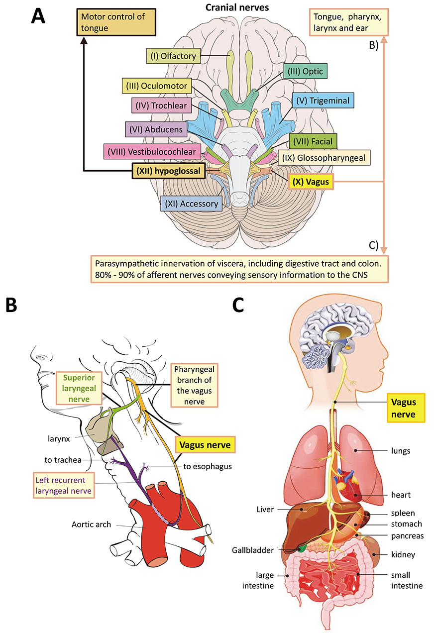
However, the hindbrain structures of the mammalian brain evolved first [48, 49]. The most basal of the twelve central nervous system (CNS) cranial nerves are XII (hypoglossal nerve, innervates the tongue), XI (Accessory nerve, innervates muscles of neck), and X (vagus nerve, main parasympathetic nerve that innervates the thorax including gut) (Fig. 1A, Ref. [50, 51]). Of these, the accessory nerve is a relatively late evolutionary development, having appeared during chordate evolution. The cell bodies of the both the hypoglossal and vagus nerve neurons are initially indistinguishable amongst the neuroblasts in the ventral hindbrain. They grow axons ventrally and dorsally respectively from mouse embryonic day e9.0 [52].

Fig. 1.

The vagus nerve. (A) The twelve cranial nerves, highlighting the positions of the vagus (X) and hypoglossal (XII) nerves in the human CNS, as a representative of vertebrates. For annotated detail see [50, 51]. The original image by Patrick J. Lynch was taken from https://commons.wikimedia.org/w/index.php?curid=15108118 under a CC BY 2.5 Creative Commons license and was altered by adding labels and annotation. It is provided under the terms of the same license. (B) Innervation by the pharyngeal branch of the vagus nerve. Original image by Wikimedia author Jkwchui. Reproduced under a Creative Commons CC BY-SA 3.0 license from https://commons.wikimedia.org/wiki/File:Recurrent_laryngeal_nerve.svg. Changes to labelling were made. The image is free to reproduce under a CC BY-SA 3.0 license. (C) Organs innervated by the vagus nerve. Note that several tissues innervated by the vagus nerve in B and C reflect gut and mouth relationships that may have been inherited from the LEUMCA. Mesodermal tissues arose first in bilaterians. Reproduced with permission from Biology Dictionary, https://biologydictionary.net/vagus-nerve/.

For our purposes these represent the evolutionarily oldest part of the vertebrate hindbrain, which is the oldest part of the entire brain [48, 53], and therefore the one most likely to share ancestral features with the LEUMCA nervous system. The hypoglossal nerve exerts motor control over the tongue, while the vagus nerve innervates tongue, pharynx, larynx as well as the entire viscera extending to the colon [50, 51] (Fig. 2, Ref. [19, 54, 55, 56, 57, 58, 59, 60]). Therefore, the vagus nerve innervates the entire gastro-intestinal tract from mouth to anus.
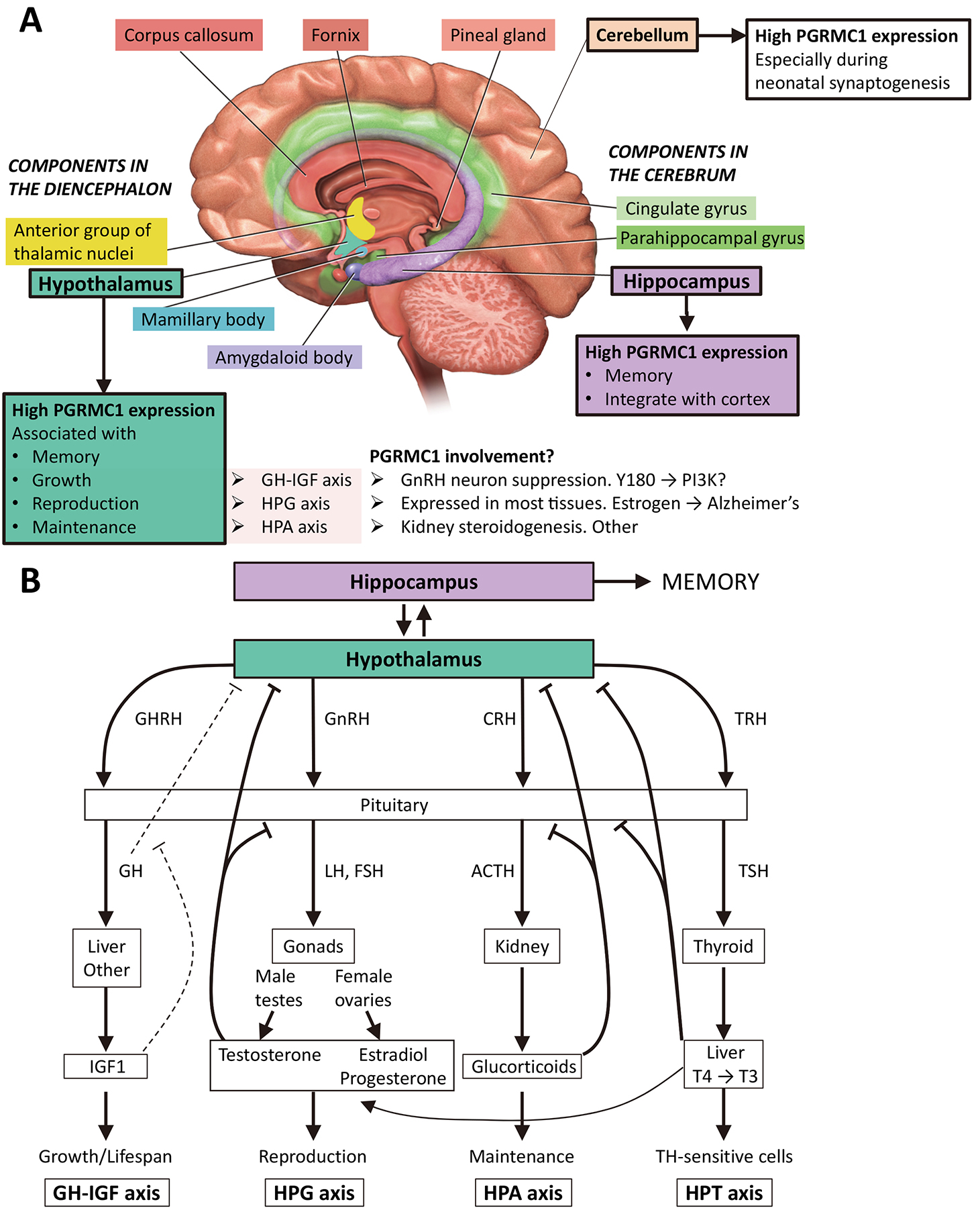
Fig. 2.

The hippocampus and hypothalamus in body metabolism and memory. (A) The image shows the limbic system, which deals with emotions and memory, and where PGRMC1 expression is reportedly highest in the adult mouse [54, 55]. The hypothalamus is depicted as associated with memory [56], as well as growth, reproduction and maintenance, which depend upon the growth hormone–insulin-like growth factor (GH-IGF) axis, the hypothalamic-pituitary-gonadal (HPG) axis, and the hypothalamic-pituitary-adrenal (HPA) axis, respectively [57]. The PI3K/Akt pathway is a powerful regulator of GH-IGF, and PGRMC1 regulation of PI3K through Y180 [19] could influence this. The HPG axis is heavily involved in learning, involving sex steroids and other mechanisms [58]. As key regulator of sterol availability PGRMC may be involved in the HPA axis, however no clear role has been identified. The limbic system image is modified from [59]. (B) Major whole-body axes of the hypothalamus. The diagram is based loosely upon [57], with addition of the hypothalamic–pituitary–thyroid (HPT) axis, which influences juvenile growth and adult fertility [60]. Note that all these systems have evolved since the last common bilaterian ancestor that was descended from the LEUMCA which acquired PGRMC1 tyrosine phosphorylated Y139 and Y180. CRH, corticotropin-releasing hormone; GH, growth hormone; GHRH, growth hormone-releasing hormone; GnRH, gonadotropin-releasing hormone; IGF1, insulin-like growth factor 1; T3, triiodothyronine; T4, thyroxine; TRH, thyrotropin releasing hormone; TSH, thyroid-stimulating hormone. T4 and T3 (numbers refer to iodines) can both be made in the thyroid. T4 can be also converted to T3 in other organs, such as the liver (shown) [60].

As such, the gut/brain neural connections of the vagus nerve, the most ancient of the vertebrate cranial nerves that primarily regulates involuntary actions via the parasympathetic nervous system, are potentially the evolutionary products of innervation that was proposed to have existed between two neural centers (apical and blastoporal) since the LEUMCA [61]. That system may have innervated the original gut and mouth of the first bilaterian and would originally have resembled the nervous arrangement of the diploblastic LEUMCA. The parasympathetic nervous system is a part of the autonomic nervous system that controls the activity of the smooth and cardiac muscles and glands, functions which must be very similar to what the urbilaterian nervous system inherited from the LEUMCA.

Perhaps that is related to the observed prominent involvement of the vagus nerve in the axis between gut microbiota and the brain [62] and its dramatic influences over CNS function [63, 64], if those neural connections have been conserved since the first neurons mediated communication between gut and sensory nerve centers in the LEUMCA. If PGRMC was ancestrally related to neural function, we expect it to feature prominently in vagus neurons, including during embryogenesis. It is fully conceivable that communication between neurons and gut epithelium from the LEUMCA provided a platform that was built upon and reformed, rather than replaced, in complex bilaterians. If so, then PGRMC1 involvement in synaptic membrane trafficking, LDLR internalization, and involvement in insulin/glucagon regulation of metabolic regulation may represent vestiges of that ancient system. PGRMC biology may form an indispensable part of the fabric of eumetazoan body architecture. To the author’s knowledge, this has not been explored. Note that this is a deductive process. Strong experimental evidence does not exist to support this hypothesis.

The nematode ventral midline-1 (Vem-1) homolog of PGRMC1 is involved in the guidance of some axons during the establishment of the ventral nerve cord. A single neuron called AVG (anterior ventral neuron G) pioneers the right ventral cord axon tract, depositing signals that will subsequently be used by other axons. Vem-1 expression was detected at gastrulation and later in early anterior head neurons, including neurons of the nerve ring and the AVG that extend axons into the ventral nerve cord. The AVG cell nucleus is near the posterior base of the brain (analogous to the origin of the vagus nerve), and its axon migrates towards the posterior, secreting the ligand netrin/UNC-6 (uncoordinated-6: uncoordinated due to faulty nervous system) which is part of an evolutionary conserved guidance system involving the receptor deleted in colorectal carcinoma (DCC)/uncoordinated-40 (UNC-40) (reviewed by [65, 66]). Subsequent axon guidance uses the secreted netrin/UNC-6 of pioneer nerves like AVG as guidance during formation of the ventral nerve cord. Vem-1 interacts physically and genetically with UNC-40, the C. elegans homolog of the netrin receptor DCC, and vem-1 gene deletion results in failure of the AVG neuron but not others to faithfully extend axons along the correct pathways. As we saw in the accompanying paper [1], the Netrin/DCC system first appeared in the LEUMCA [67], and one of the circumstances under which PGRMC1 was first identified in mammals was under the synonym of ventral midline antigen (VEMA), involved in axon migration of the embryonic mouse central nerve cord [68, 69].

Interestingly, a comparative holistic model has been recently proposed by Wang et al. [57] for coordinated cell metabolism and immune functions coordinated through the hypothalamus. An extension of that model is proposed here, where primal PGRMC biology directs profound deep-level changes to cell gene expression and metabolism, related in part to its ancient role in mitochondrial regulation.

PGRMC1 is expressed abundantly in the hypothalamus, where it is thought to exert local immunomodulatory functions [54, 55]. Wang et al. [57] discuss the hypothalamus as associated with three major whole organism feedback axes concerned with growth (growth hormone–insulin-like growth factor (GH-IGF) axis), reproduction (hypothalamic–pituitary–gonadal (HPG axis), and homeostasis (hypothalamic-pituitary-adrenal (HPA) axis). Another major axis is the hypothalamic–pituitary–thyroid (HPT), which is involved in juvenile growth and adult fertility [60] (Fig. 2B). Neurosteroidogenesis of P4, and associated reorganization of neuronal actin cytoskeleton feature prominently in the development and differentiated function of these axes [70, 71, 72, 73].

The HPA axis activity can affect memory, e.g., via stress signaling to the hippocampus [74] which involves glucocorticoid steroid hormones. PGRMC1 was originally identified under the synonym ‘inner zone antigen’ and a cytochrome P450-regulating protein associated with renal glucocorticoid steroidogenesis [75, 76], and PGRMC1 is also located in gonads [77, 78], pituitary [79], cortex, hypothalmus including hypothalamic nuclei involved in female reproduction, as well as in the hippocampus in neonatal and adult mice [55, 80]. The latter is recognized as a central component in memory formation [81]. So PGRMC1 is expressed in all the major cell types involved in the HPA axis, as we might expect if the system had evolved from a LEUMCA precursor that resembled modern cnidarians. It also inhibits gonadotropin-releasing hormone receptor (GnRH) neurons of the hypothalamus in response to P4 [82], and is expressed in the thyroid [83] and liver [41]. Remarkably, PGRMC1 is expressed in many of the tissues involved in the major whole organism feedback loops of the hypothalamus, and there is evidence of its direct involvement in many of the cell responses involved in those pathways (Fig. 2). Once more, this thought process is hypothetically deductive. The model requires experimental verification.

Another organism-wide regulatory system involves glucose homeostasis via the insulin-glucagon system. If we view the insulin response as a vestige of communication between gut, neurons, and muscle of the ancestral LEUMCA, via insulogenic secretory cells, then its manifestions in the central nervous system could be highly informative. These include (but are not limited to) glucose homeostasis [84, 85], dietary intake [86, 87, 88], neuroprotection [89], neuron growth and differentiation [90], synaptic activity [91], and memory formation [92], and could validly be added as a fifth axis to Fig. 2B. Insulin resistance leads to hippocampal dysfunction: impaired neuroplasticity and decreased cognitive function, as well as increased risk of Alzheimer’s disease (AD) [93, 94]. The relationship between insulin biology, neuronal health, and memory has indeed prompted the concept that Alzheimer’s disease can usefully be considered as type 3 diabetes [95]. The author would extend this to propose that the functioning central nervous system can be usefully considered to be dependent on ancestral PGRMC functions, and that this extends to AD and quite probably many other neuropathologies. We will pursue this below.

1.5 PGRMC1 Is Involved with the Sigma-2 Receptor and Sterol Biology

The involvement of PGRMC1 in steroidogenesis and P4 responses in the nervous system and cells associated with the female reproductive system is established and has been extensively reviewed previously [75, 76, 78, 96, 97, 98, 99, 100, 101, 102, 103, 104, 105, 106, 107, 108, 109, 110, 111, 112, 113, 114]. It will not be discussed further here beyond noting that the competing reaction to that catalyzed by lanosterol-14-demethylase (CYP51A1) and PGRMC1 involves the 24-dehydrocholesterol reductase-mediated reduction of lanosterol to dihydrolanosterol, which requires NADPH but not oxygen (see Fig. 3 in the accompanying manuscript [1]). Interestingly, this enzyme activity (under the synonym seladin-1) protects against amyloid beta (Aβ) peptide-induced neuronal apoptosis [115]. We now focus on the sigma-2 receptor (S2R) and its sterol biology.

The S2R is a pharmacological activity with possible therapeutic relevance to cancer [116, 117, 118, 119] and neurological disorders [120, 121, 122]. For reviews on the history of Sigma-1 receptor and S2R field see [118, 123, 124, 125]. A relatively detailed review of recent development in S2R biology is provided here to provide adequate background for non-versed readers for the section on Alzheimer’s disease.

S2R activity was initially localized to an 18–22 kDa [126] or 21.5 kDa [127] membrane protein. The finding that PGRMC1, with predicted molecular weight of 21.67 kDa, was cross-linked to a photoactivable S2R ligand [128] led to an initial degree of confusion as to whether PGRMC1 itself was the S2R, rather than a member of a protein complex containing S2R. It soon became apparent that whereas S2R activity was decreased by reducing PGRMC1 levels in some cell systems [118, 128, 129], in others this was not the case [130, 131, 132].

S2R-ligand-dependent affinity purification and mass spectrometry identification of associated proteins revealed that the small integral membrane protein transmembrane protein 97 (TMEM97) was crucial for S2R activity [133]. TMEM97 was previously known as meningioma-associated protein 30 (MAC30) [134]. It binds ligands within the hydrophobic interior of the lipid bilayer [135, 136]. Initial commentary on the identity of TMEM97 with S2R took the position that its molecular cloning completed the unambiguous characterization of this receptor class [137], and resolved its “identity crisis” with PGRMC1 [138]. This sentiment has become so well rooted that both NCBI (NP_055388.2) and UniProt (Q5BJF2) have now currently reannotated the TMEM97 protein as “sigma intracellular receptor 2”. In both cases the gene name is TMEM97. This altered nomenclature was adopted despite having been originally characterized with nomenclatural priority as MAC30 (meningioma is the most common type of head cancer), and the fact that S2R/TMEM97 is indeed overexpressed in a variety of other cancers [123, 139, 140].

TMEM97 is one of a suite of genes induced by sterol regulatory element-binding protein (SREBP)-2 under low cholesterol levels (the system regulated by PGRMC [41, 141, 142]), that is involved in the endosomal lysosomal compartment where it is associated with LDL cholesterol transport-regulator Niemann-Pick C1 (NPC1) [143]. It is also a member of a group of related proteins that by similarity are likely to possess sterol isomerase activity [144]. In fact, although the two human sigma receptors (S1R and S2R) are unrelated to each other, they are both related to other families with sterol isomerase domains [144], and so seem connected under the broader umbrella of sterol biology. Their overlapping ligand affinities are probably the result of convergent evolution (see [123]).

Sterol biochemistry is central to PGRMC function, whether as a member of the Insig-1/SCAP complex that senses sterol levels and regulates activation of the mevalonate pathway by sterol regulatory element binding proteins [41, 141, 145], involvement with CYP51A1 (the most conserved eukaryotic CYP450) in the 14-demethylation of lanosterol (the very first sterol modification, and therefore the earliest to evolve) [142, 146, 147], or its conferral of responsiveness to progestogens, including P4 (reviewed in [97, 147] or to other steroids such as estrogen [148, 149, 150, 151, 152]). And of course the finding that the enzymes of the entire mevalonate pathway, lanosterol cyclase, and the CYP51A/PGRMC reaction, that 14-demethylates the first sterol lanosterol, all came from bacterial rather than archaeal genes [153]. PGRMC may justifiably be thought of as a godfather of steroid biology, exerting effects that appeared at least a billion years before the appearance of nuclear steroid receptors in early animals.

It is then perhaps no surprise (in hindsight) that PGRMC1 was found to form a complex with the LDLR and TMEM97/S2R [43], which obviously provides the solution of a lipid supply problem that appeared after a vascular circulatory system and specialized tissue functions in chordates, where most steroid synthesis occurs in the liver but is consumed in the periphery. The author had highlighted the involvement of TMEM97 in sterol transport [28], and was fortunate enough to have predicted such a PGRMC1::LDLR interaction [97], which promoted Mach and colleagues [133] to investigate the potential interaction of PGRMC1, TMEM97 and LDLR. The PGRMC1/TMEM97/LDLR complex was responsible for a pathway that elevated rates of LDL internalization over and above the background constitutive levels. Since actively growing cells require more cholesterol, this was consistent with observations that S2R activity was elevated in growing cells [139]. Removal of TMEM97 from this system totally removed the binding of their S2R ligand, RHM-4 [43].

As discussed in the accompanying paper [1], the author’s laboratory has generated preliminary data that tryptophan-rich sensory protein (TSPO) may form an obligate part of a TMEM97-containing S2R ligand-binding complex, where both TSPO and TMEM97 are both required for S2R activity. TSPO and PGRMC1 both interacted with TMEM97 in proximity ligation assay, but not with each other [154]. TSPO binds heme and cholesterol [155]. That PGRMC1 and TSPO may be functionally related is further strengthened by several MAPR-related candidate phyla radiation (CPR) bacterial genes for cytb5MY proteins being found in operons that include a bacterial TSPO gene, in a phylogenetic distribution suggesting that the ancestral cytb5MY-encoding operon contained TSPO [153]. The endogenous 20(S)-hydroxycholesterol ligand of TMEM97 [136] is formed by CYP11A1 as the cholesterol steroid skeleton is transferred from the outer to inner mitochondrial membrane during the synthesis of pregnenolone, the first animal steroid hormone [156]. Deductively reconstructing, one could assume that the TMEM97 ligand system arose after the origin of animals. It remains possible that a 20-hydroxylase activity could have acted on one of the steroids from the metabolic pathways of lanosterol synthesis [1] before the evolutionary appearance of cholesterol, and later adopted cholesterol as its substrate.

That the ‘identity crisis’ of S2R may not yet be fully solved by its identity with TMEM97 is further supported, once more by publications from the Mach laboratory. Having demonstrated that TMEM97, PGRMC1 and LDLR form a complex, and that S2R ligand binding was abrogated by TMEM97 depletion [43], they also showed that S2R-induced cytotoxicity was unaffected by TMEM97 or PGRMC1 depletion. Even depleting both together had no effect on S2R-mediated cytotoxicity [139, 157]. Furthermore, while the double knockout reduced the rate of fluorescent S2R ligand SW120 uptake, the level of internalized SW120 of knockout cells eventually reached the same levels as control cells [157]. Sereti et al. [158] also concluded that S2R-ligand-mediated cytotoxicity did not correlate with levels of S2R/PGRMC1 protein abundances, and hence that the cytotoxic mode of action does not involve a TMEM97/PGRMC1 complex. One possibility is that another as-yet unidentified S2R activity such as TSPO is responsible for S2R-mediated cytotoxicity, and hence that TMEM97 represents just one S2R receptor. If so, then the identity of the further binding activity could provide new insights to address the pharmacology of pathology which TMEM97 does not exert. Another possibility is that the PGRMC2 protein could partially substitute for absent PGRMC1 function, however a perplexity of possibilities exists. For instance, S2R ligands could conceivably bind to non-protein membrane components.

As an aside, the COVID-19 disease-causing severe acute respiratory syndrome coronavirus 2 (SARS-CoV-2) protein Orf9c is present in a protein complex with TMEM97 [159]. It is interesting to speculate whether TMEM97 may be involved in the endocytic entry of virus to the cell. There is no association between PGRMC1 and SARS-CoV-2, however PGRMC1 does exert a negative influence over influenza A virus infection [160]. There, infection leads to downregulation of PGRMC1, which is associated with reduced interferon-β signaling. Such PGRMC1-modulation of the interferon (IFN) pathway could be relevant to immune response towards cancer. Note the caveat that this study presumed that all AG-205 effects were specific for PGRMC1 [160], which is not true (see the discussion in the accompanying paper [1]). It will be interesting to see whether these viral effects are related to changed membrane trafficking properties by TMEM97/S2R in the presence and absence of PGRMC1.

Finally, a closing note to the discussion of a S2R::PGRMC1 complex, PGRMC1 involvement in the Warburg effect aligns with a report that a sigma-2 receptor ligand increases aerobic glycolysis, elevating hypoxia-inducible factor 1 alpha (Hif-1α) and vascular endothelial growth factor (VEGF) [161], which is very likely to be related to PGRMC1 biology.

1.6 Neurobiology, Alzheimer’s Disease and PGRMC

PGRMC proteins may underlie many of these root-level gastrulation organizer-initiated processes in eumetazoans, including humans. Furthermore, and this may well be wrong, but AD appears to the author personally to be the disease that is most directly related to perturbations of the PGRMC signal system described here [162]. A such, its consideration can surely be broadly revealing.

1.6.1 How a PGRMC1 Researcher Became Involved in AD

The author has had only a tangential association with neuroscience research, precipitated by his development of proteomics technologies and possession of molecular cell biology signal transduction expertise in a company where others excellently performed neural cell culture [163, 164]. Nevertheless, based on having PGRMC1 expertise the author was recruited in 2013 by Cognition Therapeutics Inc. (CogRx, Pittsburgh, PA, USA) as scientific advisory board member to their AD therapeutics program (see conflict of interest statement). They had developed an anti-AD S2R ligand series which displaced synaptic oligomeric amyloid beta (Aβo), reversed synaptic damage and improved cognition in animal AD models [122]. Their synaptorestorative drug action mechanistically involved PGRMC1 membrane trafficking [165, 166, 167]. This was to the author’s knowledge the first small molecule series which demonstrated improved cognition (improved memory) in AD animal models via a synaptorestorative mechanism [122, 165, 168]. The CogRx small molecule lead compound CT1812 is currently in Phase 2 clinical trial [121, 162, 166, 169]. A related mechanism is also observed in nematodes (which have no endogenous Aβ-encoding gene) when an AD-relevant mutated Aβ protein is exogenously expressed [170].

PGRMC1 is involved in a membrane trafficking pathway which is required for the mechanism of action of synaptorestorative drug CT1812 (Note that CT1812 binds S2R, whereas Aβo possibly binds another, unidentified, target [122, 169]). The drug reverses symptoms of AD in animal models by displacing Aβo from synapses, caused by allosteric effects on TMEM97 [122] (without any detectable effect on Aβ plaque levels, consistent with plaque-directed anti-AD strategies being unproductive). In a BioRχiv preprint, Colom-Cadena et al. [171] report that soluble Aβ forms sufficiently close interactions with synaptic TMEM97 to generate Fluorescence Resonance Energy Transfer (FRET) signals. They suggested that TMEM97 may be the synaptic binding target of Aβo in AD.

Aβo is displaced from rat hippocampal synapses by CT1812, leading to improved memory/cognition, and can be detected in human cerebrospinal fluid shortly after drug administration. The latter is associated with reduced levels of many AD-associated protein isoforms in cerebrospinal fluid [166]. CT1812 also disrupted the TMEM97/Aβo FRET signal reported by Colom-Cadena et al. [171]. The United States Federal Drug Administration (FDA) has recently revised its staging guidelines for clinical trials to facilitate the development of such synaptorestorative new treatments capable of addressing early stages of predementia [172].

It was mistakenly thought that PGRMC1 was the S2R, as explained above, which is why the author was recruited by CogRx. The narrative will expound below why this may have been serendipitous because otherwise the author would not have thought about AD in depth, and how the grand-scale biology of PGRMC, as outlined here and in the accompanying paper [1], may be central to the pathobiology of AD.

1.6.2 The Scope of the AD Problem

AD is an area of critical and burgeoning importance. Worldwide, the 33 million sufferers in 2018 already carried a social cost exceeding US$1 trillion. In 2020, an estimated 700,000 people in the United States aged 65 and older had Alzheimer’s when they died [173, 174], compared with 627,039 deaths in the same country for the COVID-19 pandemic from February 2020 until July 27th 2021 [175]. Unless improved interventions are developed, the cost is expected to double by 2030, ballooning to more than 150 million cases by 2050 [176]. Governments recognize this and funding bodies in several nations have been prioritizing AD research. Large pharmaceutical companies spent more than US$75 billion on R&D for AD in 2016 and sponsored >70% of all clinical trials [177]. Prior to BioGen’s release of the largely ineffective Aducanumab in 2021 [178], the last of five FDA approvals for AD was in 2003 with the candidate-drug-to-market success rate of <0.5% among the lowest for any therapeutic area [179, 180].

Despite the growing huge market and unmet social need, the large pharmaceutical companies - the only bodies capable of bringing treatments to market - have been withdrawing from AD research (e.g., Pfizer in Jan 2018, Biogen in March 2019) after three decades of unproductive research pursuing the traditional “amyloid cascade hypothesis” [26, 168, 181, 182]. The amyloid cascade paradigm proposed that errors in the production, processing, and aggregation of Aβ caused the formation of plaques, which cause a disease that is ultimately characterized by the central deposition of insoluble senile plaques that lead to cognitive impairment. In what the author agrees to be this outmoded model, either Aβ plaques, Aβ monomers, or what are presumed to be downstream Tau neurofibrillary tangles were thought to be the causative agents of neurodegeneration [26, 181, 183, 184, 185]. However, it has long been noted that amyloid plaques are observed at the wrong time and place in the development of AD to be the causative feature [186, 187]. For recent reviews on this, as well as theories of AD aetiogenesis and therapeutic strategies, see Jeremic et al. [188]. It has been argued that subjective community bias in favor of the Amyloid Cascade Hypothesis has hampered AD research progress, even with references to an amyloid ‘cabal’ [189]. See also Herrup’s book (How Not to Study a Disease: The Story of Alzheimer’s) devoted to the topic [190, 191].

Much grant agency support continues to flow to project proposals based upon the Amyloid Cascade Hypothesis historical model, despite mice with plaques developing no memory impairment and three major beta-site amyloid precursor protein (APP) cleaving enzyme (BACE) inhibitor anti-plaque strategy drug failures in late 2018 [26, 192] (for detailed discussion see [190]). In a field where the historical success rate for discovery of new treatments has been exceedingly low and requires new impetus [26, 176, 193], the growing and tragic social burden is exacerbated by those treatments which have been approved being unable to prevent progression, but merely temporarily ameliorating some symptoms [26, 168, 178, 194]. New insights and alternative mechanisms for drug development are desperately needed [26, 194], and here, perhaps, is an example.

1.6.3 Aβ Oligomers Cause AD

CogRx [122, 169] pursued an alternative version of the amyloid hypothesis [195, 196, 197, 198], neglected by most studies at the time and all drug developments that had until then entered clinical trial. It predicted that the key neurotoxic Aβ species were soluble oligomers of Aβo in equilibrium at relatively trace levels compared to the large molar excess of plaque Aβ, and bound inappropriately to synapses to disrupt their function. This is not to propose that Aβo explains all AD etiology, but that it is more important than Aβ plaques. In hindsight, this seems to have been the correct formulation of the amyloid hypothesis, as has now been argued by multiple authors [182, 188, 199, 200, 201]. For instance, a drug called RD2 (or PRI-002) specifically disrupts Aβo leading to cognitive improvement in three separate transgenic models. It has completed Phase 1 clinical trial [202, 203, 204, 205, 206, 207].

CogRx recently demonstrated [208] that the naturally occurring human Aβ A673T mutation, that reduces the probability of AD fourfold, exerts its effects through altered monomer properties. Although the binding of mutant monomer to synapses was markedly more toxic than wild-type monomer, which involved exacerbated modulation of membrane trafficking (which they had previously shown was PGRMC1-dependent [165]), reduced affinity of the mutant Aβo monomer for the synapse resulted in an overall lower level of neurotoxicity, in line with reduced AD risk for patients carrying the mutation [208]. Altogether, the Aβo version of the revitalized amyloid hypothesis predicts that treatments directed against Aβ plaques will only succeed to the extent that they also alleviate Aβo-induced synaptic pathology, to restore synaptic function and enable cognitive processes [168]: i.e., the presence of plaques appears to be of secondary importance.

Interestingly, Biogen recently resurrected their previously abandoned Aducanumab program: a monoclonal antibody that recognizes both Aβ plaques and oligomers but not monomers [209]. Marginal positive effects were observed at higher doses after data mining of extra patients from one wing of their discontinued trials. Notably, Aducanumab efficiently lowers Aβ plaque levels (which it was designed to do) but has only marginal (if any) effects on AD disease state [178, 210, 211].

In October 2019 Biogen announced they would be filing for FDA approval, which was met with a mix of cautious optimism and skepticism by analysts at the time [168, 212, 213, 214], and after FDA approval was granted in 2021 (but only for participation in clinical trials) [178, 180, 210, 211, 215, 216]. The Biogen presumed mechanism of action for Aducanumab involves specific targeting of aggregated Aβ plaques and oligomers but not monomers [209, 217]. Positron Emission Tomography (PET) of the brain using Amyvid — a PET reagent for quantifying neuritic plaque density — showed reduced plaque density. Based solely on the assumption that plaques cause AD, Aducanumab was approved by the FDA despite none of the expert advisory committee recommending approval (ten votes against and one abstention).

It is fully possible/probable that marginally beneficial effects are due to Aβo displacement from synapses. That would resemble the mechanism of CT1812 and is in accord with the prediction that plaque-directed treatments will only work to the extent that they alleviate oligomer-induced pathology.

2. A New Alzheimer’s Hope?

In proposing that PGRMC1 may be central to AD [162], the author feels obliged to provide the community with a deeper rationale. As a non-neuroscientist, this may seem presumptuous, if not audacious. For a non-technical overview of the complexity over which the author has only limited understanding, see Herrup [190]. In the following, we walk through some AD- or neuron-related biology and make connections with the story that has been developing for PGRMC function in this and the accompanying paper [1], and never assuming that addressing PGRMC1 biology might hold all answers to AD therapy.

2.1 Glucagon Pro-Peptide Gut/Pancreas and Possible Evolutionary Involvement of PGRMC

If PGRMC-dependent processes were foundational to the evolution of the nervous system, as inherited from the LEUMCA, we could reasonably expect to see the involvement of neural innervation of mesodermal and gut structures by the vagus nerve (as discussed above), and hormonal regulation of these systems that evolved with vasculature systems. Glucose-sensing neurons are at multiple locations in peripheral and central nervous systems. The most extensively studied brain region is the hypothalamus, but the various regions are thought to be synaptically highly interconnected.

Brain glucose-sensing neurons make sympathetic and parasympathetic connections to target organs that modulate metabolism, such as liver, pancreas and visceral adipose [218], all of which are innervated by the vagus nerve. It has long been recognized that vagal stimulation can enhance pancreatic insulin release [219, 220], which the author is hypothetically relating to PGRMC function. Part of the reduced pancreatic β-cell secretion of insulin in response to glucose is mediated by β-cell glucose sensing, but also by sympathetic and parasympathetic neural release [218].

The vagus nerve plays a clear role in pancreatic activity, and also in the regulation of signals from brain regions that influence hedonic components of feeding behaviors [220]. Here, glucose homeostatic neural mechanisms are largely controlled by nutrient and hormone effects on the hypothalamus and brainstem ganglia. Conversely, hedonic drive is controlled by the mesolimbic dopaminergic (DA) system. Hormones and nutrients can directly regulate the DA system, or indirectly regulate via the hypothalamus or brain stem neurons. Hormones include satiety signals such as leptin (from adipocytes), insulin (pancreas), or glucagon-like peptide-1 (GLP-1) (from the gut, or from GLP-1-expressing neurons), or opposing hunger signals such as ghrelin (from the stomach), orexins (mostly from hypothalamus), or neuropeptide Y (sympathetic neurons), as reviewed [220].

We are here considering the modern mammalian derivative of the ancestral communication between gut, nerves and behavior that was initially associated with the evolution of the LEUMCA organizer, and the first eumetazoan nervous system. To assess the potential involvement of PGRMC in satiety control (the insulin/glucagon system), consider that PGRMC1 is involved in the regulated subcellular translocation of the following proteins to the plasma membrane: the insulin receptor and glucose transporters GLUT-4 and GLUT-1 [27], and the GLP-1 receptor in pancreatic β-cells [221]. It interacts with hexokinases (which catalyze the first step in glycolysis) to modulate Warburg metabolism [29] and affects mitochondrial form, function, and association with the endoplasmic reticulum (ER) [19, 29], which seems intimately related to metabolic regulation and glycolytic activity. PGRMC1 is expressed in the hypothalamus [54, 55, 222], which is a major regulator of a complex neuroendocrine system that regulates energy homeostasis, appetite, and satiety [223] (Fig. 2).

The insulin/glucagon system regulates not only glycolytic activity, but also the system of fatty acid storage of excess energy from glycolysis. By regulation of glycogen synthase kinase 3 (GSK-3β) [19, 224] PGRMC1 is implicated in the regulation of glycogen synthesis. That remained undemonstrated until Sabbir et al.’s [29] demonstrated reduction of glycogen levels in PGRMC1 CRISPR knockouts of HEPG2 cells.

Lipid synthesis is regulated by glucose. High glucose levels increase sterol and fatty acid synthesis via SREBP1 and SREBP2 [225]. SREBP1 is N-glycosylated in the presence of high glucose, which regulates its activity [226]. Some non-cholesterol sterol (lathosterol, cholesteronol, desmosterol) levels are good predictors of hyperglycemia and the development of type 2 diabetes [227]. PGRMC1 forms a complex with SREBP1 and SREBP cleavage activating protein (SCAP) [145]. PGRMC1 knockdown promotes dysregulation of this system leading to hepatic steatosis [41], and PGRMC1 modulates lipid homeostasis in adipose [228] and cancer breast cells [42], and in cardiac muscle cells which are highly dependent upon fatty acid oxidation [229].

Thus, excessive energy available as glucose seems to be converted to fat under the watchful eye of the PGRMC/SREBP1 system [230]. This reflects the eukaryogenic role proposed in the accompanying paper [1]. Accordingly, perturbed regulation of the PGRMC/SREBP system is thought to be related to increased fatty acid and cholesterol synthesis by antipsychotic drugs [141], perturbed lipid homeostasis and oncogenic progression in breast cancer [42], and a deletion of PGRMC2 is associated with fatty acid variations in the milk of dairy cattle [231].

As well as regulating fatty acid synthesis, SREBP activates the pentose phosphate pathway [232], the major source of reducing power to counteract oxidative stress [233] (notable in a hypothesis associating PGRMC with oxidative biology and eukaryogenesis [1]). SREBP1 is one of the major metabolic regulators induced by mammalian target of rapamycin c (mTORc) in response to low energy levels, along with Myc and Hif-1 [232] (also related to the biology of aging, which is discussed below). As we saw in the accompanying paper [1], PGRMC phospho-tyrosines and Hif-1 both entered the genome during the Sturtian glaciation in response to presumed altered metabolic requirements, coincidentally with gastrulation and the organizer [1], and this is likely to be part of the organism-wide deep biological associations of PGRMC.

In a section linking SREBPs and their regulation by PGRMC, and noting the interaction of PGRMC with membrane trafficking and components of the actin cytoskeleton, it would be remiss to omit that from Drosophila to humans SREBP1 is also regulated by mechanical stress propagated from the extracellular matrix via geranylgeranyl pyrophosphate, a key mevalonate pathway (MVP) intermediate, where RhoA-dependent actomyosin contractions inhibit the activation SREBP1 [234]. Mevalonate pathway activity and regulation is important in normal and pathological activity of many body functions, including the cardiovascular system, cancer and neurobiology [235, 236, 237, 238]. Please remember that these arguments are presented as part of a non-confirmed hypothesis. Further studies would be necessary to validate the relationships between the above features.

If neurobiology exploits some aspect of PGRMC biology inherited from the origin of eukaryotes, as adapted by the eumetazoan organizer systematics [1], it may be related to redox and/or metabolic switches and/or membrane trafficking actin cytoskeletal regulation hypothesized above to have been involved in eukaryogenesis. Membrane remodeling is one function of PGRMC1 that is active at synapses [165]. Perhaps neurogenesis requires the evocation of a PGRMC-directed cell state with cytoskeletal organization inherited from the first truly eukaryotic cell. If so, this probably involves altered mitochondrial function and glycolysis. The author stresses that this is a purely conjectural hypothesis which at this stage lacks experimental evidence.

2.2 PGRMC1 and Neuronal Cell Death

P4 clearly exerts PGRMC1-dependent effects on neurons. It induces a neuroprotective signal in primary rodent neural cultures, involving the secretion of brain-derived neurotrophic factor (BDNF) from glia cells which promotes neural survival and synaptogenic conditions. This involves PGRMC1-dependent activation of extracellular signal-regulated kinase (ERK)5 [239, 240], which can be negatively regulated by the microRNA (miRNA) let-7i that targets the PGRMC1 mRNA for destruction [241].

In a similar primary culture model, a series of publications from Hou and colleagues [242] at Shijiazhuang in China’s Hebei Province have shown that P4-mediated neuroprotection from Aβ(25–35)-induced death involved AG-205-sensitive suppression of the mitochondrial apoptotic pathway (interpreted as PGRMC1-dependent, but AG-205 does not exclusively affect PGRMC1: see discussion in the accompanying paper [1]). P4 also suppressed Aβ(25–35)-induced activation of JNK, however this was not sensitive to AG-205 treatment [242]. The same group later showed [243] that Aβ(25–35) led to reduced steroidogenic P4 production [244], that P4 treatment activated the Ras signaling pathway to protect from Aβ(25–35)-toxicity, and that the protective effect of P4 was attenuated by a lentivirally-expressed anti-PGRMC1 short hairpin RNA (shRNA), providing complementary identification of the involvement of PGRMC1 to the AG-205 sensitivity reported in the previous study [242].

In human embryonic kidney derived HEK293 cells, Sabbir [18] noted alterations in phosphorylation, ubiquitination and sumoylation of PGRMC1 following P4 treatment, and these were associated with altered glucose metabolism. The activation of the Ras pathway in neuronal survival [243] is suggestive of the involvement of tyrosine phosphorylation in this process. Although HEK293 cells are far removed from neurons, it is possible that similar processes occur in P4-treated neurons. Indeed, Hou and colleagues [20] demonstrated the involvement of PGRMC1 in elevated glycolysis in response to P4 treatment in animal and cell culture AD models. Therefore, PGRMC1 biology in this instance is closely associated with the disease state of AD. We could extrapolate from arguments developed above that Ras activation might imply PGRMC1 tyrosine phosphorylation, and that this in turn would affect PGRMC1’s membrane trafficking functions. Once more, this is conjecture.

2.3 Could a PGRMC1-Centric Model Really Explain AD?

As stated above, PGRMC1 is part of the S2R complex that is targeted by CogRx ligands such as CT1812 which allosterically alters the affinity of S2R/TMEM97 for soluble Aβo. CT1812 leads to improved cognitive function, associated with improved long-term potentiation (LTP) [121, 122, 165]. In the mammalian adult brain, PGRMC1 expression is found in the limbic system, also known as the paleomammalian cortex, and especially in the hypothalamus [54, 55] (Fig. 2). The hypothalamus has foundational nature because its precursor was present in the chordate common ancestor of vertebrates and tunicates, and therefore predated the subsequent evolution of the vertebrate brain [245].

The limbic system houses a series of separate functions associated with such central human functions as behavior, motivation, olfaction, long term memory and emotion [246]. This region of the brain consists partly of diencephalon (part of the midbrain) and of cerebrum (part of the forebrain) (Fig. 2) and is therefore a much later evolutionary development than the hindbrain. PGRMC1 is also widely expressed in the cerebellum where it is thought to be involved in synapse formation [114]. Such abundant expression levels of PGRMC1 may mean that important PGRMC neural functions inherited from primitive neurons have been retained into the more complex structures of the higher vertebrate brain.

AD symptoms of Aβ aggregates and neurofibrillary tangles containing hyperphosphorylated Tau protein are mainly localized to cortex and the limbic system [247], and a main focus of research of AD research is on the hippocampus and especially the hypothalamus which is seen as most important for acquiring episodic memory [248]. Therefore, abundant PGRMC1 expression in the hypothalamus [54, 55] corresponds to a region acutely affected by AD. The hypothalamus evolved early in chordate evolution [49], and provides a signal integration and central command station for the regulation of whole body metabolism and reproduction in response to environmental conditions. Those are just the type of functions we might expect to find PGRMC1 involved in.

Glasgow et al. [249] recently showed that LTP requires presynaptic netrin signaling to post-synaptic DCC (the same ligand-receptor pair involved in PGRMC-related axon guidance [66, 69], which appeared in the LEUMCA [1]). This induces Ca2+ signaling to induce altered gene expression which changes the composition of the α-amino-3-hydroxy-5-methyl-4-isoxazolepropionic acid (AMPA) receptor (AMPAR), which responds to glutamate as a neurotransmitter. This in turn leads to synaptic spine actin cytoskeletal remodeling associated with synaptic maturation and LTP in response to subsequent AMPAR activation.

P4 induces such synaptic actin cytoskeleton remodeling, and PGRMC1 has been implicated in the response (although whether its role was neurosteroidogenic and/or P4 binding was not clear) [73, 250]. The simplest connection between the LTP-promoting effects of CogRx molecule CT1812 and the mechanism of Glasgow et al. [249] is that a PGRMC1-DCC dependency is perturbed by A
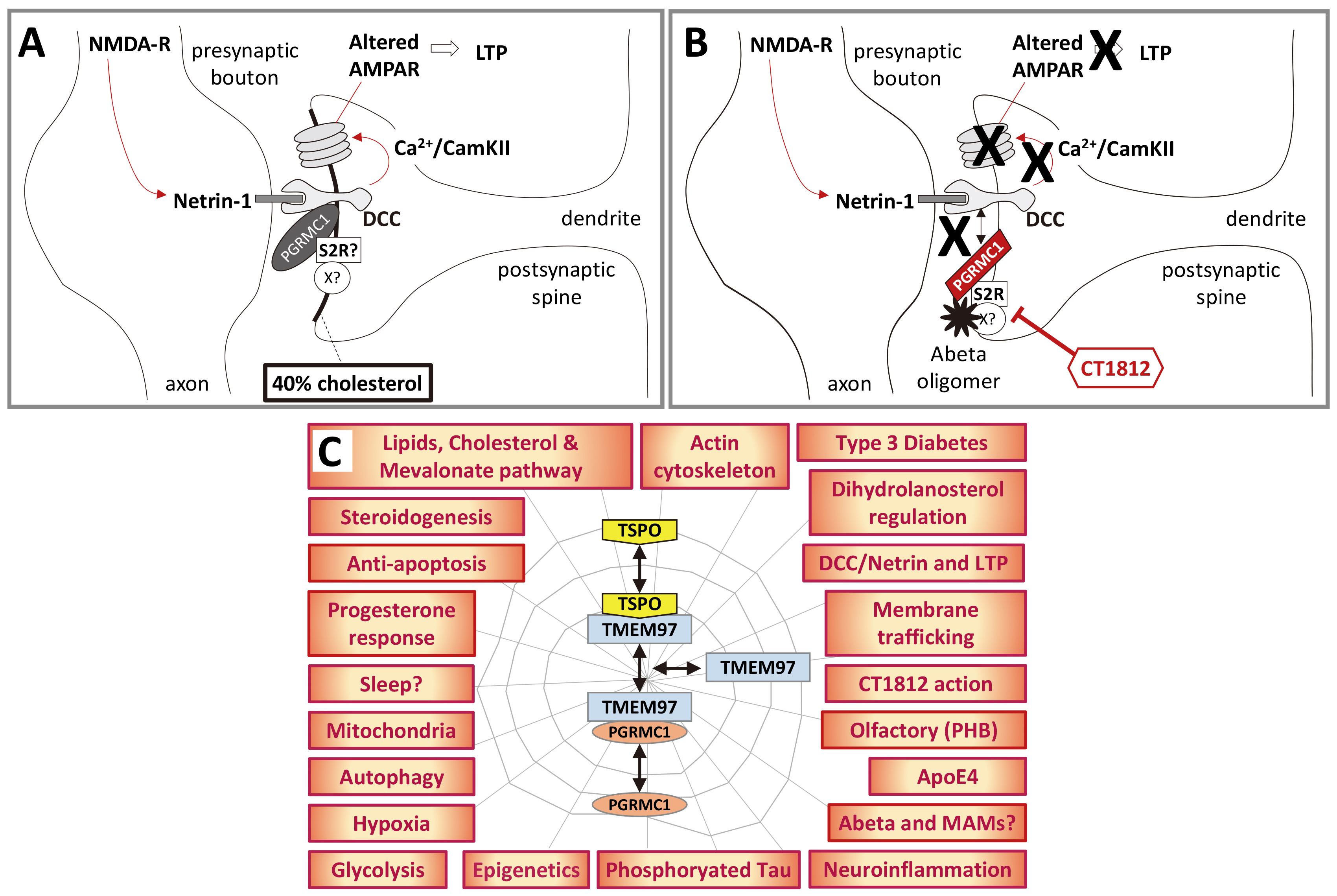
βo binding to the S2R protein complex, preventing dynamic PGRMC1 state changes, which are re-enabled by CT1812 treatment and Aβo dissociation from the synapse (Fig. 3A,B, Ref. [121, 122, 165, 249, 251]). This model potentially places PGRMC1 at the fulcral mechanistic epicenter of AD. Perturbed PGRMC1 membrane trafficking could lead to Aβo accumulation at synapses, and perturbed PGRMC1-dependent processes in the neuron, and CT1812-induced clearance of Aβo could restore PGRMC1-dependent functions.

Fig. 3.

A hypothetical model of PGRMC1 involvement in AD. (A) This crude schematic proposes that PGRMC1 is required for DCC function by incorporating the mechanism of action of LTP-promoting synaptorestorative Cognition Therapeutics drug CT1812 [121, 122, 165] with the mechanism of LTP described by Glasgow et al. [249], acknowledging that PrP [251] or other proteins in a PGRMC1/S2R complex could be the major Aβo binding site in B. (B) Aβo binding to S2R/PGRMC1 hypothetically prevents PGRMC1 from interacting appropriately with DCC, preventing LTP. Addition of CT1812 displaces Aβo, synaptorestoratively enabling PGRMC1-DCC interaction and LTP. (C) PGRMC1 biology associated with AD. See the main text for discussion.

If the last seemingly audacious statements are not fully misguided, then we predict that manifestations of AD pathology should reflect PGRMC1 biology. These would include mitochondrial effects [19, 252], PGRMC1 membrane-trafficking [28, 147], sterol biology [1, 43, 97], Warburg/glycolysis [18, 19, 20], autophagy [253, 254, 255, 256], diabetes [252], hypoxia, and epigenetics [257]. The biology should also include the known mechanism of LTP, as described above, whereby presynaptic netrin activates post-synaptic DCC, initiating Ca2+ signaling and gene expression resulting in altered subunit composition of the AMPAR. That enables the synapse to respond to subsequent input signals by increasing synaptic actin cytoskeleton, enlarging the post-synaptic bouton to strengthen the synaptic connection and facilitate learning [249] (Fig. 3A).

We also must expect the PGRMC1-centric model of AD to account for the ‘usual suspect’ key AD biomarkers apolipoprotein E4 (ApoE4), Aβ and Tau hyperphosphorylation. It should also regulate or be regulated by reelin, which controls cellular/extracellular levels of soluble Aβ [252, 258], and prevention of cell death (anti-apoptosis) would be expected to protect against AD morbidity. If that can be demonstrated then I posit that a PGRMC1-centric model for AD emerges as the leading mechanistic cause since Aβ peptides were identified as forming the plaque core three and a half decades ago [259], sowing the seeds for the Amyloid Cascade Hypothesis [183]. Recall from the accompanying paper [1] that the DHCR24 that competes with the reaction catalyzed by CYP51A1 and PGRMC1 is neuroprotective against Aβ-induced death [115]. In the following we will further examine this suite of PGRMC1-centric biological attributes in AD. These features are portrayed in Fig. 3C, as discussed in more detail below.

2.4 Warburg/Mitochondria and AD

Mitochondrial dysfunction is among the earliest pathogenic alterations found with AD, which is manifest long before the accumulation of amyloid plaques [168, 252, 260], and has even been proposed to be the driving force behind AD and worthy of drug development [261, 262, 263]. Mitochondria are central to metabolic processes and to apoptosis, and are involved in P4-dependent mechanisms of neuroprotection [264], which involve PGRMC1 [103, 114]. Lower P4 levels are associated with AD symptoms in rodent models [265] and humans [266].

We have recently shown that PGRMC1 is associated with altered metabolic changes in cancer cell culture and drives mitochondrial functional changes [19]. Wu et al. [20] have shown that so-called PGRMC1 inhibitor AG-205 blocks P4-induced neuronal glucose uptake that is required for learning. Recall that assumptions of AG-205 being PGRMC1-specific are untrue (as discussed in the accompanying paper [1]), and its effects could easily be due to other MAPR proteins, or even non-MAPR proteins. However, AG-205 does seem able to antagonize certain PGRMC1 functions [267].

Dysfunctional mitochondrial association at mitochondrial endoplasmic reticulum-associated membranes (MAMs) is associated with several neurodegenerative diseases including AD [268, 269]. The fungal cognates of MAMs are called ER-mitochondria encounter structure (ERMES) which have been shown by genetic and biochemical means to facilitate phospholipid and calcium exchange. The resemblance of eukaryotic lipids to those of bacteria, rather than the archaeal proto-eukaryotic symbiotic host cell, is related to the transfer of mitochondrial membrane lipids from mitochondria at ERMES [270, 271]. Adding to the burgeoning list of AD-phenotypes associated with PGRMC1 biology, Sabbir et al. [29] have shown that PGRMC1 knockdown disrupts mitochondria-MAM interactions in HEPG2 but not HEK293 cells. That is in striking alignment with the overarching hypothesis pursued in the accompanying paper [1], that during eukaryogenesis the original eukaryotic PGRMC function was ancestrally related to mitochondrial regulation of metabolism.

2.5 Diabetes, Autophagy, AD, and PGRMC1

Mitochondrial dysfunction is one of a suite of overlapping neurodegenerative mechanisms common to AD and diabetes [272, 273], as well of course as cancer. The cell biology of diabetes and AD are closely related [274]. The association of PGRMC1 biology with that of diabetes is described above.

As a provider of non-glucose carbon skeletons for energy production, autophagy can be considered briefly here. The involvement of PGRMC1 in autophagy was described in the accompanying paper [1]. Suffice to reiterate that autophagy levels are reduced in AD, and that increasing the level of mitophagy (a subtype of autophagy) can improve cognition in AD models [256, 275].

2.6 Are PGRMC1 Migration Responses Related to Axon Function?

Kim et al. [276], from the Korean mouse knockout collaboration, also showed that PGRMC1 is required for the growth of ductules during mammary gland development in response to P4. This may seem unrelated to neurology yet may provide useful information on the potential role of neuronal PGRMC1. As discussed elsewhere in this paper but reiterated here, the nematode VEM-1 homologue of PGRMC1 interacts with UNC-40 (one of the uncoordinated genes names due to impaired neurogenesis when they are mutated) to direct embryonic axon guidance of neurons of the central nerve cord. This function is conserved from nematodes to mammals [66, 68, 69]. Mammals have two orthologues of UNC-40. One is DCC that is involved in axon guidance [66] and long term potentiation [249], and the other is Neogenin [277, 278]. Both UNC-40 orthologs interact with their mammalian ligand Netrin.

Neogenin is also involved in Netrin-mediated axon guidance, migration of T-cells across the blood/brain barrier, inflammation, and angiogenesis [277]. It has long been known that the extending growth bud of the mammary end bud during the growth of mammary ductules involves interactions between Netrin and Neogenin [278]. With the recent discovery that this process depends upon PGRMC1 [276], we can extrapolate to hypothetically propose that this Netrin/DCC/Neogenin migration function reflects an ancient eumetazoan function related to establishing specialized communication modules between different cell types as the primitive LEUMCA body grade was stretched and elaborated by subsequent evolution. It probably involves PGRMC1 tyrosine phosphorylation acquired by the LEUMCA, and actin cytoskeletal rearrangements to mediate a migrating cell front (be it axonal extension or cell migration). Note that this is an undemonstrated hypothesis. PGRMC1/2 also promotes luteal vascularization [77], however there is no indication that this depends upon DCC. Indeed, PGRMC1 regulates VEGF expression [150, 279], which may be responsible for all PGRMC1-mediated vascularization.

Considering functions related to PGRMC1 biology, it has long been recognized that AD-associated insulin resistance and metabolic dysregulation is intertwined with Aβ and Tau pathologies [252, 280]. GSK-3β may lie at the heart of this, regulating brain glucose metabolism and being involved in Tau phosphorylation [274, 281]. We observed differential phosphorylation of GSK-3β by the PI3K/Akt pathway in response to altered phosphorylation mutations of PGRMC1 in cultured cancer cells (GSK-3β activation required PGRMC1 Y180) [19], and the GSK-3β that is involved in organizer activity is regulated by hypoxia [282] and regulates adipogenesis [283]. This biology could all be PGRMC-related, however experimental evidence would need to be demonstrated.

In striking concordance with a main hypothesis of this paper, that perturbations in AD reflect manifestations of PGRMC1 biology retained since eukaryotic origins where the ancestral archaeal host cell consumed amino acids to feed the endosymbiotic proto-mitochondrion [1, 284], substantial changes in branched chain amino acid metabolism accompany AD. Although effects are observed in samples from human cerebrospinal fluid and animal models, the underlying basis remains poorly characterized [285]. Branched chain amino acid perturbations were among those predicted by pathways analysis of cells expressing different PGRMC1 phosphorylation mutants [19]. It is unclear whether AD-specific branched chain amino acid metabolism is related to the γ-aminobutyric acid (GABA) shunt which operates in stressed cells when tricarboxylic acid (TCA) cycle activity is depressed, however the GABA shunt is associated with both AD and hypoxia [286]. We will consider hypoxia again below.

Neuropathologies like AD have been hypothesized to be essentially brain metabolic diseases related to mitochondrial function and metabolic control, which can be addressed by dietary modification and ketone body production to drive metabolism from glycolysis towards mitochondrial respiration [287]. That would be in accord with a model where ancestral PGRMC1-mediated regulation of cell metabolism was perturbed in AD. Accordingly, a 12-week modified ketogenic diet randomized crossover trial improved patient daily function and quality of life scores, and resulted in a non-significant trend towards improved cognition [288].

The PGRMC1 relevance of this concept is further strengthened by the finding that cholesterol oxidation is linked to altered glucose uptake, and cholesterol oxidation has indeed been proposed to be a main mechanistic cause of AD, leading for instance to impaired insulin-dependent glucose uptake [289]. The involvement of PGRMC1 in the mechanism of action of a drug that reverses AD symptoms [122, 165], the role of PGRMC1 in DCC interactions [66], where DCC signaling is also required for the mechanism of LTP [249], and the role of PGRMC1 in regulating glucose [18, 19, 20] and sterol [43, 97] metabolism, seem to juxtapose too many coincidences for there not to be a causal relationship between PGRMC1 and AD, as developed further below.

2.7 Why do Neurons Require Glycolysis Anyway?

All brain cells are always metabolically active. Resting oxygen consumption relies on oxidative phosphorylation. When neurons are activated their rate of glycolysis increases [290] (reviewed by [291]). This increase in glycolysis has been interpreted in terms of elevated requirements for energy associated with synaptic activity, and requirement to synthesize neurotransmitters [168, 291, 292].

However, pre-existing pools of neurotransmitters exist in synaptic vesicles ready to be fused to the presynaptic plasma membrane upon activation. If increased energy yield alone were the driving force, then the substantially higher yield of oxidative phosphorylation should be favored unless energy must be generated quickly. Glycolysis generates ATP approximately 100 times faster than oxidative phosphorylation and has been suggested to be the favored metabolic mode for rapidly dividing cells, such as cancer cells [293]. Indeed, glycolysis is required for transition through the G1/S cell cycle checkpoint, where glycolytic intermediates provide the precursors for metabolites such as nucleotides that are required prior to entering S-Phase [294]. A complex of PGRMC1 and PGRMC2 controls the G1/S checkpoint [295]. However, neural glycolysis does not easily fit these models.

While metabolic modelling and empirical measurements can accurately explain the metabolic flux of neural glucose consumption once we assume that glycolysis is involved [290, 291, 292, 296], it does not adequately explain why glycolysis is required for active neurons in the first place. The usual explanation would be that energy is required quickly, or that carbon skeletons are required for the synthesis of neurotransmitters [291], however the requirement for glycolysis does not seem to have been adequately explained. For instance, neuronal glycolysis is induced by increasing 6-phosphofructo-2-kinase (PFK2) activity, which activates the rate limiting glycolytic reaction [297]. But that only redirects the question to why does PFK2 need to be upregulated.

Perhaps the answer lies in an unexpected area to which we have been blinded by our preconceptions of the specialized nervous system, glycolysis, and energy metabolism. What if neuronal biology requires a PGRMC1 functional status (or mitochondrial state) that is proglycolytic, but glycolysis per se is not the key driving output? Rather, what if glycolytic biology permits an epigenetic cell state that is permissive for the expression of neuronal genes because of our evolutionary history and associated constraints. Once more, this is a hypothesis.

Once we accept the presence of a PGRMC-accessible switch between metabolic states in the LEUMCA, and that the LEUMCA had already developed a system of cell-type differentiation to produce specialized cells [1], it is fully conceivable that the neuronal differentiation pathway was able to evolve in the LEUMCA by employing gene combinations whose activity was originally epigenetically linked to the glycolytic state, but which were themselves not directly involved in glycolysis. This is a novel if highly speculative contrivance on the author’s part, whereby the very identity of the neuronal state could depend upon the epigenetic maintenance of glycolytic metabolism, and the connected co-expression of epigenetically linked neuronal identity-specifying genes. This hypothesis would be consistent with the ability of perturbed PGRMC1 activity to confer loss of neuronal differentiation in AD concomitant with impaired glycolysis. Over evolutionary time, the non-glycolytic enzymes would presumably have acquired allosteric metabolic regulation which now links their activity tightly to the metabolic state.

This could all be regulated by the flux of glycolytic carbons through mitochondria: the metabolic process which best fits PGRMC’s proposed ancestral role. The carbons of glucose are transformed by glycolysis to two three-carbon pyruvates, which are either converted to lactate in the cytoplasm (to be secreted) or decarboxylated via the pyruvate dehydrogenase complex to acetyl-coenzyme A (Ac-CoA) in the mitochondrial matrix. If the TCA cycle is operating, then Ac-CoA is oxidized to CO2 to generate ATP. Alternatively, mitochondrial Ac-CoA is exported (as citrate) to the cytoplasm, where it serves as the substrates for both fatty acid synthesis, and sterols via the mevalonate and isoprenoid pathways. Note that PGRMC1 binds to the Insig/SCAP/SREBP1/2 complexes which regulates SREBP activation, leading to activation of the genes for both fatty acid synthesis (SREBP1) and steroidogenesis (SREBP2) [141, 145]. Lipogenesis is also promoted by high glucose levels and glycosylation of SREBP [226].

Neurons require large supplies of cholesterol, especially in synaptic membranes [289, 298, 299], and therefore mitochondrial activity could well have been critical in establishing a neural phenotype in the LEUMCA prior to the later evolution of bilaterian circulatory systems and vertebrate lipoproteins. Having discussed the relationship between glycolysis, neurons, and steroidogenesis, we can consider steroidogenesis in some greater detail.

2.8 PGRMC, Membrane Trafficking and Sterol Biology in AD

PGRMC1 membrane trafficking includes LDLR internalization involving a complex with TMEM97 [43, 140], which has been shown by the Mach group to mediate Aβo uptake [300], and much of PGRMC1 biology revolves around sterol biology, as described above (and see [140] for TMEM97 sterol perspective). The dramatic effects we observed by mutating PGRMC1 phosphorylation sites on mitochondrial form and function, as well as changes in protein abundances that were suggestive of metabolic regulation by the insulin/glucagon system [19], led me to examine the possible role of PGRMC1 in mitochondrial origins as developed in the accompanying paper [1, 153]. CPR-regulated steroidal-modulation of the endosymbiotic proto-mitochondria and host cell co-symbionts in response to oxygen levels [1] may be still reflected in the altered mitochondrial function associated with PGRMC1-dependent aerobic glycolysis of the Warburg effect of cancer [18, 19, 29, 301], and gestational diabetes [31].

That specifically PGRMC1-mediated membrane trafficking is involved in AD is highly likely. It was shown to be required for the mechanism of action of synaptorestorative effects of CogRx small molecules [122, 165]. There is every reason to assume that it will also apply to the ‘usual suspect’ ApoE4 [302], as described below.

Neurons express high levels of the proteins that sense cholesterol levels. Neurons and astrocytes are responsible for most brain cholesterol synthesis [303]. Reduced cholesterol metabolism leads to Aβ accumulation in a mouse AD model, accompanied by changes to the mitochondrial proteome [304]. Mitochondria require a base level of cholesterol to function, however several neuropathologies are associated with elevated cholesterol levels [305].

As hypothesized in the accompanying paper [1], PGRMC1 may be eukaryogenically related to sterol transport, particularly for the mitochondrion. Mitochondrial membranes contain proportionally less cholesterol than other main membranes, however the lower amounts present are required for mitochondrial functions related to proton permeability as well as serving as substrates for steroid and bile acid synthetic pathways [306, 307]. Although it is debated whether higher or lower cellular levels of cholesterol consistently characterize AD, there is much evidence that elevated mitochondrial cholesterol levels are associated with several pathologies, including AD [303, 306, 308, 309].

In primary cultured rat cortical neurons, Hou and colleagues [310] from Hebei in China showed that oligomeric Aβ treatment (Aβ25–35 peptide) leads to an accumulation of cholesteryl ester, which was alleviated by P4 treatment. P4 treatment of APP/PS1 AD-model mice led to cognitive improvements relative to control mice. The alleviation of cholesteryl ester was blocked by treatment with so-called PGRMC1-inhibitor AG-205 but not by RU-486, the inhibitor of the classical nuclear PGR, implying that a P4-dependent function of PGRMC1 was responsible for suppressing cholesteryl ester accumulation (however recall that AG-205 is not PGRMC1-specific, and that RU-486 can act through PGRMC1). Involvement of the ERK1/2 pathway in P4-induced inhibition of cholesterol acyltransferase activity was proposed to lower cholesteryl ester levels. This paper demonstrated a direct correlation between implied effects of PGRMC1-dependent steroid biology perturbations and Aβo-induced neuropathology, noting that the neuroprotective effects of P4 in AD are all associated with MAMs.

Much mitochondrial cholesterol enters via ER contacts at MAMs, whereas extracellularly obtained sterol originating from lipoprotein endocytosis enters from a late endosomal pathway. Both the exogenous LDLR/endocytic and endogenous synthesis/MAM pathways for cholesterol transport to mitochondria are thought to involve membrane-membrane contacts with mitochondrial outer membrane and involve some of the same mitochondrial proteins [303, 307]. PGRMC1 is involved in lipoprotein [43] and Aβ [300] endocytosis, but it remains unknown whether it transfers lipids at MAMs. PGRMC1 does interact with mitochondrial ferrochelatase to regulate the last step of heme synthesis [311]. Ferrochelatase is located at MAMs [312] (although whether at the same time as PGRMC1 is unknown). It has been proposed that PGRMC1 may act as a heme chaperone between mitochondria and ER at MAMs [97, 313, 314], which remains speculative.

It remains unknown how heme transport to the ER occurs [315]. However, a hypothesized positioning of PGRMC1 at cholesterol enriched MAMs would be consistent with its largely sterol-related biology, as well as the known induction of pgrmc1 gene under conditions of low iron [316]. PGRMC1 has long been known to be associated with iron regulation, such as the regulation of hepcidin, the regulatory peptide of the ferroprotein membrane iron exporter [317]. This biology is eerily reminiscent to the CPR loci containing a PGRMC1-related cytb5MY gene, TSPO, two other cytb5 domain genes, a putative ferric reductase-related gene, and an inducible two component regulatory gene [1, 153].

PGRMC1 exerts dramatic effects on mitochondrial form (fusion/fragmentation) and metabolism [19], and several mitochondrial genes are thought to have co-evolved with PGRMC1 (or MAPR proteins) [97]. As cited above, PGRMC1 is required for the localization of mitochondria to MAMs in HepG2 but not HEK293 cells, and interacts with hexokinase (the first enzyme in the glycolytic pathway that directs glucose carbon skeletons towards the mitochondrial Krebs Cycle) [29]. Intriguing to this hypothesis, the MAM is also the site of APP cleaving enzymes BACE and γ-Secretase which produce Aβ, and symptoms including altered cholesterol metabolism and mitochondrial function have been proposed to be associated with perturbed MAM function that is central to the etiology of AD [303, 308, 309].

The further complexity of this field surpasses the scope of this work. Suffice to say here that mitochondrial function and sterol content are altered in AD, and mitochondria have been proposed as therapeutic targets, with promising preclinical results [318, 319]. Considering the pronounced effects that PGRMC1 phosphorylation can exert on mitochondria [19], it would be worthwhile to pursue this question experimentally.

2.9 PGRMC Roles in Hypoxia and Inflammation may Contribute to AD

As discussed in the accompanying paper [1], the LEUMCA (the first eumetazoan to possess the gastrulation organizer and differentiated neurons) inherited Hif-1 from its last common ancestor with placozoans. During mammalian embryogenesis of the brain, expanding radial glial cells maintain a glycolytic metabolism under the influence of Hif-1α, until the development of vasculature raises O2 levels leading to the degradation of Hif-1α which in turn promotes differentiation of neural progenitors and neurogenesis [320]. Hif-1 first appeared in the last eumetazoan and placozoan common ancestor (LEPCA, which may have been the LEUMCA) [321]. The LEUMCA developed pluripotential embryonic stem cells (PSCs) and their dependent gastrulation organizer (which originally would have just been cells adapting to survive changing conditions, without differentiating into other cell types), and hypoxia has emerged as a driver of pluripotent stem cell metabolic state and differentiation programming [322]. When we recall that the LEUMCA, which first developed neurons, probably evolved from a benthic organism in a sub-oxic environment associated with the Sturtian glaciation (see discussion in [1]), it becomes apparent that probably then, as now, neurogenesis has been intimately associated with alterations in glycolytic versus oxidative phosphorylation metabolism, and this probably extends to the function of mature neurons.

Hypoxia is recognized as a major factor in AD (associated with usual suspects), where declining oxygen leads to hypometabolism which promotes, among other thing, Aβ accumulation [323]. Hif-1α drives the induction of APP and enzymes of the amyloidogenic pathway including BACE and some components of γ-Secretase which together generate Aβ from APP. Aβ also upregulates Hif-1α, creating a potential positive feedback loop. Aβ-independent processes such as inflammation in AD are also caused by hypoxia (reviewed by [324]). It has also been proposed that elevating Hif-1α levels can be neuroprotective [325]. While the pathogenic effects of Hif-1α activity seem to be disputed, sources are essentially in agreement that AD onset is preceded by decreased oxygen levels. Whether or not an underlying inflammatory environment could contribute is unclear. Hypoxic biology, which is where it was argued that PGRMC eukaryotic biology originated [1], is central to neurodegenerative and neoplastic (cancer) diseases.

AD inflammation is also mediated by the transcription factor nuclear factor κB (NF-κB)/p65 [326]. Peluso and colleagues [295, 327] have demonstrated that a complex of PGRMC1 and PGRMC2 can cause retention of NF-κB/p65 in the cytoplasm of spontaneously immortalized granulosa cells, which would be an anti-inflammatory condition if the mechanism operated in neurons. Therefore, Aβo binding to synapses could lock PGRMC1 in a condition where it is unable to prevent the inappropriate pro-inflammatory activation of NF-κB/p65. This PGRMC1/PGRMC2 complex and NF-κB will be discussed again below in the context of cancer because it influences the G1/S checkpoint.

Pericytes have been shown to constrict brain capillaries in response to Aβo [328]. Pericyte and vascular smooth muscle cells in the vicinity of activated synapses regulate oxygen flow to active synapses, which presumably also modulates the degree of hypoxia and local metabolism in specific sub-regions within individual cells [329]. Pericytes are a diverse class of cell that form the blood brain barrier, and can contract to reduce the lumenal area of brain microvessels and reduce blood flow (reviewed by [201]). The mechanism of pericyte-mediated constriction was related to endothelin produced in response to induced reactive oxygen species production by pericyte or perhaps endothelial NADPH oxidase 4 (NOX4) following Aβ treatment [328]. Both endothelial and pericyte cells are innervated by synapses [201], so it is possible from the experimental design that Aβo interference with synaptic function induced the NOX4 inflammatory response, which does not seem to have been considered. However other mechanisms are quite possible.

2.10 PGRMC1 Effects on Epigenetics may Contribute to AD

Altered distribution of 5-methylcytosine at genomic CpG (cytosine-guanosine) sites is associated with normal embryological development and multiple diseases [330, 331], including cancer [331, 332, 333] and AD, where many enhancer CpG sites become hypomethylated [334, 335]. In a mouse model of AD inhibition of histone methyltransferases reverses histone hypermethylation and the repression of genes involved in neuronal signaling. This leads to improved synaptic function and cognitive performance of the mice [336]. This is a complex field, however AD-associated epigenetic changes at least qualitatively resemble the epigenetic changes induced by different PGRMC1 phosphorylation mutations in cancer cells [257]. To note is that PGRMC1 can be a major epigenetic regulator, CpG epigenetic enhancer regulation was adopted by bilaterians in association with the evolution of multiple different cell and tissue types, and as such is based upon the foundations of the gastrulation organizer that coincides with the evolutionary appearance of both PGRMC1 Y139/Y180 and neurons. Mutation of PGRMC1 induced a hypermethylated CpG degree that resembled embryonic stem cells [257], and epigenetics is a major feature of AD [334]. Once more, PGRMC1 biology overlaps with AD phenotype.

2.11 PGRMC on Sleep and Aβ may Contribute to AD

We saw in the accompanying paper [1] that memory formation and learning require sleep, and that sleep seems to have evolved in the LEUMCA, the animal which gave rise to cnidarians and bilaterians, and was characterized by a gastrulation organizer, the appearance of the Netrin/DCC system, the first neurons, and a circadian rhythm involving presumed neural regeneration via sleep. Sleep loss is associated with AD risk, and sleep is associated with diurnal variations in synaptic Aβ levels [181, 337].

Sleep duration is influenced by Aβo, where shorter oligomers induce wakefulness in a process requiring PGRMC1, whereas longer Aβo induce sleep via a signal cascade involving prion protein [338]. Following this logic, we would predict that PGRMC1 tyrosine phosphorylation, which appeared at the same time as neurons and sleep, should vary diurnally related to circadian rhythm. A further prediction is that the accumulation of synaptic Aβ in AD prevents PGRMC1’s normal function, leading to synaptic dysfunction. Whether this would involve a gain or loss of a PGRMC1-dependent function is unclear and requires investigation. These are firm and testable predictions of the hypothesis developed here.

2.12 PGRMC and the Usual AD Suspects: ApoE4, Aβo and Tau

For many years and as discussed above, AD research has focused on a presumed pathology involving faulty Aβ processing leading to Aβ plaques and GSK-3β-dependent hyperphosphorylated Tau intracellular neurofibrillary tangles, which lead to a cascade of effects that was thought under the conventional Amyloid Cascade Hypothesis model of AD to ultimately induce pathological memory loss, neurodegeneration, and eventually death [190, 247]. ApoE4 alleles are the strongest genetic risk associated with the development of sporadic AD, and ApoE4 has been associated with less effective maintenance of synaptic and neuronal integrity associated with neurodegeneration (reviewed by [339]). Therefore, we can identify the ‘usual suspects’ as Aβ, Tau, and ApoE4, areas of research on which exuberant levels of research funding have been expended.

ApoE is involved with the lipoprotein clearance pathways of both Tau and Aβo [339, 340, 341]. Receptors such as PGRMC1-associated LDLR [43] and LDL receptor-related protein 1 (LRP1) drive ApoE uptake and lysosomal trafficking [303, 342]. Heparan sulfate proteoglycan (HSPG) [302], also plays a role by attracting positively charged apolipoproteins on lipoprotein surfaces, via negative charges on sugar residues, to effectively increase their concentration in the vicinity of receptor. That lipoprotein-mediated transport of Aβ can mediate AD is suggested by the findings of Lam et al. [343], who showed that liver-restricted exogenous expression of human amyloid in mice led to neurodegeneration and behavioral changes resembling those of murine models of AD, providing causal evidence that a circulatory lipoprotein-Aß/capillary axis can modify neurons of the CNS in an Aβ-dependent manner.

Whether or not it is directly associated with AD etiology [190], ApoE4-mediated Aβ plaque formation is mediated through neuronal LRP1 [344]. Synaptic loss is associated with a failure to activate protein kinase C epsilon upon Aβo uptake by LRP1 [200]. Riad et al. [43] have recently shown that the PGRMC1/LDLR complex is involved in uptake of Aβ monomers and Aβo. Together, this leads to the prediction that PGRMC1 membrane trafficking will also regulate LRP1 endocytosis, as it does LDLR. Therefore, the ‘usual suspect’ markers are explicable by the new model via the proposed common denominator of LDLR/LRP1 membrane trafficking. Furthermore, this model predicts that ApoE4, the greatest genetic risk factor to develop sporadic AD [339], will be associated with mechanistically altered LDLR/LRP1 internalization that results in PGRMC1 being sequestered by synaptic Aβo into the CT1812-addressable complex with S2R, whose alleviation is synaptorestorative [122, 165]. The unavailability of PGRMC1 to perform its usual functions could then lead to neurodegeneration, by PGRMC1 being unable to modulate the plethora of functions mentioned above. In support of this surmisal, the risk of AD in females is almost exactly double that in males [345], so that the X-linked pgrmc1 gene provides a gene dosage that is double in females. Strikingly, the anti-AD lead compound CT1812 also reversed levels of AD-associated Tau phosphorylation in the cerebrospinal fluid of patients partaking in a clinical phase 2 trial [166].

As an aside here, altered S2R-associated membrane trafficking (which includes PGRMC1 and/or probably LDLR or LRP1) is involved in the pathology of α-synuclein-associated diseases, known as synucleinopathies. These include Parkinson’s disease (PD), multiple system atrophy, and dementias. Oligomeric α-synuclein interferes with synaptic membrane trafficking in PD, whereas CT1812 (the synaptorestorative CogRx AD S2R-ligand lead compound) restores synaptic function. Intriguingly, in light of the PGRMC1/autophagy association, PD is also associated with elevated levels of chaperone-associated autophagy receptor protein lysosomal-associated membrane protein-2A (LAMP-2A), and associated elevated levels of autophagy, which are also attenuated by CT1812 [167]. As discussed above, autophagy levels are also impaired in AD neurons, and elevated levels can reverse cognitive AD deficits [256, 275], consistent with the overall gross metabolic changes associated with that disease.

These results imply that the PGRMC1/S2R membrane trafficking function targeted by the S2R ligand CT1812 plays a critical role in general synaptic membrane trafficking function (not specialized to AD pathology), which is upstream of a neuronal metabolic switch. Given the pleiotropy of effects caused by PGRMC1, we can expect to encounter perturbations of this functionality to be associated with a variety of non-neuronal cell types and pathologies.

2.13 PGRMC1, Olfactory Dysfunction, Prohibitins and AD

An impaired sense of smell is one of the earliest symptoms of AD, and correlates with the presence of Aβ and phospho-Tau protein deposits in the anterior olfactory nucleus and olfactory bulb [346]. Analysis of neuroproteomics results from patient olfactory bulb tissue revealed disease-correlated differences in the relative abundances and phosphorylation states of prohibitin (PHB)-1 and PHB2 [347].

Like PGRMC1, PHBs are proteins that exhibit diverse subcellular localization and multiple pleiotropic functions [348]. In MIA PaCa-2 cells PGRMC1 is present in the same protein complexes as both PHBs and alpha-actinin (although direct protein-protein interactions have not been demonstrated) [37]. PHBs are also recruited into PGRMC1 protein complexes by progestogen treatment of estrogen receptor alpha (ESRα)/PGR-positive breast cancer cells [349].

PHBs very interestingly share much overlapping biology with PGRMC1, such as association with aurora kinase, association with alpha-actinin, modulation of the wingless-integrated (Wnt) signaling and PI3K/Akt pathways [348]. The mitochondrial PHB1/PHB2 complex that is perturbed in AD is involved in metabolic (glycolytic/gluconeogenic) regulation, the dynamics of mitochondrial fusion/fission, and mitochondrial biogenesis [346, 350, 351]. PHB1 has been discussed as a pleiotropic mediator of genes related to obesity, insulin resistance and metabolic dysregulation [352], which resembles the metabolic effects of PGRMC1. It is possible, although unproven, that the suite of symptoms induced by binding of Aβo to the S2R-TMEM97/PGRMC1 complex disrupts normal PGRMC1-PHB interactions as part of the mechanism leading to AD. PHB1 associates with Rb and can impose transcriptional repression on E2F, in a system that controls the G2/M checkpoint [353]. PHB-mediated repression involves chromatin remodeling, via recruitment of the Wilms’ tumor-1 protein (WT1)/BASP1 (brain acid soluble protein 1) repressor complex [354] and the SWI/SNF (SWItch/Sucrose Non-Fermentable) chromatin-remodeling complex [355]. If AD epigenetic changes and neuronal dedifferentiation are caused by synaptic Aβo preventing PGRMC1 from interacting appropriately with PHBs, then the available PHB-interfering ligands [348] may prove useful additions to AD therapy. These speculations provide avenues for future experimental investigation.

PHB-encoding genes are ancient, being present in both prokaryotes and eukaryotes. The reported eukaryotic distribution (plants, fungi and animals) could be consistent with presence in the last eukaryotic common ancestor (LECA) [356], however this particular question has not been addressed and remains unresolved. Therefore, it is possible that PHBs are not only involved in the PGRMC1-associated pathologies of AD, but also that they are involved in many of the eukaryotic MAPR functions described in the accompanying paper [1]. This requires future study. We will encounter PHBs again in considering cancer biology.

2.14 PGRMC1, Sigma-2 Receptor, TSPO and AD

Because the target of the Aβo-displacing synaptorestorative small S2R ligand CT1812 is TMEM97, as discussed in the accompanying paper [1], and because TMEM97 associates with both TSPO and PGRMC1 [154], then CT1812 probably also affects TSPO function. While TSPO is not known to interact with PGRMC1, it is possible that TMEM97 binds to TSPO and PGRMC1 mutually exclusively. In that sense, Aβo binding to synapses could lock either PGRMC1 or TSPO into one mode by preventing either association with TMEM97 or escape from a TMEM97 protein complex. This is relevant to AD because TSPO has long been associated with AD and has been discussed as a suitable AD drug target in its own right [357, 358, 359]. TSPO and PGRMC1 are both present in MAMs, the location of amyloid precursor processing. Upregulation of TSPO in neuropathologies including AD is associated with inflammation, and TSPO ligands have neuroprotective anti-inflammatory effects [360].

Since the putative ancestral MAPR cytb5MY protein from CPR bacteria was probably associated with an inducible redox-related operon that contained genes for TSPO as well as two other heme-binding cytb5 domain proteins and a putative ferric-reductase [1, 153], it is tempting to speculate that PGRMC1 and TSPO share a heme-dependent relationship which is related to redox perturbances in AD. However, that is unproven conjecture and both proteins appear to have acquired multiple functions during eukaryotic evolution [34, 153, 155]. Notwithstanding, our identification that TSPO is required for TMEM97-mediated S2R activity, the knowledge that S2R is the target of CT1812, and the observation that TSPO itself is strongly implicated in AD, highlight the strong overlap between PGRMC1 and AD biology.

2.15 Type 1 and Type 2 PGRMC1 Membrane Topology and AD

PGRMC1 is a single pass transmembrane protein with two positive residues (K44, R47) immediately C-terminal of its transmembrane helix. The positive charge is conserved in chordates [34]. Such positive residues are recognized by the ER membrane protein complex (EMC) and oriented towards to the cytoplasm to determine the orientation that a transmembrane helix is inserted into the membrane as the signal recognition particle ferries the nascent polypeptide/ribosome complex to the ER [361]. Therefore, PGRMC1 and PGRMC2 (which also has conserved positive charges, not shown) are predicted to be type 1 membrane proteins, with a luminal N-terminus and cytoplasmic C-terminus. The Phosphosite database (phosphosite.org) of posttranslational modifications (PTMs) has hundreds of referenced instances of PGRMC1 C-terminal PTMs consistent with a type 1 cytoplasmic C-terminus. However, in several very well characterized instances the PGRMC1 C-terminus is clearly luminal or extracellular, in type 2 topology. These include in pluripotent stem cells (PSCs) [224, 362], in Alzheimer’s neuron synapses [165], in hepatocytes [363], and in lung cancer cells [364]. In the latter, PGRMC1 was associated with the exosome pathway. PGRMC1 was also recently detected as a chondroitin sulfate-linked protein in urine [365]. The glycosaminoglycan was linked via S54 of PGRMC1, and therefore indicates a type 2 topology. It is then critical to understand what regulates membrane topology.

In neurons, Munton et al. [366] observed a fifteenfold higher levels of PGRMC1 in murine brain subcellular post-synaptic density fractions than from synaptic membranes. However, tyrosine phosphorylated PGRMC1 was only observed in the synaptosomal preparations, and not synaptic vesicles or postsynaptic density (see both Supplementary Table 1 and Supplementary Fig. 4a from Munton et al. [366]). Tyrosine phosphorylated PGRMC1 certainly has a cytoplasmic C-terminus, but AD synapses had an extracellular C-terminus [165]. It is possible that either adjacent pre- and post-synaptic neurons have different PGRMC1 topologies, that both topologies are present in single neurons, perhaps at different subcellular localities, or that altered topology is related to AD pathology. Understanding what causes PGRMC alternative membrane topologies is likely to reveal profound insights and should be investigated.

2.16 AD Summary

In summary of the above, much of PGRMC1’s biology overlaps with the pathology of AD. The author is unaware of another protein for which this overlap is so comprehensive. This represents evidence in favor of PGRMC1 playing a central mechanistic role (if not the central role) in AD pathology [162]. Stated more theatrically, PGRMC1 may be the metal of the blade of the sword that causes AD.

3. PGRMC and Cancer

It is fitting to close consideration of conventional human diseases with cancer biology, which also historically reflects how PGRMC1 phosphorylation was first discovered [301, 367], and its importance was indicated [147]. The relationship between pleiotropic PGRMC1 biology and cancer has been reviewed previously [28, 100, 253, 368, 369], and is considered here to be established. That position is underlined by the recent confirmation [18, 19] of PGRMC1’s predicted [301] role in Warburg glucose metabolism, and the discovery that PGRMC1 tyrosine phosphate acceptor Y180 affects genomic mutation rate and the state of CpG epigenetic modification [257] as well as being required for PI3K/Akt signaling and efficient subcutaneous xenograft tumor growth [19].

Our original observation of disparate PGRMC1 phosphorylation status between breast cancers with differential ESRα status [301], and the finding that Y180 drives PI3K/Akt activity [19], may be related to the recently reported “Nexus between PI3K/AKT and Estrogen Receptor Signaling in Breast Cancer” [370], which has been reported by others [371].

Recently Zhang et al. [19] demonstrated that the mitogenic effects of estrogen and norethisterone on breast cancer cells mechanistically involves the PGRMC1-mediated activation of the PI3K/Akt pathway. Interestingly, PI3K/AKT activation required PGRMC1 S181, and could be blocked by the casein kinase 2 (CK2) inhibitor quinalizarin [372], whereas our study implicated Y180 in PI3K/AKT activity. S181 is adjacent to Y180 and has been proposed to sterically block Y180 phosphorylation when phosphorylated by CK2 [19, 147, 301]. Simultaneous phosphorylation of both Y180 and S181 has never been reported [96] (https://www.phosphosite.org/proteinAction.action?id=5744, accessed 29 September 2022). The mechanistic contributions of Y180 and S181 (and the adjacent T178) towards PI3K/AKT activation require further study.

Until recently no PGRMC1 polymorphic alleles have been associated with cancer. Allelic variation does exist and has been linked to primary ovarian insufficiency as discussed in the accompanying paper [1]. The CanProVar 2.0 database [373] (accessed 20 November, 2021) lists six non-cancer specific variations, and no cancer-related variants. Osman et al. [374] reported in December 2021 that one of the 6 variants (PGRMC1-rs145582672) is significantly associated with Hodgkin lymphoma (p-value 8.56 × 10⁻¹²), a cancer of the lymphatic system.

3.1 Pleiotropic Cancer Effects

DNA methylation changes alter the level of lineage-specific cell differentiation and may progress cells towards a dedifferentiated stem cell-like status associated with cancer. In accord with the hypothesis that PGRMC1 modulation of actin cytoskeleton modulates migration, and cancer biology, Lee et al. [375] observed reduced cancer cell migration and metastasis formation in PGRMC1 knockout mice, which was associated with reduced abundance of proteins associated with epithelial-mesenchymal transition (EMT). PGRMC1 indirectly leads to MMP-9 stabilization and activity [376, 377], which favors cancer metastasis. Cai et al. [378] observed that the progestogen norethisterone (NET) increases breast cancer cell proliferation and migration in a PGRMC1-dependent manner, whereas the micro-RNA miR-181a suppresses NET-induced proliferation, migration, and enhances apoptotic propensity. In this study, NET increased the abundance of proteins in the PI3K/Akt/mTOR pathway, whereas miR-181a decreased them. Those authors had previously shown that miR-181a suppressed the progestin-dependent growth of breast cancers [379] by reducing the levels of several proteins, including PGRMC1 [380].

In recent work, radiation treatment of uterine endometrioid endometrial carcinoma patients led to approximately 30-fold reduction of PGRMC1 levels, along with modulated tumor microenvironment. Together, these were associated with higher patient disease-free survival, probably via increased infiltration by FoxP3+ and CD56+ natural killer cells [381]. Pru [368] has recently reviewed the pleiotropic effects of PGRMC proteins in cancer.

3.2 PGRMC1 and Motility in Cancer

While PGRMC1 mutations do not seem to instigate cancer progression (although PGRMC1 genomic mutations were recently associated with Hodgkin lymphoma [374]), it is highly likely that its abundance and post-translational modifications contribute dramatically to tumor biology through the types of biology described here [28], including the relaxation of epigenetic maintenance of differentiation status [257] and reversion back along a trajectory towards earlier or scrambled differentiation states. The case is particularly strong for invasion and metastasis, which involve the motility of this section topic. As cited above, in different cancer cell lines PGRMC1 is associated with increased migration and metastasis associated with markers of EMT, often involving response to steroids/progestogens [19, 382, 383, 384, 385, 386]. We showed that activated PI3K/Akt activity associated with Y180 phosphorylation status increased cell migration in scratch assays [19]. Interestingly, oroxylin A (a Chinese traditional herb) reduced cell migration of ovarian cancer cells. The effect involved upregulation of peroxisome proliferator-activated receptor gamma (PPARγ) which reduced PGRMC (1 and 2) expression [387]. PGRMC2 has also been reported to inhibit migration [388, 389], while PGRMC1 abundance in circulating cancer cells provides a useful prognostic marker for patient outcomes [390].

3.3 PGRMC1/PGRMC2 Form a Complex that Regulates Entry to the Cell Cycle

The basis of any functional difference between mammalian PGRMC1 and PGRMC2 proteins remains unresolved. These diverged before the progenitor of cartilaginous fish in vertebrate evolution, have been conserved since [2, 3], and probably therefore participate in at least one mutually exclusive vertebrate cell type-specific function. It remains unknown how such a difference may be relevant to cancer.

As already discussed in Section 2.7, a complex between PGRMC1 and PGRMC2 negatively regulates the transition through the G1/S checkpoint in spontaneously immortalized granulosa cells. The mechanism involves interaction with GTPase activating protein binding protein 2 (G3BP2) which itself binds NF-κB inhibitor alpha (Iκ-Bα). This complex retains the NF-κB/p65 transcription factor in the cytoplasm. Disrupting the PGRMC1/2 complex with G3BP2 results in NF-κB/p65 translocation to the nucleus which promoted passage through the G1/S checkpoint [295, 327]. Not only does NF-κB/p65 potentiate mitogenic transition through G1 cell cycle phase, but it is also pro-inflammatory, and is part of a transcriptional regulatory feedback pathway with Hox genes. NF-κB can induce inappropriate Hox expression which can alter cell properties, and features prominently in many cancers [391]. As reviewed by Peluso and Pru [392, 393], PGRMC2 levels transiently fall in the G1/S cell cycle stage and are reconstituted by the G2 stage. They argue that this may be related to the mechanism by which transition through the G1 checkpoint is followed by cytokinesis rather than apoptosis.

3.4 Interaction with Prohibitins

We have just seen how PGRMC1 and PGRMC2 regulate NF-κB/p65 activity to influence the G1/S checkpoint. PGRMC1 interaction with prohibitins may also affect that checkpoint. By mass spectrometric identification of proteins present in co-immunoprecipitation co-IP pellets with PGRMC1, we identified prohibitins in co-IP pellets from MIA PaCa-2 pancreatic cancer cells [37], and MCF-7 and T47D ESRα/PGR positive breast cancer cells [349]. In the breast cancer cells PHB1 and PHB2 were both associated with PGRMC1 after treatment with the progestogens NET or dydrogesterone. In the absence of progestogen treatment, the PHBs interacted with ESRα, where they inhibited its transcriptional activity. Therefore, progestogen treatment led to activation of ESRα-dependent transcription, and increased proliferation [349]. PHBs are recognized to promote tumorigenesis (for review: [394]) and therefore their regulation by PGRMC1 directly links PGRMC1 to influential cancer transcriptional regulation.

Prohibitins also mediate repression by of androgen receptor genes after treatment with androgen antagonist therapy in prostate cancer by recruiting the BRG1 core ATPase of the SWI-SNF complex. SWI-SNF destabilizes histone-DNA interactions to remodel chromatin by the removal of p300-mediated acetylation, leading to transcriptional repression [355]. Repression also involves direct deacetylation of the androgen receptor by Sirtuin (SIRT1) [395], which we will encounter below in relation to aging biology. SIRT1 is involved in transcriptional repression of ESRα [396] in the MCF7 and T47D cells employed in PGRMC1/prohibitin study [349]. PHB1 can also activate the androgen receptor in ESRα-positive breast cancers [397].

PHBs can activate anti-oxidant response element-containing promoters, including the heme-oxygenase-1 promoter [398], linking their activity with the heme-biology of PGRMC1, which binds and regulates ferrochelatase, the last and rate limiting enzyme in heme synthesis [311]. Thereby, PGRMC1 could regulate both the synthesis and degradation of heme. Recall the putative origins of MAPR proteins from an inducible CPR bacterial cytb5MY locus that encoded two other heme-binding cytb5 proteins, TSPO which can bind heme, and a putative ferric reductase. Heme biology may be an ancient feature of PGRMC proteins.

The PGRMC1/PHB complex could also directly influence the G1/S checkpoint, which has not been investigated. In MCF-7 and T47D breast cancer cells PHB1 interacts with p53 to enhance its transcriptional activation, whereas interaction with E2F suppresses its transcription [399]. PHB1 also interacts with the E2F repressor Rb, however the mechanism of transcriptional repression of E2F via PHB1 and Rb are different [353, 400]. If PGRMC1 activation by progestogens also regulates PHB interaction with these proteins, then it could be involved in PGRMC1-mediated [295, 392] regulation of progression past the G1/S checkpoint, which is dependent on E2F activity, and promote cell cycle block by p53, which induces the pan cdk inhibitor p21 in response to DNA damage. This exciting biology may be mechanistically related to the enhanced DNA mutation rate of the PGRMC1 triple mutation (S57A/Y180F/S181A)-expressing cell line (TM) of Thejer et al. [257], which would imply that PGRMC1 kinases and phosphatases can directly act on the ataxia telangiectasia mutated (ATM)/ataxia telangiectasia and Rad3-related protein (ATR) pathway that induces p53, and which was affected by the TM mutation [257]. We will revisit PGRMC1 and DNA damage in consideration of aging biology, below.

3.5 Heme Metabolism and Cancer

Of relevance to the proposed ancestral association between PGRMC1, heme biology and mitochondrial regulation [1], cyclopamine tartrate (CycT) treatment reduces the synthesis and degradation of heme. In a study using subcutaneous xenograft non-small cell lung cancer (NSCLC) cells, CycT suppressed the oxygen consumption of purified mitochondria, and reduced tumor growth in mice. PGRMC1 was among several genes involved with heme metabolism that were downregulated by CycT. Interestingly bevacizumab (an anti-VEGF mono-anticlonal antibody which inhibits angiogenesis and oxidative phosphorylation) led to even greater reduction in PGRMC1 levels [401]. Note that while hypoxia reduced PGRMC1 levels in this situation, it induced PGRMC1 in ductal carcinoma in situ breast lesions [301]. Clearly, the mechanisms underlying these processes require further investigation.

When reducing potential provided by glutathione is lowered, oxidative damage by Fenton chemistry is increased which can lead to severe oxidation of membrane lipids to the extent that membrane integrity is compromised, leading to a necrotic cell death that has been called ferroptosis. Under these conditions, You et al. [402] report that attenuation of PGRMC1 levels leads to decreased cell death, while elevated PGRMC1 levels increased death rates. It was proposed that PGRMC1 inhibits the “amino acid transport system xc- cysteine/glutamate antiporter” (xCT), thereby limiting the reducing power provided by cysteine to glutathione. It would be interesting to know whether altered redox-related PGRMC1 heme affinity and increased free ferrous heme influenced this Fenton-mediated affect.

3.6 PGRMC1 Affects Inflammation

By the proteomic detection of serum proteins related to oedema in ESRα-negative breast cancers, we confirmed the presence of inflammation [301]. A proinflammatory condition is an important component of the aggressive and often lethal pathology of triple negative breast cancers, relative to ESRα-positive tumors [403]. Our original prediction that PGRMC1 could be mechanistically involved in the malignant cell biology of ESRα-negative breast cancers (most of which are triple negative) [301], is supported by the observation that PGRMC1 is more abundant in many ESRα-negative and triple-negative [404, 405] as well as other cancers [28, 111, 406], and PGRMC1 depletion in MDA-MB-468 triple negative breast cancer (TNBC) cells alters the miRNA-mediated transcriptional regulation of thousands of genes related to cancer biology [371], which was above argued to be related to epigenetic regulation by PGRMC1. Xu et al. [407] found that PGRMC1 expression was elevated in TNBC. They proposed that PGRMC1 may affect TNBC-associated morbidity by influencing mitochondrial function, which is consistent with the main hypothesis proposed in this present work and the accompanying paper [1].

PGRMC1 activates the epidermal growth factor receptor (EGFR)-dependent PI3K/Akt pathway in breast cancer to promote inflammatory responses and tumor progression [378, 386], and interacts with ESRα [148, 149, 150, 151, 152, 371]. Lee et al. [408] show that PGRMC1 is involved in an EGFR-dependent inflammatory response in hepatocellular carcinoma, and Peluso and Pru [393] have reviewed the contribution of PGRMCs to ovarian and endometrial cancer.

Considerable other evidence points to a role of PGRMC1 in inflammation. Its levels correlate with markers of metabolism and inflammation in the hippocampus [409]. PGRMC1 is involved in the inflammatory response of the fetal membrane [410, 411] and the placenta [412]. Glycyrrhizin, a substance with anti-inflammatory properties, binds to PGRMC1 to prevent the formation of dimers and suppress tumor growth [413]. Pgrmc1 knockout mice exhibited altered hepatic metabolism and liver inflammation [229].

3.7 Cancer Summary

In addition to effects on metabolism, inflammation, epigenetics, EMT and migration, and in line with PGRMC multifunctionality, the effects on mitochondria, steroidogenesis, autophagy, and membrane trafficking described elsewhere in this and the accompanying paper [1] are also likely to play prominent cancer roles. Indeed, PGRMC1 biological attributes are associated with most of the “Hallmarks of Cancer” [414] (Fig. 4, Ref. [414]). Substantial progress has been achieved since our initial report of differential PGRMC1 phosphorylation in cancer [301], and that progress points the way towards much needed future work.

Fig. 4.

PGRMC1 affects most of the Hallmarks of Cancer. Hallmarks of Cancer are according to [414]. PGRMC1 biology is discussed in the main text. See also Fig. 5 of the accompanying paper [1] for cancer relevant biology.

4. Possible Role of PGRMC1 in Aging

Cancer is one of several ailments associated with aging, including cardiovascular disease, impaired fertility, neuropathologies such as AD, diabetes mellitus, and many more. Quite interestingly, PGRMC1 biology features prominently in many of these diseases. Sinclair argues that age-related pathologies are all symptoms or hallmarks of the aging process, and that by slowing or reversing aging, all these diseases may be suppressed by an increase of whole organism vitality associated with biological youth.

In this way of thinking, cancer, heart disease, Alzheimer’s, and other conditions we commonly associate with getting old are not necessarily diseases themselves but symptoms of something greater” [415].

Cancer can be viewed as one of several ailments that could be avoided by reversing the damage caused by the aging process, which is attracting considerable interest [416]. This includes the identification of the Horvath clock system of epigenetic regulation of aging, which can be used to measure effective biological age [417, 418], or can even be reversed [419, 420]. The Horvath aging clocks measure the methylation status of suites of CpG loci, which change in a predictable manner as the organism ages.

PGRMC1 regulates chromosomal epigenetics via the Y180 motif [257] which was acquired by and has been conserved in bilaterians since the LEUMCA and the origin of the gastrulation organizer [34]. This predated the evolution of differentiated tissues, and therefore the differentiation process that is affected by aging. Some proteins, like Sox2, involved in adult neural aging and implicated in AD are also critical in maintaining PSC pluripotency [421], which is part of the pathway leading to gastrulation organizer activity. Could the apparent overlap between PGRMC1 functions, PGRMC1 tyrosines, and the evolution of the gastrulation organizer and bilaterian aging be more than coincidence? Might PGRMC1 be a fulcral component of the aging process, or of the overarching systems that can reverse it? It must be categorically stated that this is speculation. However, such an association could explain much about PGRMC1’s long-discussed multifunctional pleiotropy [28, 98, 147]. For instance, consider that loss of brown adipose function occurs during aging [422], active brown adipose can enhance longevity [423], and the PGRMC2 knockout mouse with impaired heme chaperoning and activated heme-responsive transcriptional repressor nuclear receptor subfamily 1 group D member 1 (Rev-Erbα) was also impaired in mitochondrial thermogenesis in brown adipose tissue [314]. Therefore, PGRMC heme biology inherited from CPR bacteria at the origins of the eukaryotic kingdom may be directly related to human aging. Let us further consider the case.

4.1 Possible Role of PGRMC1 in Sleep and Neural Regeneration

As described above, sleep is thought to have originated in the LEUMCA, the first animal to have PGRMC1 Y139 and a Y180-containing C-terminus. We have seen that PGRMC1 is important in neurons, and the brain’s neurons control sleep. In fact, much of the synaptic remodeling and memory formation that is defective in AD patients occurs during sleep [424, 425, 426, 427, 428, 429, 430, 431]. Optimal sleep and circadian clock activity are intimately associated with aging processes [428, 432, 433, 434, 435, 436, 437, 438, 439], PGRMC1 Y180 stimulates GSK-3β [19], and GSK-3β activity controls a range of circadian clock proteins [440, 441, 442]. A testable prediction is that PGRMC1 post-translational modifications should change in a circadian manner. See the discussion of nicotinamide adenine dinucleotide (NAD) metabolism below to continue this line of thought.

4.2 PGRMC1 Is Involved in Metabolic Alterations Associated with Senescence

Altered mitochondrial metabolism and ketogenic metabolic profile (analogous to glucagon-induced rather than insulin-induced metabolism) in the brain accompanies reproductive senescence [443]. Such metabolic shifts in female rat hypothalamus are associated with alterations in P4 exposure, which can be addressed by post-menopausal hormone therapy [409]. In an in vivo model of ischemia, which can lead to senescence, the miRNA let-7i negatively regulates PGRMC1 levels, which in return reduces the protective effects of P4 on ischemia and vital neural functions, such as synaptogenic ability [239, 241]. Thereby, PGRMC1 is implicated in the P4-modulated control of cell metabolism and vitality that controls the propensity of neurons to enter the senescent state. A similar vitality-bestowing functionality was described by Sakamoto et al. [114] for PGRMC1 (under the synonym 25-Dx; 25 kDa protein induced by dioxin) in rat Purkinje cells.

In female reproductive tissues, Feng et al. [444] showed that PGRMC1 was responsible for the P4-dependent protection against senescence-inducing oxidative stress in chorion cells during pregnancy. Clarke et al. [445] showed that conditional ablation of pgrmc1 from female reproductive tissues promoted premature reproductive senescence, which arguably could be associated with accelerated aging of those tissues. In a recent study [446], PGRMC1 attenuation in endometrial stromal cells led to the increased expression of the transcription factor forkhead box protein O1 (FOXO1), and the appearance of senescence associated markers which could be reversed by attenuation of FOXO1 levels. Since senescence is an attribute of aging, and PGRMC1 is strongly implicated in senescent biology, these observations promote a strong argument that PGRMC1 is directly involved in aging biology.

4.3 Possible Role of PGRMC1 Effects on Metabolism and Metformin Mode of Action

Metformin inhibits the 5’-AMP-activated protein kinase (AMPK)/mTOR pathway and activates phosphatidylinositol 3,4,5-trisphosphate 3-phosphatase and dual-specificity protein phosphatase (PTEN) to inhibit the PI3K/Akt pathway [447, 448, 449, 450], which are important in the metabolic regulation and pluripotency maintenance of PSCs [451]. It is associated with reduced cancer risk and/or attenuation of cancer symptoms in a variety of cancers [452, 453, 454, 455, 456, 457, 458, 459, 460], as well as improvements in many inflammatory and age-related diseases, as well as lifespan and other hallmarks of aging [447, 448, 449, 461, 462, 463].

These metformin-improved age-related hallmarks include the accumulation of hyperphosphorylated Tau neurofibrillary tangles in AD, which are cleared by activating autophagy [464, 465]. Importantly, this is a potential common point with PGRMC1 biology. As well as being a metabolic modulator like metformin, PGRMC1 modulates autophagy via the mTOR pathway [254] and is also thought to be part of the S2R complex targeted by CogRx anti-AD drug CT1812 which causes reduction of hyperphosphorylated Tau in AD patients [166, 169]. In this system, hyperphosphorylated Tau is downstream of the S2R/PGRMC1 complex. It is, in a manner, irrelevant to treatment if we do not understand how Tau processing is impaired, if we know we can alleviate it by targeting the S2R complex. It is formally unclear whether PGRMC1 is present in the CT1812-targeted S2R complex present on the membranes of AD synapses loaded with Aβo. The complex may be in a locked conformation that prevents PGRMC1 binding, thereby causing inappropriate PGRMC1 signaling. However, as argued above, impaired PGRMC1 biology is most compatible with all cellular symptoms of AD. If PGRMC1 is part of the metformin mechanism of action, then it could also be part of its aging mechanism of action.

A PubMed search for PGRMC1 and metformin returned no results (23 November 2022). Testable predictions of the hypothesis generated here are that metformin induces post-translational changes in PGRMC1 as part of its mode of action, or that PGRMC1 mutants will interfere with metformin function.

4.4 Probable Role of PGRMC1 and DNA Damage Repair

Sinclair and colleagues have demonstrated that the inability to repair DNA damage in a sirtuin and NAD-dependent process leads to epigenetic changes associated with aging [466, 467, 468, 469, 470]. In yeast, nucleolar disruption is associated with the production of DNA damage and extrachromosomal ribosomal DNA circles which lead to cell aging, in a process which can be inhibited by translocation of sirtuin enzymes from the nucleolus [471, 472]. The basic mechanism is conserved in mammals, where sirtuins also relocate from the nucleolus to sites of DNA damage [473] in a process associated with aging [415, 474, 475]. PGRMC1 interacts with prohibitins to relieve PHB-mediated transcriptional repression of ESRα [349]. PHB-mediated transcriptional repression involves the deacetylase activity of SIRT1 [395, 476].

The yeast Dap1 homolog of PGRMC1 is a DNA damage response protein [146]. It is unclear whether Dap1 can occupy the nucleolus. PGRMC1 can localize to the nucleolus of granulosa cells and oocytes, where it is required for nucleolar localization of nucleolin, an inducer of chromatin decondensation. Nucleolin was released from the nucleolus by RNAi targeted depletion of PGRMC1, or by peroxide-induced stress [477]. Under the synonym of 25-Dx, one of the original identifications of PGRMC1 was as a gene induced in rat liver after exposure to 2,3,7,8-tetrachlorodibenzo-p-dioxin (TCDD), also known as dioxin [478], which induces DNA damage [479]. Doxorubicin, a standard of care chemotherapeutic for cancer patients also induces DNA damage [480], and elevated PGRMC1 expression is associated with doxorubicin resistance [385, 481]. PGRMC1 associates with both CYP2D6 and CYP3A4, the two CYP450s which hydroxylate and inactivate doxorubicin [40].

This represents one of the best characterized aspects of PGRMC1 function. In a variety of cell systems, overexpression of PGRMC1 leads to enhanced susceptibility to doxorubicin treatment, whereas subsequent P4 treatment has a PGRMC1-dependent protective effect [385, 482, 483]. Therefore, the best characterized DNA damage-associated function in mammalian cells is associated with PGRMC1’s CYP450 biology. When we assayed for the effects of PGRMC1 phosphorylation mutants on doxorubicin-induced death, all mutants exhibited wild-type activity with respect to doxorubicin toxicity, indicating that PGRMC1’s Y180 motif is not required for this function [257] (Fig. 5, Ref. [19, 257]).
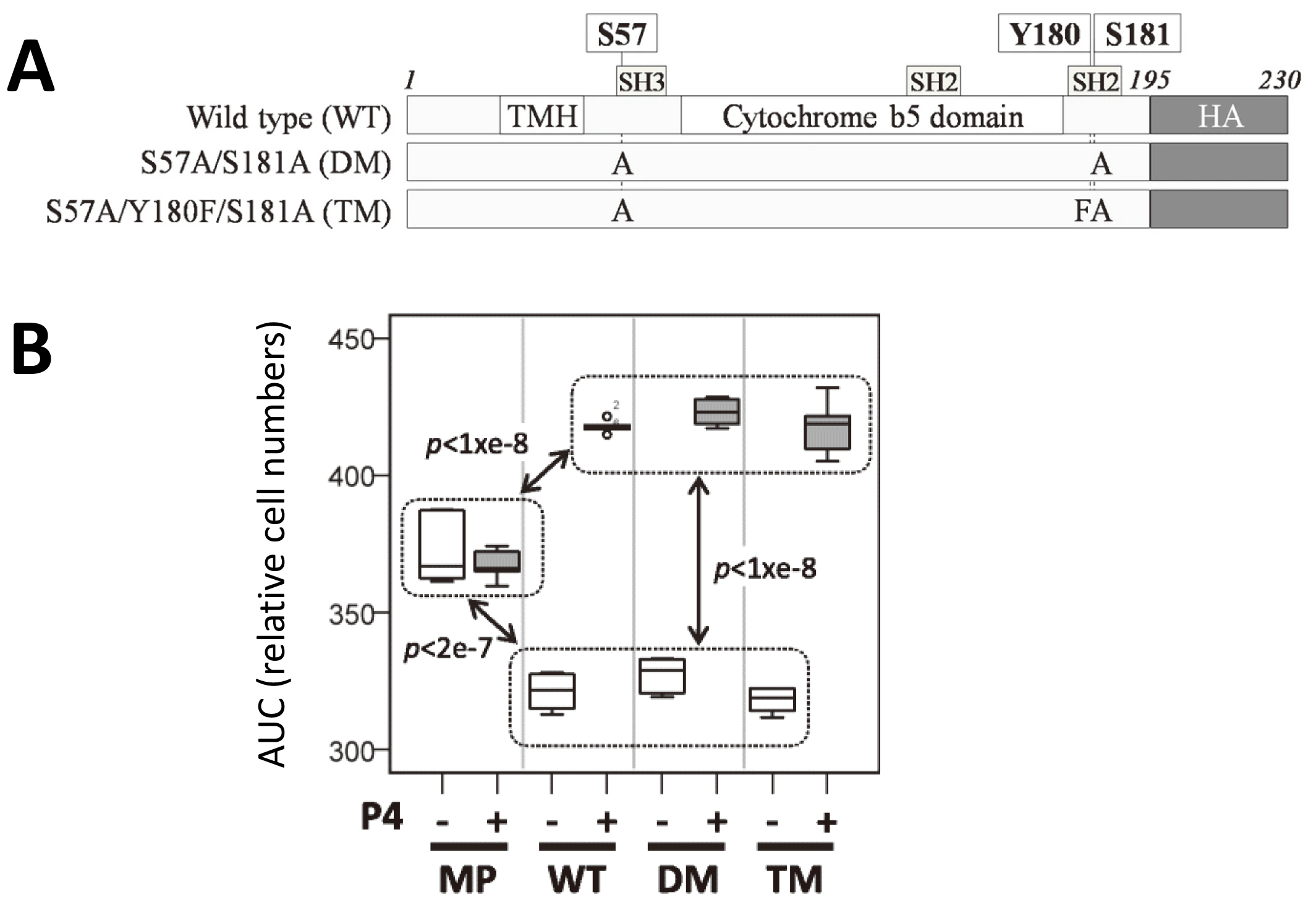
Fig. 5.

PGRMC1 phosphorylation status does not affect P4-dependent resistance to doxorubicin toxicity. (A) Schematic diagram of the PGRMC1 mutants employed in panel B. Reproduced unchanged from [19] under a Creative Commons Attribution 4.0 International License (CC BY 4.0). A copy of the license can be viewed at: https://creativecommons.org/licenses/by/4.0/. The license permits use, sharing, adaptation, distribution, and reproduction in any medium or format, as long as you give appropriate credit to the original author(s) and the source, provide a link to the Creative Commons license, and indicate if changes were made. (B) Boxplots of area under the curve (AUC) results for respective transfected cell lines expressing the PGRMC1 mutant proteins from panel A. MP, parental MIA PaCa-2 cell line. WT, DM and TM are as per panel A. For further details see [257], from which the panel is reproduced under a CC BY 4.0 Creative Commons license under conditions as per panel A. The panel is unchanged except the Y axis is relabeled to “AUC (relative cell numbers)”, representing cumulative percentage of cells relative to reference time zero as described in [257].

When we mutated different combinations of PGRMC1 S57, S180 and Y180 in stably transfected MIA PaCa-2 pancreatic cancer cells, our proteomics pathways analysis included predicted modulation of the ATM/ATR DNA repair pathways related to Ataxia Telangiectasia [257], a pathology that can be rescued by NAD replenishment in mouse models [469]. This prompted us to examine DNA mutation rates, where we detected significant differences between mutant cell lines, indicating that PGRMC1 affected genomic sequence stability [257]. This was despite finding no differences in PGRMC1-dependent resistance to doxorubicin-induced death, as discussed above. Therefore, the PGRMC1 phosphorylation mutants have identified the existence of separable PGRMC1 functions, providing the beginnings of a functionally stratified framework, where specific different PGRMC1 functions can be identified, characterized, and in the future be specifically pharmacologically addressed.

A PubMed search for PGRMC1 and sirtuin (23 November 2022) returned one result. where SIRT3 levels were lowered by oxidative stress, and further by cells with a PGRMC1 knockdown. Therefore, PGRMC1 served to maintain SIRT3 levels [444].

4.5 Possible Involvement of PGRMC1 and NAD Homeostasis

NAD synthesis leads to decreased ribosomal RNA (rRNA) transcription at the nucleolus. Depletion of the SIRT1-associated (and NAD-synthesizing) enzyme nicotinamide mononucleotide adenylyl transferase (NMNAT1, Fig. 6A, Ref. [19, 257]) increases rRNA synthesis as well as causing sensitivity to nutrient paucity and DNA damaging agents [484]. This sensitivity is at least superficially reminiscent of the sensitivity that is caused by over-expression of PGRMC1, which can be rescued and overcompensated into elevated viability by the presence of P4 as discussed above. Overarching PGRMC1 effects on ribosomal biosynthesis would be compatible with results observed for different phosphorylation mutants of PGRMC1, where pathways enrichment analysis predicted changes in proteins involved with tRNA aminoacylation, translation/elongation and T complex chaperonins [19]. Future research should investigate the contribution of PGRMC1 to NAD levels.

Fig. 6.

PGRMC1 Y180 phosphorylation is implicated in NAD homeostasis. Panels A-C of this figure are reproduced from Thejer et al. [257], with modifications, under a Creative Commons Attribution 4.0 International License (CC BY 4.0) as described in the legend to Fig. 5. Changes are described in the legend descriptions. (A) Model of the NNMT and 1-methylnicotinamide (1-MNA) pathway. The panel is modified from [257] by the addition of NAMPT (nicotinamide phosphoribosyl transferase), NMNAT1 (nicotinamide/nicotinic acid mononucleotide adenylyl transferase 1), and the black and white arrows also depicted in B and C. (B) NNMT abundance is increased by mutation of PGRMC1 Y180. Quantification of NNMT mRNA levels in WT, DM and TM cells (nomenclature follows Fig. 5A) by RT-PCR. ANOVA post-hoc Tukey’s HSD p < 0.01 (**) or p < 0.001 (***). The panel is modified from [257] by removal of parental MP cells. The white arrow indicates elevated NNMT levels in TM cells, which corresponds to the arrow in A. (C) NAMPT abundance is decreased by mutation of PGRMC1 Y180. SWATH-MS proteomic quantification of NAMPT (nicotinamide phosphoribosyl transferase). The color key schematically depicts relative abundance. For details see the data originally reported in [19] and [257]. The black arrow indicates decreased NAMPT levels in TM cells, which corresponds to the arrow in A. (D) Hypothetical model where Y180 phosphorylation leads to regeneration of NAD levels. (E) Hypothetical model where non-phosphorylated Y180 leads to 1-MNA production and falling NAD levels.

Interestingly in this respect the PGRMC1 S57A/Y180F/S180A triple mutant (TM, Fig. 5A) — which differs from the S57A/S180A double mutant (DM, Fig. 5A) only by the phosphate accepting oxygen atom of Y180 — exhibited elevated levels of nicotinamide-N-methyltransferase transferase (NNMT) mRNA and decreased levels of nicotinamide phosphoribosyl transferase (NAMPT) protein, which acts upstream of NMNAT1 in NAD salvage (Fig. 6). We would therefore predict from these results that PGRMC1 Y180 phosphorylation enhances NAD salvage from nicotinamide to elevate NAD levels. This is relevant to the prediction made in Section 2.11 above, that PGRMC1 tyrosines could be involved in sleep and circadian regulation. NAMPT is the rate-limiting enzyme of NAD synthesis in a feedback-regulated circadian metabolic pathway [485, 486].

Ramsay et al. [485] proposed that circadian regulation of NAD may be part of the mechanism that regulates a core clock. Those daily cycles could also form part of the clockwork mechanism of the epigenetic Horvath clock [487], which seems to accurately reflect the biological age of animals. It can apparently be rewound to reverse aging symptoms, and therefore its control of gene expression may be the manifestation of biological age [420]. The ability of the PGRMC1 TM mutation to convert the epigenetic state to a hypermethylated genome [257] suggests that Y180 could have direct access to the clockwork mechanism, although how it may influence the clock would require characterization.

Under the synonym Visfatin, NAMPT can be secreted as a cytokine under inflammatory and some other conditions [488, 489]. It would be interesting to see whether this correlates with PGRMC1 subcellular translocations and whether secretion correlates with type 2 PGRMC1 membrane topology.

As argued above, Y180 which regulates genomic CpG status was acquired by evolution coincidentally with the gastrulation organizer, neurons, sleep, and a variety of other eumetazoan synapomorphies. The activity of the gastrulation organizer and the eumetazoan adoption of a pre-existing system of CpG methylation to define tissue-specific gene expression are required for embryological differentiation. This produces multiple specialized cell types via a series of orchestrated progressions through lineage-specific epigenetic states which evolved after the gastrulation organizer and because of its activity, as discussed above. Furthermore, it is this eumetazoan level of epigenetic information which is thought to become corrupted during the aging process and can be modulated in an NAD-dependent manner [415, 416, 419, 469, 490].

4.6 Possible Involvement of PGRMC1 with the Epigenetic Aging Observer

We found differences in 1-methylnicotinamide (1-MNA) levels caused by PGRMC1 phosphorylation site mutants [257]. 1-MNA was more abundant in TM cells expressing a Y180F mutation, and as discussed above these cells also expressed higher levels of NNMT which produces 1-MNA, as well as lower levels of NAMPT which catalyzes the competing NAD salvage reaction [257]. These differences were associated with pronounced and specific effects on genomic CpG methylation, which was accompanied by pleiotropic changes in morphology, metabolism, and motility [19, 257].

Importantly for aging biology, these PGRMC1-dependent events affected the methylation status of CpG sites that are related to aging, as assayed by different manifestations of epigenetic aging clocks. There is a caveat that the MIA PaCa-2 cells used in the study are a genomically unstable cell culture line (one collaborating cytologist could not accurately identify homologous chromosomes by fluorescent spectral karyotyping from adjacent cells in the same culture: not shown), and genome packaging is certainly different than in vivo human cells. However, PGRMC1 phosphorylation mutants affected each of the DNAmPhenoAge Clock (Fig. 7A, Ref. [257]), DNAmAge Clock [418, 491] (Fig. 7B), and DNAmAgeSkinBlood Clock [492] (Fig. 7C). While the ‘estimated age’ Y axis of the graphs in Fig. 7 is meaningless for this cell line, the results do suggest that PGRMC1 phosphorylation status can affect epigenetics related to the aging clock. Future experiments should urgently address whether PGRMC phosphorylation sites in the T178/Y180/S181 motif, which was acquired before the evolutionary origins of bilaterally symmetrical animals, can exert corresponding influence on clock-related CpG sites during mammalian embryogenesis and adult life. Indeed, taken together, the hypothesis can be formulated whereby circadian regulation of NAD levels [493] by PGRMC1 could contribute to NAD-dependent sirtuin demethylase activity and diurnal metabolic regulation.
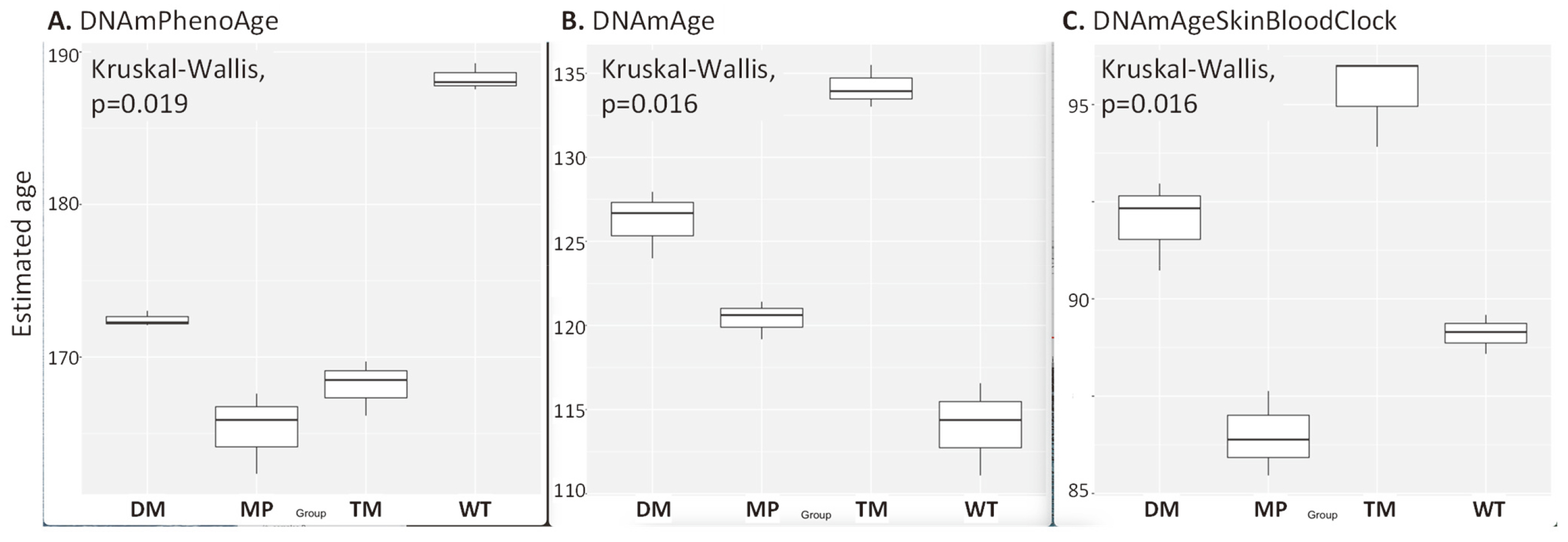
Fig. 7.

PGRMC1 phosphorylation affects CpG loci relevant to aging. Differences at different indicative sets of selected genomic CpG sites according to three different Clock Foundation standard clocks, using the publicly available methylomics data from [257] with analysis provided courtesy of J. Gordevicius, R. Brooke, and S. Horvath, The Epigenetic Clock Development Foundation. (A) DNAmPhenoAge. (B) DNAmAge. (C) DNAmAgeSkinBloodClock. Y axes represent clock-estimated ages. Note that none of the clocks was designed to estimate cell line ages. For exogenous p-PGRMC status of stable transfected cell types WT, DM, TM see Fig. 5A. MP is the parental MIA PaCa-2 cell line [257].

In this respect cnidarians, and therefore most probably the LEUMCA, exhibit sleep like behaviour [9, 494, 495]. The gastrulation organizer of the LEUMCA also generated the first synapsed nerves, and sleep is required for neural regeneration. Since the combination of PGRMC1 Y139 and a Y180-containing C-terminus also appeared first in the LEUMCA, there is a correlation between novel eumetazoan PGRMC1 functions and the onset of circadian rhythm-directed sleep. Whether the correlation is due to a functional dependence requires future exploration.

Mammalian embryogenesis produces hypomethylated naïve PSCs, which mature into hypermethylated primed PSCs, which in turn undergo sequential division and differentiation cycles to produce the differentiated cell types of the adult body. Tissue-specific multi-potent stem cells retain the ability to replace cells with certain lineage-restricted tissues in response to damage [496].

It has become clear that what used to be considered as terminally differentiated states are quite plastic, and terminology is adapting as we understand the phenomenon. Reprogramming is the process where differentiated stem cells can be induced to return to a pluripotent state (induced pluripotent stem cells: iPSCs). These have the propensity to cause cancer in adult cells [496, 497]. Transdifferentiation is the process of converting one specialized cell type to another. Transdifferentiation should be associated with minimal risk of cancer. For instance, an epithelial cell could be transdifferentiated to an advanced neural precursor in vitro and then into neurons in situ to treat neurodegenerative disorders [498, 499].

Sinclair [415] has described the targeted partial reversion of a cell to an earlier differentiation state as exdifferentiation. In this sense, fully differentiated neurons can be exdifferentiated to an earlier neural progenitor stage to repair a damaged optic nerve [420]. For this author, at least, it remains unclear whether in that instance the exdifferentiation protocol may not have reverted the cells to a primed pluripotent state which was immediately directed back along the somatic neurogenic differentiation pathway by extracellular matrix components of the environment. Aged cells could also in future potentially be exdifferentiated to younger versions of themselves, to extend lifespan [415]. The emergence of small molecules with reprogramming properties has enormous potential implications for this entire field of related processes [500]. Be that as it may, the exdifferentiation process resembles a reversion back at least towards pluripotency, and therefore to understand it we need to consider how differentiation works and whether PGRMC1 could be involved.

The induction of neuronal regenerative tissue repair and remodeling response after central nervous system damage requires both PGRMC1 protein [103, 501, 502] and, at least in response to chemical ischemia of the brain, also involves alteration of its phosphorylation status [503]. Such induction and modification probably occur at the same time, which has not been examined. Ischemic neural damage and AD share common properties, at least in part through activity of the Wnt signaling pathway and its influence on cell differentiation status [504]. PGRMC1 modulates Wnt activity [505] and cell fate determination [224] in PSCs.

The regenerative process involves the activation of quiescent stem or somatic cells to repair tissue [506], by the activation of regeneration enhancers, which dedifferentiate cells and can transdifferentiate them into other cell types. Unsurprisingly, there is overlap but not identity between regenerative and developmental enhancers [507]. PGRMC1 function is implicated above with stem cell activation and differentiation processes, and these are related to aging biology. Furthermore, PGRMC1 is required for the maintenance of primed PSC cell pluripotency in a process that involves the p53 and Wnt/
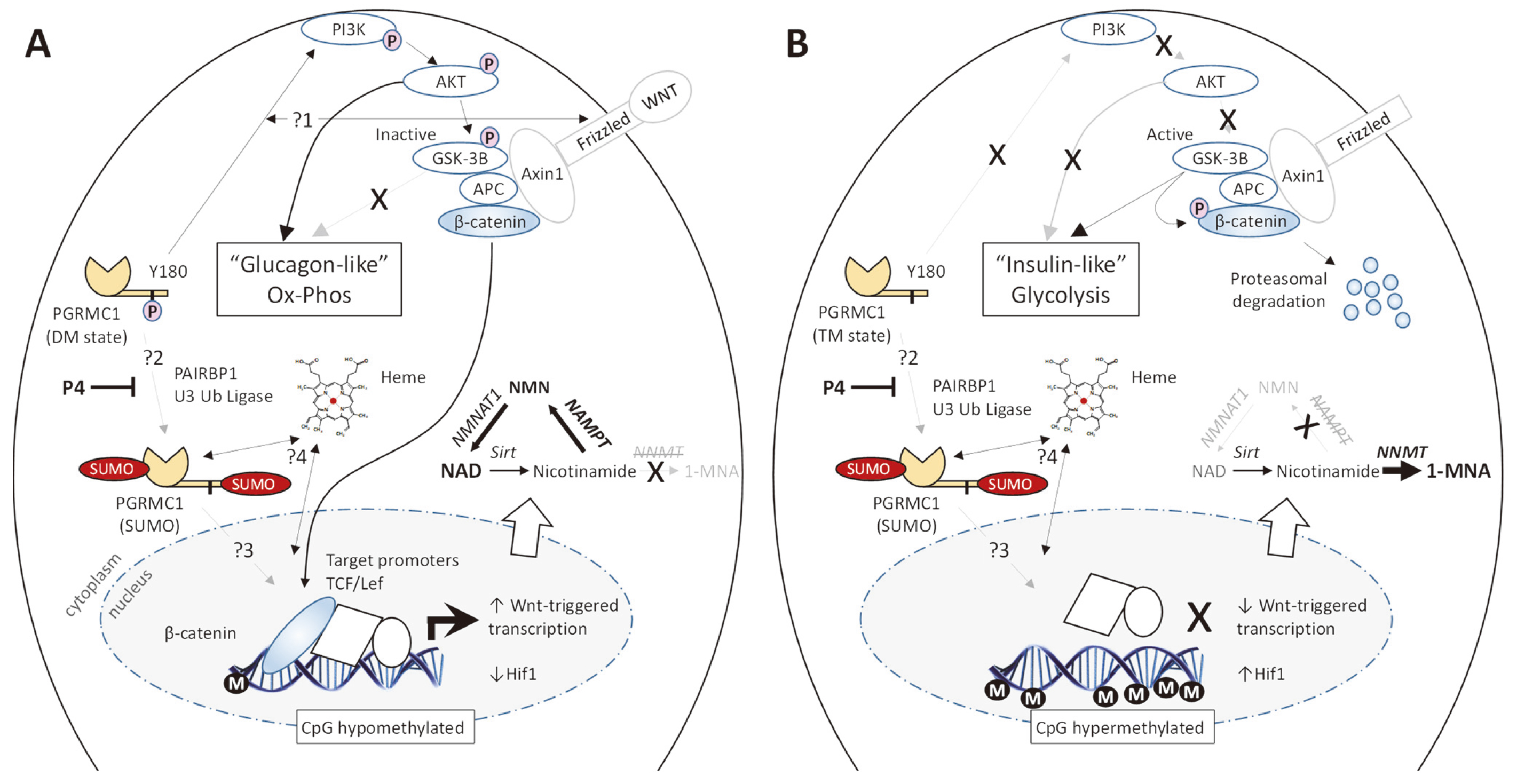
β-catenin pathways [224], and 1-MNA (Fig. 8, Ref. [19, 147, 224, 257, 301, 508, 509, 510, 511]) reportedly maintains naïve PSC status by controlling the epigenetic status [505]. Wnt/β-catenin signaling promotes self-renewal of the naïve human PSC state, and inhibits transition to the primed state [512]. PGRMC1 can directly bind to TCF/Lef sites in promoters to elevate their transcription, which can be repressed by P4 treatment [508], and where the p53 target of Wnt signaling is dependent upon nuclear heme. It is unlikely to be coincidence that PGRMC1 Y180, acquired coincidentally with the gastrulation organizer, can regulate the PI3K pathway required for Wnt signaling, PGRMC1 could conceivably provide heme to p53, and that sumoylated PGRMC1 can also bind directly to the promoters targeted by that pathway (Fig. 8) [508] (as discussed previously). In this respect note also that PGRMC2 acts as a heme chaperone, directing heme from mitochondria to the nucleus [314].

Fig. 8.

A hypothetical model for the role of PGRMC1 in Wnt signaling, NAD homeostasis, and epigenomic maintenance. The figure depicts only the biology proposed to be associated with PGRMC1. The testable model has not been experimentally validated. It is anticipated, if the model is valid, that the indicated pathways will act in concert with e.g., the Wnt, LIF/Stat3, Notch, FGF, and BMP pathways, and their effects on pluripotency transcription factors [509]. Compare the activity of the NNMT pathway to that reported for primed human PSCs, which reported histone hypermethylation [224]. Binding of sumoylated PGRMC1 to TCF/Lef binding sites follows [508]. ?1, uncertain relationship between effects of PGRMC1 phosphorylation and Wnt ligand. ?2, uncertainty which conditions lead to PGRMC1 sumoylation, therefore depicted in A and B. ?3, effects of sumo-PGRMC1 on β-catenin/TCF transcription. ?4, Modulation by heme. The conditions for ?3 and ?4 are not predicted, so they are present in both scenarios. (A) Possible effects mediated by phospho-PGRMC1 Y180 via regulation of GSK-3β activity (as seen in MIA PaCa-2 pancreatic cancer cells [19]). This condition is interpreted partly from the PGRMC1 S57A/ S181A double mutant (DM) described in [19] and [257], where phosphorylated S57 and S181 are thought to sterically impede phosphorylation of Y180 [147, 301]. β-catenin was not assayed. Whether or how PGRMC1 effects are related to the Wnt pathway is unclear (?1). “Glucagon like” refers to the unphosphorylated state of the indicated GSK-3β substrates and phosphorylated state of Akt substrates. A Y180F mutant elevated glycolysis and reduced GSK-3β phosphorylation [19] such that phosphorylated Y180 may propagate a glucagon-like (OxPhos) state. Naïve PSCs utilize either mitochondrial or glycolytic metabolism, while primed cells rely on glycolysis [510] which is associated with a Hif-1-induced metabolic switch in primed cells [511]. For the NNM pathway, see Fig. 6. M, genomic CpG methylation [257]. (B) The proposed effect of PGRMC1 Y180 dephosphorylation. This condition is interpreted partly from the PGRMC1 S57A/Y180F/S181A triple mutant (TM) described in [19] and [257]. Conventions follow A.

In the study of Kim et al. [224] the PGRMC1 C-terminus was extracellular [362], and so Y180 would not have been accessible to kinases, however Kim et al. [224] did propose that a subset of the PGRMC1 population was cytoplasmic. The overall interpretation of PGRMC1 knockdown could be confounded by interference in the system from PGRMC2, which interacts with PGRMC1 [295, 513] and could function aberrantly upon PGRMC1 knockdown: I.e., functions due to “loss of PGRMC1” could be caused by loss of restraints on PGRMC2.

We discovered [257] that PGRMC1 mutation of PGRMC1 Y180 induced nicotinamide-N-methyl transferase (NNMT), the enzyme which produces 1-MNA (Fig. 6). The association of this pathway with aging biology has been discussed previously, to which the reader is referred [257]. Taken together, it appears that PGRMC1 phosphorylation of Y180 could regulate embryonic stem cell status. However, this has not been demonstrated, and is a testable prediction.

Lu et al. [420] describe the recovery of a youthful epigenetic state in neurons by expression of three Yamanaka transcription factors (Oct4, Sox2 and Klf4) (OSK), and discuss the possible existence of an “Observer” function that enables cells to reactivate youthful epigenetic status. The ability of PGRMC1 phosphorylation mutants to apparently move the epigenetic state towards at least a pseudo-youthful hypermethylated condition (the TM containing Y180F mutation of Fig. 5 is hypermethylated to a similar extent as primed PSCs [257]) suggests that PGRMC1 could access “Observer” function. Hypothetically, the gastrulation organizer function may be related to the observer mechanism and mediate its effects at the level of chromosomal packaging which requires a PGRMC1-dependent process to maintain fidelity. Of course, a mechanistic explanation as to how PGRMC1, and especially the Y180 motif, modulates the epigenetic landscape is lacking and urgently required.

To add a hopefully novel contribution to the nature of the observer, the ability of Yamanaka factors to induce pluripotency from somatic cells implies that the promoters active in the pluripotent state exert a dominant epigenetic effect over ‘downstream’ chromosomal regions (regions which should be activated or repressed at a later stage in development because of these transcription factors). The primed embryonic PSC condition of expression corresponds to the LEUMCA epithelial cells prior to the induction of the gastrulation organizer.

Transcripts of the target genes of Yamanaka OSK factors are significantly enriched in developmental processes. Lu et al. [514] omitted the Myc Yamanaka factor. The targets of c-Myc in primed PSCs are mainly enriched in metabolism processes that are reduced in murine naïve PSCs. This is interesting because genes associated with metabolism are typical for endoderm, which is the most ancient germ layer and reflects general feeding functions. Whereas endoderm gene expression most closely resembles the ancestral monocellular state, the gametes, zygote, naïve PSCs and their derived germline cells represent highly innovative metazoan inventions [515]. Therefore, the OSK factors probably induce pluripotency by avoiding endodermal induction.

Yamanaka factors activate Nanog, Notch, Wnt, MAPK, p53, TGF-β, Hedgehog, apoptosis, axon guidance, and gap junction signaling pathways in embryonic stem cells, whereas ErbB, JAK-STAT, cytokine-cytokine receptor interaction, focal adhesion, adherens junction, cell cycle, and dorsal-ventral axis formation signaling pathways are repressed [514]. They also induce dramatic reorganization of chromatin and promoter-enhancer occupancies which precede transcriptional changes of affected genes [496].

The expression of genes is regulated by super-enhancers and insulators which govern the availability of proximal gene promoters to the transcriptional machinery. Classical enhancers bind transcription factors in the relative vicinity of the transcription start site and are enriched for the histone modification mono-methylation at lysine 4 of histone (H3H3K4me1). They can be classified into poised or active states by the respective increased abundance of either mutually exclusive lysine 27 methylation (H3K27me3) or acetylation (H3K27Ac) of histone 3, where acetylation is thought to protect against methylation [516] (for review: [517]). Therefore, histone methylases and deacetylases play critical roles in transcriptional regulation [518]. Active enhancers bind transcription factors with transactivation domains which recruit co-activators such as transcriptional activator CREB binding protein (CBP) or P300 to in turn recruit RNA polymerase II [519, 520]. In this respect, the PGRMC1-modulated NAD/1-MNA pathway (Fig. 6) affects both the level of NAD required for histone deacetylases such as Sirtuins and impinges on bulk levels of S-adenosyl methionine (SAM), the donor for cellular methylation reactions, such as of CpG or histones.

Active super-enhancers are bound by a high density of lineage-specific transcription factors, and exist in insulated chromosomal regions looped between insulators, which contain DNA binding sites for CCCCTC binding factor (CTCF). Together with cohesin, CTCF tethers chromosomal DNA into large loops. Such loops, or Topologically Associated Domains (TADs) exist between two CTCF sites which can be separated by a great many base pairs. It may be that the colocalization of active promoters and their associated RNA polymerase complexes may order nuclear architecture, and physical three-dimensional proximity to such a center may drive the rate of transcription from any promoter. By recruiting or dismissing CTCF binding sites, loops can be lengthened or shortened, thereby changing the activity of genes that resultingly find themselves in different TADs [521, 522, 523]. The order and proximity of genes separated by CTCF binding sites on a given chromosome is therefore important in how sets of genes can be either mutually or reciprocally regulated, or both, in different epigenetic circumstances.

The activity of such lineage specific super-enhancers is very probably, in the author’s opinion, closely related to the observer function. This probably involves achieving a transcriptional state where key master combinations of transcription factors are expressed that determine the root identity of a group of related more highly differentiated lineages, as achieved by establishing the activity of a certain set of super-enhancers. In this model, those super-enhancers will impose correct control of the CTCF-directed chromosomal regions (i.e., TADs). Importantly, this should involve activating/silencing of genes which should be off/on in vital/younger cells, but which may have become inappropriately regulated by the aging process. In this context, it is notable that in human granulosa cells nuclear PGRMC1 is localized in a punctate pattern, in physical proximity with the classical PGR [513]. It would be interesting to know whether CTCF is colocalized in these puncta, and to what extent sumoylation of nuclear PGRMC1 [18, 29, 393, 524] contributes to epigenetics.

The best studied differentiation system is the naïve to primed PSC transition, where certain naïve-specific enhancers are deactivated during the transition to the primed state. Enhancers that become active in the primed state were detectable in naïve cells (in a ‘poised’ state: H3K27me3), became active in primed cells (H3K27Ac), and often retained activity into the somatic state, frequently contributing to tissue-specific super-enhancers [525].

How alterations in CpG methylation affect changes in such higher order chromatin structures during development has been debated [526], however there is strong evidence that CpG methylation may be involved in the irreversible silencing of genes, such as in genomic imprinting. Studies from parental genomic imprinting sites suggest that CpG status affects the size of CTCF/condensin-structured TADs. Thereby, CTCF sites can change from being actively tethered at the base of a loop or being untethered. Intervening genes can be activated or silenced as a result [527].

Bestor et al. [526] argue that genes are sequentially activated and repressed solely by protein- and RNA-based mechanisms (including histone modifications) during developmental processes, whereas DNA methylation mediates permanent gene inactivation that originates in the germline and which usually does not change over a single lifetime. However, DNA methylation has been implicated in modulating alternative enhancers, splicing sites, and intragenic promoters. Furthermore, changes in DNA methylation in multiple disease states, as well as during development and with aging make a compelling case that changes in CpG methylation are important in gene regulation [528]. Active super-enhancers are associated with increased H3K27Ac (active state), and decreased DNA methylation. These are maintained by the presence of RNA Polymerase II-transcribed long non-coding extended RNAs (eRNAs) which participate in maintaining open chromatin structure. eRNAs require the activity of Tet (ten-eleven translocation) family methylcytosine dioxygenase DNA demethylases (which remove DNA methylation) to produce hypomethylation, which strongly associate DNA CpG hypomethylation with the establishment of active enhancers [529]. See also the references above to the Horvath clock above for functions of CpG methylation. PGRMC1 Y180 which affects CpG status was obtained in evolution at precisely the time when the reversible transition between naïve and primed PSC cell types probably evolved, which is formative to activity of the gastrulation organizer inherited by the LEUMCA.

In a possibly interesting connection between PGRMC1 and genomic methylation changes between naïve and primed PSCs, the orphan classical steroid receptor estrogen-related receptor beta (Esrrb: also known as Steroid hormone receptor ERR2, or just ERR2) binds to silenced enhancers in primed cells, displacing nucleosomes and opening up chromatin to cause epigenetic reversion to the hypomethylated naïve state [530]. We previously noticed that the abundance profile of mitochondrial proteins induced by the presence of PGRMC1 S57A/S181A DM (Fig. 5A), which is thought to permit easier Y180 phosphorylation by relieving steric hindrance of adjacent phosphorylated residues, overlapped strongly with proteins predicted to be induced by Esrrb/ERR2. The overlap profile was not perfect, and we concluded that PGRMC1 and Esrrb/ERR2 regulated overlapping sets of proteins, possibly related to shared steroid biology [19].

Given the undeniable relationship between PGRMC1 and steroid biology, it will be interesting to see whether PGRMC1 is responsible for production of the orphan ligand of Esrrb/ERR2 which is required for the naïve state. Note: (1) that we investigated Esrrb/ERR2 independently of PSC biology or aging, and (2) that classical (nuclear) steroid sensing first appeared in the LEUMCA, while receptors like Esrrb/ERR2 probably arose with the urbilaterian [2]. Conceivably, the naïve to primed PSC transition is partly due to PGRMC1-dependent changes in the levels of an unknown steroid ligand of Esrrb/ERR2. Recall again in this context the discussion above where PGRMC1 was required for the pluripotency of primed PSCs [224], while the NAD/NNMT/1-MNA pathway maintains the naïve state [505]. Esrrb/ERR2 may well be an intermediary in the mechanism. Of course, this is speculation that would require experimental validation.

Note also in this context, as detailed above, that PGRMC1 seems intimately related to mitochondrial function, and the overt regulation between glucose or lipid/amino acid (TCA cycle) respiration, and that primed cells exhibit increased mitochondrial respiration. PGRMC1 tyrosine phosphorylation (and the extended C-terminus that holds the Y180 motif) were acquired at the same time as the gastrulation organizer, sleep, and the foundational platform for cell differentiation mechanisms, making it at least plausible that PGRMC1 function is related to the differentiation processes that ultimately drive the aging process, and the mechanisms that can slow it. I.e., PGRMC1 acquired new functions at just the right time in animal evolution to affect aging, which evolved later (after the evolution of all those differentiated tissue types which age). This is another untested hypothesis, with testable predictions that, like metformin, resveratrol, and NAD supplements [415], PGRMC1 phosphorylation mutants, post-translational modifications, and/or pharmacological interference with PGRMC1 functions, could affect healthspan and lifespan.

5. PGRMC: Where Are We Now?

The previous discussion directly connects PGRMC1’s biology with all the main AD biomarkers, with multiple aspects of tumor biology (migration, EMT, epigenetic plasticity, survival, etc.), and with key feature of other diseases of aging, such as diabetes, as well as with several key pathways of aging itself. The common denominator could well be the perturbation of foundational level metabolic switches reflecting the proposed original eukaryotic function of PGRMC as a metabolic regulator [1, 153], and the adaptation of this PGRMC-based eukaryotic cellular foundation towards specialized multicellular body plans and differentiated cell types.

Fluctuations in levels or properties of the leading accepted markers for AD could be directly explained by a new model centrally involving PGRMC1 and its associated sterol and heme biology at the interface of ER and mitochondrial integration in the pathogenesis of AD. Therefore, PGRMC1 and the regulatory apparatus surrounding it may represent one of the best current directions in which to search for improved AD cures, and possibly for many other age-related illnesses.

No doubt, as stated here the model is incomplete and should be substantially refined. However, this new compass bearing has every prospect of plotting a better course to, e.g., successful AD therapies than has commanded the helm for three unsuccessful Amyloid Cascade Hypothesis-directed decades. Therein lies the justification for a PGRMC-based model of AD, and perhaps eumetazoan aging, which is nothing short of the heuristic core of a new paradigm for some of the most urgent medical issues facing modern human society. Because of the foundational level regulation proposed for PGRMC in animal biology [1], these PGRMC functions are expected to be manifest across multiple clinical indication areas.

The biology being discussed here, involving PGRMC phosphorylation and other modifications, will not be detected by many modern routine analytical techniques: especially transcriptomic-based ones. Since we discovered differential PGRMC1 phosphorylation in breast cancers [301], only Sabbir [18] from the rest of the scientific community has systematically attempted to monitor PGRMC1 phosphorylation status, which he found to change in response to P4 treatment, and where he cited the author’s publications as rationale. (See also Peluso and Pru [393]). Sabbir and the group of Peluso and collaborators have investigated sumoylation and ubiquitination [18, 29, 108, 524]. There is an urgent need to develop reagents and methods to assay the state of PGRMC post-translational status, membrane topology, and subcellular location in a variety of different organisms and their various cell types during embryological differentiation stages and adult biology.

In terms of eumetazoan biology, Archimedes may have recognized in PGRMC the fulcrum that provided sufficient evolutionary leverage to enable development of the body architecture and organization of the eumetazoan animal world, and to topple its metabolic organization in modern disease states. It appears that the PGRMC research journey will continue to illuminate many more important mechanisms for human health in the future.

Abbreviations

1-MNA, 1-methylnicotinamide; Aβ, amyloid beta; Aβo, amyloid beta oligomers; Ac-CoA, acetyl-coenzyme A; AD, Alzheimer’s disease; AMPA, α-amino-3-hydroxy-5-methyl-4-isoxazolepropionic acid; AMPAR, α-amino-3-hydroxy-5-methyl-4-isoxazolepropionic acid receptor; AMPK, 5’-AMP-activated protein kinase; ApoE4, apolipoprotein E4; APP, amyloid precursor protein; ATM, ataxia telangiectasia mutated; ATR, ataxia telangiectasia and Rad3-related protein; AVG, anterior ventral neuron G; BACE, beta-site amyloid precursor protein cleaving enzyme; BASP1, brain acid soluble protein 1; cAMP, cyclic adenosine monophosphate; CC, coiled-coil; CNS, central nervous system; CogRx, Cognition Therapeutics Inc.; CycT, cyclopamine tartrate; DA, dopaminergic; CpG, the DNA dinucleotide sequence cytosine – phosphodiester – guanosine frequently methylated in epigenetic genome regulation; CK2, casein kinase 2; CPR, candidate phyla radiation (bacteria); CREB, cAMP-response element binding protein; CTCF, CCCCTC binding factor; CYP450, cytochrome P450; DCC, deleted in colorectal carcinoma; EGFR, epidermal growth factor receptor; EMC, endoplasmic reticulum membrane protein complex; EMT, epithelial-mesenchymal/mesodermal transition; ER, endoplasmic reticulum; ERK, extracellular signal-regulated kinase; ERMES, ER-mitochondria encounter structure; eRNAs, RNA Polymerase II-transcribed long non-coding extended RNAs; ESRα, estrogen receptor alpha; Esrrb/ERR2, estrogen-related receptor beta; FDA, Federal Drug Administration; FOXO1, transcription factor forkhead box protein O1; G3BP2, GTPase activating protein binding protein 2; GABA, γ-aminobutyric acid; GLP1, glucagon-like peptide-1; GnRH, gonadotropin-releasing hormone receptor; GLUT1, facilitated glucose transporter member 1 (Solute carrier family 2); GLUT4, facilitated glucose transporter member 4 (Solute carrier family 4); GPCR, G-protein-coupled receptor; GSK-3β, glycogen synthase kinase-3β; H3K4me1, mono-methylation at lysine 4 of histone H3; H3K27Ac, acetylation at lysine 27 of histone H3; H3K27me3, tri-methylation at lysine 27 of histone H3; H3K9me2/3, di-methylation at lysine 9 of histone H3; Hif-1 α, Hypoxia-inducible factor 1 alpha; HPA, hypothalamic-pituitary-adrenal (axis); HPG, hypothalamic–pituitary–gonadal (axis); HSPG, heparan sulfate proteoglycan; HPT, hypothalamic–pituitary–thyroid (axis); IFN, interferon; Iκ-Bα, NF-κB inhibitor alpha; iPSCs, induced pluripotential stem cells; LAMP-2A, lysosomal-associated membrane protein-2A; LDL, low-density lipoprotein; LDLR, low-density lipoprotein receptor; LECA, last eukaryotic common ancestor; LEPCA, last eumetazoan and placozoan common ancestor; LEUMCA, last eumetazoan common ancestor; LRP1, low-density lipoprotein receptor-related protein 1; LTP, long-term potentiation; MAC30, meningioma-associated protein 30 (see S2R, TMEM97); MAMS, mitochondrial endoplasmic reticulum-associated membranes; MAPR, membrane-associated progesterone receptor; MIHIR, MAPR interhelical insertion region (of MAPR proteins); miRNA, micro ribonucleic acid; MMP-9, matrix metalloproteinase 9; mTORc, mammalian target of rapamycin c; MVP, mevalonate pathway; NAD, nicotinamide adenine dinucleotide; NAMP, nicotinamide phosphoribosyl transferase; NET, norethisterone; NFκB, nuclear factor κB; NMNAT1, nicotinamide mononucleotide adenylyl transferase; NNMT, nicotinamide-N-methyltransferase transferase; NOX4, NADPH oxidase 4; NPC1, Niemann-Pick C1; OSK, Oct4, Sox2 and Klf4 transcription factors; P4, progesterone; PD, Parkinson’s disease; PEPCK, phosphoenolpyruvate carboxykinase; PFK2, 6-phosphofructo-2-kinase; PGR, nuclear (classical) progesterone receptor; PGRMC, progesterone receptor membrane component; PGRMC1, progesterone receptor membrane component 1; PGRMC2, progesterone receptor membrane component 2; PHB, prohibitin; PI3K, phosphatidylinositol 3-kinase; PKA, protein kinase A; PSCs, pluripotential embryonic stem cells; PTEN, phosphatidylinositol 3,4,5-trisphosphate 3-phosphatase and dual-specificity protein phosphatase; Rev-Erbα, nuclear receptor subfamily 1 group D member 1; S1R, sigma-1 receptor; S2R, sigma-2 receptor (see MAC30, TMEM97); SARS Cov-2, severe acute respiratory syndrome coronavirus 2; SCAP, SREBP cleavage activating protein; SDS PAGE, sodium dodecyl sulphate polyacrylamide gel electrophoresis; shRNA, short hairpin RNA; SIRT, sirtuin; SIRT1, sirtuin-1; SREBP, sterol regulatory element-binding protein; SWI/SNF, SWItch/Sucrose Non-Fermentable; TADs, Topologically Associated Domains; TCA, tricarboxylic acid; Tet, ten-eleven translocation; TMEM97, Transmembrane protein 97 (see S2R, MAC30); TSPO, tryptophan-rich sensory protein; UNC-6, uncoordinated-6; UNC-40, uncoordinated-40; VEGF, vascular endothelial growth factor; Vem1, Ventral Midline-1 Ceanorhabbitis elegans homologue of PGRMC; VEMA, ventral midline antigen, a synonym of murine PGRMC1; Wnt, Cell surface receptor named after the Drosophila segment polarity gene wingless and the vertebrate homolog, integrated; WT1, Wilms’ tumor-1 protein; xCT, amino acid transport system xc- cysteine/glutamate antiporter.

Author Contributions

MAC conceptualized and wrote this work.

Ethics Approval and Consent to Participate

Not applicable.

Acknowledgment

I am grateful to Tim Cahill (R.I.P.) for Latin language assistance.

Funding

This research received no external funding.

Conflict of Interest

Michael A. Cahill is scientific advisor to and minor shareholder (<0.001%) of Cognition Therapeutics (CogRx). This work was prepared without knowledge by or influence from CogRx. There are no other competing financial interests. Given his role as Guest Editor, Michael A. Cahill was not involved in the peer-review of this article and has no access to information regarding its peer-review. Full responsibility for the editorial process for this article was delegated to Giuseppe Ingravallo and Graham Pawelec.

Publisher’s Note: IMR Press stays neutral with regard to jurisdictional claims in published maps and institutional affiliations.

References
[1]
Cahill MA. Unde venisti PGRMC? Grand-scale biology from early eukaryotes and eumetazoan animal origins. Frontiers in Bioscience-Landmark. 2022; 27: 317.
[2]
Cahill MA. The evolutionary appearance of signaling motifs in PGRMC1. Bioscience Trends. 2017; 11: 179–192.
[3]
Ren J, Chung-Davidson YW, Jia L, Li W. Genomic sequence analyses of classical and non-classical lamprey progesterone receptor genes and the inference of homologous gene evolution in metazoans. BMC Evolutionary Biology. 2019; 19: 136.
[4]
Kundu P, Blacher E, Elinav E, Pettersson S. Our Gut Microbiome: The Evolving Inner Self. Cell. 2017; 171: 1481–1493.
[5]
Stabili L, Parisi MG, Parrinello D, Cammarata M. Cnidarian Interaction with Microbial Communities: From Aid to Animal’s Health to Rejection Responses. Marine Drugs. 2018; 16: 296.
[6]
Zeibich L, Schmidt O, Drake HL. Dietary polysaccharides: fermentation potentials of a primitive gut ecosystem. Environmental Microbiology. 2019; 21: 1436–1451.
[7]
Litvak Y, Byndloss MX, Bäumler AJ. Colonocyte metabolism shapes the gut microbiota. Science. 2018; 362: eaat9076.
[8]
Han B, Sivaramakrishnan P, Lin CJ, Neve IAA, He J, Tay LWR, et al. Microbial Genetic Composition Tunes Host Longevity. Cell. 2017; 169: 1249–1262.e13.
[9]
Nath RD, Bedbrook CN, Abrams MJ, Basinger T, Bois JS, Prober DA, et al. The Jellyfish Cassiopea Exhibits a Sleep-like State. Current Biology. 2017; 27: 2984–2990.e3.
[10]
Brown R, Pang G, Husband AJ, King MG. Suppression of immunity to influenza virus infection in the respiratory tract following sleep disturbance. Regional Immunology. 1989; 2: 321–325.
[11]
Brown R, Price RJ, King MG, Husband AJ. Autochthonous intestinal bacteria and coprophagy: a possible contribution to the ontogeny and rhythmicity of slow wave sleep in mammals. Medical Hypotheses. 1988; 26: 171–175.
[12]
Brown R, Price RJ, King MG, Husband AJ. Interleukin-1 beta and muramyl dipeptide can prevent decreased antibody response associated with sleep deprivation. Brain, Behavior, and Immunity. 1989; 3: 320–330.
[13]
Brown R, Price RJ, King MG, Husband AJ. Are antibiotic effects on sleep behavior in the rat due to modulation of gut bacteria? Physiology & Behavior. 1990; 48: 561–565.
[14]
Andren J, Andrews K, Brown L, Chidgey J, Geary N, King MG, et al. Muramyl peptides and the functions of sleep. Behavioural Brain Research. 1995; 69: 85–90.
[15]
Ilie OD, Ciobica A, Riga S, Dhunna N, McKenna J, Mavroudis I, et al. Mini-Review on Lipofuscin and Aging: Focusing on The Molecular Interface, The Biological Recycling Mechanism, Oxidative Stress, and The Gut-Brain Axis Functionality. Medicina. 2020; 56: 626.
[16]
Matenchuk BA, Mandhane PJ, Kozyrskyj AL. Sleep, circadian rhythm, and gut microbiota. Sleep Medicine Reviews. 2020; 53: 101340.
[17]
DeJong EN, Surette MG, Bowdish DME. The Gut Microbiota and Unhealthy Aging: Disentangling Cause from Consequence. Cell Host & Microbe. 2020; 28: 180–189.
[18]
Sabbir MG. Progesterone induced Warburg effect in HEK293 cells is associated with post-translational modifications and proteasomal degradation of progesterone receptor membrane component 1. The Journal of Steroid Biochemistry and Molecular Biology. 2019; 191: 105376.
[19]
Thejer BM, Adhikary PP, Kaur A, Teakel SL, Van Oosterum A, Seth I, et al. PGRMC1 phosphorylation affects cell shape, motility, glycolysis, mitochondrial form and function, and tumor growth. BMC Molecular and Cell Biology. 2020; 21: 24.
[20]
Wu H, Wu ZG, Shi WJ, Gao H, Wu HH, Bian F, et al. Effects of progesterone on glucose uptake in neurons of Alzheimer’s disease animals and cell models. Life Sciences. 2019; 238: 116979.
[21]
Nishida A, Inoue R, Inatomi O, Bamba S, Naito Y, Andoh A. Gut microbiota in the pathogenesis of inflammatory bowel disease. Clinical Journal of Gastroenterology. 2018; 11: 1–10.
[22]
Wang Z, Zhao Y. Gut microbiota derived metabolites in cardiovascular health and disease. Protein & Cell. 2018; 9: 416–431.
[23]
Quigley EMM. Microbiota-Brain-Gut Axis and Neurodegenerative Diseases. Current Neurology and Neuroscience Reports. 2017; 17: 94.
[24]
Maynard C, Weinkove D. The Gut Microbiota and Ageing. Sub-Cellular Biochemistry. 2018; 90: 351–371.
[25]
Nie P, Li Z, Wang Y, Zhang Y, Zhao M, Luo J, et al. Gut microbiome interventions in human health and diseases. Medicinal Research Reviews. 2019; 39: 2286–2313.
[26]
Long JM, Holtzman DM. Alzheimer Disease: An Update on Pathobiology and Treatment Strategies. Cell. 2019; 179: 312–339.
[27]
Hampton KK, Anderson K, Frazier H, Thibault O, Craven RJ. Insulin Receptor Plasma Membrane Levels Increased by the Progesterone Receptor Membrane Component 1. Molecular Pharmacology. 2018; 94: 665–673.
[28]
Cahill MA, Jazayeri JA, Catalano SM, Toyokuni S, Kovacevic Z, Richardson DR. The emerging role of progesterone receptor membrane component 1 (PGRMC1) in cancer biology. Biochimica et Biophysica Acta. 2016; 1866: 339–349.
[29]
Sabbir MG, Taylor CG, Zahradka P. Progesterone-induced Warburg Effect is Regulated by Cell-type-specific Interaction of Progesterone Receptor Membrane Component 1 and Hexokinases. Research Square. 2020. (preprint)
[30]
Atif F, Yousuf S, Espinosa-Garcia C, Stein DG. Progesterone Modulates Mitochondrial Functions in Human Glioblastoma Cells. Molecular Neurobiology. 2021; 58: 3805–3816.
[31]
Gras F, Brunmair B, Quarre L, Szocs Z, Waldhausl W, Furnsinn C. Progesterone impairs cell respiration and suppresses a compensatory increase in glucose transport in isolated rat skeletal muscle: a non-genomic mechanism contributing to metabolic adaptation to late pregnancy? Diabetologia. 2007; 50: 2544–2552.
[32]
Lee SR, Choi WY, Heo JH, Huh J, Kim G, Lee KP, et al. Progesterone increases blood glucose via hepatic progesterone receptor membrane component 1 under limited or impaired action of insulin. Scientific Reports. 2020; 10: 16316.
[33]
Yoshitani N, Satou K, Saito K, Suzuki S, Hatanaka H, Seki M, et al. A structure-based strategy for discovery of small ligands binding to functionally unknown proteins: combination of in silico screening and surface plasmon resonance measurements. Proteomics. 2005; 5: 1472–1480.
[34]
Hehenberger E, Eitel M, Fortunato SAV, Miller DJ, Keeling PJ, Cahill MA. Early eukaryotic origins and metazoan elaboration of MAPR family proteins. Molecular Phylogenetics and Evolution. 2020; 148: 106814.
[35]
Cahill MA. Choose your partners for the next dance: implied PGRMC1 roles in membrane trafficking and mitochondrial modulation. Fertility and Sterility. 2020; 113: 938–941.
[36]
Salsano S, Gonzalez-Martin R, Quinonero A, Lopez-Martin S, Gomez-Escribano AP, Perez-Deben S, et al. Novel nonclassic progesterone receptor PGRMC1 pulldown-precipitated proteins reveal a key role during human decidualization. Fertility and Sterility. 2020; 113: 1050–1066.e7.
[37]
Teakel SL, Ludescher M, Thejer BM, Poschmann G, Forwood JK, Neubauer H, et al. Protein complexes including PGRMC1 and actin-associated proteins are disrupted by AG-205. Biochemical and Biophysical Research Communications. 2020; 524: 64–69.
[38]
Ponikwicka-Tyszko D, Chrusciel M, Stelmaszewska J, Bernaczyk P, Chrusciel P, Sztachelska M, et al. Molecular mechanisms underlying mifepristone’s agonistic action on ovarian cancer progression. EBioMedicine. 2019; 47: 170–183.
[39]
Ponikwicka-Tyszko D, Chrusciel M, Pulawska K, Bernaczyk P, Sztachelska M, Guo P, et al. Mifepristone Treatment Promotes Testicular Leydig Cell Tumor Progression in Transgenic Mice. Cancers. 2020; 12: 3263.
[40]
Kabe Y, Nakane T, Koike I, Yamamoto T, Sugiura Y, Harada E, et al. Haem-dependent dimerization of PGRMC1/Sigma-2 receptor facilitates cancer proliferation and chemoresistance. Nature Communications. 2016; 7: 11030.
[41]
Lee SR, Kwon SW, Kaya P, Lee YH, Lee JG, Kim G, et al. Loss of progesterone receptor membrane component 1 promotes hepatic steatosis via the induced de novo lipogenesis. Scientific Reports. 2018; 8: 15711.
[42]
Asperger H, Stamm N, Gierke B, Pawlak M, Hofmann U, Zanger UM, et al. Progesterone receptor membrane component 1 regulates lipid homeostasis and drives oncogenic signaling resulting in breast cancer progression. Breast Cancer Research. 2020; 22: 75.
[43]
Riad A, Zeng C, Weng CC, Winters H, Xu K, Makvandi M, et al. Sigma-2 Receptor/TMEM97 and PGRMC-1 Increase the Rate of Internalization of LDL by LDL Receptor through the Formation of a Ternary Complex. Scientific Reports. 2018; 8: 16845.
[44]
Ginsburg S, Jablonka E. The evolution of the sensitive soul: learning and the origins of consciousness (pp. 280–283). The MIT Press: Cambridge, MA, USA. 2019.
[45]
Schwab WE. The ontogeny of swimming behavior in the scyphozoan, Aurelia aurita. I. Electrophysiological analysis. The Biological Bulletin. 1977; 152: 233–250.
[46]
Galliot B, Quiquand M. A two-step process in the emergence of neurogenesis. The European Journal of Neuroscience. 2011; 34: 847–862.
[47]
Martín-Durán JM, Pang K, Børve A, Lê HS, Furu A, Cannon JT, et al. Convergent evolution of bilaterian nerve cords. Nature. 2018; 553: 45–50.
[48]
Northcutt RG. Understanding vertebrate brain evolution. Integrative and Comparative Biology. 2002; 42: 743–756.
[49]
Jerison HJ. Evolution of the brain and intelligence. Academic Press: Cambridge, MA, USA. 1973.
[50]
Yuan H, Silberstein SD. Vagus Nerve and Vagus Nerve Stimulation, a Comprehensive Review: Part I. Headache. 2016; 56: 71–78.
[51]
Yuan H, Silberstein SD. Vagus Nerve and Vagus Nerve Stimulation, a Comprehensive Review: Part II. Headache. 2016; 56: 259–266.
[52]
Tada MN, Kuratani S. Evolutionary and developmental understanding of the spinal accessory nerve. Zoological Letters. 2015; 1: 4.
[53]
Sugahara F, Murakami Y, Pascual-Anaya J, Kuratani S. Reconstructing the ancestral vertebrate brain. Development, Growth & Differentiation. 2017; 59: 163–174.
[54]
Intlekofer KA, Clements K, Woods H, Adams H, Suvorov A, Petersen SL. Progesterone receptor membrane component 1 inhibits tumor necrosis factor alpha induction of gene expression in neural cells. PLoS ONE. 2019; 14: e0215389.
[55]
Intlekofer KA, Petersen SL. Distribution of mRNAs encoding classical progestin receptor, progesterone membrane components 1 and 2, serpine mRNA binding protein 1, and progestin and ADIPOQ receptor family members 7 and 8 in rat forebrain. Neuroscience. 2011; 172: 55–65.
[56]
Burdakov D, Peleg-Raibstein D. The hypothalamus as a primary coordinator of memory updating. Physiology & Behavior. 2020; 223: 112988.
[57]
Wang A, Luan HH, Medzhitov R. An evolutionary perspective on immunometabolism. Science. 2019; 363: eaar3932.
[58]
Blair JA, McGee H, Bhatta S, Palm R, Casadesus G. Hypothalamic-pituitary-gonadal axis involvement in learning and memory and Alzheimer’s disease: more than “just” estrogen. Frontiers in Endocrinology. 2015; 6: 45.
[59]
Blausen B. Medical gallery of Blausen Medical 2014: Blausen_0614_LimbicSystem.png. WikiJournal of Medicine. 2014; Available at: https://commons.wikimedia.org/wiki/File:Blausen_0614_LimbicSystem.png (Accessed: 23 November 2022).
[60]
Krassas GE, Markou KB. The impact of thyroid diseases starting from birth on reproductive function. Hormones. 2019; 18: 365–381.
[61]
Arendt D, Tosches MA, Marlow H. From nerve net to nerve ring, nerve cord and brain–evolution of the nervous system. Nature Reviews Neuroscience. 2016; 17: 61–72.
[62]
Bonaz B, Bazin T, Pellissier S. The Vagus Nerve at the Interface of the Microbiota-Gut-Brain Axis. Frontiers in Neuroscience. 2018; 12: 49.
[63]
Breit S, Kupferberg A, Rogler G, Hasler G. Vagus Nerve as Modulator of the Brain-Gut Axis in Psychiatric and Inflammatory Disorders. Frontiers in Psychiatry. 2018; 9: 44.
[64]
Berthoud HR, Neuhuber WL. Functional and chemical anatomy of the afferent vagal system. Autonomic Neuroscience. 2000; 85: 1–17.
[65]
Hutter H. Extracellular cues and pioneers act together to guide axons in the ventral cord of C. elegans. Development. 2003; 130: 5307–5318.
[66]
Runko E, Kaprielian Z. Caenorhabditis elegans VEM-1, a novel membrane protein, regulates the guidance of ventral nerve cord-associated axons. Journal of Neuroscience. 2004; 24: 9015–9026.
[67]
Heger P, Zheng W, Rottmann A, Panfilio KA, Wiehe T. The genetic factors of bilaterian evolution. eLife. 2020; 9: e45530.
[68]
Runko E, Wideman C, Kaprielian Z. Cloning and expression of VEMA: a novel ventral midline antigen in the rat CNS. Molecular and Cellular Neurosciences. 1999; 14: 428–443.
[69]
Runko E, Kaprielian Z. Expression of Vema in the developing mouse spinal cord and optic chiasm. The Journal of Comparative Neurology. 2002; 451: 289–299.
[70]
Remage-Healey L. Frank Beach Award Winner: Steroids as neuromodulators of brain circuits and behavior. Hormones and Behavior. 2014; 66: 552–560.
[71]
Balthazart J, Choleris E, Remage-Healey L. Steroids and the brain: 50years of research, conceptual shifts and the ascent of non-classical and membrane-initiated actions. Hormones and Behavior. 2018; 99: 1–8.
[72]
Lloyd-Evans E, Waller-Evans H. Biosynthesis and signalling functions of central and peripheral nervous system neurosteroids in health and disease. Essays in Biochemistry. 2020; 64: 591–606.
[73]
Wessel L, Olbrich L, Brand-Saberi B, Theiss C. New aspects of progesterone interactions with the actin cytoskeleton and neurosteroidogenesis in the cerebellum and the neuronal growth cone. Journal of Histochemistry and Cytochemistry. 2014; 62: 835–845.
[74]
Wingenfeld K, Wolf OT. Stress, memory, and the hippocampus. Frontiers of Neurology and Neuroscience. 2014; 34: 109–120.
[75]
Min L, Takemori H, Nonaka Y, Katoh Y, Doi J, Horike N, et al. Characterization of the adrenal-specific antigen IZA (inner zone antigen) and its role in the steroidogenesis. Molecular and Cellular Endocrinology. 2004; 215: 143–148.
[76]
Ryu CS, Klein K, Zanger UM. Membrane Associated Progesterone Receptors: Promiscuous Proteins with Pleiotropic Functions - Focus on Interactions with Cytochromes P450. Frontiers in Pharmacology. 2017; 8: 159.
[77]
Peluso JJ, Liu X, Uliasz T, Pru CA, Kelp NC, Pru JK. PGRMC1/2 promotes luteal vascularization and maintains the primordial follicles of mice. Reproduction. 2018; 156: 365–373.
[78]
Peluso JJ, Pru JK. Non-canonical progesterone signaling in granulosa cell function. Reproduction. 2014; 147: R169–R178.
[79]
Bertani GR, Johnson RK, Robic A, Pomp D. Mapping of porcine ESTs obtained from the anterior pituitary. Animal Genetics. 2003; 34: 132–134.
[80]
Sun X, Hu Y, Zhou H, Wang S, Zhou C, Lin L, et al. Inhibition of progesterone receptor membrane component-1 exacerbates neonatal hypoxic-ischemic cerebral damage in male mice. Experimental Neurology. 2022; 347: 113893.
[81]
Sekeres MJ, Winocur G, Moscovitch M. The hippocampus and related neocortical structures in memory transformation. Neuroscience Letters. 2018; 680: 39–53.
[82]
Bashour NM, Wray S. Progesterone directly and rapidly inhibits GnRH neuronal activity via progesterone receptor membrane component 1. Endocrinology. 2012; 153: 4457–4469.
[83]
Crudden G, Loesel R, Craven RJ. Overexpression of the cytochrome p450 activator hpr6 (heme-1 domain protein/human progesterone receptor) in tumors. Tumour Biology. 2005; 26: 142–146.
[84]
Santos MS, Pereira EM, Carvaho AP. Stimulation of immunoreactive insulin release by glucose in rat brain synaptosomes. Neurochemical Research. 1999; 24: 33–36.
[85]
Gasparini L, Xu H. Potential roles of insulin and IGF-1 in Alzheimer’s disease. Trends in Neurosciences. 2003; 26: 404–406.
[86]
Ikeda H, West DB, Pustek JJ, Figlewicz DP, Greenwood MR, Porte D Jr, et al. Intraventricular insulin reduces food intake and body weight of lean but not obese Zucker rats. Appetite. 1986; 7: 381–386.
[87]
Konner AC, Klockener T, Bruning JC. Control of energy homeostasis by insulin and leptin: targeting the arcuate nucleus and beyond. Physiology & Behavior. 2009; 97: 632–638.
[88]
Dakic TB, Jevdjovic TV, Peric MI, Bjelobaba IM, Markelic MB, Milutinovic BS, et al. Short-term fasting promotes insulin expression in rat hypothalamus. The European Journal of Neuroscience. 2017; 46: 1730–1737.
[89]
Tanaka M, Sawada M, Yoshida S, Hanaoka F, Marunouchi T. Insulin prevents apoptosis of external granular layer neurons in rat cerebellar slice cultures. Neuroscience Letters. 1995; 199: 37–40.
[90]
Plitzko D, Rumpel S, Gottmann K. Insulin promotes functional induction of silent synapses in differentiating rat neocortical neurons. The European Journal of Neuroscience. 2001; 14: 1412–1415.
[91]
Kremerskothen J, Wendholt D, Teber I, Barnekow A. Insulin-induced expression of the activity-regulated cytoskeleton-associated gene (ARC) in human neuroblastoma cells requires p21(ras), mitogen-activated protein kinase/extracellular regulated kinase and src tyrosine kinases but is protein kinase C-independent. Neuroscience Letters. 2002; 321: 153–156.
[92]
Takadera T, Sakura N, Mohri T, Hashimoto T. Toxic effect of a beta-amyloid peptide (beta 22-35) on the hippocampal neuron and its prevention. Neuroscience Letters. 1993; 161: 41–44.
[93]
Grillo CA, Woodruff JL, Macht VA, Reagan LP. Insulin resistance and hippocampal dysfunction: Disentangling peripheral and brain causes from consequences. Experimental Neurology. 2019; 318: 71–77.
[94]
Spinelli M, Fusco S, Grassi C. Brain Insulin Resistance and Hippocampal Plasticity: Mechanisms and Biomarkers of Cognitive Decline. Frontiers in Neuroscience. 2019; 13: 788.
[95]
de la Monte SM. The Full Spectrum of Alzheimer’s Disease Is Rooted in Metabolic Derangements That Drive Type 3 Diabetes. Advances in Experimental Medicine and Biology. 2019; 1128: 45–83.
[96]
Cahill MA, Jazayeri JA, Kovacevic Z, Richardson DR. PGRMC1 regulation by phosphorylation: potential new insights in controlling biological activity. Oncotarget. 2016; 7: 50822–50827.
[97]
Cahill MA, Medlock AE. Thoughts on interactions between PGRMC1 and diverse attested and potential hydrophobic ligands. The Journal of Steroid Biochemistry and Molecular Biology. 2017; 171: 11–33.
[98]
Peluso JJ. Multiplicity of progesterone’s actions and receptors in the mammalian ovary. Biology of Reproduction. 2006; 75: 2–8.
[99]
Peluso J. Non-Genomic Actions of Progesterone in the Normal and Neoplastic Mammalian Ovary. Seminars in Reproductive Medicine. 2007; 25: 198–207.
[100]
Mueck AO, Ruan X, Seeger H, Fehm T, Neubauer H. Genomic and non-genomic actions of progestogens in the breast. The Journal of Steroid Biochemistry and Molecular Biology. 2014; 142: 62–67.
[101]
Guennoun R. Progesterone in the Brain: Hormone, Neurosteroid and Neuroprotectant. International Journal of Molecular Sciences. 2020; 21: 5271.
[102]
Guennoun R, Labombarda F, Gonzalez Deniselle MC, Liere P, De Nicola AF, Schumacher M. Progesterone and allopregnanolone in the central nervous system: response to injury and implication for neuroprotection. The Journal of Steroid Biochemistry and Molecular Biology. 2015; 146: 48–61.
[103]
Guennoun R, Meffre D, Labombarda F, Gonzalez SL, Gonzalez Deniselle MC, Stein DG, et al. The membrane-associated progesterone-binding protein 25-Dx: expression, cellular localization and up-regulation after brain and spinal cord injuries. Brain Research Reviews. 2008; 57: 493–505.
[104]
Petersen SL, Intlekofer KA, Moura-Conlon PJ, Brewer DN, Del Pino Sans J, Lopez JA. Nonclassical progesterone signalling molecules in the nervous system. Journal of Neuroendocrinology. 2013; 25: 991–1001.
[105]
Petersen SL, Intlekofer KA, Moura-Conlon PJ, Brewer DN, Del Pino Sans J, Lopez JA. Novel progesterone receptors: neural localization and possible functions. Frontiers in Neuroscience. 2013; 7: 164.
[106]
Kimura I, Nakayama Y, Konishi M, Terasawa K, Ohta M, Itoh N, et al. Functions of MAPR (membrane-associated progesterone receptor) family members as heme/steroid-binding proteins. Current Protein & Peptide Science. 2012; 13: 687–696.
[107]
Neubauer H, Ma Q, Zhou J, Yu Q, Ruan X, Seeger H, et al. Possible role of PGRMC1 in breast cancer development. Climacteric. 2013; 16: 509–513.
[108]
Peluso JJ, DeCerbo J, Lodde V. Evidence for a genomic mechanism of action for progesterone receptor membrane component-1. Steroids. 2012; 77: 1007–1012.
[109]
Wendler A, Albrecht C, Wehling M. Nongenomic actions of aldosterone and progesterone revisited. Steroids. 2012; 77: 1002–1006.
[110]
Peluso JJ. Progesterone signaling mediated through progesterone receptor membrane component-1 in ovarian cells with special emphasis on ovarian cancer. Steroids. 2011; 76: 903–909.
[111]
Rohe HJ, Ahmed IS, Twist KE, Craven RJ. PGRMC1 (progesterone receptor membrane component 1): a targetable protein with multiple functions in steroid signaling, P450 activation and drug binding. Pharmacology & Therapeutics. 2009; 121: 14–19.
[112]
Brinton RD, Thompson RF, Foy MR, Baudry M, Wang J, Finch CE, et al. Progesterone receptors: Form and function in brain. Frontiers in Neuroendocrinology. 2008; 29: 313–339.
[113]
Thomas P. Characteristics of membrane progestin receptor alpha (mPRalpha) and progesterone membrane receptor component 1 (PGMRC1) and their roles in mediating rapid progestin actions. Frontiers in Neuroendocrinology. 2008; 29: 292–312.
[114]
Sakamoto H, Ukena K, Kawata M, Tsutsui K. Expression, localization and possible actions of 25-Dx, a membraneassociated putative progesterone-binding protein, in the developing Purkinje cell of the cerebellum: a new insight into the biosynthesis, metabolism and multiple actions of progesterone as a neurosteroid. Cerebellum. 2008; 7: 18–25.
[115]
Greeve I, Hermans-Borgmeyer I, Brellinger C, Kasper D, Gomez-Isla T, Behl C, et al. The Human DIMINUTO/DWARF1 Homolog Seladin-1 Confers Resistance to Alzheimer’s Disease-Associated Neurodegeneration and Oxidative Stress. The Journal of Neuroscience. 2000; 20: 7345–7352.
[116]
Georgiadis MO, Karoutzou O, Foscolos AS, Papanastasiou I. Sigma Receptor (σR) Ligands with Antiproliferative and Anticancer Activity. Molecules. 2017; 22: 1408.
[117]
Hornick JR, Spitzer D, Goedegebuure P, Mach RH, Hawkins WG. Therapeutic targeting of pancreatic cancer utilizing sigma-2 ligands. Surgery. 2012; 152: S152–S156.
[118]
Mach RH, Zeng C, Hawkins WG. The σ2 receptor: a novel protein for the imaging and treatment of cancer. Journal of Medicinal Chemistry. 2013; 56: 7137–7160.
[119]
Huang YS, Lu HL, Zhang LJ, Wu Z. Sigma-2 receptor ligands and their perspectives in cancer diagnosis and therapy. Medicinal Research Reviews. 2014; 34: 532–566.
[120]
Davidson M, Saoud J, Staner C, Noel N, Luthringer E, Werner S, et al. Efficacy and Safety of MIN-101: A 12-Week Randomized, Double-Blind, Placebo-Controlled Trial of a New Drug in Development for the Treatment of Negative Symptoms in Schizophrenia. The American Journal of Psychiatry. 2017; 174: 1195–1202.
[121]
Grundman M, Morgan R, Lickliter JD, Schneider LS, DeKosky S, Izzo NJ, et al. A phase 1 clinical trial of the sigma-2 receptor complex allosteric antagonist CT1812, a novel therapeutic candidate for Alzheimer’s disease. Alzheimer’s and Dementia. 2019; 5: 20–26.
[122]
Izzo NJ, Staniszewski A, To L, Fa M, Teich AF, Saeed F, et al. Alzheimer’s therapeutics targeting amyloid beta 1-42 oligomers I: Abeta 42 oligomer binding to specific neuronal receptors is displaced by drug candidates that improve cognitive deficits. PLoS ONE. 2014; 9: e111898.
[123]
Schmidt HR, Kruse AC. The Molecular Function of σ Receptors: Past, Present, and Future. Trends in Pharmacological Sciences. 2019; 40: 636–654.
[124]
Zeng C, Mach RH. The Evolution of the Sigma-2 (σ2) Receptor from Obscure Binding Site to Bona Fide Therapeutic Target. Advances in Experimental Medicine and Biology. 2017; 964: 49–61.
[125]
Piechal A, Jakimiuk A, Mirowska-Guzel D. Sigma receptors and neurological disorders. Pharmacological Reports. 2021; 73: 1582–1594.
[126]
Hellewell SB, Bowen WD. A sigma-like binding site in rat pheochromocytoma (PC12) cells: decreased affinity for (+)-benzomorphans and lower molecular weight suggest a different sigma receptor form from that of guinea pig brain. Brain Research. 1990; 527: 244–253.
[127]
Hellewell SB, Bruce A, Feinstein G, Orringer J, Williams W, Bowen WD. Rat liver and kidney contain high densities of sigma 1 and sigma 2 receptors: characterization by ligand binding and photoaffinity labeling. European Journal of Pharmacology. 1994; 268: 9–18.
[128]
Xu J, Zeng C, Chu W, Pan F, Rothfuss JM, Zhang F, et al. Identification of the PGRMC1 protein complex as the putative sigma-2 receptor binding site. Nature Communications. 2011; 2: 380.
[129]
Zeng C, Garg N, Mach RH. The PGRMC1 Protein Level Correlates with the Binding Activity of a Sigma-2 Fluorescent Probe (SW120) in Rat Brain Cells. Molecular Imaging and Biology. 2016; 18: 172–179.
[130]
Chu UB, Mavlyutov TA, Chu ML, Yang H, Schulman A, Mesangeau C, et al. The Sigma-2 Receptor and Progesterone Receptor Membrane Component 1 are Different Binding Sites Derived From Independent Genes. EBioMedicine. 2015; 2: 1806–1813.
[131]
Abate C, Niso M, Infantino V, Menga A, Berardi F. Elements in support of the ‘non-identity’ of the PGRMC1 protein with the σ2 receptor. European Journal of Pharmacology. 2015; 758: 16–23.
[132]
Pati ML, Groza D, Riganti C, Kopecka J, Niso M, Berardi F, et al. Sigma-2 receptor and progesterone receptor membrane component 1 (PGRMC1) are two different proteins: Proofs by fluorescent labeling and binding of sigma-2 receptor ligands to PGRMC1. Pharmacological Research. 2017; 117: 67–74.
[133]
Alon A, Schmidt HR, Wood MD, Sahn JJ, Martin SF, Kruse AC. Identification of the gene that codes for the σ2 receptor. Proceedings of the National Academy of Sciences of the United States of America. 2017; 114: 7160–7165.
[134]
Murphy M, Pykett MJ, Harnish P, Zang KD, George DL. Identification and characterization of genes differentially expressed in meningiomas. Cell Growth and Differentiation. 1993; 4: 715–722.
[135]
Alon A, Lyu J, Braz JM, Tummino TA, Craik V, O’Meara MJ, et al. Structures of the σ2 receptor enable docking for bioactive ligand discovery. Nature. 2021; 600: 759–764.
[136]
Cheng YS, Zhang T, Ma X, Pratuangtham S, Zhang GC, Ondrus AA, et al. A proteome-wide map of 20(S)-hydroxycholesterol interactors in cell membranes. Nature Chemical Biology. 2021; 17: 1271–1280.
[137]
Kim FJ, Pasternak GW. Cloning the sigma2 receptor: Wandering 40 years to find an identity. Proceedings of the National Academy of Sciences of the United States of America. 2017; 114: 6888–6890.
[138]
Blass BE, Rogers JP. The sigma-2 (σ-2) receptor: a review of recent patent applications: 2013-2018. Expert Opinion on Therapeutic Patents. 2018; 28: 655–663.
[139]
Zeng C, Riad A, Mach RH. The Biological Function of Sigma-2 Receptor/TMEM97 and Its Utility in PET Imaging Studies in Cancer. Cancers. 2020; 12: 1877.
[140]
Yang K, Zeng C, Wang C, Sun M, Yin D, Sun T. Sigma-2 Receptor-A Potential Target for Cancer/Alzheimer’s Disease Treatment via Its Regulation of Cholesterol Homeostasis. Molecules. 2020; 25: 5439.
[141]
Cai HL, Tan QY, Jiang P, Dang RL, Xue Y, Tang MM, et al. A potential mechanism underlying atypical antipsychotics-induced lipid disturbances. Translational Psychiatry. 2015; 5: e661.
[142]
Hughes AL, Powell DW, Bard M, Eckstein J, Barbuch R, Link AJ, et al. Dap1/PGRMC1 Binds and Regulates Cytochrome P450 Enzymes. Cell Metabolism. 2007; 5: 143–149.
[143]
Bartz F, Kern L, Erz D, Zhu M, Gilbert D, Meinhof T, et al. Identification of cholesterol-regulating genes by targeted RNAi screening. Cell Metabolism. 2009; 10: 63–75.
[144]
Sanchez-Pulido L, Ponting CP. TM6SF2 and MAC30, new enzyme homologs in sterol metabolism and common metabolic disease. Frontiers in Genetics. 2014; 5: 439.
[145]
Suchanek M, Radzikowska A, Thiele C. Photo-leucine and photo-methionine allow identification of protein-protein interactions in living cells. Nature Methods. 2005; 2: 261–267.
[146]
Hand RA, Jia N, Bard M, Craven RJ. Saccharomyces cerevisiae Dap1p, a novel DNA damage response protein related to the mammalian membrane-associated progesterone receptor. Eukaryotic Cell. 2003; 2: 306–317.
[147]
Cahill MA. Progesterone receptor membrane component 1: an integrative review. Journal of Steroid Biochemistry and Molecular Biology. 2007; 105: 16–36.
[148]
Li X, Ruan X, Gu M, Mueck AO. PGRMC1 can trigger estrogen-dependent proliferation of breast cancer cells: estradiol vs. equilin vs. ethinylestradiol. Climacteric. 2019; 22: 483–488.
[149]
Neubauer H, Ruan X, Schneck H, Seeger H, Cahill MA, Liang Y, et al. Overexpression of progesterone receptor membrane component 1: possible mechanism for increased breast cancer risk with norethisterone in hormone therapy. Menopause. 2013; 20: 504–510.
[150]
Neubauer H, Adam G, Seeger H, Mueck AO, Solomayer E, Wallwiener D, et al. Membrane-initiated effects of progesterone on proliferation and activation of VEGF in breast cancer cells. Climacteric. 2009; 12: 230–239.
[151]
Schneck H, Ruan X, Seeger H, Cahill MA, Fehm T, Mueck AO, et al. Membrane-receptor initiated proliferative effects of dienogest in human breast cancer cells. Gynecological Endocrinology. 2013; 29: 160–163.
[152]
Liu S, Ruan X, Schultz S, Neubauer H, Fehm T, Seeger H, et al. Oestetrol stimulates proliferation and oestrogen receptor expression in breast cancer cell lines: comparison of four oestrogens. European Journal of Contraception and Reproductive Health Care. 2015; 20: 29–35.
[153]
Tamarit D, Teakel S, Marama M, Aragao D, Gerdes SY, Forwood JK, et al. MAPR origins reveal a new class of prokaryotic cytochrome b5 proteins and possible role in eukaryogenesis. bioRxiv. 2021. (preprint)
[154]
Thejer BM. The Role of Progesterone Receptor Membrane Component 1 (PGRMC1) in Cell Biology [PhD Thesis]. In School of Biomedical Sciences. Charles Sturt University. 2021.
[155]
Bonsack F, Sukumari-Ramesh S. TSPO: An Evolutionarily Conserved Protein with Elusive Functions. International Journal of Molecular Sciences. 2018; 19: 1694.
[156]
Morisaki M, Shikita M, Ikekawa N. Side-chain cleavage of cholesterol by gas chromatography-mass spectrometry in a selected ion monitoring mode. Methods in Enzymology. 1985; 111: 352–364.
[157]
Zeng C, Weng C, Schneider ME, Puentes L, Riad A, Xu K, et al. TMEM97 and PGRMC1 do not mediate sigma-2 ligand-induced cell death. Cell Death Discovery. 2019; 5: 58.
[158]
Sereti E, Tsimplouli C, Kalaitsidou E, Sakellaridis N, Dimas K. Study of the Relationship between Sigma Receptor Expression Levels and Some Common Sigma Ligand Activity in Cancer Using Human Cancer Cell Lines of the NCI-60 Cell Line Panel. Biomedicines. 2021; 9: 38.
[159]
Bouhaddou M, Memon D, Meyer B, White KM, Rezelj VV, Correa Marrero M, et al. The Global Phosphorylation Landscape of SARS-CoV-2 Infection. Cell. 2020; 182: 685–712.e19.
[160]
Huang K, Zhang Y, Gong W, Yang Y, Jiang L, Zhao L, et al. PGRMC1 Exerts Its Function of Anti-Influenza Virus in the Central Nervous System. Microbiology Spectrum. 2021; 9: e0073421.
[161]
Nicholson H, Mesangeau C, McCurdy CR, Bowen WD. Sigma-2 Receptors Play a Role in Cellular Metabolism: Stimulation of Glycolytic Hallmarks by CM764 in Human SK-N-SH Neuroblastoma. Journal of Pharmacology and Experimental Therapeutics. 2016; 356: 232–243.
[162]
Izzo NJ, Colom-Cadena M, Riad AA, Xu J, Singh M, Abate C, et al. Proceedings from the Fourth International Symposium on σ-2 Receptors: Role in Health and Disease. eNeuro. 2020; 7: ENEURO.0317–20.2020.
[163]
Schrattenholz A, Wozny W, Klemm M, Schroer K, Stegmann W, Cahill MA. Differential and quantitative molecular analysis of ischemia complexity reduction by isotopic labeling of proteins using a neural embryonic stem cell model. Journal of Neurological Sciences. 2005; 229: 261–267.
[164]
Vogt JA, Schroer K, Holzer K, Hunzinger C, Klemm M, Biefang-Arndt K, et al. Protein abundance quantification in embryonic stem cells using incomplete metabolic labelling with 15N amino acids, matrix-assisted laser desorption/ionisation time-of-flight mass spectrometry, and analysis of relative isotopologue abundances of peptides. Rapid Communications in Mass Spectrometry. 2003; 17: 1273–1282.
[165]
Izzo NJ, Xu J, Zeng C, Kirk MJ, Mozzoni K, Silky C, et al. Alzheimer’s therapeutics targeting amyloid beta 1-42 oligomers II: Sigma-2/PGRMC1 receptors mediate Abeta 42 oligomer binding and synaptotoxicity. PLoS ONE. 2014; 9: e111899.
[166]
Izzo NJ, Yuede CM, LaBarbera KM, Limegrover CS, Rehak C, Yurko R, et al. Preclinical and clinical biomarker studies of CT1812: A novel approach to Alzheimer’s disease modification. Alzheimer’s and Dementia. 2021; 17: 1365–1382.
[167]
Limegrover CS, Yurko R, Izzo NJ, LaBarbera KM, Rehak C, Look G, et al. Sigma-2 receptor antagonists rescue neuronal dysfunction induced by Parkinson’s patient brain-derived alpha-synuclein. Journal of Neuroscience Research. 2021; 99: 1161–1176.
[168]
Colom-Cadena M, Spires-Jones T, Zetterberg H, Blennow K, Caggiano A, DeKosky ST, et al. The clinical promise of biomarkers of synapse damage or loss in Alzheimer’s disease. Alzheimer’s Research and Therapy. 2020; 12: 21.
[169]
Rishton GM, Look GC, Ni ZJ, Zhang J, Wang Y, Huang Y, et al. Discovery of Investigational Drug CT1812, an Antagonist of the Sigma-2 Receptor Complex for Alzheimer’s Disease. ACS Medicinal Chemistry Letters. 2021; 12: 1389–1395.
[170]
Yi B, Sahn JJ, Ardestani PM, Evans AK, Scott LL, Chan JZ, et al. Small molecule modulator of sigma 2 receptor is neuroprotective and reduces cognitive deficits and neuroinflammation in experimental models of Alzheimer’s disease. Journal of Neurochemistry. 2017; 140: 561–575.
[171]
Colom-Cadena M, Tulloch J, Jackson RJ, Catterson JH, Rose J, Davies C, et al. TMEM97 increases in synapses and is a potential synaptic Aβ binding partner in human Alzheimer’s disease. bioRxiv. 2021. (preprint)
[172]
Rafii MS, Aisen PS. Alzheimer’s Disease Clinical Trials: Moving Toward Successful Prevention. CNS Drugs. 2019; 33: 99–106.
[173]
Weuve J, Hebert LE, Scherr PA, Evans DA. Deaths in the United States among persons with Alzheimer’s disease (2010-2050). Alzheimer’s and Dementia. 2014; 10: e40–e46.
[174]
2021 Alzheimer’s disease facts and figures. Alzheimer’s and Dementia. 2021; 17: 327–406.
[175]
Worldometers. 2021. Available at: https://www.worldometers.info/coronavirus/country/us/ (Accessed: 27 July 2021).
[176]
Patterson C. World Alzheimer Report 2018. The state of the art of dementia research: New frontiers. Alzheimer’s Disease International: London. 2018.
[177]
Cummings J, Reiber C, Kumar P. The price of progress: Funding and financing Alzheimer’s disease drug development. Alzheimer’s and Dementia. 2018; 4: 330–343.
[178]
Alexander GC, Karlawish J. The Problem of Aducanumab for the Treatment of Alzheimer Disease. Annals of Internal Medicine. 2021; 174: 1303–1304.
[179]
Cummings JL, Morstorf T, Zhong K. Alzheimer’s disease drug-development pipeline: few candidates, frequent failures. Alzheimer’s Research and Therapy. 2014; 6: 37.
[180]
Athar T, Al Balushi K, Khan SA. Recent advances on drug development and emerging therapeutic agents for Alzheimer’s disease. Molecular Biology Reports. 2021; 48: 5629–5645.
[181]
Nalivaeva NN, Turner AJ. Targeting amyloid clearance in Alzheimer’s disease as a therapeutic strategy. British Journal of Pharmacology. 2019; 176: 3447–3463.
[182]
Ricciarelli R, Fedele E. The Amyloid Cascade Hypothesis in Alzheimer’s Disease: It’s Time to Change Our Mind. Current Neuropharmacology. 2017; 15: 926–935.
[183]
Hardy JA, Higgins GA. Alzheimer’s disease: the amyloid cascade hypothesis. Science. 1992; 256: 184–185.
[184]
Nikseresht S, Bush AI, Ayton S. Treating Alzheimer’s disease by targeting iron. British Journal of Pharmacology. 2019; 176: 3622–3635.
[185]
Silva MVF, Loures CMG, Alves LCV, de Souza LC, Borges KBG, Carvalho MDG. Alzheimer’s disease: risk factors and potentially protective measures. Journal of Biomedical Science. 2019; 26: 33.
[186]
De Strooper B, Karran E. The Cellular Phase of Alzheimer’s Disease. Cell. 2016; 164: 603–615.
[187]
Mesulam MM. Neuroplasticity failure in Alzheimer’s disease: bridging the gap between plaques and tangles. Neuron. 1999; 24: 521–529.
[188]
Jeremic D, Jiménez-Díaz L, Navarro-López JD. Past, present and future of therapeutic strategies against amyloid-β peptides in Alzheimer’s disease: a systematic review. Ageing Research Reviews. 2021; 72: 101496.
[189]
Begley S. The maddening saga of how an Alzheimer’s ‘cabal’ thwarted progress toward a cure for decades. STAT. 2019. Available at:https://www.statnews.com/2019/2006/2025/alzheimers-cabal-thwarted-progress-toward-cure/ (Accessed: 23 November 2022).
[190]
Herrup K. How Not to Study a Disease: The Story of Alzheimer’s. The MIT Press: Cambridge, MA, USA. 2021.
[191]
Spires-Jones T. Toward a holistic model of Alzheimer’s How Not to Study a Disease: The Story of Alzheimer’s Karl Herrup MIT Press, 2021. 272 pp. Science. 2021; 374: 267.
[192]
Reardon S. Frustrated Alzheimer’s researchers seek better lab mice. Nature. 2018; 563: 611–612.
[193]
Duce JA, Zhu X, Jacobson LH, Beart PM. Therapeutics for dementia and Alzheimer’s disease: New directions for precision medicine. British Journal of Pharmacology. 2019; 176: 3409–3412.
[194]
Reynolds DS. A short perspective on the long road to effective treatments for Alzheimer’s disease. British Journal of Pharmacology. 2019; 176: 3636–3648.
[195]
Manelli AM, Bulfinch LC, Sullivan PM, LaDu MJ. Abeta42 neurotoxicity in primary co-cultures: effect of apoE isoform and Abeta conformation. Neurobiology of Aging. 2007; 28: 1139–1147.
[196]
Drouet B, Fifre A, Pincon-Raymond M, Vandekerckhove J, Rosseneu M, Gueant J, et al. ApoE protects cortical neurones against neurotoxicity induced by the non-fibrillar C-terminal domain of the amyloid-beta peptide. Journal of Neurochemistry. 2001; 76: 117–127.
[197]
Catalano SM, Dodson EC, Henze DA, Joyce JG, Krafft GA, Kinney GG. The role of amyloid-beta derived diffusible ligands (ADDLs) in Alzheimer’s disease. Current Topics in Medicinal Chemistry. 2006; 6: 597–608.
[198]
Look G, Jerecic J, Cherbavaz D, Pray T, Breach J, Crosier W, et al. Discovery of ADDL–targeting small molecule drugs for Alzheimer’s disease. Current Alzheimer Research. 2007; 4: 562–567.
[199]
Ferreira ST, Lourenco MV, Oliveira MM, De Felice FG. Soluble amyloid-beta oligomers as synaptotoxins leading to cognitive impairment in Alzheimer’s disease. Frontiers in Cellular Neuroscience. 2015; 9: 191.
[200]
Sen A, Alkon DL, Nelson TJ. Apolipoprotein E3 (ApoE3) but not ApoE4 protects against synaptic loss through increased expression of protein kinase C epsilon. Journal of Biological Chemistry. 2012; 287: 15947–15958.
[201]
Brown LS, Foster CG, Courtney JM, King NE, Howells DW, Sutherland BA. Pericytes and Neurovascular Function in the Healthy and Diseased Brain. Frontiers in Cellular Neuroscience. 2019; 13: 282.
[202]
Willbold D, Kutzsche J. Do We Need Anti-Prion Compounds to Treat Alzheimer’s Disease? Molecules. 2019; 24: 2237.
[203]
Leithold LH, Jiang N, Post J, Niemietz N, Schartmann E, Ziehm T, et al. Pharmacokinetic properties of tandem d-peptides designed for treatment of Alzheimer’s disease. European Journal of Pharmaceutical Sciences. 2016; 89: 31–38.
[204]
Elfgen A, Hupert M, Bochinsky K, Tusche M, Gonzalez de San Roman Martin E, Gering I, et al. Metabolic resistance of the D-peptide RD2 developed for direct elimination of amyloid-beta oligomers. Scientific Reports. 2019; 9: 5715.
[205]
Kutzsche J, Jurgens D, Willuweit A, Adermann K, Fuchs C, Simons S, et al. Safety and pharmacokinetics of the orally available antiprionic compound PRI-002: A single and multiple ascending dose phase I study. Alzheimer’s and Dementia. 2020; 6: e12001.
[206]
Elfgen A, Santiago-Schübel B, Hupert M, Schemmert S, Schartmann E, Tusche M, et al. Oral absorption enhancement of the amyloid-β oligomer eliminating compound RD2 by conjugation with folic acid. European Journal of Pharmaceutical Sciences. 2021; 156: 105581.
[207]
Schemmert S, Camargo LC, Honold D, Gering I, Kutzsche J, Willuweit A, et al. In Vitro and In Vivo Efficacies of the Linear and the Cyclic Version of an All-d-Enantiomeric Peptide Developed for the Treatment of Alzheimer’s Disease. International Journal of Molecular Sciences. 2021; 22: 6553.
[208]
Limegrover CS, LeVine H 3rd, Izzo NJ, Yurko R, Mozzoni K, Rehak C, et al. Alzheimer’s protection effect of A673T mutation may be driven by lower Abeta oligomer binding affinity. Journal of Neurochemistry. 2021; 157: 1316–1330.
[209]
Arndt JW, Qian F, Smith BA, Quan C, Kilambi KP, Bush MW, et al. Structural and kinetic basis for the selectivity of aducanumab for aggregated forms of amyloid-beta. Scientific Reports. 2018; 8: 6412.
[210]
Mullard A. FDA approval for Biogen’s aducanumab sparks Alzheimer disease firestorm. Nature Reviews Drug Discovery. 2021; 20: 496.
[211]
Liu KY, Schneider LS, Howard R. The need to show minimum clinically important differences in Alzheimer’s disease trials. The Lancet Psychiatry. 2021; 8: 1013–1016.
[212]
Mishra M., Roy T. Biogen, Eisai revive plans for Alzheimer’s drug, surprising market. Reuters Healthcare and Pharma. 2019. Available at: https://www.reuters.com/article/us-biogen-alzheimers-idUSKBN1X116D. (Accessed: 23 November 2022)
[213]
Carroll J. In a stunning turnaround, Biogen says that aducanumab does work for Alzheimer’s — but data mining incites controversy and questions. Endpoints News. 2019. Available at: https://endpts.com/in-a-stunning-turnaround-biogen-says-that-aducanumab-does-work-for-alzheimers-and-theyre-prepping-a-pitch-to-the-fda/ (Accessed: 23 November, 2022)
[214]
Vaz M, Silvestre S. Alzheimer’s disease: Recent treatment strategies. European Journal of Pharmacology. 2020; 887: 173554.
[215]
Decourt B, Boumelhem F, Pope ED 3rd, Shi J, Mari Z, Sabbagh MN. Critical Appraisal of Amyloid Lowering Agents in AD. Current Neurology and Neuroscience Reports. 2021; 21: 39.
[216]
Fleck LM. Alzheimer’s and Aducanumab: Unjust Profits and False Hopes. Hastings Center Report. 2021; 51: 9–11.
[217]
Avgerinos KI, Ferrucci L, Kapogiannis D. Effects of monoclonal antibodies against amyloid-beta on clinical and biomarker outcomes and adverse event risks: A systematic review and meta-analysis of phase III RCTs in Alzheimer’s disease. Ageing Research Reviews. 2021; 68: 101339.
[218]
Stanley S, Moheet A, Seaquist ER. Central Mechanisms of Glucose Sensing and Counterregulation in Defense of Hypoglycemia. Endocrine Reviews. 2019; 40: 768–788.
[219]
Bergman RN, Miller RE. Direct enhancement of insulin secretion by vagal stimulation of the isolated pancreas. The American Journal of Physiology. 1973; 225: 481–486.
[220]
Steinbusch L, Labouebe G, Thorens B. Brain glucose sensing in homeostatic and hedonic regulation. Trends in Endocrinology and Metabolism. 2015; 26: 455–466.
[221]
Zhang M, Robitaille M, Showalter AD, Huang X, Liu Y, Bhattacharjee A, et al. Progesterone receptor membrane component 1 is a functional part of the glucagon-like peptide-1 (GLP-1) receptor complex in pancreatic beta cells. Molecular and Cellular Proteomics. 2014; 13: 3049–3062.
[222]
Liu B, Arbogast LA. Gene expression profiles of intracellular and membrane progesterone receptor isoforms in the mediobasal hypothalamus during pro-oestrus. Journal of Neuroendocrinology. 2009; 21: 993–1000.
[223]
Muller HL. Management of Hypothalamic Obesity. Endocrinology and Metabolism Clinics of North America. 2020; 49: 533–552.
[224]
Kim JY, Kim SY, Choi HS, Kim MK, Lee HM, Jang YJ, et al. Progesterone Receptor Membrane Component 1 suppresses the p53 and Wnt/beta-catenin pathways to promote human pluripotent stem cell self-renewal. Scientific Reports. 2018; 8: 3048.
[225]
Kaplan M, Kerry R, Aviram M, Hayek T. High Glucose Concentration Increases Macrophage Cholesterol Biosynthesis in Diabetes through Activation of the Sterol Regulatory Element Binding Protein 1 (SREBP1): Inhibitory Effect of Insulin. Journal of Cardiovascular Pharmacology. 2008; 52: 324–332.
[226]
Cheng C, Ru P, Geng F, Liu J, Yoo JY, Wu X, et al. Glucose-Mediated N-glycosylation of SCAP Is Essential for SREBP-1 Activation and Tumor Growth. Cancer Cell. 2015; 28: 569–581.
[227]
Cederberg H, Gylling H, Miettinen TA, Paananen J, Vangipurapu J, Pihlajamaki J, et al. Non-cholesterol sterol levels predict hyperglycemia and conversion to type 2 diabetes in Finnish men. PLoS ONE. 2013; 8: e67406.
[228]
Furuhata R, Kabe Y, Kanai A, Sugiura Y, Tsugawa H, Sugiyama E, et al. Progesterone receptor membrane associated component 1 enhances obesity progression in mice by facilitating lipid accumulation in adipocytes. Communications Biology. 2020; 3: 479.
[229]
Lee SR, Heo JH, Jo SL, Kim G, Kim SJ, Yoo HJ, et al. Progesterone receptor membrane component 1 reduces cardiac steatosis and lipotoxicity via activation of fatty acid oxidation and mitochondrial respiration. Scientific Reports. 2021; 11: 8781.
[230]
Shao W, Espenshade PJ. Sugar Makes Fat by Talking to SCAP. Cancer Cell. 2015; 28: 548–549.
[231]
Duchemin SI, Visker MH, Van Arendonk JA, Bovenhuis H. A quantitative trait locus on Bos taurus autosome 17 explains a large proportion of the genetic variation in de novo synthesized milk fatty acids. Journal of Dairy Science. 2014; 97: 7276–7285.
[232]
Shaw RJ, Cantley LC. Decoding key nodes in the metabolism of cancer cells: sugar & spice and all things nice. F1000 Biology Reports. 2012; 4: 2.
[233]
Ramos-Martinez JI. The regulation of the pentose phosphate pathway: Remember Krebs. Archives of Biochemistry and Biophysics. 2017; 614: 50–52.
[234]
Bertolio R, Napoletano F, Mano M, Maurer-Stroh S, Fantuz M, Zannini A, et al. Sterol regulatory element binding protein 1 couples mechanical cues and lipid metabolism. Nature Communications. 2019; 10: 1326.
[235]
Spence D. Statins for all. British Medical Journal. 2014; 348: g1899.
[236]
Clendening JW, Pandyra A, Boutros PC, El Ghamrasni S, Khosravi F, Trentin GA, et al. Dysregulation of the mevalonate pathway promotes transformation. Proceedings of the National Academy of Sciences of the United States of America. 2010; 107: 15051–15056.
[237]
Jeong A, Suazo KF, Wood WG, Distefano MD, Li L. Isoprenoids and protein prenylation: implications in the pathogenesis and therapeutic intervention of Alzheimer’s disease. Critical Reviews in Biochemistry and Molecular Biology. 2018; 53: 279–310.
[238]
Hashemi M, Hoshyar R, Ande SR, Chen QM, Solomon C, Zuse A, et al. Mevalonate Cascade and its Regulation in Cholesterol Metabolism in Different Tissues in Health and Disease. Current Molecular Pharmacology. 2017; 10: 13–26.
[239]
Su C, Cunningham RL, Rybalchenko N, Singh M. Progesterone increases the release of brain-derived neurotrophic factor from glia via progesterone receptor membrane component 1 (Pgrmc1)-dependent ERK5 signaling. Endocrinology. 2012; 153: 4389–4400.
[240]
Sun F, Nguyen T, Jin X, Huang R, Chen Z, Cunningham RL, et al. Pgrmc1/BDNF Signaling Plays a Critical Role in Mediating Glia-Neuron Cross Talk. Endocrinology. 2016; 157: 2067–2079.
[241]
Nguyen T, Su C, Singh M. Let-7i inhibition enhances progesterone-induced functional recovery in a mouse model of ischemia. Proceedings of the National Academy of Sciences of the United States of America. 2018; 115: E9668–E9677.
[242]
Qin Y, Chen Z, Han X, Wu H, Yu Y, Wu J, et al. Progesterone attenuates Aβ (25-35)-induced neuronal toxicity via JNK inactivation and progesterone receptor membrane component 1-dependent inhibition of mitochondrial apoptotic pathway. Journal of Steroid Biochemistry and Molecular Biology. 2015; 154: 302–311.
[243]
Wu Z, Wu H, Sun S, Wu H, Shi W, Song J, et al. Progesterone attenuates Abeta25-35-induced neuronal toxicity by activating the Ras signalling pathway through progesterone receptor membrane component 1. Life Sciences. 2020; 253: 117360.
[244]
Liu S, Wu H, Xue G, Ma X, Wu J, Qin Y, et al. Metabolic alteration of neuroactive steroids and protective effect of progesterone in Alzheimer’s disease-like rats. Neural Regeneration Research. 2013; 8: 2800–2810.
[245]
Lemaire LA, Cao C, Yoon PH, Long J, Levine M. The hypothalamus predates the origin of vertebrates. Science Advances. 2021; 7: eabf7452.
[246]
Rolls ET. Limbic systems for emotion and for memory, but no single limbic system. Cortex. 2015; 62: 119–157.
[247]
Tiwari S, Atluri V, Kaushik A, Yndart A, Nair M. Alzheimer’s disease: pathogenesis, diagnostics, and therapeutics. International Journal of Nanomedicine. 2019; 14: 5541–5554.
[248]
Aggleton JP, Pralus A, Nelson AJ, Hornberger M. Thalamic pathology and memory loss in early Alzheimer’s disease: moving the focus from the medial temporal lobe to Papez circuit. Brain. 2016; 139: 1877–1890.
[249]
Glasgow SD, Labrecque S, Beamish IV, Aufmkolk S, Gibon J, Han D, et al. Activity-Dependent Netrin-1 Secretion Drives Synaptic Insertion of GluA1-Containing AMPA Receptors in the Hippocampus. Cell Reports. 2018; 25: 168–182.e6.
[250]
Olbrich L, Wessel L, Balakrishnan-Renuka A, Boing M, Brand-Saberi B, Theiss C. Rapid impact of progesterone on the neuronal growth cone. Endocrinology. 2013; 154: 3784–3795.
[251]
Smith LM, Kostylev MA, Lee S, Strittmatter SM. Systematic and standardized comparison of reported amyloid-beta receptors for sufficiency, affinity, and Alzheimer’s disease relevance. Journal of Biological Chemistry. 2019; 294: 6042–6053.
[252]
John A, Reddy PH. Synaptic basis of Alzheimer’s disease: Focus on synaptic amyloid beta, P-tau and mitochondria. Ageing Research Reviews. 2021; 65: 101208.
[253]
Hampton KK, Stewart R, Napier D, Claudio PP, Craven RJ. PGRMC1 Elevation in Multiple Cancers and Essential Role in Stem Cell Survival. Advances in Lung Cancer. 2015; 4: 37–51.
[254]
Mir SU, Schwarze SR, Jin L, Zhang J, Friend W, Miriyala S, et al. Progesterone receptor membrane component 1/Sigma-2 receptor associates with MAP1LC3B and promotes autophagy. Autophagy. 2013; 9: 1566–1578.
[255]
Zhu X, Ji M, Han Y, Guo Y, Zhu W, Gao F, et al. PGRMC1-dependent autophagy by hyperoside induces apoptosis and sensitizes ovarian cancer cells to cisplatin treatment. International Journal of Oncology. 2017; 50: 835–846.
[256]
Eshraghi M, Adlimoghaddam A, Mahmoodzadeh A, Sharifzad F, Yasavoli-Sharahi H, Lorzadeh S, et al. Alzheimer’s Disease Pathogenesis: Role of Autophagy and Mitophagy Focusing in Microglia. International Journal of Molecular Sciences. 2021; 22: 3330.
[257]
Thejer BM, Adhikary PP, Teakel SL, Fang J, Weston PA, Gurusinghe S, et al. PGRMC1 effects on metabolism, genomic mutation and CpG methylation imply crucial roles in animal biology and disease. BMC Molecular and Cell Biology. 2020; 21: 26.
[258]
Dorostkar MM, Zou C, Blazquez-Llorca L, Herms J. Analyzing dendritic spine pathology in Alzheimer’s disease: problems and opportunities. Acta Neuropathologica. 2015; 130: 1–19.
[259]
Masters CL, Simms G, Weinman NA, Multhaup G, McDonald BL, Beyreuther K. Amyloid plaque core protein in Alzheimer disease and Down syndrome. Proceedings of the National Academy of Sciences of the United States of America. 1985; 82: 4245–4249.
[260]
Pinho CM, Teixeira PF, Glaser E. Mitochondrial import and degradation of amyloid-beta peptide. Biochimica et Biophysica Acta. 2014; 1837: 1069–1074.
[261]
Albensi BC. Dysfunction of mitochondria: Implications for Alzheimer’s disease. International Review of Neurobiology. 2019; 145: 13–27.
[262]
Oliver DMA, Reddy PH. Molecular Basis of Alzheimer’s Disease: Focus on Mitochondria. Journal of Alzheimer’s Disease. 2019; 72: S95–S116.
[263]
Perez Ortiz JM, Swerdlow RH. Mitochondrial dysfunction in Alzheimer’s disease: Role in pathogenesis and novel therapeutic opportunities. British Journal of Pharmacology. 2019; 176: 3489–3507.
[264]
Schumacher M, Guennoun R, Stein DG, De Nicola AF. Progesterone: therapeutic opportunities for neuroprotection and myelin repair. Pharmacology and Therapeutics. 2007; 116: 77–106.
[265]
Carroll JC, Rosario ER, Chang L, Stanczyk FZ, Oddo S, LaFerla FM, et al. Progesterone and Estrogen Regulate Alzheimer-Like Neuropathology in Female 3xTg-AD Mice. Journal of Neuroscience. 2007; 27: 13357–13365.
[266]
Rosario ER, Chang L, Head EH, Stanczyk FZ, Pike CJ. Brain levels of sex steroid hormones in men and women during normal aging and in Alzheimer’s disease. Neurobiology of Aging. 2011; 32: 604–613.
[267]
Mir SU, Ahmed IS, Arnold S, Craven RJ. Elevated progesterone receptor membrane component 1/sigma-2 receptor levels in lung tumors and plasma from lung cancer patients. International Journal of Cancer. 2012; 131: E1–E9.
[268]
Raeisossadati R, Ferrari MFR. Mitochondria-ER Tethering in Neurodegenerative Diseases. Cellular and Molecular Neurobiology. 2022; 42: 917–930.
[269]
Veeresh P, Kaur H, Sarmah D, Mounica L, Verma G, Kotian V, et al. Endoplasmic reticulum-mitochondria crosstalk: from junction to function across neurological disorders. Annals of the New York Academy of Sciences. 2019; 1457: 41–60.
[270]
Kornmann B, Currie E, Collins SR, Schuldiner M, Nunnari J, Weissman JS, et al. An ER-mitochondria tethering complex revealed by a synthetic biology screen. Science. 2009; 325: 477–481.
[271]
Kornmann B. The endoplasmic reticulum-mitochondria encounter structure: coordinating lipid metabolism across membranes. Biological Chemistry. 2020; 401: 811–820.
[272]
Cardoso SM, Correia SC, Carvalho C, Moreira PI. Mitochondria in Alzheimer’s Disease and Diabetes-Associated Neurodegeneration: License to Heal! Handbook of Experimental Pharmacology. 2017; 240: 281–308.
[273]
Claret M, Garcia-Roves PM. Editorial: Dissecting the Role of Mitochondria in the Pathophysiology of Type-2 Diabetes and Obesity: Novel Concepts and Challenges. Current Diabetes Reviews. 2017; 13: 337.
[274]
Kandimalla R, Thirumala V, Reddy PH. Is Alzheimer’s disease a Type 3 Diabetes? A critical appraisal. Biochimica et Biophysica Acta - Molecular Basis of Disease. 2017; 1863: 1078–1089.
[275]
Fang EF, Hou Y, Palikaras K, Adriaanse BA, Kerr JS, Yang B, et al. Mitophagy inhibits amyloid-beta and tau pathology and reverses cognitive deficits in models of Alzheimer’s disease. Nature Neuroscience. 2019; 22: 401–412.
[276]
Kim G, Lee JG, Cheong SA, Yon JM, Lee MS, Hong EJ, et al. Progesterone receptor membrane component 1 is required for mammary gland developmentdagger. Biology of Reproduction. 2020; 103: 1249–1259.
[277]
Fujita Y, Yamashita T. The roles of RGMa-neogenin signaling in inflammation and angiogenesis. Inflammation and Regeneration. 2017; 37: 6.
[278]
Slorach EM, Werb Z. Epithelial morphogenesis: Netrin comes to a sticky and terminal end. Current Biology. 2003; 13: R491–R493.
[279]
Swiatek-De Lange M, Stampfl A, Hauck SM, Zischka H, Gloeckner CJ, Deeg CA, et al. Membrane-initiated effects of progesterone on calcium dependent signaling and activation of VEGF gene expression in retinal glial cells. GLIA. 2007; 55: 1061–1073.
[280]
Baglietto-Vargas D, Shi J, Yaeger DM, Ager R, LaFerla FM. Diabetes and Alzheimer’s disease crosstalk. Neuroscience and Biobehavioral Reviews. 2016; 64: 272–287.
[281]
Zhang Y, Huang NQ, Yan F, Jin H, Zhou SY, Shi JS, et al. Diabetes mellitus and Alzheimer’s disease: GSK-3beta as a potential link. Behavioural Brain Research. 2018; 339: 57–65.
[282]
Saieva L, Barreca MM, Zichittella C, Prado MG, Tripodi M, Alessandro R, et al. Hypoxia-Induced miR-675-5p Supports beta-Catenin Nuclear Localization by Regulating GSK3-beta Activity in Colorectal Cancer Cell Lines. International Journal of Molecular Sciences. 2020; 21: 3832.
[283]
Reggio A, Rosina M, Palma A, Cerquone Perpetuini A, Petrilli LL, Gargioli C, et al. Adipogenesis of skeletal muscle fibro/adipogenic progenitors is affected by the WNT5a/GSK3/beta-catenin axis. Cell Death and Differentiation. 2020; 27: 2921–2941.
[284]
Imachi H, Nobu MK, Nakahara N, Morono Y, Ogawara M, Takaki Y, et al. Isolation of an archaeon at the prokaryote-eukaryote interface. Nature. 2020; 577: 519–525.
[285]
Polis B, Samson AO. Role of the metabolism of branched-chain amino acids in the development of Alzheimer’s disease and other metabolic disorders. Neural Regeneration Research. 2020; 15: 1460–1470.
[286]
Salminen A, Jouhten P, Sarajarvi T, Haapasalo A, Hiltunen M. Hypoxia and GABA shunt activation in the pathogenesis of Alzheimer’s disease. Neurochemistry International. 2016; 92: 13–24.
[287]
Phillips MCL. Fasting as a Therapy in Neurological Disease. Nutrients. 2019; 11: 2501.
[288]
Phillips MCL, Deprez LM, Mortimer GMN, Murtagh DKJ, McCoy S, Mylchreest R, et al. Randomized crossover trial of a modified ketogenic diet in Alzheimer’s disease. Alzheimer’s Research and Therapy. 2021; 13: 51.
[289]
Gamba P, Staurenghi E, Testa G, Giannelli S, Sottero B, Leonarduzzi G. A Crosstalk Between Brain Cholesterol Oxidation and Glucose Metabolism in Alzheimer’s Disease. Frontiers in Neuroscience. 2019; 13: 556.
[290]
Diaz-Garcia CM, Yellen G. Neurons rely on glucose rather than astrocytic lactate during stimulation. Journal of Neuroscience Research. 2019; 97: 883–889.
[291]
Mergenthaler P, Lindauer U, Dienel GA, Meisel A. Sugar for the brain: the role of glucose in physiological and pathological brain function. Trends in Neurosciences. 2013; 36: 587–597.
[292]
Yu Y, Herman P, Rothman DL, Agarwal D, Hyder F. Evaluating the gray and white matter energy budgets of human brain function. Journal of Cerebral Blood Flow & Metabolism. 2018; 38: 1339–1353.
[293]
Pfeiffer T, Schuster S, Bonhoeffer S. Cooperation and competition in the evolution of ATP-producing pathways. Science. 2001; 292: 504–507.
[294]
Kalucka J, Missiaen R, Georgiadou M, Schoors S, Lange C, De Bock K, et al. Metabolic control of the cell cycle. Cell Cycle. 2015; 14: 3379–3388.
[295]
Peluso JJ, Griffin D, Liu X, Horne M. Progesterone receptor membrane component-1 (PGRMC1) and PGRMC-2 interact to suppress entry into the cell cycle in spontaneously immortalized rat granulosa cells. Biology of Reproduction. 2014; 91: 104.
[296]
Gobel B, Oltmanns KM, Chung M. Linking neuronal brain activity to the glucose metabolism. Theoretical Biology and Medical Modelling. 2013; 10: 50.
[297]
Li H, Guo X, Xu H, Woo SL, Halim V, Morgan C, et al. A role for inducible 6-phosphofructo-2-kinase in the control of neuronal glycolysis. Journal of Nutritional Biochemistry. 2013; 24: 1153–1158.
[298]
Hussain G, Wang J, Rasul A, Anwar H, Imran A, Qasim M, et al. Role of cholesterol and sphingolipids in brain development and neurological diseases. Lipids in Health and Disease. 2019; 18: 26.
[299]
Segatto M, Leboffe L, Trapani L, Pallottini V. Cholesterol homeostasis failure in the brain: implications for synaptic dysfunction and cognitive decline. Current Medicinal Chemistry. 2014; 21: 2788–2802.
[300]
Riad A, Lengyel-Zhand Z, Zeng C, Weng CC, Lee VM, Trojanowski JQ, et al. The Sigma-2 Receptor/TMEM97, PGRMC1, and LDL Receptor Complex Are Responsible for the Cellular Uptake of Abeta42 and Its Protein Aggregates. Molecular Neurobiology. 2020; 57: 3803–3813.
[301]
Neubauer H, Clare SE, Wozny W, Schwall GP, Poznanovic S, Stegmann W, et al. Breast cancer proteomics reveals correlation between estrogen receptor status and differential phosphorylation of PGRMC1. Breast Cancer Research. 2008; 10: R85.
[302]
Yamazaki Y, Zhao N, Caulfield TR, Liu CC, Bu G. Apolipoprotein E and Alzheimer disease: pathobiology and targeting strategies. Nature Reviews Neurology. 2019; 15: 501–518.
[303]
Arenas F, Garcia-Ruiz C, Fernandez-Checa JC. Intracellular Cholesterol Trafficking and Impact in Neurodegeneration. Frontiers in Molecular Neuroscience. 2017; 10: 382.
[304]
Volgyi K, Badics K, Sialana FJ, Gulyassy P, Udvari EB, Kis V, et al. Early Presymptomatic Changes in the Proteome of Mitochondria-Associated Membrane in the APP/PS1 Mouse Model of Alzheimer’s Disease. Molecular Neurobiology. 2018; 55: 7839–7857.
[305]
Torres S, Garcia-Ruiz CM, Fernandez-Checa JC. Mitochondrial Cholesterol in Alzheimer’s Disease and Niemann-Pick Type C Disease. Frontiers in Neurology. 2019; 10: 1168.
[306]
Martin LA, Kennedy BE, Karten B. Mitochondrial cholesterol: mechanisms of import and effects on mitochondrial function. Journal of Bioenergetics and Biomembranes. 2016; 48: 137–151.
[307]
Elustondo P, Martin LA, Karten B. Mitochondrial cholesterol import. Biochimica et Biophysica Acta - Molecular and Cell Biology of Lipids. 2017; 1862: 90–101.
[308]
Area-Gomez E, Schon EA. On the Pathogenesis of Alzheimer’s Disease: The MAM Hypothesis. FASEB Journal. 2017; 31: 864–867.
[309]
Area-Gomez E, Schon EA. Alzheimer Disease. Advances in Experimental Medicine and Biology. 2017; 997: 149–156.
[310]
Shi W, Wu H, Liu S, Wu Z, Wu H, Liu J, et al. Progesterone Suppresses Cholesterol Esterification in APP/PS1 mice and a cell model of Alzheimer’s Disease. Brain Research Bulletin. 2021; 173: 162–173.
[311]
Piel RB, 3rd, Shiferaw MT, Vashisht AA, Marcero JR, Praissman JL, Phillips JD, et al. A Novel Role for Progesterone Receptor Membrane Component 1 (PGRMC1): A Partner and Regulator of Ferrochelatase. Biochemistry. 2016; 55: 5204–5217.
[312]
Poston CN, Duong E, Cao Y, Bazemore-Walker CR. Proteomic analysis of lipid raft-enriched membranes isolated from internal organelles. Biochemical and Biophysical Research Communications. 2011; 415: 355–360.
[313]
McGuire MR, Mukhopadhyay D, Myers SL, Mosher EP, Brookheart RT, Kammers K, et al. Progesterone receptor membrane component 1 (PGRMC1) binds and stabilizes cytochromes P450 through a heme-independent mechanism. Journal of Biological Chemistry. 2021; 297: 101316.
[314]
Galmozzi A, Kok BP, Kim AS, Montenegro-Burke JR, Lee JY, Spreafico R, et al. PGRMC2 is an intracellular haem chaperone critical for adipocyte function. Nature. 2019; 576: 138–142.
[315]
Korolnek T, Hamza I. Like iron in the blood of the people: the requirement for heme trafficking in iron metabolism. Frontiers in Pharmacology. 2014; 5: 126.
[316]
Key J, Sen NE, Arsovic A, Kramer S, Hulse R, Khan NN, et al. Systematic Surveys of Iron Homeostasis Mechanisms Reveal Ferritin Superfamily and Nucleotide Surveillance Regulation to be Modified by PINK1 Absence. Cells. 2020; 9: 2229.
[317]
Li X, Rhee DK, Malhotra R, Mayeur C, Hurst LA, Ager E, et al. Progesterone receptor membrane component-1 regulates hepcidin biosynthesis. Journal of Clinical Investigation. 2016; 126: 389–401.
[318]
Cenini G, Voos W. Mitochondria as Potential Targets in Alzheimer Disease Therapy: An Update. Frontiers in Pharmacology. 2019; 10: 902.
[319]
Lanzillotta C, Di Domenico F, Perluigi M, Butterfield DA. Targeting Mitochondria in Alzheimer Disease: Rationale and Perspectives. CNS Drugs. 2019; 33: 957–969.
[320]
Lange C, Turrero Garcia M, Decimo I, Bifari F, Eelen G, Quaegebeur A, et al. Relief of hypoxia by angiogenesis promotes neural stem cell differentiation by targeting glycolysis. EMBO Journal. 2016; 35: 924–941.
[321]
Mills DB, Francis WR, Vargas S, Larsen M, Elemans CP, Canfield DE, et al. The last common ancestor of animals lacked the HIF pathway and respired in low-oxygen environments. eLife. 2018; 7: e31176.
[322]
Podkalicka P, Stepniewski J, Mucha O, Kachamakova-Trojanowska N, Dulak J, Loboda A. Hypoxia as a Driving Force of Pluripotent Stem Cell Reprogramming and Differentiation to Endothelial Cells. Biomolecules. 2020; 10: 1614.
[323]
Salminen A, Kauppinen A, Kaarniranta K. Hypoxia/ischemia activate processing of Amyloid Precursor Protein: impact of vascular dysfunction in the pathogenesis of Alzheimer’s disease. Journal of Neurochemistry. 2017; 140: 536–549.
[324]
Lall R, Mohammed R, Ojha U. What are the links between hypoxia and Alzheimer’s disease? Neuropsychiatric Disease and Treatment. 2019; 15: 1343–1354.
[325]
Ashok BS, Ajith TA, Sivanesan S. Hypoxia-inducible factors as neuroprotective agent in Alzheimer’s disease. Clinical and Experimental Pharmacology and Physiology. 2017; 44: 327–334.
[326]
Ju Hwang C, Choi DY, Park MH, Hong JT. NF-κB as a Key Mediator of Brain Inflammation in Alzheimer’s Disease. CNS & Neurological Disorders Drug Targets. 2019; 18: 3–10.
[327]
Peluso JJ, Pru CA, Liu X, Kelp NC, Pru JK. Progesterone receptor membrane component 1 and 2 regulate granulosa cell mitosis and survival through a NFKappaB-dependent mechanismdagger. Biology of Reproduction. 2019; 100: 1571–1580.
[328]
Nortley R, Korte N, Izquierdo P, Hirunpattarasilp C, Mishra A, Jaunmuktane Z, et al. Amyloid beta oligomers constrict human capillaries in Alzheimer’s disease via signaling to pericytes. Science. 2019; 365: eaav9518.
[329]
Rungta RL, Chaigneau E, Osmanski BF, Charpak S. Vascular Compartmentalization of Functional Hyperemia from the Synapse to the Pia. Neuron. 2018; 99: 362–375.e4.
[330]
Luo C, Hajkova P, Ecker JR. Dynamic DNA methylation: In the right place at the right time. Science. 2018; 361: 1336–1340.
[331]
Ehrlich M. DNA hypermethylation in disease: mechanisms and clinical relevance. Epigenetics. 2019; 14: 1141–1163.
[332]
De Smet C, Loriot A. DNA hypomethylation in cancer: epigenetic scars of a neoplastic journey. Epigenetics. 2010; 5: 206–213.
[333]
Pfeifer GP. Defining Driver DNA Methylation Changes in Human Cancer. International Journal of Molecular Sciences. 2018; 19: 1166.
[334]
Esposito M, Sherr GL. Epigenetic Modifications in Alzheimer’s Neuropathology and Therapeutics. Frontiers in Neuroscience. 2019; 13: 476.
[335]
Li P, Marshall L, Oh G, Jakubowski JL, Groot D, He Y, et al. Epigenetic dysregulation of enhancers in neurons is associated with Alzheimer’s disease pathology and cognitive symptoms. Nature Communications. 2019; 10: 2246.
[336]
Zheng Y, Liu A, Wang ZJ, Cao Q, Wang W, Lin L, et al. Inhibition of EHMT1/2 rescues synaptic and cognitive functions for Alzheimer’s disease. Brain. 2019; 142: 787–807.
[337]
Ngo HV, Claassen J, Dresler M. Sleep: Slow Wave Activity Predicts Amyloid-beta Accumulation. Current Biology. 2020; 30: R1371–R1373.
[338]
Ozcan GG, Lim S, Leighton P, Allison WT, Rihel J. Sleep is bi-directionally modified by amyloid beta oligomers. eLife. 2020; 9: e53995.
[339]
Tzioras M, Davies C, Newman A, Jackson R, Spires-Jones T. Invited Review: APOE at the interface of inflammation, neurodegeneration and pathological protein spread in Alzheimer’s disease. Neuropathology and Applied Neurobiology. 2019; 45: 327–346.
[340]
Marais AD. Apolipoprotein E in lipoprotein metabolism, health and cardiovascular disease. Pathology. 2019; 51: 165–176.
[341]
Shi Y, Yamada K, Liddelow SA, Smith ST, Zhao L, Luo W, et al. ApoE4 markedly exacerbates tau-mediated neurodegeneration in a mouse model of tauopathy. Nature. 2017; 549: 523–527.
[342]
Fuentealba RA, Liu Q, Zhang J, Kanekiyo T, Hu X, Lee JM, et al. Low-density lipoprotein receptor-related protein 1 (LRP1) mediates neuronal Abeta42 uptake and lysosomal trafficking. PLoS ONE. 2010; 5: e11884.
[343]
Lam V, Takechi R, Hackett MJ, Francis R, Bynevelt M, Celliers LM, et al. Synthesis of human amyloid restricted to liver results in an Alzheimer disease-like neurodegenerative phenotype. PLoS Biology. 2021; 19: e3001358.
[344]
Tachibana M, Holm ML, Liu CC, Shinohara M, Aikawa T, Oue H, et al. APOE4-mediated amyloid-beta pathology depends on its neuronal receptor LRP1. Journal of Clinical Investigation. 2019; 129: 1272–1277.
[345]
Toro CA, Zhang L, Cao J, Cai D. Sex differences in Alzheimer’s disease: Understanding the molecular impact. Brain Research. 2019; 1719: 194–207.
[346]
Attems J, Walker L, Jellinger KA. Olfactory bulb involvement in neurodegenerative diseases. Acta Neuropathologica. 2014; 127: 459–475.
[347]
Lachen-Montes M, Gonzalez-Morales A, Zelaya MV, Perez-Valderrama E, Ausin K, Ferrer I, et al. Olfactory bulb neuroproteomics reveals a chronological perturbation of survival routes and a disruption of prohibitin complex during Alzheimer’s disease progression. Scientific Reports. 2017; 7: 9115.
[348]
Wang D, Tabti R, Elderwish S, Abou-Hamdan H, Djehal A, Yu P, et al. Prohibitin ligands: a growing armamentarium to tackle cancers, osteoporosis, inflammatory, cardiac and neurological diseases. Cellular and Molecular Life Sciences. 2020; 77: 3525–3546.
[349]
Bai Y, Ludescher M, Poschmann G, Stuhler K, Wyrich M, Oles J, et al. PGRMC1 Promotes Progestin-Dependent Proliferation of Breast Cancer Cells by Binding Prohibitins Resulting in Activation of ERalpha Signaling. Cancers. 2021; 13: 5635.
[350]
Li L, Martin-Levilain J, Jimenez-Sanchez C, Karaca M, Foti M, Martinou JC, et al. In vivo stabilization of OPA1 in hepatocytes potentiates mitochondrial respiration and gluconeogenesis in a prohibitin-dependent way. Journal of Biological Chemistry. 2019; 294: 12581–12598.
[351]
Artal-Sanz M, Tavernarakis N. Prohibitin and mitochondrial biology. Trends in Endocrinology and Metabolism. 2009; 20: 394–401.
[352]
Xu YXZ, Bassi G, Mishra S. Prohibitin: a prime candidate for a pleiotropic effector that mediates sex differences in obesity, insulin resistance, and metabolic dysregulation. Biology of Sex Differences. 2019; 10: 25.
[353]
Wang S, Fusaro G, Padmanabhan J, Chellappan SP. Prohibitin co-localizes with Rb in the nucleus and recruits N-CoR and HDAC1 for transcriptional repression. Oncogene. 2002; 21: 8388–8396.
[354]
Toska E, Shandilya J, Goodfellow SJ, Medler KF, Roberts SG. Prohibitin is required for transcriptional repression by the WT1-BASP1 complex. Oncogene. 2014; 33: 5100–5108.
[355]
Dai Y, Ngo D, Jacob J, Forman LW, Faller DV. Prohibitin and the SWI/SNF ATPase subunit BRG1 are required for effective androgen antagonist-mediated transcriptional repression of androgen receptor-regulated genes. Carcinogenesis. 2008; 29: 1725–1733.
[356]
Di C, Xu W, Su Z, Yuan JS. Comparative genome analysis of PHB gene family reveals deep evolutionary origins and diverse gene function. BMC Bioinformatics. 2010; 11: S22.
[357]
Repalli J. Translocator protein (TSPO) role in aging and Alzheimer’s disease. Current Aging Science. 2014; 7: 168–175.
[358]
Boche D, Gerhard A, Rodriguez-Vieitez E, Faculty M. Prospects and challenges of imaging neuroinflammation beyond TSPO in Alzheimer’s disease. European Journal of Nuclear Medicine and Molecular Imaging. 2019; 46: 2831–2847.
[359]
Arbo BD, Ribeiro MF, Garcia-Segura LM. Development of new treatments for Alzheimer’s disease based on the modulation of translocator protein (TSPO). Ageing Research Reviews. 2019; 54: 100943.
[360]
Papadopoulos V, Lecanu L. Translocator protein (18 kDa) TSPO: an emerging therapeutic target in neurotrauma. Experimental Neurology. 2009; 219: 53–57.
[361]
Chitwood PJ, Juszkiewicz S, Guna A, Shao S, Hegde RS. EMC Is Required to Initiate Accurate Membrane Protein Topogenesis. Cell. 2018; 175: 1507–1519.e16.
[362]
Kim JY, Kim SY, Choi HS, An S, Ryu CJ. Epitope mapping of anti-PGRMC1 antibodies reveals the non-conventional membrane topology of PGRMC1 on the cell surface. Scientific Reports. 2019; 9: 653.
[363]
Nolte I, Jeckel D, Wieland FT, Sohn K. Localization and topology of ratp28, a member of a novel family of putative steroid-binding proteins. Biochimica et Biophysica Acta. 2000; 1543: 123–130.
[364]
Ahmed IS, Rohe HJ, Twist KE, Craven RJ. Pgrmc1 (progesterone receptor membrane component 1) associates with epidermal growth factor receptor and regulates erlotinib sensitivity. Journal of Biological Chemistry. 2010; 285: 24775–24782.
[365]
Ramarajan MG, Saraswat M, Budhraja R, Garapati K, Raymond K, Pandey A. Mass spectrometric analysis of chondroitin sulfate-linked peptides. Journal of Proteins and Proteomics. 2022. (Online ahead of print)
[366]
Munton RP, Tweedie-Cullen R, Livingstone-Zatchej M, Weinandy F, Waidelich M, Longo D, et al. Qualitative and quantitative analyses of protein phosphorylation in naive and stimulated mouse synaptosomal preparations. Molecular and Cellular Proteomics. 2007; 6: 283–293.
[367]
Cahill M, inventor; Schrattenholz A, inventor; Kurek R, inventor; Wallwiener D, inventor; Neubauer H, inventor; Clare S. inventor; ProteoSys AG, Mainz, Germany, assignee. Phosphorylated or non-phosphorylated MPR as diagnostic marker or therapeutic target. European Patent Office WO2007039189. Date of Publication: 12 April 2007. Available at: https://worldwide.espacenet.com/patent/search/family/036694466/publication/WO2007039189A1?q=WO2007039189.
[368]
Pru JK. Pleiotropic Actions of PGRMC Proteins in Cancer. Endocrinology. 2022; 163: bqac078.
[369]
Solairaja S, Ramalingam S, Dunna NR, Venkatabalasubramanian S. Progesterone Receptor Membrane Component 1 and its Accomplice: Emerging Therapeutic Targets in Lung Cancer. Endocrine, Metabolic & Immune Disorders Drug Targets. 2022; 22: 601–611.
[370]
Khatpe AS, Adebayo AK, Herodotou CA, Kumar B, Nakshatri H. Nexus between PI3K/AKT and Estrogen Receptor Signaling in Breast Cancer. Cancers. 2021; 13: 369.
[371]
Pedroza DA, Subramani R, Tiula K, Do A, Rashiraj N, Galvez A, et al. Crosstalk between progesterone receptor membrane component 1 and estrogen receptor alpha promotes breast cancer cell proliferation. Laboratory Investigation. 2021; 101: 733–744.
[372]
Zhang L, Ruan X, Gu M, Mueck AO. E2 + norethisterone promotes the PI3K-AKT pathway via PGRMC1 to induce breast cancer cell proliferation. Climacteric. 2022; 25: 467–475.
[373]
Zhang M, Wang B, Xu J, Wang X, Xie L, Zhang B, et al. CanProVar 2.0: An Updated Database of Human Cancer Proteome Variation. Journal of Proteome Research. 2017; 16: 421–432.
[374]
Osman Y, Elsharkawy T, Hashim TM, Alratroot JA, Alsuwat HS, Otaibi WMA, et al. Functional multigenic variations associated with hodgkin lymphoma. International Journal of Laboratory Hematology. 2021; 43: 1472–1482.
[375]
Lee SR, Lee YH, Jo SL, Heo JH, Kim G, Lee GS, et al. Absence of progesterone receptor membrane component 1 reduces migration and metastasis of breast cancer. Cell Communication and Signaling. 2021; 19: 42.
[376]
Mir SU, Jin L, Craven RJ. Neutrophil gelatinase-associated lipocalin (NGAL) expression is dependent on the tumor-associated sigma-2 receptor S2RPgrmc1. Journal of Biological Chemistry. 2012; 287: 14494–14501.
[377]
Allen TK, Nazzal MN, Feng L, Buhimschi IA, Murtha AP. Progestins Inhibit Tumor Necrosis Factor α-Induced Matrix Metalloproteinase 9 Activity via the Glucocorticoid Receptor in Primary Amnion Epithelial Cells. Reproductive Sciences. 2019; 26: 1193–1202.
[378]
Cai G, Wang Y, Houda T, Yang C, Wang L, Gu M, et al. MicroRNA-181a suppresses norethisterone-promoted tumorigenesis of breast epithelial MCF10A cells through the PGRMC1/EGFR-PI3K/Akt/mTOR signaling pathway. Translational Oncology. 2021; 14: 101068.
[379]
Gu M, Wang L, Yang C, Li X, Jia C, Croteau S, et al. Micro-RNA-181a suppresses progestin-promoted breast cancer cell growth. Maturitas. 2018; 114: 60–66.
[380]
Panda H, Chuang TD, Luo X, Chegini N. Endometrial miR-181a and miR-98 expression is altered during transition from normal into cancerous state and target PGR, PGRMC1, CYP19A1, DDX3X, and TIMP3. Journal of Clinical Endocrinology and Metabolism. 2012; 97: E1316–E1326.
[381]
Zinovkin DA, Lyzikova YA, Nadyrov EA, Petrenyov DR, Yuzugulen J, Pranjol MZI. Gamma-ray irradiation modulates PGRMC1 expression and the number of CD56+ and FoxP3+ cells in the tumor microenvironment of endometrial endometrioid adenocarcinoma. Radiation Oncology Journal. 2021; 39: 324–333.
[382]
Huang HY, Chou HC, Law CH, Chang WT, Wen TN, Liao EC, et al. Progesterone receptor membrane component 1 is involved in oral cancer cell metastasis. Journal of Cellular and Molecular Medicine. 2020; 24: 9737–9751.
[383]
Shih C, Chou H, Chen Y, Kuo W, Chan C, Lin Y, et al. Role of PGRMC1 in cell physiology of cervical cancer. Life Sciences. 2019; 231: 116541.
[384]
Tsai HW, Ho CL, Cheng SW, Lin YJ, Chen CC, Cheng PN, et al. Progesterone receptor membrane component 1 as a potential prognostic biomarker for hepatocellular carcinoma. World Journal of Gastroenterology. 2018; 24: 1152–1166.
[385]
Lin ST, May EW, Chang JF, Hu RY, Wang LH, Chan HL. PGRMC1 contributes to doxorubicin-induced chemoresistance in MES-SA uterine sarcoma. Cellular and Molecular Life Sciences. 2015; 72: 2395–2409.
[386]
Pedroza DA, Rajamanickam V, Subramani R, Bencomo A, Galvez A, Lakshmanaswamy R. Progesterone receptor membrane component 1 promotes the growth of breast cancers by altering the phosphoproteome and augmenting EGFR/PI3K/AKT signalling. British Journal of Cancer. 2020; 123: 1326–1335.
[387]
Shen JJ, Zhu XF, Xu J, Wang ZF, Gu WJ, Chen Y. Oroxylin A exerts anticancer effects on human ovarian cancer cells via the PPARgammadependent reversal of the progesterone receptor membrane component 1/2 expression profile. Oncology Reports. 2020; 43: 1309–1318.
[388]
Wendler A, Wehling M. PGRMC2, a yet uncharacterized protein with potential as tumor suppressor, migration inhibitor, and regulator of cytochrome P450 enzyme activity. Steroids. 2013; 78: 555–558.
[389]
Albrecht C, Huck V, Wehling M, Wendler A. In vitro inhibition of SKOV-3 cell migration as a distinctive feature of progesterone receptor membrane component type 2 versus type 1. Steroids. 2012; 77: 1543–1550.
[390]
Cai G, Yang X, Ruan X, Wang J, Fang Y, Wei Y, et al. Association of circulating Progesterone Receptor Membrane Component-1 (PGRMC1) with PGRMC1 expression in breast tumour tissue and with clinical breast tumour characteristics. Maturitas. 2020; 140: 64–71.
[391]
Pai P, Sukumar S. HOX genes and the NF-kappaB pathway: A convergence of developmental biology, inflammation and cancer biology. Biochimica et Biophysica Acta - Reviews on Cancer. 2020; 1874: 188450.
[392]
Griffin D, Liu X, Pru C, Pru JK, Peluso JJ. Expression of progesterone receptor membrane component-2 within the immature rat ovary and its role in regulating mitosis and apoptosis of spontaneously immortalized granulosa cells. Biology of Reproduction. 2014; 91: 36.
[393]
Peluso JJ, Pru JK. Progesterone Receptor Membrane Component (PGRMC)1 and PGRMC2 and Their Roles in Ovarian and Endometrial Cancer. Cancers. 2021; 13: 5953.
[394]
Yang J, Li B, He QY. Significance of prohibitin domain family in tumorigenesis and its implication in cancer diagnosis and treatment. Cell Death and Disease. 2018; 9: 580.
[395]
Dai Y, Ngo D, Forman LW, Qin DC, Jacob J, Faller DV. Sirtuin 1 is required for antagonist-induced transcriptional repression of androgen-responsive genes by the androgen receptor. Molecular Endocrinology. 2007; 21: 1807–1821.
[396]
Moore RL, Faller DV. SIRT1 represses estrogen-signaling, ligand-independent ERalpha-mediated transcription, and cell proliferation in estrogen-responsive breast cells. Journal of Endocrinology. 2013; 216: 273–285.
[397]
Liu P, Xu Y, Zhang W, Li Y, Tang L, Chen W, et al. Prohibitin promotes androgen receptor activation in ER-positive breast cancer. Cell Cycle. 2017; 16: 776–784.
[398]
Kathiria AS, Butcher MA, Hansen JM, Theiss AL. Nrf2 is not required for epithelial prohibitin-dependent attenuation of experimental colitis. American Journal of Physiology-Gastrointestinal and Liver Physiology. 2013; 304: G885–G896.
[399]
Fusaro G, Dasgupta P, Rastogi S, Joshi B, Chellappan S. Prohibitin induces the transcriptional activity of p53 and is exported from the nucleus upon apoptotic signaling. Journal of Biological Chemistry. 2003; 278: 47853–47861.
[400]
Wang S, Nath N, Fusaro G, Chellappan S. Rb and prohibitin target distinct regions of E2F1 for repression and respond to different upstream signals. Molecular and Cellular Biology. 1999; 19: 7447–7460.
[401]
Kalainayakan SP, Ghosh P, Dey S, Fitzgerald KE, Sohoni S, Konduri PC, et al. Cyclopamine tartrate, a modulator of hedgehog signaling and mitochondrial respiration, effectively arrests lung tumor growth and progression. Scientific Reports. 2019; 9: 1405.
[402]
You JH, Lee J, Roh JL. PGRMC1-dependent lipophagy promotes ferroptosis in paclitaxel-tolerant persister cancer cells. Journal of Experimental and Clinical Cancer Research. 2021; 40: 350.
[403]
Fabbri F, Salvi S, Bravaccini S. Know your enemy: Genetics, aging, exposomic and inflammation in the war against triple negative breast cancer. Seminars in Cancer Biology. 2020; 60: 285–293.
[404]
Willibald M, Wurster I, Meisner C, Vogel U, Seeger H, Mueck AO, et al. High Level of Progesteron Receptor Membrane Component 1 (PGRMC 1) in Tissue of Breast Cancer Patients is Associated with Worse Response to Anthracycline-Based Neoadjuvant Therapy. Hormone and Metabolic Research. 2017; 49: 595–603.
[405]
Ruan X, Zhang Y, Mueck AO, Willibald M, Seeger H, Fehm T, et al. Increased expression of progesterone receptor membrane component 1 is associated with aggressive phenotype and poor prognosis in ER-positive and negative breast cancer. Menopause. 2017; 24: 203–209.
[406]
Ahmed IS, Rohe HJ, Twist KE, Mattingly MN, Craven RJ. Progesterone receptor membrane component 1 (Pgrmc1): a heme-1 domain protein that promotes tumorigenesis and is inhibited by a small molecule. Journal of Pharmacology and Experimental Therapeutics. 2010; 333: 564–573.
[407]
Xu X, Ruan X, Zhang Y, Cai G, Ju R, Yang Y, et al. Comprehensive Analysis of the Implication of PGRMC1 in Triple-Negative Breast Cancer. Frontiers in Bioengineering and Biotechnology. 2021; 9: 714030.
[408]
Lee SR, Lee JG, Heo JH, Jo SL, Ryu J, Kim G, et al. Loss of PGRMC1 Delays the Progression of Hepatocellular Carcinoma via Suppression of Pro-Inflammatory Immune Responses. Cancers. 2021; 13: 2438.
[409]
Zhao L, Morgan TE, Mao Z, Lin S, Cadenas E, Finch CE, et al. Continuous versus cyclic progesterone exposure differentially regulates hippocampal gene expression and functional profiles. PLoS ONE. 2012; 7: e31267.
[410]
Feng L, Ransom CE, Nazzal MK, Allen TK, Li YJ, Truong T, et al. The Role of Progesterone and a Novel Progesterone Receptor, Progesterone Receptor Membrane Component 1, in the Inflammatory Response of Fetal Membranes to Ureaplasma parvum Infection. PLoS ONE. 2016; 11: e0168102.
[411]
Choi SR, Choi HE, Jo E, Choi HY, Jung S, Jang S, et al. Decreased expression of progesterone receptor membrane component 1 in fetal membranes with chorioamnionitis among women with preterm birth. Archives of Gynecology and Obstetrics. 2020; 301: 949–954.
[412]
El-Sheikh Ali H, Dini P, Scoggin K, Loux S, Fedorka C, Boakari Y, et al. Transcriptomic analysis of equine placenta reveals key regulators and pathways involved in ascending placentitisdagger. Biology of Reproduction. 2021; 104: 638–656.
[413]
Kabe Y, Koike I, Yamamoto T, Hirai M, Kanai A, Furuhata R, et al. Glycyrrhizin Derivatives Suppress Cancer Chemoresistance by Inhibiting Progesterone Receptor Membrane Component 1. Cancers. 2021; 13: 3265.
[414]
Hanahan D. Hallmarks of Cancer: New Dimensions. Cancer Discovery. 2022; 12: 31–46.
[415]
Sinclair DA, LaPlante MD. Lifespan: why we age - and why we don’t have to. Harper Collins: London. 2019.
[416]
Mkrtchyan GV, Abdelmohsen K, Andreux P, Bagdonaite I, Barzilai N, Brunak S, et al. ARDD 2020: from aging mechanisms to interventions. Aging. 2020; 12: 24484–24503.
[417]
Lu AT, Quach A, Wilson JG, Reiner AP, Aviv A, Raj K, et al. DNA methylation GrimAge strongly predicts lifespan and healthspan. Aging. 2019; 11: 303–327.
[418]
Levine ME, Lu AT, Quach A, Chen BH, Assimes TL, Bandinelli S, et al. An epigenetic biomarker of aging for lifespan and healthspan. Aging. 2018; 10: 573–591.
[419]
Kane AE, Sinclair DA. Epigenetic changes during aging and their reprogramming potential. Critical Reviews in Biochemistry and Molecular Biology. 2019; 54: 61–83.
[420]
Lu Y, Brommer B, Tian X, Krishnan A, Meer M, Wang C, et al. Reprogramming to recover youthful epigenetic information and restore vision. Nature. 2020; 588: 124–129.
[421]
Sarlak G, Vincent B. The Roles of the Stem Cell-Controlling Sox2 Transcription Factor: from Neuroectoderm Development to Alzheimer’s Disease? Molecular Neurobiology. 2016; 53: 1679–1698.
[422]
Brandao BB, Poojari A, Rabiee A. Thermogenic Fat: Development, Physiological Function, and Therapeutic Potential. International Journal of Molecular Sciences. 2021; 22: 5906.
[423]
Vatner DE, Zhang J, Oydanich M, Guers J, Katsyuba E, Yan L, et al. Enhanced longevity and metabolism by brown adipose tissue with disruption of the regulator of G protein signaling 14. Aging Cell. 2018; 17: e12751.
[424]
Frank MG. Renormalizing synapses in sleep: the clock is ticking. Biochemical Pharmacology. 2021; 191: 114533.
[425]
Helfrich RF, Lendner JD, Knight RT. Aperiodic sleep networks promote memory consolidation. Trends in Cognitive Sciences. 2021; 25: 648–659.
[426]
Akers KG, Cherasse Y, Fujita Y, Srinivasan S, Sakurai T, Sakaguchi M. Concise Review: Regulatory Influence of Sleep and Epigenetics on Adult Hippocampal Neurogenesis and Cognitive and Emotional Function. Stem Cells. 2018; 36: 969–976.
[427]
Kent BA, Mistlberger RE. Sleep and hippocampal neurogenesis: Implications for Alzheimer’s disease. Frontiers in Neuroendocrinology. 2017; 45: 35–52.
[428]
Kostin A, Alam MA, McGinty D, Alam MN. Adult hypothalamic neurogenesis and sleep-wake dysfunction in aging. Sleep. 2021; 44: zsaa173.
[429]
Sakaguchi M, Koyanagi I, Akers K, Vergara P, Srinivasan S, Sakurai T. Memory consolidation during sleep and adult hippocampal neurogenesis. Neural Regeneration Research. 2019; 14: 20–23.
[430]
Owen JE, Veasey SC. Impact of sleep disturbances on neurodegeneration: Insight from studies in animal models. Neurobiology of Disease. 2020; 139: 104820.
[431]
Vergara P, Sakaguchi M. Mechanisms Underlying Memory Consolidation by Adult-Born Neurons During Sleep. Frontiers in Cellular Neuroscience. 2020; 14: 594401.
[432]
Bah TM, Goodman J, Iliff JJ. Sleep as a Therapeutic Target in the Aging Brain. Neurotherapeutics. 2019; 16: 554–568.
[433]
Berkley AS. Sleep, Aging, and Daily Functioning. Nursing Clinics of North America. 2021; 56: 287–298.
[434]
Li J, Vitiello MV, Gooneratne NS. Sleep in Normal Aging. Sleep Medicine Clinics. 2018; 13: 1–11.
[435]
Mander BA, Winer JR, Walker MP. Sleep and Human Aging. Neuron. 2017; 94: 19–36.
[436]
Muehlroth BE, Werkle-Bergner M. Understanding the interplay of sleep and aging: Methodological challenges. Psychophysiology. 2020; 57: e13523.
[437]
Panagiotou M, Michel S, Meijer JH, Deboer T. The aging brain: sleep, the circadian clock and exercise. Biochemical Pharmacology. 2021; 191: 114563.
[438]
Romanella SM, Roe D, Paciorek R, Cappon D, Ruffini G, Menardi A, et al. Sleep, Noninvasive Brain Stimulation, and the Aging Brain: Challenges and Opportunities. Ageing Research Reviews. 2020; 61: 101067.
[439]
Romanella SM, Roe D, Tatti E, Cappon D, Paciorek R, Testani E, et al. The Sleep Side of Aging and Alzheimer’s Disease. Sleep Medicine. 2021; 77: 209–225.
[440]
Hirota T, Lewis WG, Liu AC, Lee JW, Schultz PG, Kay SA. A chemical biology approach reveals period shortening of the mammalian circadian clock by specific inhibition of GSK-3beta. Proceedings of the National Academy of Sciences of the United States of America. 2008; 105: 20746–20751.
[441]
Ko HW, Kim EY, Chiu J, Vanselow JT, Kramer A, Edery I. A hierarchical phosphorylation cascade that regulates the timing of PERIOD nuclear entry reveals novel roles for proline-directed kinases and GSK-3beta/SGG in circadian clocks. Journal of Neuroscience. 2010; 30: 12664–12675.
[442]
Wirianto M, Yang J, Kim E, Gao S, Paudel KR, Choi JM, et al. The GSK-3beta-FBXL21 Axis Contributes to Circadian TCAP Degradation and Skeletal Muscle Function. Cell Reports. 2020; 32: 108140.
[443]
Yao J, Hamilton RT, Cadenas E, Brinton RD. Decline in mitochondrial bioenergetics and shift to ketogenic profile in brain during reproductive senescence. Biochimica et Biophysica Acta. 2010; 1800: 1121–1126.
[444]
Feng L, Allen TK, Marinello WP, Murtha AP. Roles of Progesterone Receptor Membrane Component 1 in Oxidative Stress-Induced Aging in Chorion Cells. Reproductive Sciences. 2019; 26: 394–403.
[445]
Clark NC, Pru CA, Yee SP, Lydon JP, Peluso JJ, Pru JK. Conditional Ablation of Progesterone Receptor Membrane Component 2 Causes Female Premature Reproductive Senescence. Endocrinology. 2017; 158: 640–651.
[446]
Tsuru A, Yoshie M, Kojima J, Yonekawa R, Azumi M, Kusama K, et al. PGRMC1 Regulates Cellular Senescence via Modulating FOXO1 Expression in Decidualizing Endometrial Stromal Cells. Biomolecules. 2022; 12: 1046.
[447]
Amin S, Lux A, O’Callaghan F. The journey of metformin from glycaemic control to mTOR inhibition and the suppression of tumour growth. British Journal of Clinical Pharmacology. 2019; 85: 37–46.
[448]
Vazquez-Martin A, Oliveras-Ferraros C, del Barco S, Martin-Castillo B, Menendez JA. mTOR inhibitors and the anti-diabetic biguanide metformin: new insights into the molecular management of breast cancer resistance to the HER2 tyrosine kinase inhibitor lapatinib (Tykerb). Clinical and Translational Oncology. 2009; 11: 455–459.
[449]
Yue W, Yang CS, DiPaola RS, Tan XL. Repurposing of metformin and aspirin by targeting AMPK-mTOR and inflammation for pancreatic cancer prevention and treatment. Cancer Prevention Research. 2014; 7: 388–397.
[450]
Chen X, Ma J, Yao Y, Zhu J, Zhou Z, Zhao R, et al. Metformin prevents BAFF activation of Erk1/2 from B-cell proliferation and survival by impeding mTOR-PTEN/Akt signaling pathway. International Immunopharmacology. 2021; 96: 107771.
[451]
Gardner DK, Harvey AJ. Blastocyst metabolism. Reproduction, Fertility and Development. 2015; 27: 638–654.
[452]
Fatehi Hassanabad A, MacQueen KT. Molecular mechanisms underlining the role of metformin as a therapeutic agent in lung cancer. Cellular Oncology. 2021; 44: 1–18.
[453]
Berkovic MC, Mikulic D, Bilic-Curcic I, Mrzljak A. How far along are we in revealing the connection between metformin and colorectal cancer? World Journal of Gastroenterology. 2021; 27: 1362–1368.
[454]
Brancher S, Ribeiro AE, Toporcov TN, Weiderpass E. The role of metformin on lung cancer survival: the first systematic review and meta-analysis of observational studies and randomized clinical trials. Journal of Cancer Research and Clinical Oncology. 2021; 147: 2819–2836.
[455]
Chen YH, Wang PH, Chen PN, Yang SF, Hsiao YH. Molecular and Cellular Mechanisms of Metformin in Cervical Cancer. Cancers. 2021; 13: 2545.
[456]
Cheung KS, Chung KL, Leung WK. Chemopreventive Effect of Metformin on Gastric Cancer Development. Gut and Liver. 2022; 16: 147–156.
[457]
Cunha Junior AD, Bragagnoli AC, Costa FO, Carvalheira JBC. Repurposing metformin for the treatment of gastrointestinal cancer. World Journal of Gastroenterology. 2021; 27: 1883–1904.
[458]
Eibl G, Rozengurt E. Metformin: review of epidemiology and mechanisms of action in pancreatic cancer. Cancer and Metastasis Reviews. 2021; 40: 865–878.
[459]
Farkhondeh T, Amirabadizadeh A, Aramjoo H, Llorens S, Roshanravan B, Saeedi F, et al. Impact of Metformin on Cancer Biomarkers in Non-Diabetic Cancer Patients: A Systematic Review and Meta-Analysis of Clinical Trials. Current Oncology. 2021; 28: 1412–1423.
[460]
Zhang Y, Wang H, Xiao H. Metformin Actions on the Liver: Protection Mechanisms Emerging in Hepatocytes and Immune Cells against NASH-Related HCC. International Journal of Molecular Sciences. 2021; 22: 5016.
[461]
Martin-Montalvo A, Mercken EM, Mitchell SJ, Palacios HH, Mote PL, Scheibye-Knudsen M, et al. Metformin improves healthspan and lifespan in mice. Nature Communications. 2013; 4: 2192.
[462]
Kulkarni AS, Gubbi S, Barzilai N. Benefits of Metformin in Attenuating the Hallmarks of Aging. Cell Metabolism. 2020; 32: 15–30.
[463]
Piskovatska V, Storey KB, Vaiserman AM, Lushchak O. The Use of Metformin to Increase the Human Healthspan. Advances in Experimental Medicine and Biology. 2020; 1260: 319–332.
[464]
Chen JL, Luo C, Pu D, Zhang GQ, Zhao YX, Sun Y, et al. Metformin attenuates diabetes-induced tau hyperphosphorylation in vitro and in vivo by enhancing autophagic clearance. Experimental Neurology. 2019; 311: 44–56.
[465]
Chen Y, Zhao S, Fan Z, Li Z, Zhu Y, Shen T, et al. Metformin attenuates plaque-associated tau pathology and reduces amyloid-beta burden in APP/PS1 mice. Alzheimer’s Research and Therapy. 2021; 13: 40.
[466]
Sinclair DA, Oberdoerffer P. The ageing epigenome: damaged beyond repair? Ageing Research Reviews. 2009; 8: 189–198.
[467]
Kim J, Sturgill D, Tran AD, Sinclair DA, Oberdoerffer P. Controlled DNA double-strand break induction in mice reveals post-damage transcriptome stability. Nucleic Acids Research. 2016; 44: e64.
[468]
Van Meter M, Simon M, Tombline G, May A, Morello TD, Hubbard BP, et al. JNK Phosphorylates SIRT6 to Stimulate DNA Double-Strand Break Repair in Response to Oxidative Stress by Recruiting PARP1 to DNA Breaks. Cell Reports. 2016; 16: 2641–2650.
[469]
Fang EF, Kassahun H, Croteau DL, Scheibye-Knudsen M, Marosi K, Lu H, et al. NAD+ Replenishment Improves Lifespan and Healthspan in Ataxia Telangiectasia Models via Mitophagy and DNA Repair. Cell Metabolism. 2016; 24: 566–581.
[470]
Li J, Bonkowski MS, Moniot S, Zhang D, Hubbard BP, Ling AJ, et al. A conserved NAD(+) binding pocket that regulates protein-protein interactions during aging. Science. 2017; 355: 1312–1317.
[471]
Sinclair DA, Guarente L. Extrachromosomal rDNA Circles— a Cause of Aging in Yeast. Cell. 1997; 91: 1033–1042.
[472]
Sinclair DA, Mills K, Guarente L. Accelerated aging and nucleolar fragmentation in yeast sgs1 mutants. Science. 1997; 277: 1313–1316.
[473]
Lagunas-Rangel FA. Current role of mammalian sirtuins in DNA repair. DNA Repair. 2019; 80: 85–92.
[474]
Lee SH, Lee JH, Lee HY, Min KJ. Sirtuin signaling in cellular senescence and aging. BMB Reports. 2019; 52: 24–34.
[475]
Kennedy BK, Gotta M, Sinclair DA, Mills K, McNabb DS, Murthy M, et al. Redistribution of Silencing Proteins from Telomeres to the Nucleolus is Associated with Extension of Life Span in S. cerevisiae. Cell. 1997; 89: 381–391.
[476]
Dai HQ, Wang BA, Yang L, Chen JJ, Zhu GC, Sun ML, et al. TET-mediated DNA demethylation controls gastrulation by regulating Lefty-Nodal signalling. Nature. 2016; 538: 528–532.
[477]
Terzaghi L, Luciano AM, Dall’Acqua PC, Modina SC, Peluso JJ, Lodde V. PGRMC1 localization and putative function in the nucleolus of bovine granulosa cells and oocytes. Reproduction. 2018; 155: 273–282.
[478]
Selmin O, Lucier GW, Clark GC, Tritscher AM, Vanden Heuvel JP, Gastel JA, et al. Isolation and characterization of a novel gene induced by 2,3,7,8-tetrachlorodibenzo-p-dioxin in rat liver. Carcinogenesis. 1996; 17: 2609–2615.
[479]
Irvin-Barnwell EA, Benson KM, Lu M, Ragin A, Wheeler J, Hoffman R. Environmental Toxins Found Historically in the Polycythemia Vera Cluster Area and their Potential for Inducing DNA Damage. Journal of Environmental & Analytical Toxicology. 2021; 8: 10.4172/2161-0525.1000551.
[480]
van der Zanden SY, Qiao X, Neefjes J. New insights into the activities and toxicities of the old anticancer drug doxorubicin. The FEBS Journal. 2021; 288: 6095–6111.
[481]
Lin ST, Chou HC, Chang SJ, Chen YW, Lyu PC, Wang WC, et al. Proteomic analysis of proteins responsible for the development of doxorubicin resistance in human uterine cancer cells. Journal of Proteomics. 2012; 75: 5822–5847.
[482]
Friel AM, Zhang L, Pru CA, Clark NC, McCallum ML, Blok LJ, et al. Progesterone receptor membrane component 1 deficiency attenuates growth while promoting chemosensitivity of human endometrial xenograft tumors. Cancer Letters. 2015; 356: 434–442.
[483]
Peluso JJ, Romak J, Liu X. Progesterone Receptor Membrane Component-1 (PGRMC1) is the Mediator of Progesterone’s Antiapoptotic Action in Spontaneously Immortalized Granulosa Cells as Revealed by PGRMC1 Small Interfering Ribonucleic Acid Treatment and Functional Analysis of PGRMC1 Mutations. Endocrinology. 2008; 149: 534–543.
[484]
Song T, Yang L, Kabra N, Chen L, Koomen J, Haura EB, et al. The NAD+ Synthesis Enzyme Nicotinamide Mononucleotide Adenylyltransferase (NMNAT1) Regulates Ribosomal RNA Transcription. Journal of Biological Chemistry. 2013; 288: 20908–20917.
[485]
Ramsey KM, Yoshino J, Brace CS, Abrassart D, Kobayashi Y, Marcheva B, et al. Circadian clock feedback cycle through NAMPT-mediated NAD+ biosynthesis. Science. 2009; 324: 651–654.
[486]
Imai S, Guarente L. NAD+ and sirtuins in aging and disease. Trends in Cell Biology. 2014; 24: 464–471.
[487]
Horvath S. DNA methylation age of human tissues and cell types. Genome Biology. 2013; 14: R115.
[488]
Romacho T, Villalobos LA, Cercas E, Carraro R, Sanchez-Ferrer CF, Peiro C. Visfatin as a novel mediator released by inflamed human endothelial cells. PLoS ONE. 2013; 8: e78283.
[489]
Carbone F, Liberale L, Bonaventura A, Vecchie A, Casula M, Cea M, et al. Regulation and Function of Extracellular Nicotinamide Phosphoribosyltransferase/Visfatin. Comprehensive Physiology. 2017; 7: 603–621.
[490]
Gomes AP, Price NL, Ling AJ, Moslehi JJ, Montgomery MK, Rajman L, et al. Declining NAD(+) induces a pseudohypoxic state disrupting nuclear-mitochondrial communication during aging. Cell. 2013; 155: 1624–1638.
[491]
Matsuyama M, Soraas A, Yu S, Kim K, Stavrou EX, Caimi PF, et al. Analysis of epigenetic aging in vivo and in vitro: Factors controlling the speed and direction. Experimental Biology and Medicine. 2020; 245: 1543–1551.
[492]
Horvath S, Oshima J, Martin GM, Lu AT, Quach A, Cohen H, et al. Epigenetic clock for skin and blood cells applied to Hutchinson Gilford Progeria Syndrome and ex vivo studies. Aging. 2018; 10: 1758–1775.
[493]
Levine DC, Ramsey KM, Bass J. Circadian NAD(P)(H) cycles in cell metabolism. Seminars in Cell & Developmental Biology. 2022; 126: 15–26.
[494]
Anafi RC, Kayser MS, Raizen DM. Exploring phylogeny to find the function of sleep. Nature Reviews Neuroscience. 2019; 20: 109–116.
[495]
Gross M. The reasons of sleep. Current Biology. 2019; 29: R775–R777.
[496]
Sun L, Fu X, Ma G, Hutchins AP. Chromatin and Epigenetic Rearrangements in Embryonic Stem Cell Fate Transitions. Frontiers in Cell and Developmental Biology. 2021; 9: 637309.
[497]
Menon S, Shailendra S, Renda A, Longaker M, Quarto N. An Overview of Direct Somatic Reprogramming: The Ins and Outs of iPSCs. International Journal of Molecular Sciences. 2016; 17: 141.
[498]
Mollinari C, Zhao J, Lupacchini L, Garaci E, Merlo D, Pei G. Transdifferentiation: a new promise for neurodegenerative diseases. Cell Death & Disease. 2018; 9: 830.
[499]
Cieślar-Pobuda A, Knoflach V, Ringh MV, Stark J, Likus W, Siemianowicz K, et al. Transdifferentiation and reprogramming: Overview of the processes, their similarities and differences. Biochimica et Biophysica Acta-Molecular Cell Research. 2017; 1864: 1359–1369.
[500]
Zhou J, Sun J. A Revolution in Reprogramming: Small Molecules. Current Molecular Medicine. 2019; 19: 77–90.
[501]
Velardo MJ, Burger C, Williams PR, Baker HV, López MC, Mareci TH, et al. Patterns of Gene Expression Reveal a Temporally Orchestrated Wound Healing Response in the Injured Spinal Cord. Journal of Neuroscience. 2004; 24: 8562–8576.
[502]
Labombarda F, Gonzalez SL, Gonzalez Deniselle MC, Vinson GP, Schumacher M, De Nicola AF, et al. Effects of injury and progesterone treatment on progesterone receptor and progesterone binding protein 25-Dx expression in the rat spinal cord. Journal of Neurochemistry. 2003; 87: 902–913.
[503]
Sommer S, Hunzinger C, Schillo S, Klemm M, Biefang-Arndt K, Schwall G, et al. Molecular Analysis of Homocysteic Acid-Induced Neuronal Stress. Journal of Proteome Research. 2004; 3: 572–581.
[504]
Kriska J, Hermanova Z, Knotek T, Tureckova J, Anderova M. On the Common Journey of Neural Cells through Ischemic Brain Injury and Alzheimer’s Disease. International Journal of Molecular Sciences. 2021; 22: 9689.
[505]
Sperber H, Mathieu J, Wang Y, Ferreccio A, Hesson J, Xu Z, et al. The metabolome regulates the epigenetic landscape during naive-to-primed human embryonic stem cell transition. Nature Cell Biology. 2015; 17: 1523–1535.
[506]
Matsubara S, Matsuda T, Nakashima K. Regulation of Adult Mammalian Neural Stem Cells and Neurogenesis by Cell Extrinsic and Intrinsic Factors. Cells. 2021; 10: 1145.
[507]
Rodriguez AM, Kang J. Regeneration enhancers: Starting a journey to unravel regulatory events in tissue regeneration. Seminars in Cell & Developmental Biology. 2020; 97: 47–54.
[508]
Peluso JJ. Progesterone receptor membrane component 1 and its role in ovarian follicle growth. Frontiers in Neuroscience. 2013; 7: 99.
[509]
Hackett JA, Surani MA. Regulatory Principles of Pluripotency: from the Ground State up. Cell Stem Cell. 2014; 15: 416–430.
[510]
Ghosh A, Som A. Decoding molecular markers and transcriptional circuitry of naive and primed states of human pluripotency. Stem Cell Research. 2021; 53: 102334.
[511]
Zhou W, Choi M, Margineantu D, Margaretha L, Hesson J, Cavanaugh C, et al. HIF1alpha induced switch from bivalent to exclusively glycolytic metabolism during ESC-to-EpiSC/hESC transition. EMBO Journal. 2012; 31: 2103–2116.
[512]
Xu Z, Robitaille AM, Berndt JD, Davidson KC, Fischer KA, Mathieu J, et al. Wnt/beta-catenin signaling promotes self-renewal and inhibits the primed state transition in naive human embryonic stem cells. Proceedings of the National Academy of Sciences of the United States of America. 2016; 113: E6382–E6390.
[513]
Sueldo C, Liu X, Peluso JJ. Progestin and AdipoQ Receptor 7, Progesterone Membrane Receptor Component 1 (PGRMC1), and PGRMC2 and Their Role in Regulating Progesterone’s Ability to Suppress Human Granulosa/Luteal Cells from Entering into the Cell Cycle. Biology of Reproduction. 2015; 93: 63.
[514]
Liu X, Huang J, Chen T, Wang Y, Xin S, Li J, et al. Yamanaka factors critically regulate the developmental signaling network in mouse embryonic stem cells. Cell Research. 2008; 18: 1177–1189.
[515]
Hashimshony T, Feder M, Levin M, Hall BK, Yanai I. Spatiotemporal transcriptomics reveals the evolutionary history of the endoderm germ layer. Nature. 2015; 519: 219–222.
[516]
Tie F, Banerjee R, Stratton CA, Prasad-Sinha J, Stepanik V, Zlobin A, et al. CBP-mediated acetylation of histone H3 lysine 27 antagonizes Drosophila Polycomb silencing. Development. 2009; 136: 3131–3141.
[517]
Agrawal P, Rao S. Super-Enhancers and CTCF in Early Embryonic Cell Fate Decisions. Frontiers in Cell and Developmental Biology. 2021; 9: 653669.
[518]
Kouzarides T. Chromatin Modifications and their Function. Cell. 2007; 128: 693–705.
[519]
Agrawal P, Blinka S, Pulakanti K, Reimer MH Jr, Stelloh C, Meyer AE, et al. Genome editing demonstrates that the -5 kb Nanog enhancer regulates Nanog expression by modulating RNAPII initiation and/or recruitment. Journal of Biological Chemistry. 2021; 296: 100189.
[520]
Tobias IC, Abatti LE, Moorthy SD, Mullany S, Taylor T, Khader N, et al. Transcriptional enhancers: from prediction to functional assessment on a genome-wide scale. Genome. 2021; 64: 426–448.
[521]
Herold M, Bartkuhn M, Renkawitz R. CTCF: insights into insulator function during development. Development. 2012; 139: 1045–1057.
[522]
Shin HY. The structural and functional roles of CTCF in the regulation of cell type-specific and human disease-associated super-enhancers. Genes & Genomics. 2019; 41: 257–265.
[523]
Cook PR, Marenduzzo D. Transcription-driven genome organization: a model for chromosome structure and the regulation of gene expression tested through simulations. Nucleic Acids Research. 2018; 46: 9895–9906.
[524]
Peluso JJ, Lodde V, Liu X. Progesterone Regulation of Progesterone Receptor Membrane Component 1 (PGRMC1) Sumoylation and Transcriptional Activity in Spontaneously Immortalized Granulosa Cells. Endocrinology. 2012; 153: 3929–3939.
[525]
Factor DC, Corradin O, Zentner GE, Saiakhova A, Song L, Chenoweth JG, et al. Epigenomic comparison reveals activation of ”seed” enhancers during transition from naive to primed pluripotency. Cell Stem Cell. 2014; 14: 854–863.
[526]
Bestor TH, Edwards JR, Boulard M. Notes on the role of dynamic DNA methylation in mammalian development. Proceedings of the National Academy of Sciences of the United States of America. 2015; 112: 6796–6799.
[527]
Noordermeer D, Feil R. Differential 3D chromatin organization and gene activity in genomic imprinting. Current Opinion in Genetics & Development. 2020; 61: 17–24.
[528]
Recillas-Targa F. New insights on the role of DNA methylation from a global view. Frontiers in Bioscience. 2017; 22: 644–668.
[529]
Pulakanti K, Pinello L, Stelloh C, Blinka S, Allred J, Milanovich S, et al. Enhancer transcribed RNAs arise from hypomethylated, Tet-occupied genomic regions. Epigenetics. 2013; 8: 1303–1320.
[530]
Adachi K, Kopp W, Wu G, Heising S, Greber B, Stehling M, et al. Esrrb Unlocks Silenced Enhancers for Reprogramming to Naive Pluripotency. Cell Stem Cell. 2018; 23: 266–275.e6.
Share
Back to top