

 , Silvia Romano 1,2,†, Umberto Galderisi 2, Fabrizia Sepe 1, Gianfranco Peluso 1,3, Raffaele Conte 1,4,*
 , Anna Calarco 1
1 Research Institute on Terrestrial Ecosystems (IRET)-CNR, 80131 Naples, Italy
2 Department of Experimental Medicine, University of Campania “Luigi Vanvitelli”, 80138 Naples, Italy
3 Faculty of Medicine and Surgery, Saint Camillus International University of Health Sciences, 00131 Rome, Italy
4 National Biodiversity Future Center (NBFC), 90133 Palermo, Italy
†These authors contributed equally.
Abstract
Regenerative medicine is an evolving field that seeks to restore or replace damaged tissues and organs through the activation of endogenous repair pathways or the application of engineered therapeutic strategies. Within this paradigm, drug delivery systems (DDSs) serve as essential mediators for localized, sustained, and controlled release of bioactive agents that stimulate and support tissue regeneration. The advent of biotechnology has catalyzed the development of innovative DDSs based on biologically derived materials, offering improved biocompatibility, biodegradability, and functional versatility. This review presents a critical analysis of recent advances in the design and application of biotechnologically derived materials, such as recombinant collagen, elastin, and silk, as well as microbial biosynthesized polysaccharides, including bacterial cellulose, hyaluronic acid, and alginate, as drug delivery platforms in regenerative medicine. Thus, a systematic approach was adopted based on recent peer-reviewed studies to evaluate the physicochemical properties and biofunctional characteristics of these materials. The results indicate that recombinant proteins offer tunable mechanical and biochemical properties, exhibit tunable mechanical moduli ranging from ~0.5 to 50 kPa, mimicking native extracellular matrix components; meanwhile, microbial polysaccharides demonstrate high water retention (above 90%), structural flexibility, and bioadhesive potential, making these polysaccharides highly suitable for soft tissue engineering. These materials also enable encapsulation of growth factors, nucleic acids, and small-molecule drugs, facilitating spatiotemporal release and degradation half-lives between 1 and 6 weeks, aligned with tissue-specific repair processes. Therefore, biotechnologically derived DDSs represent a promising frontier for regenerative medicine, merging the precision of recombinant engineering with the scalability of microbial fermentation.
Graphical Abstract
Keywords
- regenerative medicine
- drug delivery systems
- recombinant proteins
- microbial biosynthesis
- tissue engineering
- biopolymers
- controlled release
- scaffold integration
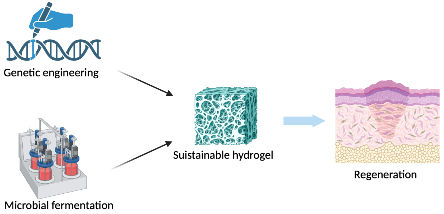
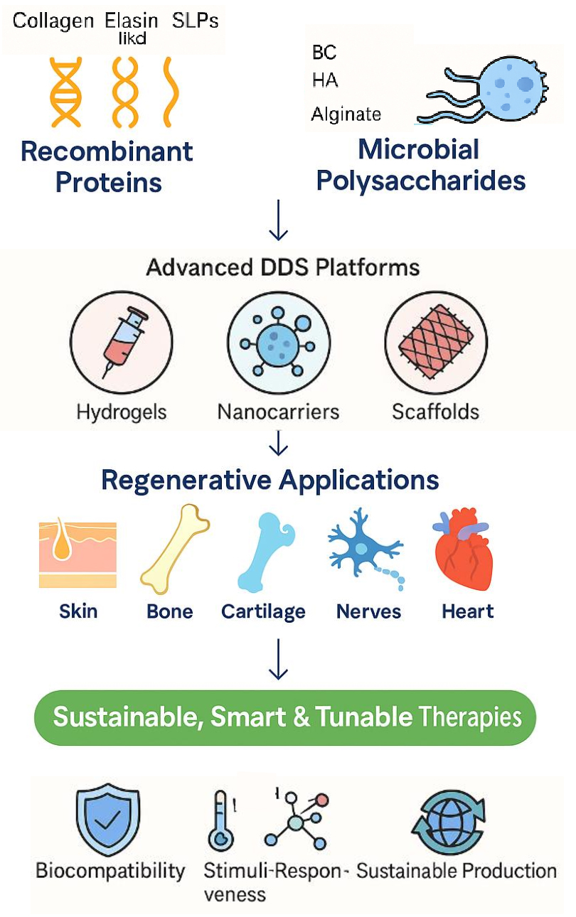
Regenerative medicine is an emerging and highly interdisciplinary field that focuses on restoring the structure and function of damaged or diseased tissues and organs by stimulating the body’s own repair mechanisms or by integrating biological substitutes [1]. Regenerative medicine has evolved significantly over the past two decades to include a wide range of strategies like cell-based therapies, engineered scaffolds, gene editing, and the controlled delivery of therapeutic agents to damaged sites [1]. The clinical promise of regenerative medicine lies in its ability to provide long-term, curative treatments for conditions that are currently managed with palliative approaches, such as chronic wounds, musculoskeletal injuries, cardiovascular diseases, and neurodegenerative disorders [2]. At the basis of many regenerative strategies lies the need for precise, localized, and sustained delivery of bioactive molecules that can guide cellular behavior and orchestrate the complex processes involved in tissue repair [3]. These molecules—ranging from small-molecule drugs and peptides to proteins, genes, and even living cells—must be delivered in a temporally and spatially controlled manner to achieve therapeutic efficacy without systemic toxicity. This is particularly important given the inherently dynamic and multifactorial nature of tissue regeneration, which involves sequential stages of inflammation, cell recruitment, proliferation, extracellular matrix remodeling, and functional integration [4]. In this context, drug delivery systems (DDS) play an essential role as mediators that enable the fine-tuned release of regenerative cues. Traditional DDS, often based on synthetic polymers or inert carriers, have demonstrated the potential to improve drug pharmacokinetics and minimize off-target effects. However, their limited biocompatibility, lack of biological signaling, and unpredictable degradation profiles have restricted their effectiveness in tissue regeneration applications [5]. Moreover, the mechanical and structural mismatch between many synthetic DDS and the native extracellular matrix (ECM) can lead to poor integration with host tissue [5]. Then, the research on this field shifted toward the development of biologically inspired and biofunctional delivery systems that not only serve as passive carriers but actively participate in the healing process [5]. Biotechnology has emerged as a powerful enabler of this new generation of drug delivery platforms. Through the use of recombinant DNA technology, microbial biosynthesis, and metabolic engineering, researchers can design and produce advanced biomaterials with high reproducibility, tailorability, and functionality. These biotechnologically derived materials offer unique advantages in terms of bioactivity, mechanical tunability, immunomodulation, and compatibility with complex biological environments [6]. One of the most promising approaches involves the use of recombinant protein-based materials, which closely mimic natural ECM components while allowing for molecular customization. Proteins such as collagen, elastin-like polypeptides (ELPs), and silk fibroin have been extensively studied for their role in tissue structure, biomechanics, and cellular signaling [7]. By expressing these proteins in microbial or eukaryotic systems, it is possible to generate variants with precisely defined amino acid sequences, mechanical properties, degradation kinetics, and functional motifs. For instance, recombinant collagen can be modified to incorporate integrin-binding domains (e.g., Arginine–Glycine–Aspartic Acid (RGD) sequences), protease-cleavable linkers for controlled degradation, or even therapeutic fusion proteins for dual-functionality [8]. ELPs can be engineered to exhibit thermo-responsive behavior, enabling the creation of injectable gels that solidify at body temperature and serve as in situ depots for drug release [9]. Similarly, recombinant silk fibroin can be processed into a range of formats—fibers, sponges, films, and nanoparticles—each with unique loading and release characteristics [10]. In parallel, microbial biosynthesis and fermentation technologies have unlocked access to a suite of polysaccharide-based biomaterials that are highly attractive for regenerative DDS. These include bacterial cellulose (BC), hyaluronic acid (HA), and alginate, each of which exhibits a combination of structural versatility, biocompatibility, and functional adaptability. Bacterial cellulose, synthesized by strains such as Komagataeibacter xylinus, forms an ultra-pure, nanofibrillar hydrogel with high tensile strength, remarkable water retention capacity, and a morphology similar to native collagen networks [11]. Its three-dimensional architecture supports cell adhesion and proliferation, making it ideal for wound healing and soft tissue regeneration [11]. Hyaluronic acid, a naturally occurring glycosaminoglycan, plays a central role in cell migration, inflammation, angiogenesis, and ECM remodeling. Microbial production of HA, especially through Streptococcus species, has enabled its large-scale and contamination-free synthesis [12]. HA can be chemically modified to tune its viscosity, degradation rate, and bioactivity, and it serves as an excellent matrix for delivering growth factors, stem cells, and genes. Its viscoelastic properties make it particularly suitable for applications in cartilage repair and ophthalmology [13]. Alginate, a linear polysaccharide composed of mannuronic and guluronic acid residues, is widely used in biomedical applications due to its mild gelation with divalent cations (e.g., Ca2+). Produced by microbial fermentation or extracted from brown algae, alginate hydrogels can encapsulate a variety of drugs and biologics while protecting them from degradation [14]. Its low immunogenicity and ease of functionalization enable its use in injectable formulations, microbeads, and multilayered scaffolds tailored for specific regenerative applications [14]. These biotechnologically derived materials can be further engineered to exhibit stimuli-responsive behavior, releasing their payloads in response to environmental cues such as pH changes, enzymatic activity, redox potential, or mechanical stress [15]. For example, pH-responsive hydrogels typically exploit the acidic microenvironments of inflamed or ischemic tissues, where extracellular pH falls from physiological 7.4 to ~6.5–6.8, and in more severe hypoxic or tumor-like regions to as low as 6.2–6.5. Moreover, the biotechnological production of these materials also aligns with principles of sustainability and scalability, as microbial fermentation and recombinant expression systems permit a controlled, large-scale manufacturing with minimal reliance on animal-derived components. This not only ensures batch-to-batch reproducibility and regulatory compliance but also reduces the risk of immunogenicity and contamination, facilitating translation into clinical practice [16]. The aim of this review is to provide a detailed and critical overview of biotechnologically derived materials—specifically those based on recombinant proteins and microbial polysaccharides—as platforms for drug delivery in tissue regeneration. Their characteristics and their biological performance in relevant regenerative models will be examined. Fig. 1 visually recaps the scope of the review.
 Fig. 1.
Scope of the review. Use of Biotechnologically derived materials for tissue regeneration. Created in BioRender Sepe, F (2026) https://BioRender.com/uzysmmx.
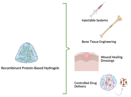
Advancements in synthetic biology have revolutionized the design of protein-based polymers (PBPs), offering new possibilities for regenerative medicine through the creation of biomimetic hydrogels with precise structural and functional properties. These engineered proteins—designed from natural or synthetic amino acid sequences—provide several key advantages over conventional synthetic polymers. They are inherently biocompatible, enzymatically degradable, and produce non-toxic metabolites, making them particularly well-suited for clinical use in tissue repair and regeneration [17, 18]. Additionally, PBPs can be processed under mild, aqueous conditions, preserving bioactivity and supporting the encapsulation of cells, growth factors, and therapeutic agents [19]. Genetic engineering allows for meticulous control over the amino acid sequence, molecular weight, and functional group presentation of these protein polymers. This precision enables the rational design of hydrogels with tunable mechanical properties, controlled degradation rates, and specific biological functionalities. Recombinant DNA technologies also make it possible to incorporate cell adhesion motifs, growth factor-binding domains, and enzymatically cleavable sequences directly into the protein backbone, thereby enhancing cellular integration, tissue-specific remodeling, and responsiveness to the local microenvironment [20, 21]. Scalable microbial systems, such as E. coli, are commonly employed for the high-yield, cost-effective production of these proteins with exceptional purity and consistency [22]. Particular attention has been directed toward PBPs inspired by structural proteins like silk fibroin, elastin, and collagen—each known for their repetitive sequences and capacity for self-assembly into ordered, hierarchical structures [7]. These intrinsic features endow the resulting hydrogels with a combination of mechanical strength, elasticity, and biological activity that is essential for supporting cell viability, guiding tissue organization, and facilitating regenerative outcomes. As a platform for regenerative medicine, genetically engineered protein hydrogels enable the development of tailored microenvironments that emulate the native ECM. Their modular design supports the precise delivery of bioactive cues, modulation of immune responses, and synchronization with tissue healing phases. These features make them highly adaptable for applications ranging from skin and nerve regeneration to musculoskeletal repair and vascular tissue engineering (Fig. 2) [23]. In addition, recombinant proteins intended for regenerative applications undergo multistep purification to ensure the removal of host cell proteins (HCPs), host cell DNA (hcDNA), and other process-related impurities. Standard approaches include affinity chromatography (e.g., His-tag/Ni-NTA or antibody-based capture), ion-exchange and size-exclusion chromatography for removal of aggregates and nucleic acids, and ultrafiltration/diafiltration to reduce endotoxins and residual media components. In microbial systems such as E. coli, additional treatments such as enzymatic nuclease digestion (e.g., Benzonase®) are applied to degrade residual DNA, while yeast and mammalian systems rely on optimized downstream processing pipelines to limit glycosylation heterogeneity.
 Fig. 2.
Applications of hydrogels from genetically engineered sustainable proteins. Created in BioRender. Sepe, F(2026) https://BioRender.com/n1scx6y.
Collagen, the most abundant protein in the ECM of animal tissues, plays a
fundamental role in maintaining structural integrity and modulating cellular
activities essential for tissue homeostasis and repair [24]. Of the 29 known
collagen types, types I, II, and III account for over 90% of total collagen in
the human body. These proteins provide mechanical strength and structural support
to various tissues, including skin, bone, cartilage, and connective tissue [25].
Collagen’s structure is based on repeating Gly-X-Y amino acid sequences, where
glycine occupies every third position to allow tight packing of the three
| Device | Applications in regeneration | Key features | Reference |
| RCPhC1-MA, RCPhC1-NB, RCPhC1-SH (functionalized recombinant type I collagen) | High-resolution 3D-printed scaffolds for tissue engineering | Tunable chemistry, sub-micrometer resolution via 2PP, supports cell proliferation | [35] |
| rhCol III + MSC-EVs hydrogel | Skin wound healing and inflammation modulation | Immunomodulation via M2 polarization, enhanced angiogenesis, promotes cell migration | [36] |
| Recombinant collagen–calcium carbonate hybrid nanoparticles | Controlled drug delivery in tissue repair | pH-responsive release, high drug loading, porous nanostructure, biocompatibility | [37] |
| rHCIII hydrogel with hADSCs | Diabetic wound healing and stem cell delivery | Prolonged cell viability, enhanced stem cell retention and regenerative outcome in vivo | [38] |
| rhCol III (recombinant type III collagen) | Photoaged skin regeneration | Increases collagen content and elasticity, reduces skin thickening | [39] |
RCPhC1-MA, recombinant type I collagen protein with methacrylamide; RCPhC1-NB, recombinant type I collagen protein with norbornene; RCPhC1-SH, recombinant type I collagen protein with thiol; 2PP, two-photon polymerization; rhCol III, recombinant type III collagen; MSC-EVs, mesenchymal stem cell-derived exosomes; rHCIII, recombinant type III collagen; hADSCs, human adipose-derived stem cells.
Elastin is a crucial extracellular matrix protein that imparts elasticity and
resilience to dynamic tissues such as blood vessels, lungs, and skin. Its
structural architecture is composed of insoluble fibrillar networks stabilized by
hydrophobic interactions, and is characterized by repetitive peptide sequences
like elastin and elastin-like polypeptides (VPGG, VPGVG, and APGVGV). These
properties not only contribute to the mechanical function of elastin but also
support critical biological processes such as cell adhesion, proliferation, and
migration—making it highly relevant for regenerative medicine applications
[40]. However, the clinical translation of native elastin is hindered by several
limitations: it is insoluble in aqueous media, potentially immunogenic when
isolated from animal sources, and presents difficulties in processing and
modification. Moreover, synthetic analogs are often expensive and inconsistent in
structure [41]. A transformative advancement occurred when Urry and collaborators
[42] identified VPGVG as a recurrent motif in natural elastin, leading to the
development of elastin-like polypeptides (ELPs)—genetically engineered
biopolymers mimicking elastin’s hydrophobic domain [43]. ELPs are typically
produced via recombinant techniques, predominantly in Escherichia coli,
due to its high-yield expression and scalability, although Pichia
pastoris has also been utilized [44]. These recombinant polymers are generally
composed of repeating pentapeptide units of the sequence Val-Pro-Gly-X-Gly
(VPGXG), where X is any amino acid except proline. This modularity allows ELPs to
replicate key elastin functions while enabling tunable mechanical and biochemical
properties [45]. One of the most compelling features of ELPs in regenerative
applications is their reversible phase transition behavior in aqueous solution.
ELPs are soluble below a defined transition temperature (Tt), but undergo
aggregation and phase separation above it. This thermoresponsive property is
programmable through the choice and arrangement of guest residues, polymer
length, and overall hydrophobicity, making ELPs ideal candidates for in
situ forming biomaterials [46]. For tissue engineering purposes, researchers
have developed multiblock ELP copolymers by linking hydrophobic and hydrophilic
segments (e.g., [VPGIG]ₙ₁–[VPGSG]ₙ₂), or appending hydrophobic terminal blocks
to facilitate self-assembly and mechanical reinforcement [47]. ELPs have also
been functionalized with bioactive peptide sequences or engineered into fusion
constructs to enhance cellular responses such as adhesion, angiogenesis, and
matrix remodeling—key factors in tissue regeneration [48]. The injectable and
thermoresponsive nature of ELPs makes them particularly useful in forming
depot-like structures that provide controlled, localized, and sustained release
of therapeutic agents. For instance, fusion of ELP to therapeutic proteins like
interferon-alpha (IFN-
| Device | Applications in regeneration | Key features | Reference |
| ELP-IFN- |
Sustained cytokine delivery for tissue repair | Extended half-life, depot formation, zero-order release kinetics, enhanced tissue accumulation | [49, 50] |
| REDV-ELP Nanofibers | Vascular grafts | Promotes endothelialization, reduces platelet adhesion, supports smooth muscle contractility | [51] |
| Silk-ELP Nanocomposite with Nanocellulose Layer | Bilayer skin substitute | High mechanical strength, antibacterial activity, supports skin regeneration | [52] |
| Collagen-ELP Recombinant Scaffold | Dermal regeneration | Improved mechanical durability, biocompatibility, and wound healing performance | [53] |
| ELP-pH-Responsive Dox Conjugate with CPP | Targeted delivery in acidic wound environments | pH-triggered release, enhanced cellular uptake, genetic CPP encoding for cell penetration | [54] |
| Crosslinked ELP Nanovesicles with pAzF | Stable delivery system for bioactive molecules | UV-induced crosslinking, increased stability, controlled release, tunable size and hydrophobicity | [55] |
| ELP-K12 + CpG Depot | Immunomodulatory system for wound healing support | Electrostatic binding of CpG, sustained release (up to 3 weeks), local immune activation | [56] |
IFN-
Silk-like polymers (SLPs) represent a prominent class of genetically engineered biomaterials with extensive applications in regenerative medicine. These polymers are composed of repeating sequences such as Gly–Ala–Gly–Ala–Gly–Ser, mimicking the structure of natural silk proteins produced by insects and spiders. Native silks, including those from Bombyx mori and Nephila clavipes, are well known for their impressive mechanical strength, elasticity, biocompatibility, and biodegradability [57, 58]. Among expression systems, Escherichia coli is frequently used for recombinant silk production due to its cost-effectiveness, rapid growth, and ease of genetic manipulation [59]. Genetically engineered SLPs often replicate silk motifs derived from the major ampullate gland proteins, with typical repetitive sequences such as [GGAGQGGYGGLGSQGAGRGGLGGQGGAG] and [GPGGYGGPGQQGPGGYAPGQQPSGPGS] from N. clavipes [60]. These sequences are frequently modified to regulate crystallinity or to introduce biologically active motifs such as RGD to enhance cell adhesion. Due to their low solubility and limited flexibility, silk domains are often copolymerized with elastin-like motifs, resulting in silk-elastin-like proteins (SELPs) that combine mechanical resilience with water solubility and flexibility [61]. This configuration is advantageous for producing injectable biomaterials and drug delivery vehicles with tunable degradation profiles [62, 63, 64]. For instance, Florczak et al. [65] demonstrated that MS1 silk gels functionalized with the H2.1 peptide and loaded with doxorubicin selectively targeted Her2-positive breast cancer cells in vivo, significantly reducing tumor volume with minimal systemic toxicity. Notably, unloaded particles showed no adverse effects in healthy mice, and detailed histology revealed no damage to major organs [65]. Herold et al. [66] further developed recombinant silk particles (eADF4(C16)) chemically linked with doxorubicin via a pH-sensitive hydrazine linker. These particles exhibited minimal drug release at physiological pH, while delivering the full drug payload under acidic conditions typical of intracellular vesicles, enabling precise intracellular drug release [66]. Similarly, Mulinti et al. [67] developed enzyme-responsive nanospheres using a recombinant spider silk copolymer with a thrombin-sensitive linker to release vancomycin in response to Staphylococcus aureus infection. These nanocarriers demonstrated strong antibacterial effects and potential for managing drug-resistant infections in tissue repair [67]. Lian et al. [68] created a nanofibrous membrane by electrospinning recombinant silk protein blended with sodium hydrogen sulfide (NaHS). The resulting scaffold sustained hydrogen sulfide (H₂S) release, supported endothelial progenitor cell (EPC) function, and significantly accelerated wound healing in a murine skin injury model [68]. Further regenerative applications have focused on hybrid constructs combining silk fibroin and recombinant spidroins. For example, composite hydrogels fabricated from silk fibroin and recombinant silk proteins functionalized with fibronectin motifs, growth factors (e.g., Fibroblast Growth Factor (FGF)), and antimicrobial peptides exhibited enhanced cell adhesion, antimicrobial activity, and stimulation of dermal cell proliferation—supporting the development of bilayered skin grafts [69, 70]. Other innovations include recombinant spidroins enriched with SIBLING-derived peptide tags (e.g., osteopontin and bone sialoprotein), which enhanced calcium phosphate mineralization and osteoblast adhesion—demonstrating strong potential for tendon–bone interface regeneration [71]. Additionally, eADF4(C16)-based scaffolds rich in carboxylic acid residues were shown to promote calcium ion binding and mineralization. Culturing mesenchymal stem cells (MSCs) on these hydrogels resulted in elevated alkaline phosphatase activity, highlighting their osteoinductive capacity [72]. Another engineered scaffold featured a chimeric fusion protein incorporating a spider silk-inspired sequence and a hyaluronic acid-binding motif (VTK), which guided osteogenic differentiation of bone marrow-derived human MSCs, underlining its applicability for bone tissue engineering [73]. Finally, a composite construct was developed by integrating decellularized extracellular matrix (dECM) derived from equine cartilage with a silk-based hydrogel. This composite served both as a structural scaffold and a bioadhesive to promote integration with host tissue. Mechanical and biological analyses confirmed its suitability for supporting chondral regeneration under load-bearing conditions, emphasizing the value of SELP-based hydrogels in developing advanced regenerative therapies [74]. Table 3 (Ref. [65, 66, 67, 68, 69, 70, 71, 72, 73, 74]) summarizes the described applications.
| Device | Applications in regeneration | Key features | Reference |
| H2.1-MS1 Silk Gel Particles | Targeted delivery for breast cancer tissue regeneration post-resection | Selective cytotoxicity to Her2+ cells, low systemic toxicity, in vivo efficacy | [65] |
| eADF4(C16) Silk Particles with Hydrazine-Doxorubicin Linker | pH-sensitive drug delivery for tissue repair in acidic microenvironments | Controlled release, pH-responsive linker, enhanced cellular uptake | [66] |
| Thrombin-Responsive Silk Nanospheres | Infection-responsive antibiotic delivery for regenerating infected tissues | Enzyme-triggered release, effective antibacterial activity | [67] |
| Recombinant Major Ampullate Spidroin (rMaSp)/NaHS Nanofibrous Membrane | Skin wound healing and vascular tissue regeneration | Sustained H₂S release, EPC support, improved healing outcomes | [68] |
| Silk Fibroin + Recombinant Spidroin Hydrogel | Skin tissue engineering, wound dressing | Enhanced adhesion, antimicrobial, growth factor delivery (a general-purpose, multifunctional platform with antimicrobial activity and growth factor delivery, suitable for wound healing applications) | [69] |
| Recombinant Spidroin with Fibronectin Motif | Burn wound repair and dermal reconstruction | Improved fibroblast attachment, scaffold bioactivity (a targeted, bioactive scaffold, optimized for fibroblast integration and dermal reconstruction, making it especially valuable for burn injuries or full-thickness skin loss) | [70] |
| Spider Silk–SIBLING Hybrid Gel | Regeneration of tendon–bone interface | Enhanced mineralization, osteoblast adhesion | [71] |
| eADF4(C16)-Based Hydrogel | Bone tissue engineering | Calcium-binding capacity, ALP upregulation in MSCs | [72] |
| Silk-Spidroin-HA Binding Domain Hydrogel | Osteogenic differentiation of hMSCs | ECM mimetic, osteoinductive signaling | [73] |
| dECM + SELP Hydrogel | Cartilage regeneration and integration with host tissue | High mechanical resistance, adhesion, tailored microenvironment | [74] |
eADF4(C16), recombinant silk particles; NaHS, sodium hydrogen sulfide; EPC, endothelial progenitor cell; MSCs, mesenchymal stem cells; ALP, alkaline phosphatase; hMSCs, human mesenchymal stem cells; ECM, extracellular matrix; HA, hyaluronic acid; dECM, decellularized extracellular matrix; SELP, silk-elastin-like protein.
Hydrogels derived from the controlled fermentation of sustainable biomaterials
represent a cutting-edge approach in regenerative medicine, offering a
biocompatible and functional alternative to conventional synthetic polymers.
These bioengineered hydrogels not only provide essential mechanical support to
damaged or developing tissues but also actively modulate the cellular
microenvironment by delivering biochemical cues and bioactive molecules. This
dynamic interaction allows for the precise regulation of fundamental processes
such as cell proliferation, migration, and differentiation—key drivers in the
repair and regeneration of complex tissues and organs [75]. A cornerstone of this
technology lies in the microbial biosynthesis of polysaccharides through the
fermentation of carbohydrate-rich substrates. Specific microbial strains are
harnessed to produce polymers with distinct and tunable mechanical, rheological,
and biological properties. These polymers are then enzymatically engineered to
incorporate specific functional groups or bioactive domains that mimic native ECM
components. Such functionalization enhances cell–matrix interactions and enables
the localized, sustained release of growth factors and signaling molecules
critical for tissue regeneration [75]. Natural polysaccharides produced via
fermentation—such as xanthan gum, cellulose, and hyaluronic acid—are
increasingly utilized as base materials, either alone or in composite
formulations to construct hydrogels with customizable features. Their combined
physicochemical and biological properties allow the creation of smart hydrogel
platforms that respond to physiological stimuli and support complex regenerative
processes with high spatial and temporal precision (Fig. 3) [76]. These hydrogels
are particularly suited for applications in soft tissue engineering, wound
healing, and the regeneration of cartilage, bone, and neural tissues. Their
adaptability, biocompatibility, and ability to mimic native ECM structures make
them highly effective scaffolds for promoting endogenous repair mechanisms and
guiding tissue morphogenesis. As such, fermentation-derived hydrogels are rapidly
becoming integral tools in the design of next-generation biomaterials for
advanced regenerative therapies. However, Bacterial-derived materials may contain
trace contaminants such as lipoteichoic acids, peptidoglycan fragments, or
residual DNA that activate Toll-like receptors (TLR2/TLR4), triggering unwanted
inflammatory responses. To reduce these effects, several strategies have been
developed: advanced downstream purification (ultrafiltration, enzymatic
digestion, endotoxin-removal resins) to minimize immunogenic residues; chemical
modifications (e.g., esterification, methacrylation) that mask TLR recognition
sites; and the use of alternative production platforms such as GRAS organisms
(Bacillus subtilis) or cell-free enzymatic synthesis, which eliminate
streptococcal endotoxins entirely. Additionally, maintaining HA in the
high-molecular-weight form (
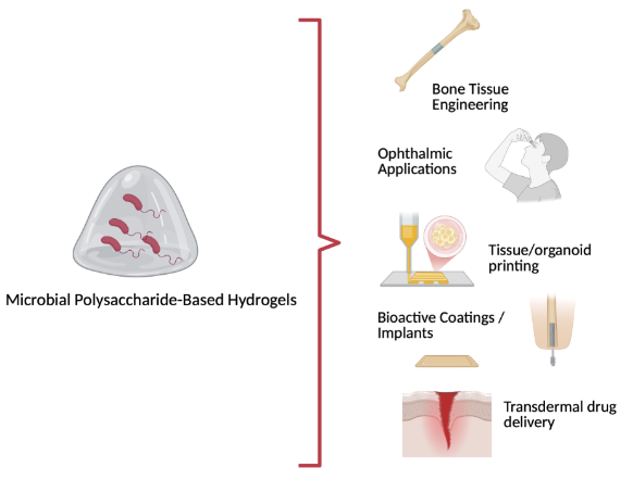 Fig. 3.
Application of hydrogels obtained from controlled fermentation of sustainable biomaterials. Created in BioRender. Sepe, F (2026) https://BioRender.com/rg4isxk.
Bacterial cellulose (BC) is a polysaccharide macromolecule with the same
chemical structure as plant-derived cellulose, yet it lacks associated plant
components such as lignin, pectin, and hemicellulose that are commonly found in
plant cell walls [77]. Structurally distinct due to its unique nanoscale fiber
organization, BC exhibits superior mechanical strength, elevated crystallinity,
and enhanced purity compared to its plant-based counterpart [78]. It is composed
of
| Device | Applications in regeneration | Key features | Reference |
| BC/CS/nHA Nanocomposite Scaffold | Bone tissue regeneration | Enhanced mechanical strength, controlled degradation, high water retention, supports osteoblast proliferation | [80] |
| BC/CS/Alg Hydrogel Scaffold | Bone and cartilage regeneration | Dense fibrous network, good swelling behavior, bioactivity, protein adsorption, and controlled drug release | [81] |
| BC/DCECM Composite Scaffold (NHS/EDC crosslinked) | Cartilage tissue regeneration | Hierarchical porous structure, strong chondrocyte adhesion, elasticity, shape memory, mimics native cartilage | [82] |
| BC/PVA Composite Hydrogel | Corneal stroma regeneration | High transparency, biocompatibility, water retention, stable morphology, maintains corneal integrity | [83] |
| BC/Quince Seed Mucilage Composite Scaffold | Soft tissue regeneration | Improved swelling capacity, enhanced fibroblast adhesion and proliferation | [84] |
| BC Nanoparticles (BCNPs) | Drug delivery for regenerative therapies | Biodegradable, thermally stable, capable of sustained protein release | [85] |
| BC loaded with Microemulsified Glucocorticoids | Local anti-inflammatory delivery during regeneration | Uniform drug incorporation, high and tunable drug permeation, preserved anti-inflammatory activity | [86] |
BC, bacterial cellulose; CS, chitosan; nHA, nano-hydroxyapatite; Alg, alginate; DCECM, decellularized cartilage extracellular matrix; NHS, N-hydroxysuccinimide; EDC, N-[3-(dimethylamino)propyl]-N′-ethyl carbodiimide hydrochloride; PVA, poly(vinyl alcohol).
Hyaluronic acid (HA) is a high molecular weight glycosaminoglycan extensively
used in regenerative medicine due to its unique physicochemical and biological
properties. While HA is naturally present in the ECM of vertebrate tissues, its
biotechnological production via microbial fermentation—particularly from
Streptococcus equi subsp. zooepidemicus and recombinant
platforms such as Lactococcus lactis, Bacillus subtilis,
Corynebacterium glutamicum, Escherichia coli, and
Pichia pastoris—has become the preferred method for obtaining
pharmaceutical-grade HA. This shift ensures enhanced biosafety, scalability, and
product standardization while eliminating risks associated with animal-derived
HA, such as immunogenicity and pathogen contamination [87, 88]. Structurally, HA
consists of repeating disaccharide units of D-glucuronic acid and
N-acetyl-D-glucosamine linked via
| Device | Applications in regeneration | Key features | Reference |
| HA-based Injectable Hydrogels | Bone, cartilage, nerve, muscle, skin regeneration | Biocompatible, biodegradable; tunable viscoelasticity; supports cell proliferation and differentiation | [93] |
| Thermo-responsive HA Hydrogels | Osteoarthritis treatment, cartilage repair | Injectable liquid-to-gel transition at body temperature; minimally invasive; supports chondrocyte viability | [97] |
| HA-based Viscosupplementation Devices | Joint lubrication and osteoarthritis management | Restores synovial fluid viscoelasticity; anti-inflammatory; improves joint mobility | [98] |
| HA/Inorganic Composite Scaffolds | Bone regeneration | Combines mechanical strength with bioactivity; promotes osteogenesis and vascularization | [99] |
| Injectable HA Stem Cell Hydrogels | Myocardial regeneration | Encapsulates MSCs; promotes cell adhesion and differentiation in situ | [100] |
| HA-based Nerve Conduits and Scaffolds | Spinal cord injury repair | Supports axon growth and functional recovery; biocompatible scaffold | [101] |
Alginate is a naturally occurring polysaccharide predominantly found in brown
algae (Phaeophyceae), where it serves as a key structural component of cell
walls, providing both mechanical stability and osmotic regulation [102]. Although
bacterial production is also possible, most commercially available alginate is
extracted from algal sources such as Laminaria, Macrocystis, Ascophyllum, and
Sargassum. Its high viscosity and gelling capacity make alginate a valuable
material, particularly in the food and pharmaceutical industries [103]. Due to
its sustainable origin, biocompatibility, and biodegradability, alginate has
gained considerable attention in biotechnology and biomedicine, where it is used
in drug delivery systems, scaffolds for tissue regeneration, and 3D bioprinting
applications [104]. Alginate is recognized as safe by the FDA, further supporting
its role in regenerative medicine [105]. Traditional industrial alginate
extraction from seaweed involves multiple steps: raw material pretreatment,
alkaline solubilization, precipitation with acids or alcohols, followed by drying
and milling. However, environmental concerns related to seaweed harvesting and
variability in the alginate’s composition—affected by seasonal and geographic
factors—complicate standardization, especially in pharmaceutical contexts. As
an alternative, bacterial systems such as Pseudomonas and Azotobacter species
have been explored for more controlled alginate biosynthesis [106]. Among these,
Azotobacter vinelandii stands out for its capacity to produce alginate with high
molecular weight and rheological stability. This non-pathogenic bacterium
naturally synthesizes alginate as both a carbon reserve and a nitrogenase
protective barrier. The complete genome of A. vinelandii facilitates genetic
modification, enabling strains that produce alginates with customized properties.
For instance, oxygen tension and growth rate during fermentation significantly
affect polymer molecular weight, and limiting oxygen availability can promote
synthesis of highly homogeneous polymers [106, 107]. Chemically, alginate is a
linear anionic copolymer composed of
| Device | Applications in regeneration | Key features | Reference |
| Injectable alginate-hydroxyapatite hydrogel with gelatin microspheres | Bone regeneration | Enhanced mechanical stability, osteointegration, injectable formulation | [113] |
| Alginate hydrogel enriched with platelets in a chitosan–chondroitin sulfate–silk fibroin scaffold | Cartilage regeneration | Mimics native ECM, promotes type II collagen expression, supports metabolic activity | [114] |
| Biodegradable alginate–chitosan hydrogel with vitamin E | Skin tissue engineering | High porosity, improved cell proliferation, enhanced wound healing and tissue regeneration | [115] |
| Microfluidic-spun calcium alginate fibrous scaffold | Chronic wound healing | ECM-mimicking structure, excellent biocompatibility, promotes healing in chronic wounds | [116] |
| Sulfated alginate–polyurethane elastomer | Cardiovascular applications | Anticoagulant properties, improved endothelial cell adhesion, anti-inflammatory, self-healing, flexible | [117] |
| Alginate–fullerenol hydrogel | Cardiac regeneration post-MI | Injectable antioxidant system, scavenges ROS, improves cardiac function recovery | [118] |
| Alginate hydrogel with anisotropic capillary structures | Spinal cord injury repair | Oriented axonal growth, supports neuronal regeneration, non-inflammatory, promotes in vivo integration | [119, 120] |
Biotechnologically derived materials are redefining the landscape of DDS in regenerative medicine. These innovative platforms, based on recombinant proteins and microbially produced polysaccharides, offer a broad range of advantages compared to traditional synthetic carriers. Their molecular precision, biocompatibility, and the ability to integrate bioactive functionalities make them ideal candidates for guiding complex regenerative processes in a temporally and spatially controlled manner. One of the primary benefits of these systems is their biomimetics. Materials such as recombinant collagen, elastin-like polypeptides, silk fibroin, bacterial cellulose, hyaluronic acid, and alginate closely replicate the structure and biochemical cues of the native ECM. This enables enhanced cellular adhesion, migration, proliferation, and differentiation—critical processes for effective tissue repair. Additionally, the programmable nature of these biomaterials allows for the fine-tuning of mechanical properties, degradation rates, and therapeutic release profiles, offering unmatched versatility across different tissue types and clinical contexts. These materials also support stimuli-responsive drug delivery, releasing therapeutic agents in response to local environmental cues such as enzymatic activity, pH changes, or mechanical stress. This level of control ensures high therapeutic efficacy while minimizing systemic side effects. Furthermore, their compatibility with advanced fabrication technologies—including nanoparticle encapsulation, layer-by-layer assembly, and 3D bioprinting—enables the creation of hierarchically structured systems that can deliver multiple agents in a coordinated, tissue-specific manner. Importantly, the production of these materials through microbial fermentation and recombinant expression aligns with principles of safety, reproducibility, and scalability [121, 122]. The ability to manufacture these systems under defined, animal-free conditions minimizes immunogenic risks and facilitates compliance with regulatory standards, enhancing their translational potential for clinical use. The continued evolution of these systems, along with the expansion of the detection techniques used for their analysis (Table 7), is being driven by progress in material science and bioengineering, enabling the development of modular DDS that can be adapted to meet specific regenerative needs.
| Material | Main characterization techniques | Obtained informations |
| Recombinant Collagen | FTIR; Circular Dichroism (CD); DSC; Rheology; SEM/TEM | Triple helix confirmation; thermal stability; viscoelasticity; fibrillar morphology |
| Recombinant Elastin | FTIR; NMR; Rheology; AFM; Dynamic Light Scattering (DLS) | Secondary structure; self-assembly; nanostructural elasticity; size distribution |
| Recombinant Silk | FTIR; XRD; DSC; Rheology; Mechanical Tensile Testing | |
| Bacterial Cellulose | FTIR; XRD; SEM/AFM; TGA; Mechanical Tensile Testing | Degree of crystallinity; fiber morphology; thermal stability; elastic modulus |
| Hyaluronic Acid (HA) | FTIR; NMR; SEC-MALS; Rheology; Zeta Potential | Crosslinking verification; molecular weight distribution; viscoelastic moduli; surface charge |
| Alginate | FTIR; ¹H NMR; Rheology; SEM; ICP-OES (per Ca²⁺ crosslinking) | Crosslinking confirmation |
FTIR, Fourier Transform Infrared Spectroscopy; CD, Circular Dichroism; DSC, Differential Scanning Calorimetry; SEM, Scanning Electron Microscopy; TEM, Transmission Electron Microscopy; NMR, Nuclear Magnetic Resonance Spectroscopy; AFM, Atomic Force Microscopy; DLS, Dynamic Light Scattering; XRD, X-ray Diffraction; TGA, Thermogravimetric Analysis; SEC-MALS, Size Exclusion Chromatography – Multi-Angle Light Scattering; ICP-OES, Inductively Coupled Plasma – Optical Emission Spectroscopy.
DDS, Drug Delivery Systems; ECM, extracellular matrix; ELPs, elastin-like polypeptides; BC, Bacterial cellulose; HA, Hyaluronic acid; SLPs, Silk-like polymers.
SY, SR, FS, GP, and UG contributed to the study conception and design. Material preparation and data collection were performed by AC and RC. The first draft of the manuscript was written by SY, SR, and FS. Supervision was provided by AC, RC, GP, and UG. GP and UG also contributed to the design of the research study, preparation of figures and tables, and literature search. All authors read and approved the final manuscript. All authors contributed to the editorial changes in the manuscript. All authors have participated sufficiently in the work to take public responsibility for appropriate portions of the content and agreed to be accountable for all aspects of the work in ensuring that questions related to its accuracy or integrity.
Not applicable.
We would like to acknowledge Orsolina Petillo for data organization support (IRET NA, CNR).
The work was supported by the National Recovery and Resilience Plan (NRRP), Mission 4 Component 2 Investment 1.4—Call for tender No. 3138 of 16 December 2021, rectified by Decree n. 3175 of 18 December 2021 of the Italian Ministry of University and Research funded by the European Union—NextGenerationEU, Award Number: Project code CN_00000033; Concession Decree No. 1034 of 17 June 2022 adopted by the Italian Ministry of University and Research, CUP B83C22002930006, Project title “National Biodiversity Future Center—NBFC”; and National Recovery and Resilience Plan (NRRP), Mission 4, Component 2, Investment 1.1, Call for tender No. 1409 published on 14 September 2022 by the Italian Ministry of University and Research (MUR), funded by the European Union—Next Generation EU, (PRIN 2022 PNRR) CUP B53D23031790001, Injectable Hydrogels for treatment of periodontal disease (HYPE)—P2022RZ8WM—Grant Assignment Decree No. n. 1369 adopted on 01/09/2023 1 September 2023 by the Italian Ministry of Ministry of University and Research (MUR).
The authors declare no conflict of interest.
References
Publisher’s Note: IMR Press stays neutral with regard to jurisdictional claims in published maps and institutional affiliations.