IMR Press / FBE / Volume 14 / Issue 4 / DOI: 10.31083/j.fbe1404027
Open Access Review
Natural and Semi-Synthetic Flavonoid Anti-SARS-CoV-2 Agents for the Treatment of Long COVID-19 Disease and Neurodegenerative Disorders of Cognitive Decline
Show Less
1 Raymond Purves Laboratory, Institute of Bone and Joint Research, Kolling Institute of Medical Research, Faculty of Health and Science, University of Sydney at Royal North Shore Hospital, St. Leonards, NSW 2065, Australia
2 Graduate School of Biomedical Engineering, University of NSW, Sydney, NSW 2052, Australia
3 Sydney Medical School, Northern Campus, University of Sydney at Royal North Shore Hospital, St. Leonards, NSW 2065, Australia
*Correspondence: james.melrose@sydney.edu.au (James Melrose)
Academic Editor: Hongwei Yao
Front. Biosci. (Elite Ed) 2022, 14(4), 27; https://doi.org/10.31083/j.fbe1404027
Submitted: 10 May 2022 | Revised: 13 June 2022 | Accepted: 16 June 2022 | Published: 9 October 2022
Copyright: © 2022 The Author(s). Published by IMR Press.
This is an open access article under the CC BY 4.0 license.
Abstract

The aim of this review is to highlight the beneficial attributes of flavonoids, a diverse family of widely-distributed polyphenolic phytochemicals that have beneficial cell and tissue protective properties. Phytochemicals are widely distributed in plants, herbs and shrubs used in traditional complimentary medical formulations for centuries. The bioactive components that convey beneficial medicinal effects in these complex herbal preparations are now being identified using network pharmacology and molecular docking procedures that identify their molecular targets. Flavonoids have anti-oxidant, anti-inflammatory, antiviral, antibacterial and anti-cancer properties that have inspired the development of potent multifunctional derivatised flavonoids of improved efficacy. The antiviral properties of flavonoids and the emergence of the severe acute respiratory syndrome (SARS-CoV-2) pandemic has resulted in a resurgence of interest in phytochemicals in the search for efficacious compounds that can prevent viral infection or replication, with many promising plant compounds identified. Promising semi-synthetic flavonoid derivatives have also been developed that inhibit multiple pathological neurodegenerative processes; these offer considerable promise in the treatment of diseases of cognitive decline. Clinical trials are currently being undertaken to evaluate the efficacy of dietary supplements rich in flavonoids for the treatment of virally-mediated diseases. Such trials are expected to identify flavonoids with cell and tissue protective properties that can be harnessed in biomedical applications that may serve as supportive adjunctive procedures to conventional anti-viral drug therapies against diseases such as COVID-19.

Keywords
flavones
chalcones
anti-viral phytochemicals
SARS-CoV-2
long COVID disease
anti-inflammatory
anti-oxidant
neuroinflammation
neuroprotection
cognition and memory
Nrf2
Alzheimer's disease
ARDS
Parkinson's disease
1. Introduction
Aim of the Study

The aim of this review was to highlight the tissue and cell protective properties of flavones and chalcones as anti-viral compounds that prevent SARS-CoV-2 infection and replication through inhibition of key enzymes of the viral genome such as RNA-dependent RNA polymerase (RdRp), 3CL main protease (3CL Pro Main ) and PL protease, involved in SARS-CoV-2 replication [1, 2, 3, 4, 5]. These flavones/chalcones also counter primary bacterial infections and multi drug resistant (MDR) bacterial strains that have emerged as secondary infections in long COVID disease. Development of semi-synthetic analog flavonoid derivatives inspired by these natural flavonoids also show promise in ameliorating neurologic deficits such as brain fogging, inability to concentrate and focus on problem solving and the general cognitive decline observed in long COVID disease. In addition, they are promising agents for the treatment of neurodegenerative diseases of cognitive decline such as Alzheimer’s disease (AD) and Parkinson’s disease (PD).

2. Flavone and Chalcone Phytochemical Biomedicines

Plants containing beneficial flavonoid polyphenolic antioxidant, anti-inflammatory neuroprotective compounds have been used in traditional complimentary medicine for thousands of years [4, 5, 6, 7, 8]. With the emergence of the SARS-CoV-2 pandemic, a search of compounds displaying SARS-CoV-2 inhibitory activity from literature searches of ScienceDirect, PubMed, Scopus, and Google Scholar databases in 2021 supplemented by data from articles in community surveys, case reports, and articles describing the use of antiviral herbal medicines in Traditional Chinese, Vietnamese, Thai and Indian Asian medicine has uncovered a number of promising plants and efficacious anti-viral compounds [6]. A total of 91 plant taxa contain anti-viral compounds with potency against SARS-CoV-2. Advanced screening and activity profiling of these compounds using in-silico computational docking simulations and X-ray crystallography have been undertaken, as well as assessment of their bioactivities in SARS-CoV-2-infected VERO cells. In vitro biochemical analyses of their enzyme inhibitory activities has further confirmed their potential. Network machine learning has also been employed to identify anti-viral compounds, their efficacy and molecular targets, and to search for foods rich in these compounds [9]. A large range of dietary phytochemicals have been identified; a few selected examples of these are presented in Table 1 (Ref. [6, 10, 11, 12, 13, 14, 15, 16, 17, 18, 19]).

Table 1.Selected Examples of Flavone and Chalcone Rich Foods.
Flavone sub-class Flavonoid/chalcone examples Food Source Ref
Flavonol (+) catechin/(-)epicatechin, epigallocatechin Green and black tea [10, 11]
Flavone luteolin, rutin, chrysin, apigenin red wine, capsicum, fruit skins, buckwheat [12, 13]
Flavonol kaempferol, quercetin, myrecetin, tamarixetin red wine, onion, olive oil, red-black berries, grapefruit [12, 13, 14]
Flavanone naringin, naringenin, taxifolin, hesperidin Citrus fruit flesh and skin [15, 16, 17, 18]
Isoflavone genistein, diadzin Soybean [19]
Chalcone Panduratin A Pomegranate, citrus flesh and peel, SE Asian medicinal herbs [6]
3. Chalcone and Flavone Biodiversity

The chalcones and flavones are a diverse group of polyphenolic heterocyclic organic phytochemicals with roles in the defense of plants from parasites and are volatile scented insect attractants that promote pollination, provide flower colouration, protect plants from damaging UV radiation, and provide temperature stress properties to plants [20]. Flavone and chalcone compounds are useful therapeutic components in plant and herbal preparations that have been used in traditional Thai, Chinese, Ayurvedic and Australian First Nation medical practices for centuries [21, 22, 23]. Flavones and chalcones display anti-inflammatory activity through the inhibition of lipoxygenase (LOX) and cyclooxygenase (COX) activity, and regulate nitric oxide and prostaglandin tissue levels. They also suppress nuclear factor (NF)- κ B activation, downregulate TNF production [24], display antiviral activity against HIV [25], dengue virus [26] and coronaviruses [21, 24, 27], and have antibacterial and antifungal properties.

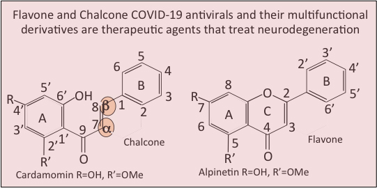
The structure and ring numbering systems of chalcones and flavones are shown in Fig. 1. Hybrid chalcone-flavone desmoflavans have also been identified (Fig. 1). Chalcones and flavones occur in plants as glycoside and aglycone forms. Two examples of these, namely rutin and hesperidin, are illustrated in Fig. 2g,k. The related Sofalcone and metochalcone are also shown (Fig. 2m,n). Dietary flavone and chalcone glycosides are converted to their aglycone forms when ingested and are then conjugated to glucuronic acid to form 7-O- and 3-O-glucuronate glycoforms. These are the forms that circulate in plasma. The 7-O-glucuronate glycoform is more bioavailable than the 3-O-glycoform. Hesperitin-7-O-glucuronate is an active pharmacologic flavonoid that exerts hypotensive, vasodilatory and anti-inflammatory effects on the endothelium, similar to the hesperetin aglycone however the 3-O-glucuronate glycoform is less active [28]. The increased bioavailability of hesperitin-7-O-glucuronate leads to an improved prevention of bone loss in ovariectomised rats [29].
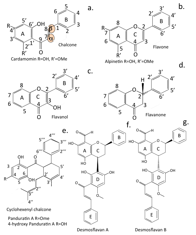
Fig. 1.

Comparison of the generic structures of chalcone and flavone showing their ring numbering system. The structures of flavonol and flavanone are also shown and cyclohexenyl chalcone (Panduratin) and the hybrid desmoflavan A and B. The reactive
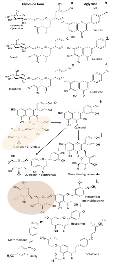
 α and β unsaturated carbonyl residues in chalcone are highlighted.

Fig. 2.

Flavone and Chalcone diversity. Structural depiction of the glycoside and aglycone forms of luteolin (a,b), baicalin and baicalein (c,d) and scutellarin and scutellarein (e,f). Structure of the glycoside form of quercetin (rutin) found in plant tissues (g) showing how it is converted to the aglycone form (h) when ingested and conjugated with glucuronate in plasma (i,j). The glycoside form of hesperidin (k) and its aglycone form, hesperitin (l) are also shown and two further licenced forms of hesperidin, metochalcone (m) and sofalcone (n). The rutinose (6-O- α -L rhamnosyl-D-glucose) disaccharide component of rutin is highlighted.

Flavonoids are a diverse group of phytochemicals that have been grouped into families based on their structures (Fig. 3). Peterson [30] screened 72 flavonoids for their ability to interact with the SARS-CoV-2 3CLPro protease main active site using in-silico molecular docking. The 14 best inhibitors were listed (Fig. 4, Ref. [30]), with further studies confirming the inhibitory activity [24, 31, 32]. The IC
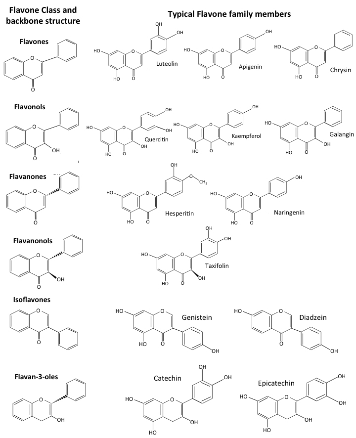
 50 values for several flavonoids have been compared, the components attached to their ring structures influence their biological activity (Fig. 5, Ref. [33, 34]). Two further engineered chalcone analog derivatives (11a, 11b) have been designed against the SARS-CoV-2 3CLPro main active site (Fig. 5c). Compound 11a and 11b are the two most effective 3CLPro inhibitors known, exhibiting 96–100% inhibition at a concentration of 1 μ M (Fig. 5b). C-terminal aldehyde groups in compound 11a and 11b covalently attach to the Cys 145 moiety in the MPro catalytic dyad to provide this potent inhibitory activity.

Fig. 3.

Comparison of the structures of flavonoid forms showing the generic structures of flavone, flavonols, flavanones, flavanonols, iso-flavones and flavan-3- ols and representative members.
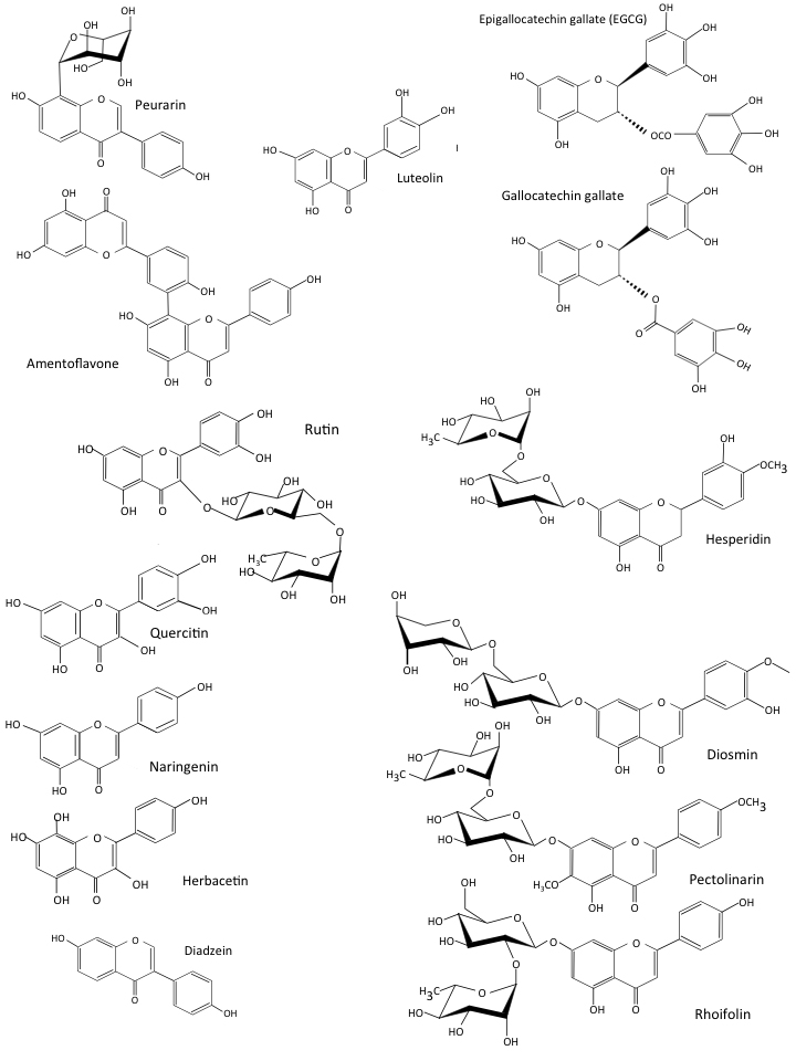
Fig. 4.

The 14 most inhibitory flavones identified by In-silico molecular docking procedures of 72 inhibitory COVID-19 flavonoids [30].
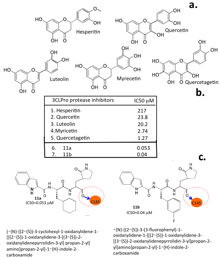
Fig. 5.

Inhibitory flavonoids. Structures of selected flavonoids that display inhibitory activity for 3CLPro of CoV-2 (a) and their IC 50 values (b) and compounds 11a and 11b which were synthesised to target the 3CLPro active site inspired by the properties of these native flavonoids (c). 11a and 11b are two of the most potent inhibitors of 3CLPro that have been developed. The cysteine residue of the catalytic dyad of 3CLPro that 11a and 11b interact with is highlighted. Figure constructed from data provided in [33, 34].

Chalcones consist of a ketone composed of two aromatic rings linked by an aliphatic carbon bridge containing two unsaturated carbonyl residues [35]. Conjugated double bonds in these ring structures and a de-centralised Pi-electron system which can donate or accept outer shell electrons, make these compounds highly interactive. Natural and semi-synthetic chalcones and flavones are of considerable interest as therapeutic agents in biomedicine. The central unsaturated alpha and beta carbonyl residues in the chalcones which attach its two aromatic rings together are interactive with bioactive function-defining cysteine residues in proteins. Many of the flavonoids induce Nrf2 (nuclear factor erythroid-related factor-2) expression and are interactive with androgen and oestrogen receptors (ARs, ERs), peroxisome proliferator-activated receptor (PPAR- γ ) and β -catenin/Wnt cell signaling. The anti-inflammatory properties of chalcones arise from their inhibitory properties over LOX, COX, interleukins, NO synthase, prostaglandins and NF- κ B expression [36]. Naturally occurring hybrid desmosflavans A and B also have anti-oxidant properties, inhibit LOX and have anti-tumor properties [37].

4. Privileged Status of Chalcone as a Module for Medicinal Compound Development

Chalcone [(2E)-1, 3-diphenylprop-2-en-1-one] is an important scaffolding molecule amenable to derivatization with a diverse range of functional groups through varied linkage chemistries, making it a key intermediate in the synthesis of new and more efficient drugs that are of major importance in medicinal chemistry. Chalcone is considered a privileged structure and represents a template that can be used to synthesize compounds displaying a wide range of pharmacological activities, including anti-inflammatory, anti-microbial, anti-oxidant, anti-viral, anti-diabetic, anti-malarial and cytotoxic anti-tumor activities [38, 39]. Novel chalcones have been synthesised with CNS receptor interactive properties that equip them with anti-anxiety, anti-depression and analgesic properties [40]. Chalcones with vasodilatory properties [41], anti-hypertensive, anti-anginal, anti-arrhythmic and cardioprotective agents have also been developed.

5. Natural Anti-Viral Phytochemicals

SARS-CoV-2 3CLPro has major roles to play in viral replication and is inhibited to a variable degree by many natural plant compounds. These include biflavonoids [42], flavonoids [42, 43, 44], isoflavones [44], triterpenes [45, 46], phyto-sterols [47], lignans [46], indole alkaloids [44, 48, 49], glucosinolates [44], anthraquinones [50], phenanthrenes [51], phloro-tannins [52], chalcones [53], diaryl heptanoids [46], and propanoids [54]. Of all plant anti-viral studies that have been conducted, those on flavones and chalcones represent almost half of all studies so far conducted [reviewed in [55]]. It was beyond the scope of this review to examine all the aforementioned anti-viral phytochemicals ( > 8000 compounds). Flavones and chalcones were focused on since these represent in ~50% of all anti-viral phytochemical studies so far conducted.

6. Licensed Chalcones

Hesperidin methylchalcone, metochalcone and sofalcone are currently licensed for clinical use (Fig. 3). Hesperidin methylchalcone has vasodilatory properties and has been used to treat venous insufficiency for five decades. Metochalcone and Sofalcone are useful in the treatment of Helicobacter pylori-induced gastric inflammation and have been used for decades to treat gastritis and gastric ulcers in Japan [1, 2, 3].

6.1 Hesperidin Methylchalcone

Hesperidin is a member of the chalcone sub-category of plant flavanones, and is composed of an aglycone (hesperitin) linked to a disaccharide (rutinose). Hesperidin and hesperitin occur naturally in citrus fruits [56]. Hesperetin is reported to interact strongly with membranes; hesperidin may be sterically hindered in such interactions due to its rutinose side chain [57]. In a double-blind cross-over clinical trial in healthy volunteers, enzymatic removal of rutinose from hesperidin improved its bioavailability [58]. Rutinose is a 6-O- α -L-rhamnosyl-D-glucose disaccharide. Clinical trials on hesperidin have examined the action of hesperidin in chronic venous insufficiency, leg pain and lymphatic edema [59]. In combination with Ruscus aculeatus extract and ascorbic acid, hesperidin safely and effectively treated chronic venous deficiency [60]. Mounting evidence indicates that hesperidin and hesperitin prevent neuroinflammation [61, 62] and development of neurodegenerative diseases [56]. Cell and animal models of neurodegenerative disease show hesperidin improves neural growth factor delivery and endogenous antioxidant defence. Hesperidin-enriched dietary supplements improve health through anti-inflammatory properties, and ability to improve cerebral blood flow, cognition, and memory [61, 62, 63, 64]. The angiotensin-converting enzyme ACE-2, a carboxypeptidase that degrades angiotensin II into angiotensin 1-7, is a receptor for SARS-CoV-2. Molecular docking studies show hesperidin binds to ACE-2 and inhibits enzymatic activity [65].

6.2 Metochalcone

Several metochalcone analogues display potent activity against drug resistant forms of Helicobacter pylori, inhibiting cellular adhesion and invasion of gastric epithelial cells. Metochalcone reduces H.pylori-induced gastric inflammation by reducing NF- κ B activation, and secretion of IL-8 [66]. Based on a computational model of the colchicine binding site on β -tubulin, chalcone derivatives were designed to inhibit tubulin assembly and mitotis [67] and shown to provide cytotoxic properties against human cancer cell lines [67]. Molecular docking studies revealed the chalcone scaffold could fit the colchicine binding site on β -tubulin. Attachment of a 3,4,5-trimethoxyphenyl ring next to the carbonyl group on metochalcone promoted this colchicine-mimicking tubulin interaction [67] and improved cytotoxicity against murine acute lymphoblastic leukemia. The most potent chalcones display growth inhibition at nanomolar concentrations. Microtubule destabilisation and mitotic arrest provide potent inhibitory activity against human cervical and breast cancer cell migration [67]. This derivatisation step also improved inhibitory activity against Helicobacter pylori-induced inflammation in human gastric epithelial cells [66].

6.3 Sofalcone

Sofalcone also has mucosal protective properties, inhibits growth of H. pylori and has been used to treat gastritis and gastric ulcers in Japan for decades. These protective properties stem from activation of the cytoprotective and anti-inflammatory nuclear factor-erythroid 2 (NF-E2) p45-related factor 2 (Nrf2)-heme oxygenase (HO)-1 pathway [68]. Sofalcone disrupts binding of the Kelch-like ECH-associated protein 1 (KEAP1), a cytosolic repressor of Nrf2 activation [68, 69] and increases VEGF via an Nrf2-HO-1 dependent pathway in gastric epithelial cells [70]. KEAP1 is a tumor and metastasis suppressor gene [71]. Sofalcone has been used to treat pre-eclampsia, where the cytoprotective and anti-inflammatory Nrf2-HO-1 pathway is induced in primary trophoblasts and human umbilical vein endothelial cells (HUVECs) [72]. Sofalcone promotes nuclear translocation of NF-E2 and transactivation of NF-E2 responsive genes, decreasing secretion of soluble fms-like tyrosine kinase-1 (sFlt-1) and endoglin by primary human trophoblasts. This potently suppresses endothelial cell dysfunction, blocks TNF α -induced monocyte adhesion and VCAM- 1 expression in HUVECs [72].

7. Interactive Properties of Flavonoids that Contribute to Their Anti-Viral Properties

With the emergence of the coronavirus pandemics of the last five decades, plant extracts have been extensively screened in the search for phytochemicals that impede viral infection and replication [73]. Many plant flavones and chalcones display properties that block viral attachment to host cells while others specifically target enzymes responsible for viral replication. Hesperidin, quercetagetin, and myricetin are examples of phytochemicals that strongly bind to the active site of RdRp, inhibiting its enzymatic activity and viral replication [74, 75]. In-silico molecular binding studies have also identified a number of flavonoids that interact with the catalytic site of SARS-CoV-2 3 CL Pro inhibiting its enzymatic activity [24, 30, 31, 32] and Spike-ACE2 interaction. They can also inhibit helicase and topoisomerase [76, 77, 78, 79, 80, 81]. The RecQ helicase family (nsp13) unravel double-stranded DNA, producing ssRNA required for viral replication, transcription and translation. They also facilitate DNA repair from UV light damage through recombination processes that maintain genomic stability and integrity. A number of flavones that inhibit helicase also disrupt SARS-CoV-2 replication [82, 83, 84]. Anti-tumor studies with flavones have found many that inhibit topoisomerase I and II [85, 86, 87, 88].

8. Screening for Anti-Viral Phytochemicals

Network machine learning has also been applied in the design of new SARS-CoV-2 drugs and the re-purposing of existing drugs for the treatment of SARS-CoV-2 [89, 90, 91, 92, 93]. Advanced computer software was developed to investigate molecular docking events in SARS-CoV-2 interactions with anti-viral compounds [94, 95, 96]. This methodology facilitated a systematic analysis of the interactive chemical determinants of anti-viral phytochemicals that determine SARS-CoV-2 spike glycoprotein interactions [97]. These functional interactive groups on phytochemicals can be modified to obtain a more efficacious anti-viral compound [98]. Chalcones and flavones are amenable structural templates for the synthesis of phytochemical libraries of varied structure to evaluate viral binding. AI-based computational simulation for drug design and large-scale inhibitor screening have also been applied to optimize such evaluations [99]. Homology modeling studies and in-silico studies employing advanced computational molecular docking software and x-ray crystallography have identified phytochemicals that interfere with the Spike glycoprotein interaction with the human ACE2 receptor [73]. The identification of TMPRSS2 (transmembrane serine protease 2), TMPRSS4 and furin cleavage sites in the Spike glycoprotein, which prime it for fusion with the host cell plasma membrane, have identified further targets of interest in anti-viral strategies. Phytochemical inhibitors of TMPRSS2 and furin have also now been identified [100].

9. The Impact of Coronaviruses on Human Health and Well-Being

Coronaviruses (CoVs) are enveloped viruses of the Nidovirales order, Coronaviridae family. Bats, dogs, cats and humans can all be infected with these viruses [101]. Seven species of CoVs have so far been identified, four of these produce relatively mild symptoms of the common cold [102]. Severe acute respiratory syndrome (SARS-CoV), Middle East respiratory syndrome (MERS-CoV) and SARS-CoV-2 induce high impact life-threatening diseases [102]. The appearance of the SARS-CoV pandemic in 2002–2003 resulted in 774 deaths and 8098 cases of infection in 26 countries. Ten years later, MERS-CoV emerged as a sixth coronavirus. Infections with this virus across 27 countries in the Middle East, Asia, North Africa and Europe resulted in 2040 infections and 712 deaths. The emergence of a seventh coronavirus (SARS-CoV-2) has lead to the COVID-19 global health pandemic. SARS-CoV-2 is closely related to SARS-CoV but is far more infectious and has significantly greater health consequences. As at 8 June 2022, more than 535 million SARS-CoV-2 cases and 6.3 million deaths in 223 countries had been reported (www.worldometers.info/coronavirus/). A highly infectious delta variant (B 1.617.2) of SARS-CoV-2 emerged in India in 2020 and rapidly became the dominant strain. On 24 November 2021, a further highly infectious SARS-CoV-2 variant (B.1.1.529/BA.1) was reported, which has had a significant global impact [103]. The World Health Organization Technical Advisory Group on SARS-CoV-2 Virus Evolution designated this B.1.1.529, the fifth coronavirus variant, and named it Omicron [104]. This is the most infectious form of SARS-CoV-2 so far identified. Of major concern are the 32 mutations in Omicron located within its Spike protein with 15 of these located in the receptor binding region [105]. The high infectivity rate of Omicron suggest that it uses an extensive range of cell surface binding sites in addition to the ACE2 receptor and neuropilin-1 (Nrp-1) on host cells to effect infection of host cells. Fig. 6 depicts the structure of a SARS-CoV-2 viral particle, its genomic organization and the open reading frames (ORFs) that encode non-structural proteins (Nsps) that have important roles to play in CoV-2 replication.
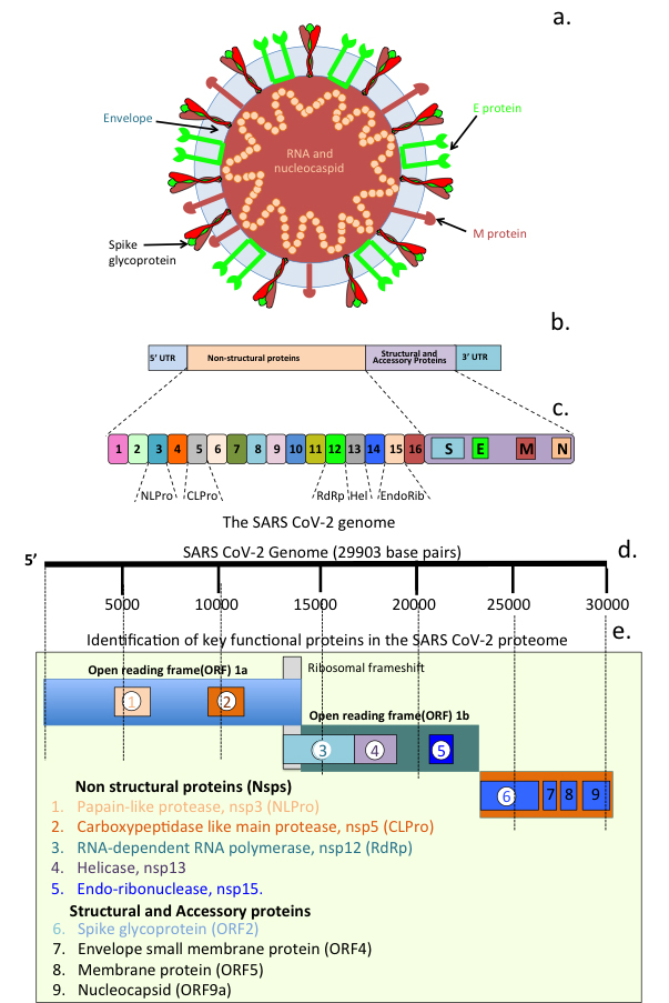
Fig. 6.

SARS Cov-2 structural organization and its genome. Schematic of a SARS-CoV-2 viral particle showing the structural organization of the nucleocapsid and viral RNA, viral envelope and Spike glycoprotein (a). Viral genomic organization (b) and open reading frames (ORFs) showing regions encoding the major non structural proteins (Nsps 1-6) and viral particle structural and envelope small membrane, membrane and nucleocapsid accessory proteins 7-9 (c).

In order to enter cells, viruses need to attach to and activate envelope glycoproteins by host cell proteases. Host cell surface TMPRSS2 plays a crucial role in the activation of SARS-CoV-2 spike protein, facilitating the rapid infection of these cells [106]. This activity of host cell proteases is essential for viral infectivity and constitutes a logical target for therapeutic intervention to prevent infection. Host cell entry is the first step in the viral life cycle with the Spike glycoprotein binding to host cell receptors, conformational reorganisation of the S1 sub-domain upon internal cleavages in this region by TMPRSS2 or furin facilitate the fusing of the viral membrane with the host cell plasma membrane to effect host cell entry. The SARS-CoV Spike protein is also the major target of the neutralizing antibody response of SARS-CoV-2 vaccines. ACE2 is the primary host receptor for SARS-CoV-2 and SARS-CoV however these related viruses have vastly different infection rates, suggesting the involvement of factors in addition to ACE2 that promote SARS-CoV-2 infection. Neuropilin-1 (Nrp-1) is another host cell receptor that SARS-CoV-2 uses for cellular attachment. Nrp-1 is processed by furin exposing a C-end rule motif (CendR) that binds to the SARS-CoV-2 spike protein and is internalised by endocytosis [107, 108, 109, 110]. The even greater infectivity of the Omicron CoV-2 variant is highly suggestive that this viral form may utilise additional host cell surface proteins to effect host cell infection that have yet to be identified.

Table 2 [6, 22, 27, 65, 84, 111, 112, 113, 114, 115, 116, 117, 118, 119, 120, 121, 122, 123, 124, 125, 126, 127, 128, 129, 130, 131, 132, 133, 134, 135, 136, 137, 138, 139, 140, 141, 142, 143, 144, 145, 146, 147, 148, 149, 150, 151, 152, 153, 154, 155, 156, 157, 158, 159, 160, 161, 162, 163, 164, 165, 166, 167, 168, 169, 170, 171, 172, 173, 174, 175, 176, 177, 178, 179, 180, 181, 182, 183, 184, 185, 186, 187, 188, 189, 190, 191, 192, 193, 194, 195] reviews the properties and mode of action of a selected number of naturally-occurring plant flavones, chalcones and analog derivatives.

Table 2.Examples of Bioactive Chalcones and Flavones and their derivatised analog forms used to treat SARS-CoV-2 infection and neurodegeneration in disorders of cognitive decline.
Compound Properties/Mode of Action Ref
Naturally occurring flavones/chalcones
Phenolic compounds Inhibition of SARS 3CLPro activity, cell ular anti-oxidant, anti-inflammatory activity. Panduratin A inhibits SARS-CoV-2 infection at pre-entry and post-infection phases. Multi targeting chalcones show promise in the treatment of AD. [6, 22, 65, 111, 112, 113, 114, 115]
Panduratin A
Flavonoids
Chalcones
Quercetin Block RNA dependent RNA polymerase activity, inhibit SARS-CoV-2 cell entry. Quercetin inhibits ACE2 enzymatic activity. Molecular docking studies show rutin binds to SARS-CoV-2 M Pro , RdRp, PL Pro , and S-proteins with Ki values between 5.66 μ M and 6.54 μ M [27, 113, 116, 117, 118]
Rutin
Myrcetin Interference with the ATPase activity of nsp13 inhibitis helicase activity, viral replication and SARS 3CLPro enzymatic activity. Analog aglycone and glycoside forms of scutellarin have therapeutic anti-viral properties. [84, 113, 119]
Scuttellarein
Glycirrhizin Inhibition of SARS-CoV-2 adsorption to host cells through interactions with Spike protein antagonises host cell ACE2 interactions, tissue anti-oxidant, anti-inflammatory activities [120, 121, 122, 123, 124]
Quercetin Inhibition of SARS-CoV-2 3CLPro enzymatic activity, Spike protein interactions blocks viral host cell entry, inhibition of nsp15 endoribonuclease atttenuates viral replication. Molecular docking studies show Catechin targets 3CLpro, CTSL, RBD of S protein, NSP6 and nucleocapsid protein. CoV-2: Spike ACE2 interactions inhibited. [125, 126, 127, 128, 129, 130, 131, 132, 133, 134]
Epigallocatechin
Gallate Gallocatechin
Catechin
Chrysin Chrysin has anti-oxidant and immunomodulatory properties. Inhibits NFkB pathway as a PPAR γ -agonist, inhibits COX-2, MPO activity, reduces, IL-1 β , IL-8, iNOS levels [135, 136, 137]
Kaempferol Inhibition of movement of metabolites through viral 3a ion channels inhibits viral replication [138]
Luteolin Binding to Spike protein inhibits viral attachment to host cells, also displays inhibitory activity against SARS-CoV-2 3CL pro. Has anti-oxidant activity, inhibits MAPK, NFκB pathways, reduces COX-2, TNF α , INOS, IL-6,IL-1 β , production, and MPO activity [139, 140, 141, 142]
Kaempferol, luteolin Kaempferol and luteolin have monoamine oxidase inhibitory activity therapeutic agents in neurodegenerative disorders [143, 144, 145]
Hesperidin/hesperitin Vasodilatory, used to treat stress induced H.pylori gastric ulcer, ulcerative colitis, gastric/mucosal infections. Supports innate and acquired immune responses, binds to SARS-CoV-2 3CL pro, blocks CoV-2 entry into host cells. Promising agents for treatment of neurodegenerative disorders. [2, 146, 147, 148, 149]
Induces Nrf2 and tissue protection.
Licochalcone B Multifunctional, inhibits A β 42 self-aggregation (IC 50 = 2.16 ± 0.24 μ M), disaggregates pre-formed A β 42 fibrils, reduces metal-ion-induced A β 42 aggregation through metal chelation. Protects SH-SY5Y cells from H 2 O 2 -induced cell death. [150, 151, 152, 153]
Flavokawin Suppresses NF-κB-mediated inflammation and cancer
Butein An anti-oxidant flavonoid, hepato-protective, anti-tumour activity against a range of cancer types [154, 155, 156]
Xanthoangelol Anti-oxidant, anti-inflammatory, anti-cancer, anti-bacterial properties, neuroprotective. Induces apoptosis in neuroblastoma and leukemia tumour cells [157, 158, 159]
Scutellarin Multifunctional phenolic herbal flavonoid, interacts with SARS-CoV-2 3CL pro and endoribonuclease (NSP15) to disrupt viral replication. [160, 161]
4-Hydroxyderricin Produced by Angelica keiskei, anti-tumour activity through induction of Caspase mediated apoptosis of leukemia cells
Cardamonin Anti-oxidant, anti-inflammatory chalcone used in the treatment of gastric, colonic and breast cancer [162]
Isoliquiritigenin Antiinflammatory, anti-oxidant, anti-cancer, hepato- and, cardio protective, potent MAO inhibitor, has potential in the treatment of neurodegenerative disorders. identified as a bioactive component of the Chinese herbal Qing Fei Pai Du decoction, used to treat COVID-19 and fatty liver disease. [163, 164, 165, 166]
Naringenin Anti-oxidant, anti-cancer, suppresses allergic asthma, cholinesterase inhibitor. Inhibits ERK and NFκB pathway COX-2, iNOS, TNF α expression, IL-1 β , IL-6, MPO activity [167, 168, 169, 170, 181, 172]
Analog Chalcone/Flavone derivatives
Tris chalcones A novel class of fluoro-substituted tris-chalcone AChE and BuChE inhibitors, K i values of 1.09–6.84 nM (AChE), 8.30–32.30 nM (BChE), treatment of leukemia, epilepsy, AD.
Bis chalcones Carbonic anhydrase inhibitors [173]
Chalcone metal co-ordination complexes Metallopharmaceuticals have improved efficacy through enhanced pharmacokinetic pharmacodynamics. Carbonyl, hydroxyl, phenolic oxygen in heterocyclic chalcone ring facilitate metal coordination. Cu (II)-cardamonin, is a potent antitumour agent, induces DNA damage, microtubule disruption, ROS inducing apoptosis, activation of caspase-3/7, PARP cleavage. Downregulation of Mcl-1 inhibits Akt signalling. Platinum (IV) chalcones are cytotoxic in Cisplatin resistant tumour cells, mitochondrial membrane collapse, induces apoptosis, intracellular ROS in tumour cells. [174, 175, 176, 177, 178, 179, 180]
Ferulic acid –O-alkylamines Anti-oxidant, impressive inhibitor of BuChE, inhibits and disaggregates self-induced A β aggregation, MAO-B inhibitor, antioxidant, neuroprotective, reverses scopolamine-induced memory loss . [181]
Dimethylamino chalcone-O-alkylamines Impressive dimethylamino chalcone-O-alkylamines multifunctional compounds, inhibit/disaggregate A β aggregation, selective AChE /MAO-B inhibitors biometal chelators, promising therapeutic properties in treatment of AD. Compound TM-6 potently inhibits self-induced A β aggregation (IC 50 = 0.88 μ M), disaggregates self-induced A β aggregation (95.1%, 25 μ M), remarkable antioxidant, AChE (IC 50 = 0.13 μ M) and MAO-B inhibitor (IC 50 = 1.0 μ M)., neuroprotectant, crosses blood-brain barrier, non-toxic up to 1000 mg/kg improves scopolamine-induced memory loss [180, 182, 183]
4-hydroxy-chalcones, bis-chalcone ethers antioxidant, LOX, AChE inhibitory activity, potent inhibitors of lipid peroxidation multifunctional compounds for treatment of AD. [184]
chalcone-O-carbamates inhibits AChE/BChE, MAO-A/MAO-B, A β 1 - 42 aggregation and assembly, metal chelating, neuroprotective against H 2 O 2 PC12 cell injury. [160, 161]
Scutellarein-O-alkylamine analogs Multifunctional, metal chelating, anti-oxidant, inhibits self-induced, Cu(2+) and AChE-induced A β aggregation, protects against peroxide-induced PC12 cell injury and scopolamine-induced memory loss. [160, 161]
Halogenated coumarin-chalcones MAO, AChE, BuChE, and BACE-1 inhibitor, non-toxic to Vero cells up to 100 μ g/mL, attenuated H 2 O 2 -induced cellular damage via ROS scavenging properties. [185]
Derivatised Hesperitin analogs Improved inhibition of AChE, selectivity for BuChE, inhibits self-induced A β aggregation. Neuroprotective against H₂O₂-induced cell death, non-toxic to neurons. 7-O-1, 2, 3-triazole hesperetins excellent BuChE inhibitor, anti-inflammatory, reduces NO production, blocks NF-κB signaling, inhibits phosphorylation of P65, improved learning and memory recovery in scopolamine treated AD mice. 7-O-amide hesperetins, strong antioxidants, anti-A β self-aggregative and anti-inflammatory compounds, inhibit iNOS and COX-2 expression, prevent LPS-mediated inflammation, reduces scopolamine induced cognitive impairment. [149, 186, 187]
Structure based anti-viral drugs targeting the SARS-CoV-2 main protease active site Inspired by anti-viral inhibitory activities of flavones and chalcones through virtual screening of ChEMBL database*. Compounds 11a, 11b target CoV-2 MPro active site. X-ray crystallography shows C-terminal aldehyde groups of 11a and 11b covalently attach to the Cys 145 moeity in MPro catalytic dyad, potent anti-virals 11a 100% and 11b 96% inhibition of 3CL MPro at a concentration of 1 μ M. [33]
Selenium chalcones Anti-tumour, inhibit tubulin polymerisation, thioredoxin reductase. potent anti-cancer agents, anti-viral properties. Ebselen has potent anti-bacterial activity against MDR C. difficile targets the transpeptidase Ldt Mt2 protease, acts synergistically with Remedesivir to eradicate SARS-CoV-2 and MDR bacterial infections in long COVID disease. [188, 189, 190, 191, 192, 193, 194, 195]
Abbreviations used: ACE2, Angiotensin converting enzyme-2; AChE, Acetylcholinesterase; AD, Alzheimer’s disease; A β , Amyloid beta; BACE-1, Beta-secretase 1, also known as beta-site amyloid precursor protein cleaving enzyme 1; BuChe, Butyrylcholinesterase; CTSL, Cathepsin-L; 3CLPro, ChEMBL database, a manually curated chemical database maintained by the European Bioinformatics Institute; 3-chymotrypsin like main protease (3CL Pro ); CoV-2, Coronavirus-2; COX-2, Cyclooxygenase-2 ; ERK, Extracellular signal regulated kinase; Ldt Mt2 protease, L,D-transpeptidase from Mycobacterium tuberculosis; IL-6, Interleukin-6; IL-1 β , Interleukin-1 beta; LOX, Lysyl oxidase; MAO-B, Monoamine oxidase-B; MDR, multi drug resistant; MAPK, Mitogen activated protein kinase; MPO, Myeloperoxidase; Nsp6, non-structural protein-6; iNOS, Inducible isoform of Nitric Oxide synthase; NF κ B, Nuclear factor kappa-light-chain-enhancer of activated B cells; Nrf2, Nuclear factor erythroid 2-related factor 2; PL Pro , Papain-like protease; RBD, Receptor binding domain; RdRp, RNA dependent RNA polymerase; SARS, Severe acute respiratory syndrome; Spike glycoprotein(S); SH-SY5Y, a subcloned cell line derived from the SK-N-SH neuroblastoma cell line; TNF α , tumour necrosis factor alpha.
10. Naturally Occurring Chalcones and Flavones used to Treat Neurodegeneration

The neuroprotective properties of chalcones and flavones have been attributed to their anti-oxidant and anti-inflammatory properties and ability to induce Nrf2 expression [61, 64]; flavonoids also induce neurogenesis and neural differentiation [196]. Besides having an ability to induce Nrf2 (Fig. 7, Ref. [197]) [198], flavonoids regulate the production of inflammatory mediators, inhibit endothelial activation and the NLRP3 inflammasome and toll-like receptors (TLRs). Flavones also counter mitochondrial dysfunction [199] in neurodegenerative disorders [200].
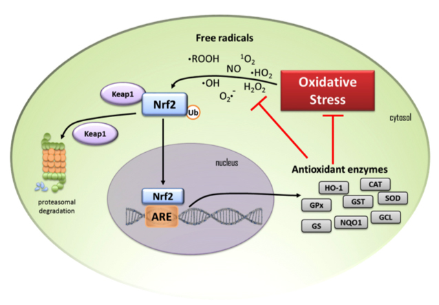
Fig. 7.

Schematic depiction of a cell and the Nrf 2 cell signalling pathway showing the anti-oxidant enzymes that are induced by oxidant stress. (1) Under homeostatic conditions cytosolic Nrf2 transcription factor is maintained at low levels by proteasomal degradation under control of the Keap1 protein complex. (2) When cells are exposed to oxidative stress free radicals result in the release of Nrf2 from Keap1 to escape proteasomal degradation and it translocates to the nucleus where it binds to the oxidant response element (ARE) and anti-oxidant enzymes are induced. (3) These include heme oxygenase-1 (HO-1), glutathione peroxidase (GPx), glutathione-S- transferase (GST), superoxide dismutase (SOD), catalase (CAT), glutathione reductase (GR), NAD(P)H quinone oxidoreductase (NQO1), glutamine-cysteine ligase (GCL) and glutathione synthetase (GS). These enzymes diminish oxidative stress on the cell and reduce free radical levels. (4) Black arrows in the schematic depict activation pathways, (5) red T-bars signify the blocking steps induced by transcription of anti-oxidant enzymes. Figure reproduced from [197] by open access Creative Common CC BY license.

10.1 Licochalcone A and B

Licochalcone A and B from liquorice root (Glycyrrhiza glabra or Glycyrrhiza inflata) are bioactive anti-tumour chalcones [201] that up-regulate the Nrf2 anti-oxidant pathway [202] and attenuate neuronal injury in a rat model of stroke [153]. Licochalcone B is neuroprotective, inhibits amyloid β 42 self-aggregation (IC 50 = 2.16 μ M), disaggregates pre-formed A β 42 fibrils, and reduces metal-induced A β 42 aggregation through its metal ion chelating properties [150]. Quercetin [203, 204], luteolin [205], myrcetin [206], apigenin [207], chrysin [208] and catechins [209] also induce Nrf2 which provides anti-inflammatory and anti-oxidant protection to tissues [210].

10.2 Quercetin

Quercetin is neuroprotective, enhances neuronal viability, promotes neurogenesis [203, 211] and can modulate/inhibit a number of cell signaling pathways including Nrf2, PON2 (paraoxonase-2), JNK (c-Jun N-terminal kinase), TNF- α , peroxisome proliferator-activated receptor γ coactivator 1- α , mitogen-activated protein kinases (MAPKs), CREB (Cyclic AMP response element binding protein) and PI3K/Akt (Phosphoinositide 3- kinase) [203]. Quercetin’s beneficial therapeutic properties in AD stem from its ability to protect neurons from oxidative stress mediated by lipid peroxidation, and it also inhibits fibril formation from amyloid- β proteins, counters deleterious inflammatory cytokine production prevalent in neuroinflammation [212].

10.3 Chrysin

Chrysin exhibits anti-oxidative effects on dopaminergic neurons in PD by increasing Nrf2 expression [208], reduces neuron NO levels intracellularly and regulates neuronal anti-oxidant pathways. Chrysin promotes dopaminergic neuronal survival by upregulating the activation of myocyte enhancer factor 2D (MEF2D), suppresses the upregulation of c-caspase and Bax and downregulates the anti-apoptotic protein Bcl 2 and enhanced neuronal survival through production of neurotrophic factors. Chrysin’s anti-inflammatory properties increase dopamine levels through inhibition of monoamino-oxidase B activity restoring behavioral deficits in animal models of PD [213].

10.4 Catechins

Oxidative stress and inflammation are major contributors to the pathogenesis of neurodegenerative diseases. Catechins are powerful antioxidants with free radical scavenging properties that have roles to play in the management of neurodegenerative diseases. Catechins modulate cellular processes mediated through NF- κ B and Nrf2 signaling pathways to regulate neuroinflammation [209].

10.5 Luteolin

Luteolin’s anti-oxidant, anti-inflammatory properties and ability to induce Nrf2 are neuroprotective [214, 215] and counter neuroinflammation following brain trauma [216] downregulating the TLR4/TRAF6/NF- κ B pathway after intracerebral hemorrhage and cerebral ischemia [217, 218].

10.6 Myrcetin

Myrcetin has beneficial properties in the treatment of cerebral ischemia and AD and has multifunctional properties regulating the expression of Hippo, MAPK, GSK-3 β , PI3K/AKT/mTOR, STAT3, TLR, I κ B/NF- κ B, Nrf2/HO-1, ACE, eNOS / NO and AChE [219].

10.7 Apigenin

Apigenin’s antioxidant properties regulate redox cell signaling pathways involving NF- κ B, Nrf2, MAPK, and P13/Akt. Apigenin also has metal chelating, antiamyloidogenic, fibril-destabilization activity and free radical scavenging properties that provide tissue protection in chronic inflammation, metal induced oxidative stress, and in neurodegenerative diseases [207, 220, 221, 222].

10.8 Epigallocatechin Gallate (EGCG)

In-vitro, in-silico and x-ray crystallographic studies show EGCG exerts anti-oxidative health benefits to neural tissues [205]. Surface plasmon resonance and computational docking simulations demonstrate EGCG’s direct binding to pro-inflammatory chemokines blocking the recruitment of inflammatory cells into tissues, regulating inflammatory diseases [223]. EGCG also inhibits amyloid plaque formation in AD and aggregation of A β peptides. EGCG’s metal chelating properties inhibit amyloid fibril formation in AD. In-silico docking simulation and in-vitro studies demonstrate the AChE inhibitory properties of EGCG’s and beneficial effects in AD [224].

10.9 Genistein

Genistein modulates pathogenic events in neurodegeneration and is neuroprotective, attenuates amyloid-beta-induced cognitive impairment in rats in an in-vivo model of A β toxicity [225]. Genisteins mechanism of action lies in its ability to regulate Akt and Tau protein phosphorylation to inhibit amyloid fibril deposition [226]. Genistein improves impaired spatial learning and memory by regulating cAMP/CREB and BDNF-TrkB-PI3K/Akt cell signaling pathways [227] and also regulates mitochondrial enzymatic activity and oxidative phosphorylation countering neurodegenerative mitochondrial misfunction [228].

10.10 Cardamonin

Cardamonin induces Nrf-2 expression and its neuroprotective anti-oxidant enzyme systems [229], attenuates inflammation and oxidative damage in IL-1 stimulated chondrocytes in OA [230] and significantly up-regulates seleno- anti-oxidant enzymes induced by Nrf2 [231].

10.11 Hesperidin/Hesperitin

Hesperidin’s anti-oxidant, anti-inflammatory and neuroprotective properties are useful in the treatment of neurodegenerative conditions [232] and memory impairment in AD, PD, MS, and ALS. Hesperidin glycoside and its aglycone form, hesperitin, have been developed into multifunctional derivatives of higher efficacy [233]. A multi-tier flavonone screening protocol employing molecular docking for BACE1 inhibitory, and anti-amyloidogenic and antioxidant activities have demonstrated hesperidin derivatives as potent AD therapeutics [234].

10.12 Hesperidin

Hesperidin is a high affinity BACE1 inhibitor completely inhibiting BACE1 at a concentration of 500 nM and provides complete inhibition of amyloid fibril formation [234, 235]. Inhibition of BACE1 by hesperidin acts upstream of the APP processing that generates A β protein required for fibril aggregate assembly into plaques in AD brains. Inhibition of BACE1 and A β aggregation occurs by binding close to the catalytic aspartate dyad constraining BACE1 activity preventing APP recognition to inhibit amyloid fibril formation, A β 25 - 35 induced ROS generation and mitochondrial dysfunction [235]. Mitochondrial dysfunction and oxidative stress also induce pathological neurodegenerative changes contributing to the development of AD [236]. Hesperidin inhibits A β -induced cognitive dysfunction, improves learning and reverses memory deficits improving locomotor activity. Increased phosphorylation of GSK-3 β by hesperidin, improves cognitive function in the APPswe/PS1dE9 transgenic mouse model of AD [236]. A limited number of human clinical trials have shown that hesperidin-enriched dietary supplements significantly improved cerebral blood flow, cognition, and memory performance [63].

Cerebral ischaemic injury and degenerative pathology in AD are linked, hesperidin down-regulates Bcl-2, Akt/PI3K protecting against A β 25 - 35 -induced apoptotic neurotoxic effects [63]. Oxidative stress and inflammation have pivotal roles in the pathophysiology of AD and are attenuated by hesperidin in APP/PS1 mice resulting in a reduction in ROS, LPO, and increased activity of HO-1, SOD, catalase, and GSH and inhibits neuroinflammation by decreasing TNF- α and NF- κ B activity [237]. A decrease in phosphorylation of Akt and GSK-3 β by hesperidin is neuroprotective in APP/PS1 mice [238]. Hesperidin has anti-inflammatory, anti-oxidative and neuroprotective properties in an adult male Sprague Dawley AD rat model induced by scopolamine and reduced memory loss and decreased serum TNF- α and IL-1 β levels, [8]. Inhibition of amyloid A β -42 and AChE activity in the hippocampus and prefrontal cortex also preserved normal brain tissue architecture and function [239].

10.13 Hesperitin

Intracerebroventricular injection of hesperetin 24 hours after injection of A β 1-42 in mice has been used as a model of AD. Hesperetin significantly attenuated oxidative stress and expression of Nrf2/HO-1, LPO and ROS production in the hippocampus, cortex, and in HT22 neural cell cultures and had a strong antiapoptotic neuroprotective effect, inhibited oxidative stress, neuroinflammation, and cognitive decline countering neurodegeneration and memory impairment [64]. Inhibition of the oligomerization of A β or tau peptide into fibrils by heparitin, reduces scopolamine-induced cognitive decline [240].

11. Upregulation of Nrf2 by Flavonoids Provides Anti-Oxidant Cell Protective Properties

NF-E2-related factor 2 (Nrf2) is a master regulator of numerous cytoprotective genes [241, 242]. After translation, the Nrf2 protein is rapidly degraded by the ubiquitin-proteasome system in the cytoplasm [243]. Kelch-like ECH-associated protein 1 (Keap1) is a component of the Cullin 3 (CUL3)-based E3 ubiquitin ligase complex and controls the stability and accumulation of Nrf2 (Fig. 7). Table 3 (Ref. [197, 225, 226, 227, 228, 229, 230, 231, 232, 233, 234, 235, 236, 237, 238, 239, 240, 241, 242, 243, 244, 245, 246, 247, 248, 249, 250, 251, 252, 253, 254, 255, 256, 257, 258, 259, 260, 261, 262, 263, 264, 265, 266, 267, 268, 269, 270, 271, 272, 273, 274, 275, 276, 277, 278, 279, 280, 281, 282, 283, 284, 285, 286, 287, 288, 289, 290, 291, 292, 293, 294, 295, 296, 297, 298, 299, 300, 301, 302, 303, 304, 305, 306, 307, 308, 309, 310, 311, 312, 313, 314, 315, 316, 317, 318, 319, 320, 321, 322, 323, 324, 325, 326, 327, 328, 329, 330]) illustrates the diversity of flavonoids that up-regulate Nrf2 to exert a cell and tissue protective effect.

Table 3.Flavonoids that display cytoprotective, anti-inflammatory and anti-oxidant properties through the upregulation of Nrf2.
Flavonoid Ref Flavonoid Ref
Acacetin [244] Licocalchlcone A. [245]
Apigenin [246] Liquiritin [247]
Artocarmitin B [248] Limonin [249]
Baicalein [250] Luteolin [251, 252, 253, 254, 255, 256, 257, 258]
Baicalin [259, 260, 261] Malvidin-3-O-Glucoside [262]
Biochanin A [263, 264] Morin [265]
Cardamonin [266, 267] Naringenin [268, 269]
Cynaroside [270] Natural/synthetic chalcones [271]
Chrysin [272, 273] Neobavaisoflavone [274]
Chrysoeriol [275] Nobiletin [276]
Cyanidin-3-glucoside [277] Orientin [278]
Daidzein [279] Peurarin [280, 281]
Dihydromyrecetin [282] Phloretin [263]
Epigallocatechin Gallate [283] Pinocembrin [284, 285]
(-)-Epicatechin [286] Pinocembrin-7-methylether [287]
Formononetin [288] Punicalagin [289]
Galangin [290, 291, 292, 293] Quercetin [294, 295, 296, 297, 298]
Gallocatechin [299] Scutellarin [300]
Genistein [301, 302, 303] Silychristin A [304]
Hesperidin [305, 306] Silymarin [307]
Hesperitin [308, 309, 310] Theaflavin [311, 312, 313]
Hyperoside [314] 6,7,4’-Trihydroxyflavanone [315]
Icariin [316, 317, 318] Vitexin [319]
Icaritin [320] Wogonin [321, 322]
Kaempferol [323, 324, 325, 326, 327] Xanthohumol [328, 329]
Kushenol [330]
12. Multifunctional AD Therapeutic Chalcone Derivatives
12.1 4-Hydroxy-Chalcones and Bis-Chalcone Ether Derivatives

Diversely-substituted 4-hydroxy-chalcones and a series of bis-chalcone ether derivatives with antioxidative properties, lipoxygenase (LOX) and AChE inhibitory activity are potent in vitro inhibitors of lipid peroxidation and potential new multifunctional AD compounds [184]. Multifunctional 4-hydroxy chalcones inhibit self-induced A β 1 - 42 aggregation (45.9–94.5% at 20 μ M) and disassemble self-induced A β 1 - 42 fibril aggregates. The Cu 2 + - chelating properties of these compounds contribute to their ability to inhibit assembly and disaggregation of A β fibrils. The most active derivative (3 g) had low cytotoxicity, significantly reversed A β 1 - 42 -induced SH-SY5Y cell damage and ameliorated scopolamine-induced memory impairment in mice [331].

12.2 Dimethylamino Chalcone-O-Alkylamines Derivatives

Dimethylamino chalcone-O-alkylamines derivatives inhibit A
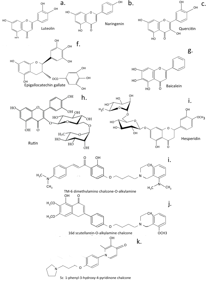
 β assembly and disaggregate established A β fibrils, are AChE inhibitors, biometal chelators and selectively inhibit MAO-B. Compound TM-6 showed the greatest inhibitory activity against self-induced A β aggregation displaying 95.1% inhibition at 25 μ M and was a remarkable antioxidant, good AChE (IC 50 = 0.13 μ M) and MAO-B (IC 50 = 1.0 μ M) inhibitor, neuroprotectant Cu 2 + chelator, inhibiting Cu 2 + -induced A β aggregation (95.3%, 25 μ M) and assembly of A β fibrils (88.1%, 25 μ M). TM-6 could cross the blood-brain barrier had low toxicity in mice at doses of up to 1000 mg/kg and improved scopolamine-induced memory impairment [182, 183, 332] (Fig. 8).

Fig. 8.

Naturally occurring flavonoids and multifunctional flavonoids developed from for the treatment of neurodegeneration.

12.3 Anti-Oxidant Chalcone-O-Carbamates

Anti-oxidant chalcone-O-carbamates are multitargeting compounds that inhibit AChE/BChE and MAO-A/MAO-B, A β 1 - 42 aggregation/assembly and have metal-chelating and neuroprotective properties against peroxide induced PC12 cell injury. Compounds 5b and 5h had highly selective BChE inhibitory activity (IC 50 values of 3.1 μ M and 1.2 μ M, respectively) and MAO-B inhibitory potency (IC 50 values of 1.3 μ M and 3.7 μ M), inhibited self-induced A β 1 - 42 aggregation (63.9% and 53.1% inhibition for 5b and 5h), were permeable to the BBB and improved scopolamine-induced cognitive impairment. Compound 5b was the best multifunctional therapeutic agent for the treatment of AD [182].

12.4 Scutellarein-O-Alkylamine Analogs

Scutellarein-O-alkylamine analogs have metal chelating properties, anti-oxidative activity, and inhibit self-induced, Cu 2 + -induced and human AChE-induced A β 1 - 40 aggregation. Compound 16d binds simultaneously to the catalytic active and peripheral anionic sites of AChE, protecting against peroxide-induced PC12 cell injury, had low toxicity in SH-SY5Y cells and significantly reversed murine scopolamine-induced memory loss [333].

12.5 Ferulic Acid-O-Alkylamines

Ferulic acid-O-alkylamines are anti-AD agents with impressive inhibitory activity against BuChE, inhibition/disaggregation of self-induced A β aggregation antioxidants. Compound 7f had an IC 50 value of 0.021 μ M for equine, 8.63 μ M for rat and 0.07 μ M for human BuChE, and was also a good AChE inhibitor (IC 50 = 2.13 μ M for electric eel, 1.8 μ M for rat and 3.82 μ M for human erythrocyte AChE). Compound 7f inhibited self-induced A β 1 - 42 aggregation (50.8 ± 0.82%), disaggregated self-assembled A β 1 - 42 fibrils (38.7 ± 0.65%), modest antioxidant activity, protected against H 2 O 2 -induced PC12 cell injury, and had low toxicity [181]. Further novel multifunctional chalcone-O-alkylamines inhibit AChE (IC 50 = 1.3 ± 0.01 μ M) and BuChe (IC 50 = 1.2 ± 0.09 μ M). Compound 23c was a selective MAO-B inhibitor (IC 50 value of 0.57 ± 0.01 μ M), had antioxidant neuroprotective properties and could inhibit self-induced A β 1 - 42 aggregation. 23c was a selective metal chelator disaggregator of Cu 2 + -induced A β 1 - 42 aggregation and could cross the BBB, improving scopolamine-induced memory impairment. Molecular modeling showed 23c binds to the active site of AChE and BuChE, MAO-B [332].

12.6 Halogenated Coumarin-Chalcones

Halogenated coumarin-chalcones inhibit MAO s, AChE, BuChE, and BACE-1. Compound CC2 potently inhibited MAO-B (IC 50 = 0.51 μ M), CC1 displayed an IC 50 of 0.69 μ M. CC2 and CC3 inhibited BuChE (IC 50 7.00 and 11.8 μ M). CC1 and CC2 could cross the BBB were non-toxic and attenuated H 2 O 2 -induced cellular damage via ROS scavenging properties [185].

12.7 Monoamine Oxidase Inhibitors

Monoamine Oxidase inhibitors regulate monoamine neurotransmitters, oxidative stress, A β aggregation, AChE inhibition, and are anti-ROS and metal ion chelator multitargeting agents of value in the treatment of AD [150, 334].

12.8 Derivatised Forms of Hesperitin

Hesperetin derivatives are AChE dual-site inhibitors displaying strong inhibitory activity against AChE, high selectivity for BuChE and inhibit self-induced β -amyloid (A β ) aggregation. Compound 4f significantly protected PC12 neurons against H 2 O 2 -induced cell death, and was non-cytotoxic to SH-SY5Y neurons [335].

12.9 Multifunctional 7-O-1, 2, 3-Triazole Hesperetins

A series of 7-O-1, 2, 3-triazole hesperetins inhibit BuChE, are anti-neuroinflammatory, and neuroprotective. Compound a8 (7-O-((1-(3-chlorobenzyl)-1H-1,2,3-triazol-4-yl)methyl)hesperetin) displayed excellent anti-BuChE inhibitory activity (IC 50 = 3.08 ± 0.29 μ M) and anti-neuroinflammatory activity lowering NO production by blocking the NF- κ B signaling pathway inhibiting the phosphorylation of P65, a8 had remarkable neuroprotective properties, lacked neurotoxicity and inhibited self-mediated A β 1 - 42 aggregation, chelated biometals, and was permeable to the BBB [149] and improved learning and memory recovery in scopolamine treated AD mice.

12.10 7-O-Amide Hesperetins

7-O-amide hesperetins inhibit BuChE and are neuroprotectors. Compound 7c (7-O-(4-(morpholinoethyl)-acetamide) hesperetin) was the most effective BuChE inhibitor (IC 50 = 0.28 ± 0.05 μ M) [187]. 4d, 4e and 7c are anti-inflammatory strong antioxidants and inhibited A β self-aggregation. 7c inhibited iNOS and COX-2 expression, prevented LPS-mediated inflammation, was a Cu 2 + and Zn 2 + chelator, penetrated the BBB and reduced scopolamine induced cognitive impairment.

13. Traditional Chinese Medicinal Formulations used to Treat AD

Traditional Chinese herbal preparations have been used for centuries in complementary alternative medicine [336, 337]. Attempts have been made to better understand their chemical components to determine if they can be applied in Western medicine. Claims have been made that herbal medications can successfully combat COVID-19 infections [338, 339]. Network pharmacology, molecular docking and in-vitro cell based investigations have identified a number of active components in these herbal preparations that could potentially provide a therapeutic effect [340, 341]. Chinese herbal preparations used to treat AD have also undergone similar assessments to identify their active therapeutic components and their molecular targets.

13.1 LeZhe

LeZhe is purported to be a nerve calmative detoxifying antipyretic agent useful in the prevention and treatment of age dependent AD [342]. Network pharmacology and molecular docking studies have been employed to identify LeZhe’s active components and their molecular targets and these have been evaluated in PC12 primary hippocampal neural cultures where injury had been induced using A β 25 - 35 . A total of 105 active compounds and 38 molecular target proteins were identified. The main bioactive compounds of LeZhe include alkaloids such as berberine, the aromatic amide aurantiomide, coumaroyl tyramine, trans-syringin and 3-dimethyl phillyrin phenylpropanoid [342]. The molecular targets identified included protein kinase B (AKT), phosphoinositide 3-kinase (PI3K), tyrosine-protein kinase JAK1 (JAK1), mammalian target of rapamycin (mTOR), TNF- α , neuronal NOS (NOS1), and cholinergic function-related proteins, including α 4-nicotinic acetylcholine receptor ( α 4 nAChR) and Muscarinic acetylcholine receptor M1 (Muscarinic M1). Inflammation and cholinergic dysfunction were thus central features of this interactive network. The LeZhe compounds significantly improved PC12 cell survival and inhibited apoptosis of A β 25 - 35 injured primary hippocampal neuron cell cultures through a complex multi-compound-multi-target-multi-pathway regulatory network [343].

13.2 Chaihu Shugan San

Chaihu Shugan San (CSS) is another well-known herbal antidepressant used in traditional Chinese medicine. Modern pharmacological and clinical evidence indicate that CSS could also be beneficial in the treatment of cognitive dysfunction in AD. Active compounds in CSS have been screened using the Traditional Chinese Medicine Systems Pharmacology database. Compound-related targets retrieved using the SwissTarget Prediction database facilitated the identification of major depressive disorder (MDD)-related targets The CSS compounds were examined in cumulative unpredictable mild stress (CUMS) mice. Molecular docking analyses determined the binding affinities of the bioactive CSS compounds [344]. Elucidation of multi-target mechanisms of action for CSS using network pharmacology analysis identified a total of 152 active compounds, 520 predicted biological targets and 160 AD-related targets [345] regulating PI3K-Akt, MAPK and HIF signaling pathways. Pre-treatment of neural cell cultures with CSS reduced A β -induced neural cell death and apoptosis in differentiated PC12 cells, increased phosphorylation of Akt, decreased Bax expression and pGSK3 β /GSK3 β levels in the hippocampus of CUMS mice showing the PI3K/Akt signaling pathway provided the CSS protective effect. The active flavonoid compounds identified included quercetin and luteolin, which showed good docking scores for the PI3K protein. Quercetin, luteolin, and kaempferol are probable active compounds in CSS which warrant further examination in the treatment of the MDD features of AD.

13.3 Qing Fei Pai Du and Ma Xing Shi Gan

Qing Fei Pai Du and Ma Xing Shi Gan anti-viral decoctions used to treat COVID-19 and AD in Traditional Chinese Medicine are of considerable complexity, however molecular networking of mass spectrometry data has been used to identify a number of bio-active flavone and chalcone compounds present in these formulations [346]. Hesperidin, glycyrrhizic acid, baicalin, baicalein, naringin, phillyrin, quercetin, luteolin, kaempferol, licochalcone B and mangiferin were all present [346]. Further studies are required to fully decipher all the therapeutic bioactive component combinations in these formulations and their pharmacological interactions.

14. Potential of Metal Co-Ordination Flavones as Anti-Viral Compounds

Highly active chalcone metal co-ordination complexes were originally developed to treat drug resistant solid tumours. Metallopharmaceuticals have amplified therapeutic modulatory pharmacokinetic and pharmacodynamic properties against cell receptors [178]. Carbonyl, hydroxyl, phenolic oxygen in heterocyclic ring structures in chalcones and flavones have excellent chelating properties on the preparation of metal coordination complexes. These have improved therapeutic and catalytic activities that have found successful application in the treatment of drug resistant tumours but have not been extensively examined for their anti-viral properties. Platinum(IV) complexed chalcones have potent anti-tumour activity and low cytotoxicity, inducing G2/M phase arrest and apoptosis in A549 cancer cells. Collapse of mitochondrial membrane potential, elevated expression of apoptosis-related proteins and reactive oxygen species all contribute to inhibition of tumour growth [177]. Metal coordination complexes prepared with chalcones and flavones represent a novel area of application in anti-viral development that needs to be explored further. Zinc not only inhibits the SARS-CoV-2 Mpro with nanomolar affinity, but also inhibits viral replication [347]. Furthermore, the natural ionophore quercetin increases the anti-viral potency of Zn 2 + . The highly conserved catalytic dyad of Mpro in SARS-CoV, MERS-CoV and variant forms of SARS-CoV-2 suggests Zn 2 + mediated inhibition of Mpro may be of wider application in anti-viral therapeutics. Gallium also occurs in trace amounts in zinc ores and displays strong anti-inflammatory and antiviral activity against the influenza A H1N1 virus, HIV and SARS-CoV-2 thus represents another candidate for development of therapeutic anti-viral metal co-ordination complexes [348].

15. Assessment of Selenium-Derivatised Flavones and Chalcones as Anti-Viral Agents

While selenium is a non-metal, it can also inhibit viral replication. Ebselen is an active seleno-organic anti-viral against zika, influenza A, HCV, and HIV-1, and SARS-CoV-2 [194, 349]. Selenium interacts with thiol groups in proteins and this may represent a mechanism whereby it inhibits SARS-CoV-2 Mpro activity and viral replication [350]. Selenium-substituted chrysin and quercetin, developed as anti-cancer agents, also display anti-viral properties that need further evaluation (Fig. 9).
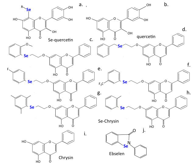
Fig. 9.

Selenium substituted flavonoids of improved efficacy.

16. Long COVID Disease

Critically ill COVID-19 patients suffering from acute respiratory distress syndrome (ARDS) show lung injury and haemolysis. Heme is a prosthetic group crucial for the function of the oxygen-trapping haemoglobin and the energy-producing cytochromes of the electron transport chain of mitochondria. Haemolysis generates free heme in ARDS patients promoting adhesion molecule expression, leukocyte recruitment, vascular permeabilization, platelet and complement activation, thrombosis, and fibrosis. Heme is degraded by the anti-inflammatory enzyme heme oxygenase-1 (HO-1) generating biliverdin/bilirubin, iron/ferritin, and carbon monoxide. Free heme contributes to many of the inflammatory aspects of critically ill COVID-19 patients, thus induction of HO-1 may be protective and a therapeutic target in COVID-19 patients reducing long-term fibrotic changes in lung tissues [351]. HO-activity not only degrades injurious heme, but its effector molecules possess anti-oxidative and anti-inflammatory properties of potential benefit to ARDS patients [352]. 4-Anilinoquinolinyl chalcone upregulates HO-1 expression and has beneficial anti-inflammatory and anti-oxidant properties typical of the chalcone family [353]. Novel chalcones display anti-inflammatory and anti-oxidant effects in-vitro and after LPS induced acute lung injury [354]. Studies are warranted with these chalcone derivatives for the treatment of long COVID-19 disease [355]. Promotion of tissue fibrosis in COVID-19 infections results in fibrotic changes in liver and lung tissues [356] and leads to long-term pulmonary fibrosis and associated breathing difficulties. A number of chalcones (Panduratin A) present in Thai and Chinese herbal medicines [6, 357] and identified in pomegranate [358] display beneficial anti-inflammatory properties and reduce tissue fibrosis in a similar manner to Pirfenidone [359, 360], a long-standing anti-fibrosis medication. These are worthy of further investigation in the management of long COVID-19 disease.

A screen of extracts from 122 Thai traditional medicinal plants for anti-viral and specifically anti-SARS-CoV-2 compounds identified Panduratin A from Boesenbergia rotunda. This plant is also known as Chinese keys, finger-root, lesser galangal or Chinese ginger and is found in SE Asia and China. Panduratin-A (2,6-dihydroxy-4-methoxyphenyl)[(1R,2S,6R)-3-methyl-2-(3-methylbut-2-en-1-yl)-6-phenylcyclohex-3-en-1-yl]methanone) is a potent non-toxic anti-inflammatory chalcone that strongly inhibits NO (IC 50 : 0.175 μ M) and PGE-2 (IC 50 : 0.0195 μ M), suppresses iNOS and COX-2 expression and has anti-tumour activity against A549 human non-small cell lung cancer cells (IC 50 of 5 μ g/mL) arresting tumour cell proliferation and inducing apoptosis [361]. Panduratin A administered to Vero E6 cells infected with SARS-CoV-2 displayed an IC 50 at 3.62 μ g/mL (IC 50 of 5.3 μ M) [6]. Panduratin A is also a component of pomegranate and has been used for over a thousand years as a fruit with medicinal properties and has been proposed as a functional superfood [362, 363, 364]. Extracts of pomegranate peel have marked antioxidant properties [365] containing diverse phenolic compounds, which scavenge free radicals and inhibit lipid peroxidation [362, 366]. Panduratin A acts in conjunction with punicalagin, an anti-proliferative, apoptotic, anti-oxidant phytochemical [367]. Punicalagin (2,3-(S)-hexahydroxydiphenoyl-4,6-(S,S)-gallagyl-D-glucose), a polycyclic phenolic phytochemical inhibits SARS-CoV-2 3CL-protease in-vitro, displaying an IC 50 of 6.192 μ g/mL and when combined with zinc sulphate it displays enhanced 3CL-protease inhibitory activity as a metal transition complex [368, 369].

16.1 The Impact of COVID-19 on AD and Dementia Patients

The cost of dementia in Australia in 2016 was estimated at $14.25 billion and is escalating [370] with an increased incidence of AD and dementia in global ageing populations [371]. In 2010, the cost of treating dementia in the USA was estimated at $200 billion. The COVID-19 pandemic has had a disproportionately negative impact on people affected by AD and dementia. Individuals affected with dementia may have a reduced capacity to understand and comply with pandemic health care restrictions and thus potentially represent a spreader risk for COVID-19 infection [372]. With present day AD/dementia patient numbers of 47 million projected to triple by 2050 compounded by the impact of the present day COVID-19 pandemic there is a clear need to develop therapeutics that target oxidative stress, neuroinflammation, cholesterol metabolism, amyloid plaque formation, and adverse regulatory effects on neurotransmitters and vascular factors to combat this progressive and debilitating neurodegenerative disorder.

Of particular concern are the cognitive deficits that have been reported in patients who have recovered from COVID-19 respiratory disease. This includes an inability to concentrate and a fogging of thought processes impairing concentration for tasks at hand and the solving of problems and feelings of long-term anxiety and insecurity [373, 374, 375, 376, 377]. Particularly disturbing are emerging reports of COVID-19 causing a reduction in IQ in children. Long-term fatigue associated with long covid patients impacts on the development of neuro-psychiatric disorders [378, 379, 380]. AD is the sixth-leading cause of death and is present in 70% of all cases of dementia. The global burden of AD is expected to accelerate from 26.6 million cases in 2006 to 106.8 million by 2050, estimated worldwide costs of dementia were US$ 604 billion in 2010 so this projected increase in the number of AD and dementia patients will make a significant impact on healthcare resources.

16.2 Bacterial Infections Associated with Long COVID Disease

Secondary bacterial infections have been observed in long COVID disease and this may involve MDR bacterial strains. The attainment of bacterial antibiotic resistance is a serious healthcare problem [381] and one that has been acknowledged by the WHO with their publication of the dirty dozen list of MDR pathogenic bacteria [http://www.who.int/news-room/detail/27-02-2017-who-publishes-list-of-bacteria-for-which-new-antibiotics-areurgentlyneeded (accessed 12 January 2018)]. MDR bacterial infections have been compounded by the COVID-19 pandemic and the emergence of the MDR strains of Clostridium difficile and Mycobacterium tuberculosis in long COVID bacterial infections [382, 383, 384, 385, 386]. Inappropriate administration of antibiotics to long COVID-19 patients despite the fact that this is not a bacterial infection may be inappropriate even when these are administered as a preventative measure against potential secondary bacterial infections that may occur and may actually result in these patients acquiring troublesome antibiotic resistant bacterial strains [387]. Antibiotic resistance is a serious problem and a major public health concern. Multi drug resistant Mycobacterium tuberculosis bacterial strains causing tuberculosis (TB) have emerged in the COVID-19 pandemic. These may not be responsive to any antibiotic currently available, leading to lethal pneumonia as a secondary respiratory infection of COVID-19, although other organs can also be effected including the brain, 150,000 TB infections are reported annually with lethal consequences in 40% of these patients [388, 389, 390, 391]. Clostridium difficile has also emerged during the COVID-19 pandemic as an additional MDR gut bacterium with serious health impact. Some positive developments have also emerged on how to combat such infections. Phage therapy is a therapeutic which is proving effective against a number of MDR bacteria and secondary infections occurring with COVID-19 [392, 393, 394, 395, 396, 397, 398]. Ebselen has been used as an anti-cancer, anti-bacterial and anti-viral SARS-CoV-2 main protease inhibitor [189]. Ebselen has potent anti-bacterial activity against antibiotic resistant C. difficile where it targets the transpeptidase Ldt Mt2 protease [188, 189, 190] and can act synergistically with the CoV-2 replication inhibitor Remedesivir to eradicate both SARS-CoV-2 and MDR bacterial infections [191]. Flavonoids are active against MDR bacteria and are a promising and underappreciated reservoir to counter antibiotic resistance. The antimycobacterial and anti-inflammatory activities of substituted chalcones have also been used in the development of anti-tuberculosis therapeutic treatments [399]. Flavonoids have been widely utilized in traditional medical practices to combat bacterial infections [17, 399, 400] with some approaches focusing specifically on how to combat MDR bacteria [400, 401, 402, 403, 404]. Combination therapies with antibiotics [405] and approaches examining how the antiviral and immunomodulatory properties of flavonoids can be harnessed in the treatment of respiratory diseases have also been examined [406].

Clostridium difficile (now renamed as Clostridioides difficile) is a problematic bacterium that has recently attained antibiotic-resistant status. Antibiotic resistant Clostridium difficile spore-forming bacteria are frequently found in the bowel. Infections with C.difficile are lethal in 30% of patients. Faecal transplant therapy has been used to treat these infections [407, 408, 409, 410, 411, 412]. This is a phage-mediated therapy that is used to treat antibiotic resistant bacterial infection and is a useful approach harnessing protective aspects of the human microbiome; a 80% cure rate is reported for faecal transplant therapy [407, 408, 409, 410, 411, 412]. While faecal transplantation is a highly effective modern development in Western medicine it is not a new technique. In traditional Chinese medicine, Ge Hong in the 4th century used faecal transfer as a therapeutic approach for the treatment of chronic diarrhea. In the 16th century, another famous Chinese physician, Li Shizhen, described the use of fresh or fermented faecal products, called “yellow soup” to treat severe diarrhea, fever, pain and constipation [413]. A series of publications have appeared in Western medical circles advocating this treatment [188, 414, 415, 416, 417] and guidelines on this methodology have also been published [410, 411]. A faecal enema may be a more acceptable route of administration for phage therapy rather than “yellow soup”.

Dietary supplements or diets rich in flavonoid and chalcone components may be of benefit in the treatment of long COVID disease and neurological disorders [418, 419]. A recent study comparing the impact of diet versus drugs on cellular metabolism found nutrition had a much stronger impact than drugs on many cellular processes [420]. This pre-clinical study showed that diet could be more powerful than drugs in keeping conditions like diabetes, immune dysfunction, stroke and heart disease at bay. Diet is a powerful medicine, involving nutrient-signaling pathways that affect the gut microbiome [421, 422, 423]. The formation of a healthy microbiome in early childhood, is important to the establishment and maintenance of health in later life. Studies have suggested that COVID-19 may impact the microbiome composition and diversity, increasing the incidence of allergic and autoimmune disorders, especially in children [424]. The full impact of the gut microbiome on the attainment of tolerance to certain foods and the neurological pathways that train innate immune responses is, however, incompletely understood. Dietary flavonoids have been shown to interact with the microbiome [425] and the gut microbiome has emerged as a key conduit in mental health and a promising target for interventions [426, 427, 428]. Dietary flavones and chalcones can have important cell regulatory and tissue protective properties positively impacting on a number of diseases, many studies have shown how flavones and chalcones can impact diabetes, liver fibrosis, cancers and bacterial infections and these can also be beneficially regulated by dietary control [11, 429, 430, 431, 432]. Pre-clinical studies have also shown that neurological disorders such as AD, PD, ALS, MS and autism can also benefit from dietary flavones and chalcones and related compounds which regulate mitochondrial activity and pathways that can generate oxidative stress. Dietary components need to be taken seriously in the overall scheme of improving and maintaining a healthy cellular metabolic environment in tissues. There therefore is a scientific basis to the use of superfoods rich in flavonoid dietary components to positively aid in tissue protection and cellular functions that maintain tissue homeostasis and combat disease. Nutrient-sensing pathways influence metabolic health and aging, offering the possibility that diet might be used therapeutically. For example, dietary composition powerfully impacts on the hepatic proteome, not only on its metabolic profile [420] but on fundamental processes such as mitochondrial function and RNA splicing. This also needs to be considered in other tissue contexts in health and disease and in the specific context of viral infection could represent a supportive adjunct to conventional anti-viral therapeutic treatments.

17. The Potential Application of Flavonoid Supplements in Biomedicine

Flavonoid supplements have emerged as possible approaches in the treatment of COVID-19 and neurodegeneration based on their cell and tissue protective properties as already discussed.

Flavonoids the Gut Microbiome and the Gut-Brain and Gut-Lung Axes

Flavonoid supplements have emerged as putative nutritional or therapeutic adjunct approaches for the treatment of COVID-19 [24] and neurodegeneration based on their antioxidant, antiviral, anti-inflammatory, immunomodulatory effects and ability to promote a healthy gut microbiome [212, 433]. Flavonoid-modifying enzymes are encoded in gut bacteria however little is known of the active flavonoid components that they generate from dietary flavonoids and polyphenolic compounds and how these exert disease prevention and beneficial effects on the health of tissues.

Intestinal microbiota can indirectly modulate airway physiology and immunity. COVID-19 patients have been observed to exhibit a specific imbalance in their gut microbiome closely associated with CoV-2 disease pathophysiology [433]. Rebalancing the intestinal microbiome using probiotics has been suggested as an effective therapeutic approach against COVID-19.

Lactobacillus plantarum, Bifidobacterium longum and Lactococcus lactis ssp. lactis, exhibit robust anti-infective properties against respiratory RNA viruses [434]. Furthermore, L. plantarum is capable of expressing viral antigens including the spike protein of SARS-CoV-2 and is capable of inducing protective immune responses in the gut and respiratory tract and of modulating innate and adaptive immune responses. This has led to L-plantarum being suggested as a potential adjuvant delivery system for the development of SARS-CoV-2 oral vaccines [435]. The gut microbiome is influenced by dietary flavonoids and these can have disease modifying health promoting benefits. Dietary polyphenolic compounds have beneficial properties on the gut microbiome and feed-on effects on neurodegenerative disorders through the gut-brain axis. Hesperidin has been used clinically for decades due to its anti-inflammatory gut mucosal protective and anti-bacterial properties against Helicobacter pylori which can produce ulcers in the colon and stomach [436, 437]. Myrecetin [438], kaempferol [439], naringin [440], quercetin [441, 442] and luteolin [443] beneficially modulating the colon microbiome. Flavonoids thus have a number of beneficial health promoting properties exerted through the gut-lung, gut-liver and gut-brain axes [444, 445, 446, 447, 448, 449, 450]. Functional screening of metagenome and genome libraries has also been employed to detect flavonoid-modifying enzymes that generate bioactive components from dietary flavonoids [451] and bacterial species that convert dietary flavonoids have been identified [450]. However this is an emerging area and much more research is required to better understand the health promoting properties of flavonoids and polyphenolic substances delivered by the gut-lung and gut-brain axes, this may represent a new therapeutic frontier [451, 452, 453, 454, 455, 456]. A number of recent studies have shown the potential of dietary flavonoids to treat neurodegenerative conditions [457, 458], depression, anxiety and cognitive dysfunction [459, 460, 461, 462] and Alzheimer’s disease [463, 464, 465, 466, 467, 468].

Chinese traditional medicine is claimed to effectively alleviate COVID-19 disease symptoms, delay disease progression and reduce death rates however much more research is required to de-mystify their therapeutic effects and the active components responsible for their purported effects [469, 470]. The herbal formulations used in Chinese complementary medicine are complex mixtures of bioactive compounds and attempts are now being made to identify individual bioactive components and their molecular targets [469]. Oral administration of the Chinese herbal medicine Qingfei Paidu decoction regulates plasma TNF- α , IL-1 β , IL-18 and IL-8 levels and aids in the re-balancing of inflammatory components in the CoV-2 cytokine storm [471]. Xiaoyaosan, a classic traditional Chinese medicine containing eight Chinese herbs, has been used to treat depression for thousands of years however the bioactive components that provide neurological improvement await identification [472]. Changes in the gut microbiome of treated individuals that display clinical neurological improvement establishes a functional linkage of these herbal medications in the gut-brain axis [473].

18. Concluding Remarks

This review has documented the beneficial health-promoting and tissue-protective attributes of dietary flavones and chalcones and the semi-synthetic multifunctional analog derivatives that have been developed from them. Plant and herbal formulations containing flavonoids have been used in traditional Chinese, Thai, Ayurvedic and Australian First Nation alternative medicinal practices for many generations and some of their bioactive components and molecular targets are now being deciphered using network pharmacology. Flavones and chalcones have antioxidant, anti-inflammatory, anti-viral and anti-bacterial health promoting properties that combat SARS-CoV-2 infection, long COVID disease and neurodegeneration. Flavonoids not only obstruct the Spike ACE-2 interaction to restrict infection but also target key enzymes essential for viral replication. Multifunctional flavonoid derivatives have been designed to target multiple targets with high binding efficiency including molecular targets responsible for neural changes reported in long COVID disease. Flavonoids also induce Nrf2 expression with tissue and cell protective properties addressing aspects of long COVID disease such as inflammation and hemolysis which release injurious free heme into tissues. Ebselen, a Selenium substituted cysteine reactive antioxidant phytochemical has been used as an anti-bacterial, anti-viral SARS-CoV-2 Main protease inhibitor and is also active against MDR C. difficile secondary infections that have emerged in long COVID disease. Ebselen synergises with the CoV-2 replication inhibitor Remdesivir to eradicate both SARS-CoV-2 and MDR bacterial infections. Flavonoids are thus versatile multifunctional therapeutics and can be prepared with varied novel structures that can potentially target emerging new SARS-CoV-2 variants and may be used in combination with conventional anti-viral drug therapies to improve health and well-being.

Author Contributions

JM conceived the study, JM and MMS both wrote the original draft and edited subsequent versions. Both authors approved the final version of the manuscript.

Ethics Approval and Consent to Participate

Not applicable.

Acknowledgment

Not applicable.

Funding

This research was funded by the Melrose Personal Fund, Sydney, Australia.

Conflict of Interest

The authors declare no conflict of interest.

References
[1]
Parhiz H, Roohbakhsh A, Soltani F, Rezaee R, Iranshahi M. Antioxidant and Anti-Inflammatory Properties of the Citrus Flavonoids Hesperidin and Hesperetin: an Updated Review of their Molecular Mechanisms and Experimental Models. Phytotherapy Research. 2015; 29: 323–331.
[2]
Guazelli CFS, Fattori V, Ferraz CR, Borghi SM, Casagrande R, Baracat MM, et al. Antioxidant and anti-inflammatory effects of hesperidin methyl chalcone in experimental ulcerative colitis. Chemico-Biological Interactions. 2021; 333: 109315.
[3]
Jawien A, Bouskela E, Allaert FA, Nicolaïdes AN. The place of Ruscus extract, hesperidin methyl chalcone, and vitamin C in the management of chronic venous disease. International Angiology. 2017; 36: 31–41.
[4]
Iriti M, Vitalini S, Fico G, Faoro F. Neuroprotective herbs and foods from different traditional medicines and diets. Molecules. 2010; 15: 3517–3755.
[5]
Ji H, Li X, Zhang H. Natural products and drug discovery. Can thousands of years of ancient medical knowledge lead us to new and powerful drug combinations in the fight against cancer and dementia?. EMBO Reports. 2009; 10: 194–200.
[6]
Kanjanasirirat P, Suksatu A, Manopwisedjaroen S, Munyoo B, Tuchinda P, Jearawuttanakul K, et al. High-content screening of Thai medicinal plants reveals Boesenbergia rotunda extract and its component Panduratin a as anti-SARS-CoV-2 agents. Scientific Reports. 2020; 10: 19963.
[7]
Lu Q, Li R, Yang Y, Zhang Y, Zhao Q, Li J. Ingredients with anti-inflammatory effect from medicine food homology plants. Food Chemistry. 2022; 368: 130610.
[8]
Yatoo MI, Gopalakrishnan A, Saxena A, Parray OR, Tufani NA, Chakraborty S, et al. Anti-Inflammatory Drugs and Herbs with Special Emphasis on Herbal Medicines for Countering Inflammatory Diseases and Disorders - a Review. Recent Patents on Inflammation & Allergy Drug Discovery. 2018; 12: 39–58.
[9]
Laponogov I, Gonzalez G, Shepherd M, Qureshi A, Veselkov D, Charkoftaki G, et al. Network machine learning maps phytochemically rich “Hyperfoods” to fight COVID-19. Human Genomics. 2021; 15: 1.
[10]
Samynathan R, Thiruvengadam M, Nile SH, Shariati MA, Rebezov M, Mishra RK, et al. Recent insights on tea metabolites, their biosynthesis and chemo-preventing effects: A review. Critical Reviews in Food Science and Nutrition. 2021. (in press)
[11]
Bag S, Mondal A, Majumder A, Banik A. Tea and its phytochemicals: Hidden health benefits & modulation of signaling cascade by phytochemicals. Food Chemistry. 2021; 371: 131098.
[12]
Peng Y, Chu Q, Liu F, Ye J. Determination of Phenolic Constituents of Biological Interest in Red Wine by Capillary Electrophoresis with Electrochemical Detection. Journal of Agricultural and Food Chemistry. 2004; 52: 153–156.
[13]
Stewart AJ, Bozonnet S, Mullen W, Jenkins GI, Lean MEJ, Crozier A. Occurrence of Flavonols in Tomatoes and Tomato-Based Products. Journal of Agricultural and Food Chemistry. 2000; 48: 2663–2669.
[14]
López M, Martı́nez F, Del Valle C, Orte C, Miró M. Analysis of phenolic constituents of biological interest in red wines by high-performance liquid chromatography. Journal of Chromatography A. 2001; 922: 359–363.
[15]
Rouseff RL, Martin SF, Youtsey CO. Quantitative survey of narirutin, naringin, hesperidin, and neohesperidin in citrus. Journal of Agricultural and Food Chemistry. 1987; 35: 1027–1030.
[16]
Liu N, Li X, Zhao P, Zhang X, Qiao O, Huang L, et al. A review of chemical constituents and health-promoting effects of citrus peels. Food Chemistry. 2021; 365: 130585.
[17]
Alam F, Mohammadin K, Shafique Z, Amjad ST, Asad MHHB. Citrus flavonoids as potential therapeutic agents: a review. Phytotherapy Research. 2022; 36: 1417–1441.
[18]
Ghanbari-Movahed M, Jackson G, Farzaei MH, Bishayee A. A Systematic Review of the Preventive and Therapeutic Effects of Naringin Against Human Malignancies. Frontiers in Pharmacology. 2021; 12: 639840.
[19]
Privatti RT, Capellini MC, Thomazini M, Favaro-Trindade CS, Rodrigues CEC. Profile and content of isoflavones on flaked and extruded soybeans and okara submitted to different drying methods. Food Chemistry. 2022; 380: 132168.
[20]
Buer CS, Imin N, Djordjevic MA. Flavonoids: New Roles for Old Molecules. Journal of Integrative Plant Biology. 2010; 52: 98–111.
[21]
Elkhalifa D, Al-Hashimi I, Al Moustafa A, Khalil A. A comprehensive review on the antiviral activities of chalcones. Journal of Drug Targeting. 2021; 29: 403–419.
[22]
Prasansuklab A, Theerasri A, Rangsinth P, Sillapachaiyaporn C, Chuchawankul S, Tencomnao T. Anti-COVID-19 drug candidates: a review on potential biological activities of natural products in the management of new coronavirus infection. Journal of Traditional and Complementary Medicine. 2021; 11: 144–157.
[23]
Zakaryan H, Arabyan E, Oo A, Zandi K. Flavonoids: promising natural compounds against viral infections. Archives of Virology. 2017; 162: 2539–2551.
[24]
Alzaabi MM, Hamdy R, Ashmawy NS, Hamoda AM, Alkhayat F, Khademi NN, et al. Flavonoids are promising safe therapy against COVID-19. Phytochemistry Reviews. 2022; 21: 291–312.
[25]
Tewtrakul S, Subhadhirasakul S, Puripattanavong J, Panphadung T. HIV-1 protease inhibitory substances from Boesenbergia pandurata Holtt. Songklanakarin Journal of Science and Technology. 2003; 25: 503–508.
[26]
Kiat TS, Pippen R, Yusof R, Ibrahim H, Khalid N, Rahman NA. Inhibitory activity of cyclohexenyl chalcone derivatives and flavonoids of fingerroot, Boesenbergia rotunda (L.), towards dengue-2 virus NS3 protease. Bioorganic & Medicinal Chemistry Letters. 2006; 16: 3337–3340.
[27]
Russo M, Moccia S, Spagnuolo C, Tedesco I, Russo GL. Roles of flavonoids against coronavirus infection. Chemico-Biological Interactions. 2020; 328: 109211.
[28]
Yamamoto M, Jokura H, Hashizume K, Ominami H, Shibuya Y, Suzuki A, et al. Hesperidin metabolite hesperetin-7-O-glucuronide, but not hesperetin-3′-O-glucuronide, exerts hypotensive, vasodilatory, and anti-inflammatory activities. Food & Function. 2013; 4: 1346–1351.
[29]
Habauzit V, Nielsen I, Gil-Izquierdo A, Trzeciakiewicz A, Morand C, Chee W, et al. Increased bioavailability of hesperetin-7-glucoside compared with hesperidin results in more efficient prevention of bone loss in adult ovariectomised rats. British Journal of Nutrition. 2009; 102: 976–984.
[30]
Peterson L. COVID-19 and Flavonoids: In Silico Molecular Dynamics Docking to the Active Catalytic Site of SARS-CoV and SARS-CoV-2 Main Protease. SSRN. 2020. Available at: https://ssrn.com/abstract=3599426orhttp://dx.doi.org/10.2139/ssrn.3599426 (Accessed: 16 May 2021).
[31]
Jo S, Kim S, Kim DY, Kim M, Shin DH. Flavonoids with inhibitory activity against SARS-CoV-2 3CLpro. Journal of Enzyme Inhibition and Medicinal Chemistry. 2020; 35: 1539–1544.
[32]
Jo S, Kim S, Shin DH, Kim M. Inhibition of SARS-CoV 3CL protease by flavonoids. Journal of Enzyme Inhibition and Medicinal Chemistry. 2020; 35: 145–151.
[33]
Dai W, Zhang B, Jiang X, Su H, Li J, Zhao Y, et al. Structure-based design of antiviral drug candidates targeting the SARS-CoV-2 main protease. Science. 2020; 368: 1331–1335.
[34]
Mengist H, Dilnessa T, Jin T. Structural Basis of Potential Inhibitors Targeting SARS-CoV-2 Main Protease. Frontiers in Chemistry. 2021; 9: 622898.
[35]
Rani A, Anand A, Kumar K, Kumar V. Recent developments in biological aspects of chalcones: the odyssey continues. Expert Opinion on Drug Discovery. 2019; 14: 249–288.
[36]
Kar Mahapatra D, Asati V, Bharti SK. An updated patent review of therapeutic applications of chalcone derivatives (2014-present). Expert Opinion on Therapeutic Patents. 2019; 29: 385–406.
[37]
Bajgai SP, Prachyawarakorn V, Mahidol C, Ruchirawat S, Kittakoop P. Hybrid flavan-chalcones, aromatase and lipoxygenase inhibitors, from Desmos cochinchinensis. Phytochemistry. 2011; 72: 2062–2067.
[38]
Matos MJ, Vazquez-Rodriguez S, Uriarte E, Santana L. Potential pharmacological uses of chalcones: a patent review (from June 2011 – 2014). Expert Opinion on Therapeutic Patents. 2015; 25: 351–366.
[39]
Zhuang C, Zhang W, Sheng C, Zhang W, Xing C, Miao Z. Chalcone: a Privileged Structure in Medicinal Chemistry. Chemical Reviews. 2017; 117: 7762–7810.
[40]
Higgs J, Wasowski C, Marcos A, Jukič M, Paván CH, Gobec S, et al. Chalcone derivatives: synthesis, in vitro and in vivo evaluation of their anti-anxiety, anti-depression and analgesic effects. Heliyon. 2019; 5: e01376.
[41]
Mohd Sabri MS, Chuan Wei O, Mun Fei Y. Synthesis, Characterisation and Vasolidation Properties of Indanone-based Chalcones. Journal of Physical Science. 2018; 29: 99–106.
[42]
Ryu YB, Jeong HJ, Kim JH, Kim YM, Park J, Kim D, et al. Biflavonoids from Torreya nucifera displaying SARS-CoV 3CLpro inhibition. Bioorganic & Medicinal Chemistry. 2010; 18: 7940–7947.
[43]
Chen L, Li J, Luo C, Liu H, Xu W, Chen G, et al. Binding interaction of quercetin-3- β -galactoside and its synthetic derivatives with SARS-CoV 3CLpro: Structure–activity relationship studies reveal salient pharmacophore features. Bioorganic & Medicinal Chemistry. 2006; 14: 8295–8306.
[44]
Lin C, Tsai F, Tsai C, Lai C, Wan L, Ho T, et al. Anti-SARS coronavirus 3C-like protease effects of Isatis indigotica root and plant-derived phenolic compounds. Antiviral Research. 2005; 68: 36–42.
[45]
Ryu YB, Park S, Kim YM, Lee J, Seo WD, Chang JS, et al. SARS-CoV 3CLpro inhibitory effects of quinone-methide triterpenes from Tripterygium regelii. Bioorganic & Medicinal Chemistry Letters. 2010; 20: 1873–1876.
[46]
Wen C, Kuo Y, Jan J, Liang P, Wang S, Liu H, et al. Specific Plant Terpenoids and Lignoids Possess Potent Antiviral Activities against Severe Acute Respiratory Syndrome Coronavirus. Journal of Medicinal Chemistry. 2007; 50: 4087–4095.
[47]
Chen C, Zuckerman DM, Brantley S, Sharpe M, Childress K, Hoiczyk E, et al. Sambucus nigra extracts inhibit infectious bronchitis virus at an early point during replication. BMC Veterinary Research. 2014; 10: 24.
[48]
Chen L, Wang Y, Lin YW, Chou S, Chen S, Liu LT, et al. Synthesis and evaluation of isatin derivatives as effective SARS coronavirus 3CL protease inhibitors. Bioorganic & Medicinal Chemistry Letters. 2005; 15: 3058–3062.
[49]
Liu W, Zhu H, Niu G, Shi E, Chen J, Sun B, et al. Synthesis, modification and docking studies of 5-sulfonyl isatin derivatives as SARS-CoV 3C-like protease inhibitors. Bioorganic & Medicinal Chemistry. 2014; 22: 292–302.
[50]
Schmidtke M, Schacke M, Helbig B, Wutzler P, Meier C, Makarov V, et al. Antiviral activity of phenolic polymers and cycloSal-pronucleotides against a SARS-associated coronavirus. Journal of Chemotherapy. 2005; 14: 16–21.
[51]
Park J, Kim JH, Kim YM, Jeong HJ, Kim DW, Park KH, et al. Tanshinones as selective and slow-binding inhibitors for SARS-CoV cysteine proteases. Bioorganic & Medicinal Chemistry. 2012; 20: 5928–5935.
[52]
Park J, Kim JH, Kwon JM, Kwon H, Jeong HJ, Kim YM, et al. Dieckol, a SARS-CoV 3CLpro inhibitor, isolated from the edible brown algae Ecklonia cava. Bioorganic & Medicinal Chemistry. 2013; 21: 3730–3737.
[53]
Park JY, Ko JA, Kim DW, Kim YM, Kwon HJ, Jeong HJ, et al. Chalcones isolated from Angelica keiskeiinhibit cysteine proteases of SARS-CoV. Journal of Enzyme Inhibition and Medicinal Chemistry. 2016; 31: 23–30.
[54]
Park JY, Yuk HJ, Ryu HW, Lim SH, Kim KS, Park KH, et al. Evaluation of polyphenols from Broussonetia papyrifera as coronavirus protease inhibitors. Journal of Enzyme Inhibition and Medicinal Chemistry. 2017; 32: 504–515.
[55]
Vougogiannopoulou K, Corona A, Tramontano E, Alexis MN, Skaltsounis AL. Natural and Nature-Derived Products Targeting Human Coronaviruses. Molecules. 2021; 26: 448.
[56]
Roohbakhsh A, Parhiz H, Soltani F, Rezaee R, Iranshahi M. Neuropharmacological properties and pharmacokinetics of the citrus flavonoids hesperidin and hesperetin — a mini-review. Life Sciences. 2014; 113: 1–6.
[57]
Londoño-Londoño J, Lima VRD, Jaramillo C, Creczynski-pasa T. Hesperidin and hesperetin membrane interaction: Understanding the role of 7-O-glycoside moiety in flavonoids. Archives of Biochemistry and Biophysics. 2010; 499: 6–16.
[58]
Nielsen ILF, Chee WSS, Poulsen L, Offord-Cavin E, Rasmussen SE, Frederiksen H, et al. Bioavailability is Improved by Enzymatic Modification of the Citrus Flavonoid Hesperidin in Humans: a Randomized, Double-Blind, Crossover Trial. The Journal of Nutrition. 2006; 136: 404–408.
[59]
Boisseau M. Pharmacological targets of drugs employed in chronic venous and lymphatic insufficiency. International Angiology. 2002; 21: 33–39.
[60]
Allaert F. Combination of Ruscus aculeatus extract, hesperidin methyl chalcone and ascorbic acid: a comprehensive review of their pharmacological and clinical effects and of the pathophysiology of chronic venous disease. International Angiology. 2016; 35: 111–116.
[61]
Scoditti E. Neuroinflammation and Neurodegeneration: The Promising Protective Role of the Citrus Flavanone Hesperetin. Nutrients. 2020; 12: 2336.
[62]
Muhammad T, Ikram M, Ullah R, Rehman SU, Kim MO. Hesperetin, a Citrus Flavonoid, Attenuates LPS-Induced Neuroinflammation, Apoptosis and Memory Impairments by Modulating TLR4/NF-κB Signaling. Nutrients. 2019; 11: 648.
[63]
Hajialyani M, Hosein Farzaei M, Echeverría J, Nabavi SM, Uriarte E, Sobarzo-Sánchez E. Hesperidin as a Neuroprotective Agent: A Review of Animal and Clinical Evidence. Molecules. 2019; 24: 648.
[64]
Ikram M, Muhammad T, Rehman SU, Khan A, Jo MG, Ali T, et al. Hesperetin Confers Neuroprotection by Regulating Nrf2/TLR4/NF-κB Signaling in an a β Mouse Model. Molecular Neurobiology. 2019; 56: 6293–6309.
[65]
Junior AG, Tolouei SEL, dos Reis Lívero FA, Gasparotto F, Boeing T, de Souza P. Natural Agents Modulating ACE-2: a Review of Compounds with Potential against SARS-CoV-2 Infections. Current Pharmaceutical Design. 2021; 27: 1588–1596.
[66]
Lai C, Rao YK, Fang S, Sing Y, Tzeng Y. Identification of 3′,4′,5′-trimethoxychalcone analogues as potent inhibitors of Helicobacter pylori-induced inflammation in human gastric epithelial cells. Bioorganic & Medicinal Chemistry Letters. 2010; 20: 5462–5465.
[67]
Salum LB, Altei WF, Chiaradia LD, Cordeiro MNS, Canevarolo RR, Melo CPS, et al. Cytotoxic 3,4,5-trimethoxychalcones as mitotic arresters and cell migration inhibitors. European Journal of Medicinal Chemistry. 2013; 63: 501–510.
[68]
Kim W, Lee H, Kim S, Joo S, Jeong S, Yoo J, et al. Sofalcone, a gastroprotective drug, covalently binds to KEAP1 to activate Nrf2 resulting in anti-colitic activity. European Journal of Pharmacology. 2019; 865: 172722.
[69]
Kim W, Kim S, Ju S, Lee H, Jeong S, Yoo J, et al. Colon-Targeted Delivery Facilitates the Therapeutic Switching of Sofalcone, a Gastroprotective Agent, to an Anticolitic Drug via Nrf2 Activation. Molecular Pharmaceutics. 2019; 16: 4007–4016.
[70]
Shibuya A, Onda K, Kawahara H, Uchiyama Y, Nakayama H, Omi T, et al. Sofalcone, a gastric mucosa protective agent, increases vascular endothelial growth factor via the Nrf2-heme-oxygenase-1 dependent pathway in gastric epithelial cells. Biochemical and Biophysical Research Communications. 2010; 398: 581–584.
[71]
Lignitto L, LeBoeuf SE, Homer H, Jiang S, Askenazi M, Karakousi TR, et al. Nrf2 Activation Promotes Lung Cancer Metastasis by Inhibiting the Degradation of Bach1. Cell. 2019; 178: 316–329.e18.
[72]
Onda K, Tong S, Nakahara A, Kondo M, Monchusho H, Hirano T, et al. Sofalcone Upregulates the Nuclear Factor (Erythroid-Derived 2)–Like 2/Heme Oxygenase-1 Pathway, Reduces Soluble fms–Like Tyrosine Kinase-1, and Quenches Endothelial Dysfunction: Potential Therapeutic for Preeclampsia. Hypertension. 2015; 65: 855–862.
[73]
Zhang Z, Zhang Y, Li X, Zhang H, Xiao S, Deng F, et al. A cell-based large-scale screening of natural compounds for inhibitors of SARS-CoV-2. Signal Transduction and Targeted Therapy. 2020; 5: 218.
[74]
Mouffouk C, Mouffouk S, Mouffouk S, Hambaba L, Haba H. Flavonols as potential antiviral drugs targeting SARS-CoV-2 proteases (3CLpro and PLpro), spike protein, RNA-dependent RNA polymerase (RdRp) and angiotensin-converting enzyme II receptor (ACE2). European Journal of Pharmacology. 2021; 891: 173759.
[75]
Singh S, Sk MF, Sonawane A, Kar P, Sadhukhan S. Plant-derived natural polyphenols as potential antiviral drugs against SARS-CoV-2 via RNA-dependent RNA polymerase (RdRp) inhibition: an in-silico analysis. Journal of Biomolecular Structure and Dynamics. 2021; 39: 6249–6264.
[76]
Amin SA, Banerjee S, Ghosh K, Gayen S, Jha T. Protease targeted COVID-19 drug discovery and its challenges: Insight into viral main protease (Mpro) and papain-like protease (PLpro) inhibitors. Bioorganic & Medicinal Chemistry. 2021; 29: 115860.
[77]
Hajbabaie R, Harper MT, Rahman T. Establishing an Analogue Based In Silico Pipeline in the Pursuit of Novel Inhibitory Scaffolds against the SARS Coronavirus 2 Papain-Like Protease. Molecules. 2021; 26: 1134.
[78]
Jamalan M, Barzegari E, Gholami-Borujeni F. Structure-Based Screening to Discover New Inhibitors for Papain-like Proteinase of SARS-CoV-2: An In Silico Study. Journal of Proteome Research. 2021; 20: 1015–1026.
[79]
Choudhary S, Silakari O. Scaffold morphing of arbidol (umifenovir) in search of multi-targeting therapy halting the interaction of SARS-CoV-2 with ACE2 and other proteases involved in COVID-19. Virus Research. 2020; 289: 198146.
[80]
Sharifkashani S, Bafrani MA, Khaboushan AS, Pirzadeh M, Kheirandish A, Yavarpour_Bali H, et al. Angiotensin-converting enzyme 2 (ACE2) receptor and SARS-CoV-2: Potential therapeutic targeting. European Journal of Pharmacology. 2020; 884: 173455.
[81]
Sivaraman H, Er SY, Choong YK, Gavor E, Sivaraman J. Structural Basis of SARS-CoV-2– and SARS-CoV–Receptor Binding and Small-Molecule Blockers as Potential Therapeutics. Annual Review of Pharmacology and Toxicology. 2021; 61: 465–493.
[82]
Keum Y, Jeong Y. Development of chemical inhibitors of the SARS coronavirus: Viral helicase as a potential target. Biochemical Pharmacology. 2012; 84: 1351–1358.
[83]
Xu H. Flavones inhibit the hexameric replicative helicase RepA. Nucleic Acids Research. 2001; 29: 5058–5066.
[84]
Yu M, Lee J, Lee JM, Kim Y, Chin Y, Jee J, et al. Identification of myricetin and scutellarein as novel chemical inhibitors of the SARS coronavirus helicase, nsP13. Bioorganic & Medicinal Chemistry Letters. 2012; 22: 4049–4054.
[85]
Bensasson RV, Zoete V, Jossang A, Bodo B, Arimondo PB, Land EJ. Potency of inhibition of human DNA topoisomerase i by flavones assessed through physicochemical parameters. Free Radical Biology and Medicine. 2011; 51: 1406–1410.
[86]
Chowdhury AR, Sharma S, Mandal S, Goswami A, Mukhopadhyay S, Majumder HK. Luteolin, an emerging anti-cancer flavonoid, poisons eukaryotic DNA topoisomerase I. Biochemical Journal. 2002; 366: 653–661.
[87]
WEBB M, EBELER S. Comparative analysis of topoisomerase IB inhibition and DNA intercalation by flavonoids and similar compounds: structural determinates of activity. Biochemical Journal. 2004; 384: 527–541.
[88]
Zahir A, Jossang A, Bodo B, Provost J, Cosson JP, Sévenet T. DNA topoisomerase I inhibitors: cytotoxic flavones from Lethedon tannaensis. Journal of Natural Products. 1996; 59: 701–703.
[89]
Kaushal K, Sarma P, Rana SV, Medhi B, Naithani M. Emerging role of artificial intelligence in therapeutics for COVID-19: a systematic review. Journal of Biomolecular Structure and Dynamics. 2020; 10: 1–16.
[90]
Ke Y, Peng T, Yeh T, Huang W, Chang S, Wu S, et al. Artificial intelligence approach fighting COVID-19 with repurposing drugs. Biomedical Journal. 2020; 43: 355–362.
[91]
Mohanty S, Harun AI Rashid M, Mridul M, Mohanty C, Swayamsiddha S. Application of Artificial Intelligence in COVID-19 drug repurposing. Diabetes & Metabolic Syndrome: Clinical Research & Reviews. 2020; 14: 1027–1031.
[92]
Prasad K, Kumar V. Artificial intelligence-driven drug repurposing and structural biology for SARS-CoV-2. Current Research in Pharmacology and Drug Discovery. 2021; 2: 100042.
[93]
Zhou Y, Wang F, Tang J, Nussinov R, Cheng F. Artificial intelligence in COVID-19 drug repurposing. The Lancet Digital Health. 2020; 2: e667–e676.
[94]
Kumar Verma A, Kumar V, Singh S, Goswami BC, Camps I, Sekar A, et al. Repurposing potential of Ayurvedic medicinal plants derived active principles against SARS-CoV-2 associated target proteins revealed by molecular docking, molecular dynamics and MM-PBSA studies. Biomedicine & Pharmacotherapy. 2021; 137: 111356.
[95]
Suručić R, Tubić B, Stojiljković MP, Djuric DM, Travar M, Grabež M, et al. Computational study of pomegranate peel extract polyphenols as potential inhibitors of SARS-CoV-2 virus internalization. Molecular and Cellular Biochemistry. 2021; 476: 1179–1193.
[96]
Zothantluanga JH, Gogoi N, Shakya A, Chetia D, Lalthanzara H. Computational guided identification of potential leads from Acacia pennata (L.) Willd. as inhibitors for cellular entry and viral replication of SARS-CoV-2. Future Journal of Pharmaceutical Sciences. 2021; 7: 201.
[97]
Kushwaha P, Singh AK, Bansal T, Yadav A, Prajapati KS, Shuaib M, et al. Identification of Natural Inhibitors Against SARS-CoV-2 Drugable Targets Using Molecular Docking, Molecular Dynamics Simulation, and MM-PBSA Approach. Frontiers in Cellular and Infection Microbiology. 2021; 11: 730288.
[98]
Muhseen Z, Hameed AR, Al-Hasani HMH, Ahmad S, Li G. Computational Determination of Potential Multiprotein Targeting Natural Compounds for Rational Drug Design Against SARS-COV-2. Molecules. 2021; 26: 674.
[99]
Kim C. Anti-SARS-CoV-2 Natural Products as Potentially Therapeutic Agents. Frontiers in Pharmacology. 2021; 12: 590509.
[100]
de Souza AA, de Andrade DM, Siqueira FDS, Di Iorio JF, Veloso MP, Coelho CDM, et al. Semysinthetic biflavonoid Morelloflavone-7,4′,7″,3‴,4‴-penta-O-butanoyl is a more potent inhibitor of Proprotein Convertases Subtilisin/Kexin PC1/3 than Kex2 and Furin. Biochimica et Biophysica Acta (BBA) - General Subjects. 2021; 1865: 130016.
[101]
Fehr AR, Perlman S. Coronaviruses: an Overview of their Replication and Pathogenesis. Coronaviruses. 2015; 11: 1–23.
[102]
Rabaan A, Al-Ahmed SH, Haque S, Sah R, Tiwari R, Malik YS, et al. SARS-CoV-2, SARS-CoV, and MERS-COV: a comparative overview. InfezMed. 2020; 28: 174–184.
[103]
Callaway E. Heavily mutated Omicron variant puts scientists on alert. Nature. 2021; 600: 21–21.
[104]
WHO. Classification of Omicron (B.1.1.529): SARS-CoV-2 Variant of Concern. 2021. Available at: https://wwwwhoint/news/item/26-11-2021-classification-of-omicron-(b11529)-sars-cov-2-variant-of-concern (Accessed: February 2022).
[105]
GISAID. 2021. Available at: https://http://www.gisaid.org/hcov19-variants/ (Accessed: July 2021).
[106]
Seyedpour S, Khodaei B, Loghman AH, Seyedpour N, Kisomi MF, Balibegloo M, et al. Targeted therapy strategies against SARS-CoV-2 cell entry mechanisms: a systematic review of in vitro and in vivo studies. Journal of Cellular Physiology. 2021; 236: 2364–2392.
[107]
Chekol Abebe E, Mengie Ayele T, Tilahun Muche Z, Asmamaw Dejenie T. Neuropilin 1: A Novel Entry Factor for SARS-CoV-2 Infection and a Potential Therapeutic Target. Biologics. 2021; 15: 143–152.
[108]
Cantuti-Castelvetri L, Ojha R, Pedro LD, Djannatian M, Franz J, Kuivanen S, et al. Neuropilin-1 facilitates SARS-CoV-2 cell entry and infectivity. Science. 2020; 370: 856–860.
[109]
Daly JL, Simonetti B, Klein K, Chen K, Williamson MK, Antón-Plágaro C, et al. Neuropilin-1 is a host factor for SARS-CoV-2 infection. Science. 2020; 370: 861–865.
[110]
Davies J, Randeva HS, Chatha K, Hall M, Spandidos DA, Karteris E, et al. Neuropilin 1 as a new potential SARS CoV 2 infection mediator implicated in the neurologic features and central nervous system involvement of COVID 19. Molecular Medicine Reports. 2020; 22: 4221–4226.
[111]
Khan T, Khan MA, Mashwani Z, Ullah N, Nadhman A. Therapeutic potential of medicinal plants against COVID-19: the role of antiviral medicinal metabolites. Biocatalysis and Agricultural Biotechnology. 2021; 31: 101890.
[112]
Wang T, Li Q, Bi K. Bioactive flavonoids in medicinal plants: Structure, activity and biological fate. Asian Journal of Pharmaceutical Sciences. 2018; 13: 12–23.
[113]
Yan S, Xie M, Wang Y, Xiao Q, Ding N, Li Y. Semi-synthesis of a series natural flavonoids and flavonoid glycosides from scutellarin. Tetrahedron. 2020; 76: 130950.
[114]
Zhang X, Rakesh KP, Bukhari SNA, Balakrishna M, Manukumar HM, Qin H. Multi-targetable chalcone analogs to treat deadly Alzheimer’s disease: Current view and upcoming advice. Bioorganic Chemistry. 2018; 80: 86–93.
[115]
Goc A, Sumera W, Rath M, Niedzwiecki A. Phenolic compounds disrupt spike-mediated receptor-binding and entry of SARS-CoV-2 pseudo-virions. PLoS ONE. 2021; 16: e0253489.
[116]
Liu X, Raghuvanshi R, Ceylan FD, Bolling BW. Quercetin and its Metabolites Inhibit Recombinant Human Angiotensin-Converting Enzyme 2 (ACE2) Activity. Journal of Agricultural and Food Chemistry. 2020; 68: 13982–13989.
[117]
Rahman F, Tabrez S, Ali R, Alqahtani AS, Ahmed MZ, Rub A. Molecular docking analysis of rutin reveals possible inhibition of SARS-CoV-2 vital proteins. Journal of Traditional and Complementary Medicine. 2021; 11: 173–179.
[118]
Rakshit G, Dagur P, Satpathy S, Patra A, Jain A, Ghosh M. Flavonoids as potential therapeutics against novel coronavirus disease-2019 (nCOVID-19). Journal of Biomolecular Structure and Dynamics. 2021; 8: 1–13.
[119]
Xiao T, Cui M, Zheng C, Wang M, Sun R, Gao D, et al. Myricetin Inhibits SARS-CoV-2 Viral Replication by Targeting Mpro and Ameliorates Pulmonary Inflammation. Frontiers in Pharmacology. 2021; 12: 669642.
[120]
Diomede L, Beeg M, Gamba A, Fumagalli O, Gobbi M, Salmona M. Can Antiviral Activity of Licorice Help Fight COVID-19 Infection?. Biomolecules. 2021; 11: 855.
[121]
Sun Z, He G, Huang N, Thilakavathy K, Lim JCW, Kumar SS, et al. Glycyrrhizic Acid: A Natural Plant Ingredient as a Drug Candidate to Treat COVID-19. Frontiers in Pharmacology. 2021; 12: 707205.
[122]
Tian X, Gan W, Nie Y, Ying R, Tan Y, Chen J, et al. Clinical efficacy and security of glycyrrhizic acid preparation in the treatment of anti-SARS-CoV-2 drug-induced liver injury: a protocol of systematic review and meta-analysis. BMJ Open. 2021; 11: e051484.
[123]
Zhao Z, Xiao Y, Xu L, Liu Y, Jiang G, Wang W, et al. Glycyrrhizic Acid Nanoparticles as Antiviral and Anti-inflammatory Agents for COVID-19 Treatment. ACS Applied Materials & Interfaces. 2021; 13: 20995–21006.
[124]
Zheng W, Huang X, Lai Y, Liu X, Jiang Y, Zhan S. Glycyrrhizic Acid for COVID-19: Findings of Targeting Pivotal Inflammatory Pathways Triggered by SARS-CoV-2. Frontiers in Pharmacology. 2021; 12: 631206.
[125]
Bhardwaj V, Singh R, Sharma J, Rajendran V, Purohit R, Kumar S. Bioactive Molecules of Tea as Potential Inhibitors for RNA-Dependent RNA Polymerase of SARS-CoV-2. Frontiers in Medicine (Lausanne). 2021; 8: 684020.
[126]
Chourasia M, Koppula PR, Battu A, Ouseph MM, Singh AK. EGCG, a Green Tea Catechin, as a Potential Therapeutic Agent for Symptomatic and Asymptomatic SARS-CoV-2 Infection. Molecules. 2021; 26: 1200.
[127]
Du A, Zheng R, Disoma C, Li S, Chen Z, Li S, et al. Epigallocatechin-3-gallate, an active ingredient of Traditional Chinese Medicines, inhibits the 3CLpro activity of SARS-CoV-2. International Journal of Biological Macromolecules. 2021; 176: 1–12.
[128]
Ghosh R, Chakraborty A, Biswas A, Chowdhuri S. Evaluation of green tea polyphenols as novel corona virus (SARS CoV-2) main protease (Mpro) inhibitors - an in silico docking and molecular dynamics simulation study. Journal of Biomolecular Structure and Dynamics. 2021; 39: 4362–4374.
[129]
Hasan A, Jannat K, Bondhon TA, Jahan R, Hossan MS, de Lourdes Pereira M, et al. Can Antimalarial Phytochemicals be a Possible Cure for COVID-19? Molecular Docking Studies of Some Phytochemicals to SARS-CoV-2 3C-like Protease. Infectious Disorders - Drug Targets. 2022; 22: e290721195143.
[130]
Hong S, Seo SH, Woo S, Kwon Y, Song M, Ha N. Epigallocatechin Gallate Inhibits the Uridylate-Specific Endoribonuclease Nsp15 and Efficiently Neutralizes the SARS-CoV-2 Strain. Journal of Agricultural and Food Chemistry. 2021; 69: 5948–5954.
[131]
Mhatre S, Gurav N, Shah M, Patravale V. Entry-inhibitory role of catechins against SARS-CoV-2 and its UK variant. Computers in Biology and Medicine. 2021; 135: 104560.
[132]
Mishra CB, Pandey P, Sharma RD, Malik MZ, Mongre RK, Lynn AM, et al. Identifying the natural polyphenol catechin as a multi-targeted agent against SARS-CoV-2 for the plausible therapy of COVID-19: an integrated computational approach. Briefings in Bioinformatics. 2021; 22: 1346–1360.
[133]
Nguyen TTH, Woo H, Kang H, Nguyen VD, Kim Y, Kim D, et al. Flavonoid-mediated inhibition of SARS coronavirus 3C-like protease expressed in Pichia pastoris. Biotechnology Letters. 2012; 34: 831–838.
[134]
Yang C, Wu CJ, Chien CY, Chien CT. Green Tea Polyphenol Catechins Inhibit Coronavirus Replication and Potentiate the Adaptive Immunity and Autophagy-Dependent Protective Mechanism to Improve Acute Lung Injury in Mice. Antioxidants (Basel). 2021; 10: 928.
[135]
Sassi A, Mokdad Bzéouich I, Mustapha N, Maatouk M, Ghedira K, Chekir-Ghedira L. Immunomodulatory potential of hesperetin and chrysin through the cellular and humoral response. European Journal of Pharmacology. 2017; 812: 91–96.
[136]
Shen Y, Tian P, Li D, Wu Y, Wan C, Yang T, et al. Chrysin suppresses cigarette smoke-induced airway inflammation in mice. International Journal of Clinical and Experimental Medicine. 2015; 8: 2001–2008.
[137]
Zeinali M, Rezaee SA, Hosseinzadeh H. An overview on immunoregulatory and anti-inflammatory properties of chrysin and flavonoids substances. Biomedicine & Pharmacotherapy. 2017; 92: 998–1009.
[138]
Schwarz S, Sauter D, Wang K, Zhang R, Sun B, Karioti A, et al. Kaempferol derivatives as antiviral drugs against the 3a channel protein of coronavirus. Planta Medica. 2014; 80: 177–182.
[139]
Shawan MMAK, Halder SK, Hasan MA. Luteolin and abyssinone II as potential inhibitors of SARS-CoV-2: an in silico molecular modeling approach in battling the COVID-19 outbreak. Bulletin of the National Research Centre. 2021; 45: 27.
[140]
Kim S, Saba E, Kim B, Yang W, Park Y, Shin HJ, et al. Luteolin attenuates airway inflammation by inducing the transition of CD4+CD25– to CD4+CD25+ regulatory T cells. European Journal of Pharmacology. 2018; 820: 53–64.
[141]
Kuo MY, Liao MF, Chen FL, Li YC, Yang ML, Lin RH, et al. Luteolin attenuates the pulmonary inflammatory response involves abilities of antioxidation and inhibition of MAPK and NFkB pathways in mice with endotoxin-induced acute lung injury. Food and Chemical Toxicology. 2011; 49: 2660–2666.
[142]
Liu B, Yu H, Baiyun R, Lu J, Li S, Bing Q, et al. Protective effects of dietary luteolin against mercuric chloride-induced lung injury in mice: involvement of AKT/Nrf2 and NF-kB pathways. Food and Chemical Toxicology 2018; 113: 296–302.
[143]
Gidaro MC, Astorino C, Petzer A, Carradori S, Alcaro F, Costa G, et al. Kaempferol as Selective Human MAO-a Inhibitor: Analytical Detection in Calabrian Red Wines, Biological and Molecular Modeling Studies. Journal of Agricultural and Food Chemistry. 2016; 64: 1394–1400.
[144]
Margret AA, Begum TN, Parthasarathy S, Suvaithenamudhan S. A Strategy to Employ Clitoria ternatea as a Prospective Brain Drug Confronting Monoamine Oxidase (MAO) against Neurodegenerative Diseases and Depression. Natural Products and Bioprospecting. 2015; 5: 293–306.
[145]
Park S, Paudel P, Wagle A, Seong SH, Kim HR, Fauzi FM, et al. Luteolin, a Potent Human Monoamine Oxidase-A Inhibitor and Dopamine D4 and Vasopressin V1A Receptor Antagonist. Journal of Agricultural and Food Chemistry. 2020; 68: 10719–10729.
[146]
Elshazly SM, Abd El Motteleb DM, Ibrahim IAAE. Hesperidin protects against stress induced gastric ulcer through regulation of peroxisome proliferator activator receptor gamma in diabetic rats. Chemico-Biological Interactions. 2018; 291: 153–161.
[147]
Haggag YA, El-Ashmawy NE, Okasha KM. Is hesperidin essential for prophylaxis and treatment of COVID-19 Infection? Medical Hypotheses. 2020; 144: 109957.
[148]
Joshi R, Jagdale SS, Bansode SB, Shankar SS, Tellis MB, Pandya VK, et al. Discovery of potential multi-target-directed ligands by targeting host-specific SARS-CoV-2 structurally conserved main protease. Journal of Biomolecular Structure and Dynamics. 2021; 39: 3099–3114.
[149]
Wang M, Fang L, Liu T, Chen X, Zheng Y, Zhang Y, et al. Discovery of 7-O-1, 2, 3-triazole hesperetin derivatives as multi-target-directed ligands against Alzheimer’s disease. Chemico-Biological Interactions. 2021; 342: 109489.
[150]
Cao Y, Xu W, Huang Y, Zeng X. Licochalcone B, a chalcone derivative from Glycyrrhiza inflata, as a multifunctional agent for the treatment of Alzheimer’s disease. Natural Product Research. 2020; 34: 736–739.
[151]
Li Q, Feng H, Wang H, Wang Y, Mou W, Xu G, et al. Licochalcone B specifically inhibits the NLRP3 inflammasome by disrupting NEK7‐NLRP3 interaction. EMBO Reports. 2022; 23: e53499.
[152]
Yuan X, Li T, Xiao E, Zhao H, Li Y, Fu S, et al. Licochalcone B inhibits growth of bladder cancer cells by arresting cell cycle progression and inducing apoptosis. Food and Chemical Toxicology. 2014; 65: 242–251.
[153]
Zhou B, Wang H, Zhang B, Zhang L. Licochalcone B attenuates neuronal injury through anti-oxidant effect and enhancement of Nrf2 pathway in MCAO rat model of stroke. International Immunopharmacology. 2021; 100: 108073.
[154]
Ishikawa C, Senba M, Mori N. Butein inhibits NF-κB, AP-1 and Akt activation in adult T-cell leukemia/lymphoma. International Journal of Oncology. 2017; 51: 633–643.
[155]
Jayasooriya RGPT, Molagoda IMN, Park C, Jeong J, Choi YH, Moon D, et al. Molecular chemotherapeutic potential of butein: a concise review. Food and Chemical Toxicology. 2018; 112: 1–10.
[156]
Yang P, Hu DN, Kao YH, Lin IC, Liu FS. Butein induces apoptotic cell death of human cervical cancer cells. Oncology Letters. 2018; 16: 6615–6623.
[157]
Chao M, Gao C, Huang Y. Xanthoangelol alleviates cerebral ischemia reperfusion injury in rats. The Anatomical Record. 2021; 304: 602–612.
[158]
Meier D, Hernández MV, van Geelen L, Muharini R, Proksch P, Bandow JE, et al. The plant-derived chalcone Xanthoangelol targets the membrane of Gram-positive bacteria. Bioorganic & Medicinal Chemistry. 2019; 27: 115151.
[159]
Tabata K, Motani K, Takayanagi N, Nishimura R, Asami S, Kimura Y, et al. Xanthoangelol, a Major Chalcone Constituent of Angelica keiskei, Induces Apoptosis in Neuroblastoma and Leukemia Cells. Biological and Pharmaceutical Bulletin. 2005; 28: 1404–1407.
[160]
Sharma A, Goyal S, Yadav AK, Kumar P, Gupta L. In-silico screening of plant-derived antivirals against main protease, 3CLpro and endoribonuclease, NSP15 proteins of SARS-CoV-2. Journal of Biomolecular Structure and Dynamics. 2020; 8: 1–15.
[161]
Wang L, Ma Q. Clinical benefits and pharmacology of scutellarin: a comprehensive review. Pharmacology & Therapeutics. 2018; 190: 105–127.
[162]
Hou G, Yuan X, Li Y, Hou G, Liu X. Cardamonin, a natural chalcone, reduces 5-fluorouracil resistance of gastric cancer cells through targeting Wnt/ β -catenin signal pathway. Investigational New Drugs. 2020; 38: 329–339.
[163]
Peng F, Du Q, Peng C, Wang N, Tang H, Xie X, et al. A Review: the Pharmacology of Isoliquiritigenin. Phytotherapy Research. 2015; 29: 969–977.
[164]
Prajapati R, Seong SH, Park SE, Paudel P, Jung HA, Choi JS. Isoliquiritigenin, a potent human monoamine oxidase inhibitor, modulates dopamine D1, D3, and vasopressin V1a receptors. Scientific Reports. 2021; 11: 23528.
[165]
Xu F, Hou T, Shen A, Jin H, Xiao Y, Yu W, et al. Mechanism deconvolution of Qing Fei Pai Du decoction for treatment of Coronavirus Disease 2019 (COVID-19) by label-free integrative pharmacology assays. Journal of Ethnopharmacology. 2021; 280: 114488.
[166]
Zhang L, Yang SY, Qi-Li FR, Liu XX, Zhang WT, Peng C, et al. Administration of isoliquiritigenin prevents nonalcoholic fatty liver disease through a novel IQGAP2-CREB-SIRT1 axis. Phytotherapy Research. 2021; 35: 3898–3915.
[167]
Iwamura C, Shinoda K, Yoshimura M, Watanabe Y, Obata A, Nakayama T. Naringenin Chalcone Suppresses Allergic Asthma by Inhibiting the Type-2 Function of CD4 T Cells. Allergology International. 2010; 59: 67–73.
[168]
Tran T, Vo T, Vo T, Cao T, Tran T. Synthesis and Evaluation of the Acetylcholinesterase Inhibitory Activities of some Flavonoids Derived from Naringenin. The Scientific World Journal. 2021; 2021: 4817900.
[169]
Veiko AG, Lapshina EA, Zavodnik IB. Comparative analysis of molecular properties and reactions with oxidants for quercetin, catechin, and naringenin. Molecular and Cellular Biochemistry. 2021; 476: 4287–4299.
[170]
Zeya B, Nafees S, Imtiyaz K, Uroog L, Fakhri KU, Rizvi MMA. Diosmin in combination with naringenin enhances apoptosis in colon cancer cells. Oncology Reports. 2022; 47: 4.
[171]
Ali R, Shahid A, Ali N, Hasan SK, Majed F, Sultana S. Amelioration of Benzo[a]pyrene-induced oxidative stress and pulmonary toxicity by Naringenin in Wistar rats: A plausible role of COX-2 and NF-κB. Human & Experimental Toxicology. 2017; 36: 349–364.
[172]
Fouad AA, Albuali WH, Jresat I. Protective Effect of Naringenin against Lipopolysaccharide-Induced Acute Lung Injury in Rats. Pharmacology. 2016; 97: 224–232.
[173]
Arslan T, Çelik G, Çelik H, Şentürk M, Yaylı N, Ekinci D. Synthesis and Biological Evaluation of Novel Bischalcone Derivatives as Carbonic Anhydrase Inhibitors. Archiv der Pharmazie. 2016; 349: 741–748.
[174]
Campbell KJ, Dhayade S, Ferrari N, Sims AH, Johnson E, Mason SM, et al. MCL-1 is a prognostic indicator and drug target in breast cancer. Cell Death & Disease. 2018; 9: 19.
[175]
Deng J, Sanchez T, Al-Mawsawi LQ, Dayam R, Yunes RA, Garofalo A, et al. Discovery of structurally diverse HIV-1 integrase inhibitors based on a chalcone pharmacophore. Bioorganic & Medicinal Chemistry. 2007; 15: 4985–5002.
[176]
Hossan M, Break MKB, Bradshaw TD, Collins HM, Wiart C, Khoo TJ, et al. Novel Semi-Synthetic Cu (II)-Cardamonin Complex Exerts Potent Anticancer Activity against Triple-Negative Breast and Pancreatic Cancer Cells via Inhibition of the Akt Signaling Pathway. Molecules. 2021; 26: 2166.
[177]
Huang X, Liu Z, Wang M, Yin X, Wang Y, Dai L, et al. Platinum(IV) complexes conjugated with chalcone analogs as dual targeting anticancer agents: in vitro and in vivo studies. Bioorganic Chemistry. 2020; 105: 104430.
[178]
Mahapatra DK, Bharti SK, Asati V, Singh SK. Perspectives of medicinally privileged chalcone based metal coordination compounds for biomedical applications. European Journal of Medicinal Chemistry. 2019; 174: 142–158.
[179]
Monserrat J, Al-Safi RI, Tiwari KN, Quentin L, Chabot GG, Vessières A, et al. Ferrocenyl chalcone difluoridoborates inhibit HIV-1 integrase and display low activity towards cancer and endothelial cells. Bioorganic & Medicinal Chemistry Letters. 2011; 21
[180]
Mouscadet J, Delelis O, Marcelin A, Tchertanov L. Resistance to HIV-1 integrase inhibitors: a structural perspective. Drug Resistance Updates. 2010; 13: 139–150.
[181]
Sang Z, Pan W, Wang K, Ma Q, Yu L, Yang Y, et al. Design, synthesis and evaluation of novel ferulic acid- O -alkylamine derivatives as potential multifunctional agents for the treatment of Alzheimer’s disease. European Journal of Medicinal Chemistry. 2017; 130: 379–392.
[182]
Sang Z, Wang K, Shi J, Liu W, Tan Z. Design, synthesis, in-silico and biological evaluation of novel chalcone-O-carbamate derivatives as multifunctional agents for the treatment of Alzheimer’s disease. European Journal of Medicinal Chemistry. 2019; 178: 726–739.
[183]
Sang Z, Song Q, Cao Z, Deng Y, Tan Z, Zhang L. Design, synthesis and evaluation of novel dimethylamino chalcone-O-alkylamines derivatives as potential multifunctional agents against Alzheimer’s disease. European Journal of Medicinal Chemistry. 2021; 216: 113310.
[184]
Liargkova T, Hadjipavlou-Litina DJ, Koukoulitsa C, Voulgari E, Avgoustakis C. Simple chalcones and bis-chalcones ethers as possible pleiotropic agents. Journal of Enzyme Inhibition and Medicinal Chemistry. 2016; 31: 302–313.
[185]
Rehuman NA, Oh JM, Nath LR, Khames A, Abdelgawad MA, Gambacorta N, et al. Halogenated Coumarin–Chalcones as Multifunctional Monoamine Oxidase-B and Butyrylcholinesterase Inhibitors. ACS Omega. 2021; 6: 28182–28193.
[186]
Jung K, Park J, Han Y, Lee YH, Shin SY, Lim Y. Synthesis and biological evaluation of hesperetin derivatives as agents inducing apoptosis. Bioorganic & Medicinal Chemistry. 2017; 25: 397–407.
[187]
Wu M, Zhu X, Zhang Y, Wang M, Liu T, Han J, et al. Biological evaluation of 7-O-amide hesperetin derivatives as multitarget-directed ligands for the treatment of Alzheimer’s disease. Chemico-Biological Interactions. 2021; 334: 109350.
[188]
de Munnik M, Lohans CT, Lang PA, Langley GW, Malla TR, Tumber A, et al. Targeting the Mycobacterium tuberculosis transpeptidase LdtMt2 with cysteine-reactive inhibitors including ebselen. Chemical Communications Journal. 2019; 55: 10214*7.
[189]
Nogara P, Omage FB, Bolzan GR, Delgado CP, Aschner M, Orian L, et al. In silico Studies on the Interaction between Mpro and PLpro From SARS-CoV-2 and Ebselen, its Metabolites and Derivatives. Molecular Informatics. 2021; 40: e2100028.
[190]
Amporndanai K, Meng X, Shang W, Jin Z, Rogers M, Zhao Y, et al. Inhibition mechanism of SARS-CoV-2 main protease by ebselen and its derivatives. Nature Communications. 2021; 12: 3061.
[191]
Chen T, Fei C, Chen Y, Sargsyan K, Chang C, Yuan HS, et al. Synergistic Inhibition of SARS-CoV-2 Replication Using Disulfiram/Ebselen and Remdesivir. ACS Pharmacology & Translational Science. 2021; 4: 898–907.
[192]
Martins IL, Charneira C, Gandin V, Ferreira da Silva JL, Justino GC, Telo JP, et al. Selenium-Containing Chrysin and Quercetin Derivatives: Attractive Scaffolds for Cancer Therapy. Journal of Medicinal Chemistry. 2015; 58: 4250–4265.
[193]
Yan J, Xu Y, Jin X, Zhang Q, Ouyang F, Han L, et al. Structure modification and biological evaluation of indole-chalcone derivatives as anti-tumor agents through dual targeting tubulin and TrxR. European Journal of Medicinal Chemistry. 2022; 227: 113897.
[194]
Zhang J, Saad R, Taylor EW, Rayman MP. Selenium and selenoproteins in viral infection with potential relevance to COVID-19. Redox Biology. 2020; 37: 101715.
[195]
Zhang S, An B, Li J, Hu J, Huang L, Li X, et al. Synthesis and evaluation of selenium-containing indole chalcone and diarylketone derivatives as tubulin polymerization inhibition agents. Organic & Biomolecular Chemistry. 2017; 15: 7404–7410.
[196]
Antunes MS, Cattelan Souza L, Ladd FVL, Ladd AABL, Moreira AL, Bortolotto VC, et al. Hesperidin Ameliorates Anxiety-Depressive-Like Behavior in 6-OHDA Model of Parkinson’s Disease by Regulating Striatal Cytokine and Neurotrophic Factors Levels and Dopaminergic Innervation Loss in the Striatum of Mice. Molecular Neurobiology. 2020; 57: 3027–3041.
[197]
Francisqueti-Ferron F, Ferron AJT, Garcia JL, Silva CCVA, Costa MR, Gregolin CS, et al. Basic Concepts on the Role of Nuclear Factor Erythroid-Derived 2-Like 2 (Nrf2) in Age-Related Diseases. International Journal of Molecular Sciences. 2019; 20: 3208.
[198]
Habtemariam S. The Nrf2/HO-1 Axis as Targets for Flavanones: Neuroprotection by Pinocembrin, Naringenin, and Eriodictyol. Oxidative Medicine and Cellular Longevity. 2019; 2019: 4724920.
[199]
Denzer I, Münch G, Friedland K. Modulation of mitochondrial dysfunction in neurodegenerative diseases via activation of nuclear factor erythroid-2-related factor 2 by food-derived compounds. Pharmacological Research. 2016; 103: 80–94.
[200]
Khan H, Tundis R, Ullah H, Aschner M, Belwal T, Mirzaei H, et al. Flavonoids targeting NRF2 in neurodegenerative disorders. Food and Chemical Toxicology. 2020; 146: 111817.
[201]
Fu Y, Hsieh T, Guo J, Kunicki J, Lee MYWT, Darzynkiewicz Z, et al. Licochalcone-a, a novel flavonoid isolated from licorice root (Glycyrrhiza glabra), causes G2 and late-G1 arrests in androgen-independent PC-3 prostate cancer cells. Biochemical and Biophysical Research Communications. 2004; 322: 263–270.
[202]
Lv H, Xiao Q, Zhou J, Feng H, Liu G, Ci X. Licochalcone A Upregulates Nrf2 Antioxidant Pathway and Thereby Alleviates Acetaminophen-Induced Hepatotoxicity. Frontiers in Pharmacology. 2018; 9: 147.
[203]
Grewal AK, Singh TG, Sharma D, Sharma V, Singh M, Rahman MH, et al. Mechanistic insights and perspectives involved in neuroprotective action of quercetin. Biomedicine & Pharmacotherapy. 2021; 140: 111729.
[204]
Zaplatic E, Bule M, Shah SZA, Uddin MS, Niaz K. Molecular mechanisms underlying protective role of quercetin in attenuating Alzheimer’s disease. Life Sciences. 2019; 224: 109–119.
[205]
Saeki K, Hayakawa S, Nakano S, Ito S, Oishi Y, Suzuki Y, et al. In Vitro and In Silico Studies of the Molecular Interactions of Epigallocatechin-3-O-gallate (EGCG) with Proteins That Explain the Health Benefits of Green Tea. Molecules. 2018; 23: 1295.
[206]
Song X, Tan L, Wang M, Ren C, Guo C, Yang B, et al. Myricetin: a review of the most recent research. Biomedicine & Pharmacotherapy. 2021; 134: 111017.
[207]
Kashyap P, Shikha D, Thakur M, Aneja A. Functionality of apigenin as a potent antioxidant with emphasis on bioavailability, metabolism, action mechanism and in vitro and in vivo studies: a review. Journal of Food Biochemistry. 2022; 46: e13950.
[208]
Angelopoulou E, Pyrgelis E, Piperi C. Neuroprotective potential of chrysin in Parkinson’s disease: Molecular mechanisms and clinical implications. Neurochemistry International. 2020; 132: 104612.
[209]
Farkhondeh T, Yazdi HS, Samarghandian S. The Protective Effects of Green Tea Catechins in the Management of Neurodegenerative Diseases: a Review. Current Drug Discovery Technologies. 2019; 16: 57–65.
[210]
Liskova A, Samec M, Koklesova L, Samuel SM, Zhai K, Al-Ishaq RK, et al. Flavonoids against the SARS-CoV-2 induced inflammatory storm. Biomedicine & Pharmacotherapy. 2021; 138: 111430.
[211]
Barreca D, Bellocco E, D’;Onofrio G, Fazel Nabavi S, Daglia M, Rastrelli L, et al. Neuroprotective Effects of Quercetin: from Chemistry to Medicine. CNS & Neurological Disorders - Drug Targets. 2016; 15: 964–975.
[212]
Khan H, Ullah H, Aschner M, Cheang WS, Akkol EK. Neuroprotective Effects of Quercetin in Alzheimer’s Disease. Biomolecules. 2019; 10: 59.
[213]
Guo B, Zheng C, Cai W, Cheng J, Wang H, Li H, et al. Multifunction of Chrysin in Parkinson’s Model: Anti-Neuronal Apoptosis, Neuroprotection via Activation of MEF2D, and Inhibition of Monoamine Oxidase-B. Journal of Agricultural and Food Chemistry. 2016; 64: 5324–5333.
[214]
Ahmad S, Jo MH, Ikram M, Khan A, Kim MO. Deciphering the Potential Neuroprotective Effects of Luteolin against A β 1-42-Induced Alzheimer’s Disease. International Journal of Molecular Sciences. 2021; 22: 9583.
[215]
Sun G, Sun X, Wang M, Ye J, Si J, Xu H, et al. Oxidative stress suppression by luteolin-induced heme oxygenase-1 expression. Toxicology and Applied Pharmacology. 2012; 265: 229–240.
[216]
Kempuraj D, Thangavel R, Kempuraj DD, Ahmed ME, Selvakumar GP, Raikwar SP, et al. Neuroprotective effects of flavone luteolin in neuroinflammation and neurotrauma. BioFactors. 2021; 47: 190–197.
[217]
Yang Y, Tan X, Xu J, Wang T, Liang T, Xu X, et al. Luteolin alleviates neuroinflammation via downregulating the TLR4/TRAF6/NF-κB pathway after intracerebral hemorrhage. Biomedicine & Pharmacotherapy. 2020; 126: 110044.
[218]
Li Q, Tian Z, Wang M, Kou J, Wang C, Rong X, et al. Luteoloside attenuates neuroinflammation in focal cerebral ischemia in rats via regulation of the PPAR γ /Nrf2/NF-κB signaling pathway. International Immunopharmacology. 2019; 66: 309–316.
[219]
Dajas F, Rivera-Megret F, Blasina F, Arredondo F, Abin-Carriquiry JA, Costa G, et al. Neuroprotection by flavonoids. Brazilian Journal of Medical and Biological Research. 2003; 36: 1613–1620.
[220]
Kim M, Jung J, Jeong NY, Chung H. The natural plant flavonoid apigenin is a strong antioxidant that effectively delays peripheral neurodegenerative processes. Anatomical Science International. 2019; 94: 285–294.
[221]
Simunkova M, Alwasel SH, Alhazza IM, Jomova K, Kollar V, Rusko M, et al. Management of oxidative stress and other pathologies in Alzheimer’s disease. Archives of Toxicology. 2019; 93: 2491–2513.
[222]
Venigalla M, Sonego S, Gyengesi E, Sharman MJ, Münch G. Novel promising therapeutics against chronic neuroinflammation and neurodegeneration in Alzheimer’s disease. Neurochemistry International. 2016; 95: 63–74.
[223]
Qin S, Alcorn JF, Craigo JK, Tjoeng C, Tarwater PM, Kolls JK, et al. Epigallocatechin-3-Gallate Reduces Airway Inflammation in Mice through Binding to Proinflammatory Chemokines and Inhibiting Inflammatory Cell Recruitment. The Journal of Immunology. 2011; 186: 3693–3700.
[224]
Ali B, M.S. Jamal Q, Shams S, A. Al-Wabel N, U. Siddiqui M, A. Alzohairy M, et al. In Silico Analysis of Green Tea Polyphenols as Inhibitors of AChE and BChE Enzymes in Alzheimer’s Disease Treatment. CNS & Neurological Disorders - Drug Targets. 2016; 15: 624–628.
[225]
Petry FDS, Hoppe JB, Klein CP, dos Santos BG, Hözer RM, Bifi F, et al. Genistein attenuates amyloid-beta-induced cognitive impairment in rats by modulation of hippocampal synaptotoxicity and hyperphosphorylation of Tau. The Journal of Nutritional Biochemistry. 2021; 87: 108525.
[226]
Petry FDS, Coelho BP, Gaelzer MM, Kreutz F, Guma FTCR, Salbego CG, et al. Genistein protects against amyloid‐beta‐induced toxicity in SH‐SY5Y cells by regulation of Akt and Tau phosphorylation. Phytotherapy Research. 2020; 34: 796–807.
[227]
Jiang T, Wang X, Ding C, Du X. Genistein attenuates isoflurane-induced neurotoxicity and improves impaired spatial learning and memory by regulating cAMP/CREB and BDNF-TrkB-PI3K/Akt signaling. The Korean Journal of Physiology & Pharmacology. 2017; 21: 579.
[228]
de Oliveira MR. Evidence for genistein as a mitochondriotropic molecule. Mitochondrion. 2016; 29: 35–44.
[229]
Peng S, Hou Y, Yao J, Fang J. Activation of Nrf2-driven antioxidant enzymes by cardamonin confers neuroprotection of PC12 cells against oxidative damage. Food & Function. 2017; 8: 997–1007.
[230]
Peng Y, Lu JW, Lee CH, Lee HS, Chu YH, Ho YJ, et al. Cardamonin Attenuates Inflammation and Oxidative Stress in Interleukin-1 β -Stimulated Osteoarthritis Chondrocyte through the Nrf2 Pathway. Antioxidants. 2021; 10: 862.
[231]
De Spirt S, Eckers A, Wehrend C, Micoogullari M, Sies H, Stahl W, et al. Interplay between the chalcone cardamonin and selenium in the biosynthesis of Nrf2-regulated antioxidant enzymes in intestinal Caco-2 cells. Free Radical Biology and Medicine. 2016; 91: 164–171.
[232]
Nones J, e Spohr TCLDS, Gomes FCA. Hesperidin, a Flavone Glycoside, as Mediator of Neuronal Survival. Neurochemical Research. 2011; 36: 1776–1784.
[233]
Khan A, Ikram M, Hahm JR, Kim MO. Antioxidant and Anti-Inflammatory Effects of Citrus Flavonoid Hesperetin: Special Focus on Neurological Disorders. Antioxidants. 2020; 9: 609.
[234]
Chakraborty S, Bandyopadhyay J, Chakraborty S, Basu S. Multi-target screening mines hesperidin as a multi-potent inhibitor: Implication in Alzheimer’s disease therapeutics. European Journal of Medicinal Chemistry. 2016; 121: 810–822.
[235]
Chakraborty S, Rakshit J, Bandyopadhyay J, Basu S. Multi-target inhibition ability of neohesperidin dictates its neuroprotective activity: Implication in Alzheimer’s disease therapeutics. International Journal of Biological Macromolecules. 2021; 176: 315–324.
[236]
Wang D, Liu L, Zhu X, Wu W, Wang Y. Hesperidin Alleviates Cognitive Impairment, Mitochondrial Dysfunction and Oxidative Stress in a Mouse Model of Alzheimer’s Disease. Cellular and Molecular Neurobiology. 2014; 34: 1209–1221.
[237]
Wang J, Yuan Y, Zhang P, Zhang H, Liu X, Zhang Y. Neohesperidin Prevents a β 25–35-Induced Apoptosis in Primary Cultured Hippocampal Neurons by Blocking the S-Nitrosylation of Protein-Disulphide Isomerase. Neurochemical Research. 2018; 43: 1736–1744.
[238]
Hong Y, An Z. Hesperidin attenuates learning and memory deficits in APP/PS1 mice through activation of Akt/Nrf2 signaling and inhibition of RAGE/NF-κB signaling. Archives of Pharmacal Research. 2018; 41: 655–663.
[239]
Mandour DA, Bendary MA, Alsemeh AE. Histological and imunohistochemical alterations of hippocampus and prefrontal cortex in a rat model of Alzheimer like-disease with a preferential role of the flavonoid “hesperidin”. Journal of Molecular Histology. 2021; 52: 1043–1065.
[240]
Ishola IO, Jacinta AA, Adeyemi OO. Cortico-hippocampal memory enhancing activity of hesperetin on scopolamine-induced amnesia in mice: role of antioxidant defense system, cholinergic neurotransmission and expression of BDNF. Metabolic Brain Disease. 2019; 34: 979–989.
[241]
Itoh K, Chiba T, Takahashi S, Ishii T, Igarashi K, Katoh Y, et al. An Nrf2/Small Maf Heterodimer Mediates the Induction of Phase II Detoxifying Enzyme Genes through Antioxidant Response Elements. Biochemical and Biophysical Research Communications. 1997; 236: 313–322.
[242]
McMahon M, Itoh K, Yamamoto M, Chanas SA, Henderson CJ, McLellan LI, et al. The Cap’n’Collar basic leucine zipper transcription factor Nrf2 (NF-E2 p45-related factor 2) controls both constitutive and inducible expression of intestinal detoxification and glutathione biosynthetic enzymes. Cancer Research. 2001; 691: 3299–3307.
[243]
Kobayashi A, Kang M, Okawa H, Ohtsuji M, Zenke Y, Chiba T, et al. Oxidative Stress Sensor Keap1 Functions as an Adaptor for Cul3-Based E3 Ligase to Regulate Proteasomal Degradation of Nrf2. Molecular and Cellular Biology. 2004; 24: 7130–7139.
[244]
Wang H, Jiang Z, Pang Z, Zhou T, Gu Y. Acacetin Alleviates Inflammation and Matrix Degradation in Nucleus Pulposus Cells and Ameliorates Intervertebral Disc Degeneration in vivo. Drug Design, Development and Therapy. 2020; 14: 4801–4013.
[245]
Luo Z, Li T, Gao Q, Chen Y, Su G, Zhao Y. Impact of licochalcone a on the progression of diabetic nephropathy in type 2 diabetes mellitus of C57BL/6 mice. Food & Function. 2021; 12: 10676–10689.
[246]
Zhang Y, Yang Y, Yu H, Li M, Hang L, Xu X. Apigenin Protects Mouse Retina against Oxidative Damage by Regulating the Nrf2 Pathway and Autophagy. Oxidative Medicine and Cellular Longevity. 2020; 2020: 9420704.
[247]
Li X, Qin X, Tian J, Gao X, Wu X, Du G, et al. Liquiritin protects PC12 cells from corticosterone-induced neurotoxicity via regulation of metabolic disorders, attenuation ERK1/2-NF-κB pathway, activation Nrf2-Keap1 pathway, and inhibition mitochondrial apoptosis pathway. Food and Chemical Toxicology. 2020; 146: 111801.
[248]
Wu X, Chen X, Zhou M, Hu H, Zhang J, Wang X, et al. Artocarmitin B enhances intracellular antioxidant capacity via activation of Nrf2 signaling pathway in human lung epithelial cells. Chemico-Biological Interactions. 2019; 310: 108741.
[249]
Ishak NIM, Mohamed S, Madzuki IN, Mustapha NM, Esa NM. Limonin modulated immune and inflammatory responses to suppress colorectal adenocarcinoma in mice model. Naunyn-Schmiedeberg’S Archives of Pharmacology. 2021; 394: 1907–1915.
[250]
Dong Y, Xing Y, Sun J, Sun W, Xu Y, Quan C. Baicalein Alleviates Liver Oxidative Stress and Apoptosis Induced by High-Level Glucose through the Activation of the PERK/Nrf2 Signaling Pathway. Molecules. 2020; 25: 599.
[251]
Albarakati AJA, Baty RS, Aljoudi AM, Habotta OA, Elmahallawy EK, Kassab RB, et al. Luteolin protects against lead acetate-induced nephrotoxicity through antioxidant, anti-inflammatory, anti-apoptotic, and Nrf2/HO-1 signaling pathways. Molecular Biology Reports. 2020; 47: 2591–2603.
[252]
Alekhya Sita G, Gowthami M, Srikanth G, Krishna MM, Rama Sireesha K, Sajjarao M, Nagarjuna K, et al. Protective role of luteolin against bisphenol A-induced renal toxicity through suppressing oxidative stress, inflammation, and upregulating Nrf2/ARE/ HO-1 pathway. IUBMB Life. 2019; 71: 1041–1047.
[253]
Awoyomi OV, Adeoye YD, Oyagbemi AA, Ajibade TO, Asenuga ER, Gbadamosi IT, et al. Luteolin mitigates potassium dichromate‐induced nephrotoxicity, cardiotoxicity and genotoxicity through modulation of Kim‐1/Nrf2 signaling pathways. Environmental Toxicology. 2021; 36: 2146–2160.
[254]
Chen L, Cheng HL, Kuan YH, Liang TJ, Chao YY, Lin HC. Therapeutic Potential of Luteolin on Impaired Wound Healing in Streptozotocin-Induced Rats. Biomedicines. 2021; 9: 761.
[255]
Fu J, Xu W, Zhang Y, Sun H, Zhao J. Luteolin Modulates the NF-E2-Related Factor 2/Glutamate–Cysteine Ligase Pathway in Rats with Spinal Cord Injury. Journal of Medicinal Food. 2021; 24: 218–225.
[256]
Jegal KH, Kim EO, Kim JK, Park SM, Jung DH, Lee GH, et al. Luteolin prevents liver from tunicamycin-induced endoplasmic reticulum stress via nuclear factor erythroid 2-related factor 2-dependent sestrin 2 induction. Toxicology and Applied Pharmacology. 2020; 399: 115036.
[257]
Kang KA, Piao MJ, Hyun YJ, Zhen AX, Cho SJ, Ahn MJ, et al. Luteolin promotes apoptotic cell death via upregulation of Nrf2 expression by DNA demethylase and the interaction of Nrf2 with p53 in human colon cancer cells. Experimental & Molecular Medicine. 2019; 51: 1–14.
[258]
Yuan J, Che S, Zhang L, Ruan Z. Reparative Effects of Ethanol-Induced Intestinal Barrier Injury by Flavonoid Luteolin via MAPK/NF-κB/MLCK and Nrf2 Signaling Pathways. Journal of Agricultural and Food Chemistry. 2021; 69: 4101–4110.
[259]
Ishfaq M, Chen C, Bao J, Zhang W, Wu Z, Wang J, et al. Baicalin ameliorates oxidative stress and apoptosis by restoring mitochondrial dynamics in the spleen of chickens via the opposite modulation of NF-κB and Nrf2/HO-1 signaling pathway during Mycoplasma gallisepticum infection. Poultry Science. 2019; 98: 6296–6310.
[260]
Wang X, Yu J, Sun Y, Wang H, Shan H, Wang S. Baicalin protects LPS-induced blood–brain barrier damage and activates Nrf2-mediated antioxidant stress pathway. International Immunopharmacology. 2021; 96: 107725.
[261]
Yu H, Chen B, Ren Q. Baicalin relieves hypoxia-aroused H9c2 cell apoptosis by activating Nrf2/HO-1-mediated HIF1 α /BNIP3 pathway. Artificial Cells, Nanomedicine, and Biotechnology. 2019; 47: 3657–3663.
[262]
Xu Y, Ke H, Li Y, Xie L, Su H, Xie J, et al. Malvidin-3-O-Glucoside from Blueberry Ameliorates Nonalcoholic Fatty Liver Disease by Regulating Transcription Factor EB-Mediated Lysosomal Function and Activating the Nrf2/ARE Signaling Pathway. Journal of Agricultural and Food Chemistry. 2021; 69: 4663–4673.
[263]
Singh G, Thaker R, Sharma A, Parmar D. Therapeutic effects of biochanin a, phloretin, and epigallocatechin-3-gallate in reducing oxidative stress in arsenic-intoxicated mice. Environmental Science and Pollution Research. 2021; 28: 20517–20536.
[264]
Xue Z, Gao X, Yu W, Zhang Q, Song W, Li S, et al. Biochanin a alleviates oxidative damage caused by the urban particulate matter. Food & Function. 2021; 12: 1958–1972.
[265]
Arab HH, Saad MA, El-Sahar AE, Al-Shorbagy MY. Mechanistic perspective of morin protection against ketoprofen-induced gastric mucosal injury: Targeting HMGB1/RAGE/NF-κB, DJ-1/Nrf2/HO-1 and PI3K/mTOR pathways. Archives of Biochemistry and Biophysics. 2020; 693: 108552.
[266]
Qi W, Boliang W, Xiaoxi T, Guoqiang F, Jianbo X, Gang W. Cardamonin protects against doxorubicin-induced cardiotoxicity in mice by restraining oxidative stress and inflammation associated with Nrf2 signaling. Biomedicine & Pharmacotherapy. 2020; 122: 109547.
[267]
Xie C, Ma H, Shi Y, Li J, Wu H, Wang B, et al. Cardamonin protects nucleus pulposus cells against IL-1 β -induced inflammation and catabolism via Nrf2/NF-κB axis. Food & Function Journal. 2021; 12: 2703–2714.
[268]
Chen W, Ye Y, Wu Z, Lin J, Wang Y, Ding Q, et al. Temporary Upregulation of Nrf2 by Naringenin Alleviates Oxidative Damage in the Retina and ARPE-19 Cells. Oxidative Medicine and Cellular Longevity. 2021; 2021: 4053276.
[269]
Tseng Y, Hsu H, Lee T, Chang W, Lo Y. Naringenin, a dietary flavanone, enhances insulin-like growth factor 1 receptor-mediated antioxidant defense and attenuates methylglyoxal-induced neurite damage and apoptotic death. Nutritional Neuroscience. 2021; 24: 71–81.
[270]
Feng J, Liu Z, Chen H, Zhang M, Ma X, Han Q, et al. Protective effect of cynaroside on sepsis-induced multiple organ injury through Nrf2/HO-1-dependent macrophage polarization. European Journal of Pharmacology. 2021; 911: 174522.
[271]
Egbujor MC, Saha S, Buttari B, Profumo E, Saso L. Activation of Nrf2 signaling pathway by natural and synthetic chalcones: a therapeutic road map for oxidative stress. Expert Review of Clinical Pharmacology. 2021; 14: 465–480.
[272]
Xingyue L, Shuang L, Qiang W, Jinjuan F, Yongjian Y. Chrysin Ameliorates Sepsis-Induced Cardiac Dysfunction through Upregulating Nfr2/Heme Oxygenase 1 Pathway. Journal of Cardiovascular Pharmacology. 2021; 77: 491–500.
[273]
Yuvaraj S, Ramprasath T, Saravanan B, Vasudevan V, Sasikumar S, Selvam GS. Chrysin attenuates high-fat-diet-induced myocardial oxidative stress via upregulating eNOS and Nrf2 target genes in rats. Molecular and Cellular Biochemistry. 2021; 476: 2719–2727.
[274]
Zhu Z, Wang X, Wang Z, Zhao Z, Zhou P, Gao X. Neobavaisoflavone protects osteoblasts from dexamethasone-induced oxidative stress by upregulating the CRNDE-mediated Nrf2/HO-1 signaling pathway. Drug Development Research. 2021; 82: 1044–1054.
[275]
Kim M, Kwon SY, Woo SY, Seo WD, Kim DY. Antioxidative Effects of Chrysoeriol via Activation of the Nrf2 Signaling Pathway and Modulation of Mitochondrial Function. Molecules. 2021; 26: 313.
[276]
Amarsanaa K, Kim H, Ko E, Jo J, Jung S. Nobiletin Exhibits Neuroprotective Effects against Mitochondrial Complex i Inhibition via Regulating Apoptotic Signaling. Experimental Neurobiology. 2021; 30: 73–86.
[277]
Sukprasansap M, Chanvorachote P, Tencomnao T. Cyanidin-3-glucoside activates Nrf2-antioxidant response element and protects against glutamate-induced oxidative and endoplasmic reticulum stress in HT22 hippocampal neuronal cells. BMC Complementary Medicine and Therapies. 2020; 20: 46.
[278]
Xiao Q, Cui Y, Zhao Y, Liu L, Wang H, Yang L. Orientin relieves lipopolysaccharide-induced acute lung injury in mice: the involvement of its anti-inflammatory and anti-oxidant properties. International Immunopharmacology. 2021; 90: 107189.
[279]
Yu Z, Yang L, Deng S, Liang M. Daidzein ameliorates LPS-induced hepatocyte injury by inhibiting inflammation and oxidative stress. European Journal of Pharmacology. 2020; 885: 173399.
[280]
Li X, Zhang J, Zhang X, Dong M. Puerarin suppresses MPP+/MPTP-induced oxidative stress through an Nrf2-dependent mechanism. Food and Chemical Toxicology. 2020; 144: 111644.
[281]
Yu Z, Wan X, Xiao M, Zheng C, Zhou X. Puerarin induces Nrf2 as a cytoprotective mechanism to prevent cadmium-induced autophagy inhibition and NLRP3 inflammasome activation in AML12 hepatic cells. Journal of Inorganic Biochemistry. 2021; 217: 111389.
[282]
Guo Z, Chen X, Huang Z, Chen D, Yu B, Chen H, et al. Dietary dihydromyricetin supplementation enhances antioxidant capacity and improves lipid metabolism in finishing pigs. Food & Function. 2021; 12: 6925–6935.
[283]
Arafa MH, Atteia HH. Protective Role of Epigallocatechin Gallate in a Rat Model of Cisplatin-Induced Cerebral Inflammation and Oxidative Damage: Impact of Modulating NF-κB and Nrf2. Neurotoxicity Research. 2020; 37: 380–396.
[284]
Chen X, Wan W, Guo Y, Ye T, Fo Y, Sun Y, et al. Pinocembrin ameliorates post-infarct heart failure through activation of Nrf2/HO-1 signaling pathway. Molecular Medicine. 2021; 27: 100.
[285]
Gu J, Huang H, Liu C, Jiang B, Li M, Liu L, et al. Pinocembrin inhibited cardiomyocyte pyroptosis against doxorubicin-induced cardiac dysfunction via regulating Nrf2/Sirt3 signaling pathway. International Immunopharmacology. 2021; 95: 107533.
[286]
Tian X, Xue Y, Xie G, Zhou Y, Xiao H, Ding F, et al. (−)-Epicatechin ameliorates cigarette smoke-induced lung inflammation via inhibiting ROS/NLRP3 inflammasome pathway in rats with COPD. Toxicology and Applied Pharmacology. 2021; 429: 115674.
[287]
Zou Z, Fu J, Dang Y, Zhang Q, Wang X, Chen H, et al. Pinocembrin-7-Methylether Protects SH-SY5Y Cells against 6-Hydroxydopamine-Induced Neurotoxicity via Modulating Nrf2 Induction through AKT and ERK Pathways. Neurotoxicity Research. 2021; 39: 1323–1337.
[288]
Zhuang K, Jiang X, Liu R, Ye C, Wang Y, Wang Y, et al. Formononetin Activates the Nrf2/ARE Signaling Pathway Via Sirt1 to Improve Diabetic Renal Fibrosis. Frontiers in Pharmacology. 2021; 11: 616378.
[289]
Luo J, Long Y, Ren G, Zhang Y, Chen J, Huang R, et al. Punicalagin Reversed the Hepatic Injury of Tetrachloromethane by Antioxidation and Enhancement of Autophagy. Journal of Medicinal Food. 2019; 22: 1271–1279.
[290]
Arab HH, Ashour AM, Eid AH, Arafa EA, Al Khabbaz HJ, Abd El-Aal SA. Targeting oxidative stress, apoptosis, and autophagy by galangin mitigates cadmium-induced renal damage: Role of SIRT1/Nrf2 and AMPK/mTOR pathways. Life Sciences. 2022; 291: 120300.
[291]
Salama SA, Elshafey MM. Galangin mitigates iron overload-triggered liver injury: up-regulation of PPAR γ and Nrf2 signaling, and abrogation of the inflammatory responses. Life Sciences. 2021; 283: 119856.
[292]
Sangaraju R, Alavala S, Nalban N, Jerald MK, Sistla R. Galangin ameliorates Imiquimod-Induced psoriasis-like skin inflammation in BALB/c mice via down regulating NF-κB and activation of Nrf2 signaling pathways. International Immunopharmacology. 2021; 96: 107754.
[293]
Song Y, Liu Y, Li D, Yang S, Wang Q, Liu Y, et al. Galangin ameliorates severe acute pancreatitis in mice by activating the nuclear factor E2-related factor 2/heme oxygenase 1 pathway. Biomedicine & Pharmacotherapy. 2021; 144: 112293.
[294]
Guan Y, Wang J, Wu X, Song L, Wang Y, Gong M, et al. Quercetin reverses chronic unpredictable mild stress-induced depression-like behavior in vivo by involving nuclear factor-E2-related factor 2. Brain Research. 2021; 1772: 147661.
[295]
Lei L, Chai Y, Lin H, Chen C, Zhao M, Xiong W, et al. Dihydroquercetin Activates AMPK/Nrf2/HO-1 Signaling in Macrophages and Attenuates Inflammation in LPS-Induced Endotoxemic Mice. Frontiers in Pharmacology. 2020; 11: 662.
[296]
Li M, Xue Y, Yu H, Mao D. Quercetin alleviated H2 O2 -induced apoptosis and steroidogenic impairment in goat luteinized granulosa cells. The Journal of Biochemical and Molecular Toxicology. 2020; 15: e22527.
[297]
Sun L, Xu G, Dong Y, Li M, Yang L, Lu W. Quercetin Protects Against Lipopolysaccharide-Induced Intestinal Oxidative Stress in Broiler Chickens through Activation of Nrf2 Pathway. Molecules. 2020; 25: 1053.
[298]
Zhao X, Gong L, Wang C, Liu M, Hu N, Dai X, et al. Quercetin mitigates ethanol-induced hepatic steatosis in zebrafish via P2X7R-mediated PI3K/ Keap1/Nrf2 signaling pathway. Journal of Ethnopharmacology. 2021; 268: 113569.
[299]
Vendidandala NR, Yin TP, Nelli G, Pasupuleti VR, Nyamathulla S, Mokhtar SI. Gallocatechin‑silver nanoparticle impregnated cotton gauze patches enhance wound healing in diabetic rats by suppressing oxidative stress and inflammation via modulating the Nrf2/HO-1 and TLR4/NF-κB pathways. Life Sciences. 2021; 286: 120019.
[300]
Wu H, Jia L. Scutellarin attenuates hypoxia/reoxygenation injury in hepatocytes by inhibiting apoptosis and oxidative stress through regulating Keap1/Nrf2/are signaling. Bioscience Reports. 2019; 39: BSR20192501.
[301]
Guo J, Yang G, He Y, Xu H, Fan H, An J, et al. Involvement of α 7nAChR in the Protective Effects of Genistein against β -Amyloid-Induced Oxidative Stress in Neurons via a PI3K/Akt/Nrf2 Pathway-Related Mechanism. Cellular and Molecular Neurobiology. 2021; 41: 377–393.
[302]
Wang L, Li A, Liu Y, Zhan S, Zhong L, Du Y, et al. Genistein protects against acetaminophen-induced liver toxicity through augmentation of SIRT1 with induction of Nrf2 signalling. Biochemical and Biophysical Research Communications. 2020; 527: 90–97.
[303]
Yi S, Chen S, Xiang J, Tan J, Huang K, Zhang H, et al. Genistein exerts a cell-protective effect via Nrf2/HO-1/ /PI3K signaling in Ab25-35-induced Alzheimer’s disease models in vitro. Folia Histochemica et Cytobiologica. 2021; 59: 49–56.
[304]
Wang J, Zhang X, Zhang L, Yan T, Wu B, Xu F, et al. Silychristin a activates Nrf2-HO-1/SOD2 pathway to reduce apoptosis and improve GLP-1 production through upregulation of estrogen receptor α in GLUTag cells. European Journal of Pharmacology. 2020; 881: 173236.
[305]
Guo K, Ren J, Gu G, Wang G, Gong W, Wu X, et al. Hesperidin Protects against Intestinal Inflammation by Restoring Intestinal Barrier Function and up‐Regulating Treg Cells. Molecular Nutrition & Food Research. 2019; 63: 1800975.
[306]
Xin X, Li Y, Liu H. Hesperidin ameliorates hypobaric hypoxia-induced retinal impairment through activation of Nrf2/HO-1 pathway and inhibition of apoptosis. Scientific Reports. 2020; 10: 19426.
[307]
Yardım A, Kucukler S, Özdemir S, Çomaklı S, Caglayan C, Kandemir FM, et al. Silymarin alleviates docetaxel-induced central and peripheral neurotoxicity by reducing oxidative stress, inflammation and apoptosis in rats. Gene. 2021; 769: 145239.
[308]
Chen Y, Kong L, Tang Z, Zhang Y, Liu Y, Wang T, et al. Hesperetin ameliorates diabetic nephropathy in rats by activating Nrf2/are/glyoxalase 1 pathway. Biomedicine & Pharmacotherapy. 2019; 111: 1166–1175.
[309]
Li J, Wang T, Liu P, Yang F, Wang X, Zheng W, et al. Hesperetin ameliorates hepatic oxidative stress and inflammation via the PI3K/AKT-Nrf2-ARE pathway in oleic acid-induced HepG2 cells and a rat model of high-fat diet-induced NAFLD. Food & Function. 2021; 12: 3898–3918.
[310]
Lin Z, Fu C, Yan Z, Wu Y, Zhan J, Lou Z, et al. The protective effect of hesperetin in osteoarthritis: an in vitro and in vivo study. Food & Function. 2020; 11: 2654–2666.
[311]
Zeng J, Deng Z, Zou Y, Liu C, Fu H, Gu Y, et al. Theaflavin alleviates oxidative injury and atherosclerosis progress via activating microRNA-24-mediated Nrf2/HO-1 signal. Phytotherapy Research. 2021; 35: 3418–3427.
[312]
Li Z, Zhu J, Wan Z, Li G, Chen L, Guo Y. Theaflavin ameliorates renal ischemia/reperfusion injury by activating the Nrf2 signalling pathway in vivo and in vitro. Biomedicine & Pharmacotherapy. 2021; 134: 111097.
[313]
Zhou R, Li X, Li L, Zhang H. Theaflavins alleviate sevoflurane-induced neurocytotoxicity via Nrf2 signaling pathway. International Journal of Neuroscience. 2020; 130: 1–8.
[314]
Sun K, Luo J, Jing X, Xiang W, Guo J, Yao X, et al. Hyperoside ameliorates the progression of osteoarthritis: an in vitro and in vivo study. Phytomedicine. 2021; 80: 153387.
[315]
Lee H, Jeong GS. 6,7,4’-Trihydroxyflavanone Mitigates Methamphetamine-Induced Neurotoxicity in SH-SY5y Cells via Nrf2/heme Oxyganase-1 and PI3K/Akt/mTOR Signaling Pathways. Molecules. 2021; 26: 2442.
[316]
Zhao H, You X, Chen Q, Yang S, Ma Q, He Y, et al. Icariin Improves Age-Related Testicular Dysfunction by Alleviating Sertoli Cell Injury via Upregulation of the ER α /Nrf2-Signaling Pathway. Frontiers in Pharmacology. 2020; 11: 677.
[317]
Zuo S, Zou W, Wu RM, Yang J, Fan JN, Zhao XK, et al. Icariin Alleviates IL-1 β -Induced Matrix Degradation By Activating The Nrf2/ARE Pathway In Human Chondrocytes. Drug Design, Development and Therapy. 2019; 13: 3949–3961.
[318]
Hua W, Li S, Luo R, Wu X, Zhang Y, Liao Z, et al. Icariin protects human nucleus pulposus cells from hydrogen peroxide-induced mitochondria-mediated apoptosis by activating nuclear factor erythroid 2-related factor 2. Biochimica Et Biophysica Acta (BBA) - Molecular Basis of Disease. 2020; 1866: 165575.
[319]
Kim SY, Jin W, Sood A, Montgomery DW, Grant OC, Fuster MM, et al. Characterization of heparin and severe acute respiratory syndrome-related coronavirus 2 (SARS-CoV-2) spike glycoprotein binding interactions. Antiviral Research. 2020; 181: 104873.
[320]
Xu Y, Lu X, Zhang L, Wang L, Zhang G, Yao J, et al. Icaritin activates Nrf2/Keap1 signaling to protect neuronal cells from oxidative stress. Chemical Biology & Drug Design. 2021; 97: 111–120.
[321]
Dai J, Guo W, Tan Y, Niu K, Zhang J, Liu C, et al. Wogonin alleviates liver injury in sepsis through Nrf2‐mediated NF‐κB signalling suppression. Journal of Cellular and Molecular Medicine. 2021; 25: 5782–5798.
[322]
Yu W, Xu Z, Gao Q, Xu Y, Wang B, Dai Y. Protective role of wogonin against cadmium induced testicular toxicity: Involvement of antioxidant, anti-inflammatory and anti-apoptotic pathways. Life Sciences. 2020; 258: 118192.
[323]
Alshehri A, El-Kott AF, Eleawa SM, El-Gerbed MSA, Khalifa HS, El-Kenawy AE, et al. Kaempferol protects against streptozotocin-induced diabetic cardiomyopathy in rats by a hypoglycemic effect and upregulating SIRT1. Journal of Physiology and Pharmacology. 2021. (in press)
[324]
Alshehri AS, El-kott AF, El-Gerbed MSA, El-Kenawy AE, Albadrani GM, Khalifa HS. Kaempferol prevents cadmium chloride-induced liver damage by upregulating Nrf2 and suppressing NF-κB and keap1. Environmental Science and Pollution Research. 2022; 29: 13917–13929.
[325]
Feng Z, Wang C, Yue, Jin, Meng Q, Wu J, et al. Kaempferol-induced GPER upregulation attenuates atherosclerosis via the PI3K/AKT/Nrf2 pathway. Pharmaceutical Biology. 2021; 59: 1104–1114.
[326]
Jin Y, Zhai Z, Jia H, Lai J, Si X, Wu Z. Kaempferol attenuates diquat-induced oxidative damage and apoptosis in intestinal porcine epithelial cells. Food & Function. 2021; 12: 6889–6899.
[327]
Rajendran P, Ammar RB, Al-Saeedi FJ, Mohamed ME, ElNaggar MA, Al-Ramadan SY, et al. Kaempferol Inhibits Zearalenone-Induced Oxidative Stress and Apoptosis via the PI3K/Akt-Mediated Nrf2 Signaling Pathway: In Vitro and In Vivo Studies. International Journal of Molecular Sciences. 2020; 22: 217.
[328]
Chen X, Li Z, Hong H, Wang N, Chen J, Lu S, et al. Xanthohumol suppresses inflammation in chondrocytes and ameliorates osteoarthritis in mice. Biomedicine & Pharmacotherapy. 2021; 137: 111238.
[329]
Wang W, Chen Z, Zheng T, Zhang M. Xanthohumol alleviates T2DM-induced liver steatosis and fibrosis by mediating the NRF2/RAGE/NF-κB signaling pathway. Future Medicinal Chemistry. 2021; 13: 2069–2081.
[330]
Cho B, Che DN, Kim JS, Kim JH, Shin JY, Kang HJ, et al. In vitro Anti-Inflammatory and Anti-Oxidative Stress Activities of Kushenol C Isolated from the Roots of Sophoraflavescens. Molecules. 2020; 25: 1768.
[331]
Wang X, Zhou L, Tan R, Liang G, Fang S, Li W, et al. Design, Synthesis, and Evaluation of Chalcone Derivatives as Multifunctional Agents against Alzheimer’s Disease. Chemistry & Biodiversity. 2021; 18: e2100341.
[332]
Bai P, Wang K, Zhang P, Shi J, Cheng X, Zhang Q, et al. Development of chalcone-O-alkylamine derivatives as multifunctional agents against Alzheimer’s disease. European Journal of Medicinal Chemistry. 2019; 183: 111737.
[333]
Sang Z, Qiang X, Li Y, Yuan W, Liu Q, Shi Y, et al. Design, synthesis and evaluation of scutellarein-O-alkylamines as multifunctional agents for the treatment of Alzheimer’s disease. European Journal of Medicinal Chemistry. 2015; 94: 348–366.
[334]
Zhang C, Lv Y, Bai R, Xie Y. Structural exploration of multifunctional monoamine oxidase B inhibitors as potential drug candidates against Alzheimer’s disease. Bioorganic Chemistry. 2021; 114: 105070.
[335]
Li B, Huang AL, Zhang YL, Li Z, Ding HW, Huang C, Meng XM, et al. Design, Synthesis and Evaluation of Hesperetin Derivatives as Potential Multifunctional Anti-Alzheimer Agents. Molecules. 2017; 22: 1067.
[336]
Lu A. Theory of traditional Chinese medicine and therapeutic method of diseases. World Journal of Gastroenterology. 2004; 10: 1854.
[337]
Jiang W. Therapeutic wisdom in traditional Chinese medicine: a perspective from modern science. Trends in Pharmacological Sciences. 2005; 26: 558–563.
[338]
Lee DYW, Li QY, Liu J, Efferth T. Traditional Chinese herbal medicine at the forefront battle against COVID-19: Clinical experience and scientific basis. Phytomedicine. 2021; 80: 153337.
[339]
Wu XV, Dong Y, Chi Y, Yu M, Wang W. Traditional Chinese Medicine as a complementary therapy in combat with COVID‐19—a review of evidence‐based research and clinical practice. Journal of Advanced Nursing. 2021; 77: 1635–1644.
[340]
Choudhry N, Zhao X, Xu D, Zanin M, Chen W, Yang Z, et al. Chinese Therapeutic Strategy for Fighting COVID-19 and Potential Small-Molecule Inhibitors against Severe Acute Respiratory Syndrome Coronavirus 2 (SARS-CoV-2). Journal of Medicinal Chemistry. 2020; 63: 13205–13227.
[341]
Li Y, Chu F, Li P, Johnson N, Li T, Wang Y, et al. Potential effect of Maxing Shigan decoction against coronavirus disease 2019 (COVID-19) revealed by network pharmacology and experimental verification. Journal of Ethnopharmacology. 2021; 271: 113854.
[342]
Xu X, Saadeldeen FSA, Xu L, Zhao Y, Wei J, Wang HD, et al. The Mechanism of Phillyrin from the Leaves of Forsythia suspensa for Improving Insulin Resistance. BioMed Research International. 2019; 2019: 3176483.
[343]
Zhou F, He K, Guan Y, Yang X, Chen Y, Sun M, et al. Network pharmacology-based strategy to investigate pharmacological mechanisms of Tinospora sinensis for treatment of Alzheimer’s disease. Journal of Ethnopharmacology. 2020; 259: 112940.
[344]
Zeng Q, Li L, Siu W, Jin Y, Cao M, Li W, et al. A combined molecular biology and network pharmacology approach to investigate the multi-target mechanisms of Chaihu Shugan San on Alzheimer’s disease. Biomedicine & Pharmacotherapy. 2019; 120: 109370.
[345]
Zhang S, Lu Y, Chen W, Shi W, Zhao Q, Zhao J, et al. Network Pharmacology and Experimental Evidence: PI3K/AKT Signaling Pathway is Involved in the Antidepressive Roles of Chaihu Shugan San. Drug Design, Development and Therapy. 2021; 15: 3425–3441.
[346]
Yang R, Liu H, Bai C, Wang Y, Zhang X, Guo R, et al. Chemical composition and pharmacological mechanism of Qingfei Paidu Decoction and Ma Xing Shi Gan Decoction against Coronavirus Disease 2019 (COVID-19): in silico and experimental study. Pharmacological Research. 2020; 157: 104820.
[347]
Panchariya L, Khan WA, Kuila S, Sonkar K, Sahoo S, Ghoshal A, et al. Zinc2+ ion inhibits SARS-CoV-2 main protease and viral replication in vitro. Chemical Communications. 2021; 57: 10083–10086.
[348]
Bernstein L, Zhang L. Gallium maltolate has in vitro antiviral activity against SARS-CoV-2 and is a potential treatment for COVID-19. Antiviral Chemistry & Chemotherapy. 2020; 28: 2040206620983780.
[349]
Sarkar C, Abdalla M, Mondal M, Khalipha ABR, Ali N. Ebselen suitably interacts with the potential SARS-CoV-2 targets: an in-silico approach. Journal of Biomolecular Structure and Dynamics. 2021; 30: 1–16.
[350]
Sies H, Parnham MJ. Potential therapeutic use of ebselen for COVID-19 and other respiratory viral infections. Free Radical Biology and Medicine. 2020; 156: 107–112.
[351]
Lundvig D, Immenschuh S, Wagener FA. Heme oxygenase, inflammation, and fibrosis: the good, the bad, and the ugly? Frontiers in Pharmacology. 2012; 3: 81.
[352]
Wagener F, Pickkers P, Peterson SJ, Immenschuh S, Abraham NG. Targeting the Heme-Heme Oxygenase System to Prevent Severe Complications Following COVID-19 Infections. Antioxidants. 2020; 9: E540.
[353]
Kao Y, Chen YS, Tang KW, Lee JC, Tseng CH, Tzeng CC, et al. Discovery of 4-Anilinoquinolinylchalcone Derivatives as Potential NRF2 Activators. Molecules. 2020; 25: 3133.
[354]
Lin Y, Zhang M, Lu Q, Xie J, Wu J, Chen C. A novel chalcone derivative exerts anti-inflammatory and anti-oxidant effects after acute lung injury. Aging. 2019; 11: 7805–7816.
[355]
Seifirad S. Pirfenidone: a novel hypothetical treatment for COVID-19. Medical Hypotheses. 2020; 144: 110005.
[356]
Ibáñez-Samaniego L, Bighelli F, Usón C, Caravaca C, Fernández Carrillo C, Romero M, et al. Elevation of Liver Fibrosis Index FIB-4 is Associated with Poor Clinical Outcomes in Patients with COVID-19. The Journal of Infectious Diseases. 2020; 222: 726–733.
[357]
Nwafor E, Lu P, Liu Y, Peng H, Qin H, Zhang K, et al. Active Components from Traditional Herbal Medicine for the Potential Therapeutics of Idiopathic Pulmonary Fibrosis: a Systemic Review. The American Journal of Chinese Medicine. 2021; 49: 1093–1114.
[358]
Shaikh S, Bhandary YP. Therapeutic properties of Punica granatum L (pomegranate) and its applications in lung-based diseases: A detailed review. Journal of Food Biochemistry. 2021; 45: e13684.
[359]
Li X, Ding Z, Wu Z, Xu Y, Yao H, Lin K. Targeting the TGF- β signaling pathway for fibrosis therapy: a patent review (2015–2020). Expert Opinion on Therapeutic Patents. 2021; 31: 723–743.
[360]
Shah PV, Balani P, Lopez AR, Nobleza CMN, Siddiqui M, Khan S. A Review of Pirfenidone as an Anti-Fibrotic in Idiopathic Pulmonary Fibrosis and its Probable Role in other Diseases. Cureus. 2021; 13: e12482.
[361]
Yun J, Kwon H, Hwang JK. In vitro anti-inflammatory activity of panduratin A isolated from Kaempferia pandurata in RAW264.7 cells. Planta Medica. 2003; 69: 1102–1108.
[362]
Wu S, Tian L. Diverse phytochemicals and bioactivities in the ancient fruit and modern functional food pomegranate (Punica granatum). Molecules. 2017; 22: E1606.
[363]
Longtin R. The Pomegranate: Nature’s Power Fruit? JNCI Journal of the National Cancer Institute. 2003; 95: 346–348.
[364]
Saeed M, Naveed M, BiBi J, Kamboh AA, Arain MA, Shah QA, et al. The Promising Pharmacological Effects and Therapeutic/Medicinal Applications of Punica Granatum L. (Pomegranate) as a Functional Food in Humans and Animals. Recent Patents on Inflammation & Allergy Drug Discovery. 2018; 12: 24–38.
[365]
Negi PS, Jayaprakasha GK, Jena BS. Antioxidant and antimutagenic activities of pomegranate peel extracts. Food Chemistry. 2003; 80: 393–397.
[366]
Salama SM, AlRashdi AS, Abdulla MA, Hassandarvish P, Bilgen M. Protective activity of Panduratin a against Thioacetamide-induced oxidative damage: demonstration with in vitro experiments using WRL-68 liver cell line. BMC Complementary and Alternative Medicine. 2013; 13: 279.
[367]
Seeram N, Adams L, Henning S, Niu Y, Zhang Y, Nair M, et al. In vitro antiproliferative, apoptotic and antioxidant activities of punicalagin, ellagic acid and a total pomegranate tannin extract are enhanced in combination with other polyphenols as found in pomegranate juice. The Journal of Nutritional Biochemistry. 2005; 16: 360–367.
[368]
Saadh M, Almaaytah AM, Alaraj M, Dababneh MF, Sa’adeh I, Aldalaen SM, et al. Punicalagin and zinc (II) ions inhibit the activity of SARS-CoV-2 3CL-protease in vitro. European Review for Medical and Pharmacological Sciences. 2021; 25: 3908–3913.
[369]
Du R, Cooper L, Chen Z, Lee H, Rong L, Cui Q. Discovery of chebulagic acid and punicalagin as novel allosteric inhibitors of SARS-CoV-2 3CLpro. Antiviral Research. 2021; 190: 105075.
[370]
Brown L, Hansnata E, La, H-A. Economic Cost of Dementia in Australia 2016-2056: Report prepared for Alzheimer’s Australia. 2017. Available at: https://www.dementia.org.au/files/NATIONAL/documents/The-economic-cost-of-dementia-in-Australia-2016-to-2056.pdf (Accessed: 4 July 2020).
[371]
Prince M, Wimo A, Guerchet M, Ali GC, Wu YTPM. ‘World Alzheimer Report 2015,’ The Global Impact of Dementia: An Analysis of Prevalence, Incidence, Cost and Trends. Alzheimer’s Disease International (ADI): London. 2015.
[372]
Liu KY, Howard R, Banerjee S, Comas-Herrera A, Goddard J, Knapp M, et al. Dementia wellbeing and COVID-19: Review and expert consensus on current research and knowledge gaps. International Journal of Geriatric Psychiatry. 2021; 36: 1597–1639.
[373]
Palakshappa JA, Krall JTW, Belfield LT, Files DC. Long-Term Outcomes in Acute Respiratory Distress Syndrome: Epidemiology, Mechanisms, and Patient Evaluation. Critical Care Clinics. 2021; 37: 895–911.
[374]
Schou TM, Joca S, Wegener G, Bay-Richter C. Psychiatric and neuropsychiatric sequelae of COVID-19 – a systematic review. Brain, Behavior, and Immunity. 2021; 97: 328–348.
[375]
Yong SJ. Long COVID or post-COVID-19 syndrome: putative pathophysiology, risk factors, and treatments. Infectious Diseases. 2021; 53: 737–754.
[376]
Yang F, Zhao H, Liu H, Wu X, Li Y. Manifestations and mechanisms of central nervous system damage caused by SARS-CoV-2. Brain Research Bulletin. 2021; 177: 155–163.
[377]
Hampshire A, Trender W, Chamberlain SR, Jolly AE, Grant JE, Patrick F, et al. Cognitive deficits in people who have recovered from COVID-19. EClinicalMedicine. 2021; 39: 101044.
[378]
Méndez R, Balanzá-Martínez V, Luperdi SC, Estrada I, Latorre A, González-Jiménez P, et al. Long-term neuropsychiatric outcomes in COVID-19 survivors: a 1-year longitudinal study. Journal of Internal Medicine. 2022; 291: 247–251.
[379]
Jesuthasan A, Massey F, Manji H, Zandi MS, Wiethoff S. Emerging potential mechanisms and predispositions to the neurological manifestations of COVID-19. Journal of the Neurological Sciences. 2021; 428: 117608.
[380]
Aiyegbusi OL, Hughes SE, Turner G, Rivera SC, McMullan C, Chandan JS, et al. Symptoms, complications and management of long COVID: a review. Journal of the Royal Society of Medicine. 2021; 114: 428–442.
[381]
Bottalico L, Charitos IA, Potenza MA, Montagnani M, Santacroce L. The war against bacteria, from the past to present and beyond. Expert Review of Anti-Infective Therapy. 2022; 20: 681–706.
[382]
Jian Z, Zeng L, Xu T, Sun S, Yan S, Yang L, et al. Antibiotic resistance genes in bacteria: Occurrence, spread, and control. Journal of Basic Microbiology. 2021; 61: 1049–1070.
[383]
Waterer G, Pickens CI, Wunderink R. Antibiotic-resistant bacteria: COVID-19 hasn’t made the challenge go away. Respirology. 2021; 26: 1024–4026.
[384]
Ghosh S, Bornman C, Zafer MM. Antimicrobial Resistance Threats in the emerging COVID-19 pandemic: where do we stand? Journal of Infection and Public Health. 2021; 14: 555–560.
[385]
Keating JA, McKinley L, Safdar N. Coronavirus disease 2019 (COVID-19) and antibiotic stewardship: Using a systems engineering approach to maintain patient safety. Infection Control & Hospital Epidemiology. 2021; 42: 1416–1418.
[386]
Miao Q, Ma Y, Ling Y, Jin W, Su Y, Wang Q, et al. Evaluation of superinfection, antimicrobial usage, and airway microbiome with metagenomic sequencing in COVID-19 patients: a cohort study in Shanghai. Journal of Microbiology, Immunology and Infection. 2021; 54: 808–815.
[387]
Diacon AH, Guerrero-Bustamante CA, Rosenkranz B, Rubio Pomar FJ, Vanker N, Hatfull GF. Mycobacteriophages to Treat Tuberculosis: Dream or Delusion? Respiration. 2022; 101: 1–15.
[388]
Parums D. Editorial: Updates from the World Health Organization (WHO) on Global Treatment Recommendations for Drug-Susceptible and Multidrug-Resistant Tuberculosis. Medical Science Monitor. 2021; 27: e934292.
[389]
Ortiz-Martinez Y, Mejia-Alzate C, Vega-Vera A, Fajardo-Rivero JE, Rodriguez-Morales AJ. Drug-resistant tuberculosis and COVID-19 co-infection: A systematic review of case reports. International Journal of Mycobacteriology. 2021; 10: 214–215.
[390]
Tiberi S, Vjecha MJ, Zumla A, Galvin J, Migliori GB, Zumla A. Accelerating development of new shorter TB treatment regimens in anticipation of a resurgence of multi-drug resistant TB due to the COVID-19 pandemic. International Journal of Infectious Diseases. 2021; 113: S96–S99.
[391]
Lehman S, Mearns G, Rankin D, Cole RA, Smrekar F, Branston SD, et al. Design and Preclinical Development of a Phage Product for the Treatment of Antibiotic-Resistant Staphylococcus aureus Infections. Viruses. 2019; 11: 88.
[392]
Loh B, Wang X, Hua X, Luo J, Wen T, Zhang L, et al. Complete Genome Sequences of Bacteriophages Kaya, Guyu, Kopi, and TehO, which Target Clinical Strains of Pseudomonas aeruginosa. Microbiology Resource Announcements. 2021; 10: e0104321
[393]
Bhargava K, Nath G, Bhargava A, Aseri GK, Jain N. Phage therapeutics: from promises to practices and prospectives. Applied Microbiology and Biotechnology. 2021; 105: 9047–9067.
[394]
Mousavi SM, Babakhani S, Moradi L, Karami S, Shahbandeh M, Mirshekar M, et al. Bacteriophage as a Novel Therapeutic Weapon for Killing Colistin-Resistant Multi-Drug-Resistant and Extensively Drug-Resistant Gram-Negative Bacteria. Current Microbiology. 2021; 78: 4023–4036.
[395]
Wu N, Dai J, Guo M, Li J, Zhou X, Li F, et al. Pre-optimized phage therapy on secondary Acinetobacter baumannii infection in four critical COVID-19 patients. Emerging Microbes & Infections. 2021; 10: 612–618.
[396]
Li P, Wang H, Li M, Qi W, Qi Z, Chen W, et al. Characterization and genome analysis of a broad lytic spectrum bacteriophage P479 against multidrug-resistant Escherichia coli. Virus Research. 2022; 308: 198628.
[397]
Wu N, Chen L, Zhu T. Phage therapy for secondary bacterial infections with COVID-19. Current Opinion in Virology. 2022; 52: 9–14.
[398]
Ventura T, Calixto SD, de Azevedo Abrahim-Vieira B, de Souza AM, Mello MV, Rodrigues CR, et al. Antimycobacterial and anti-inflammatory activities of substituted chalcones focusing on an anti-tuberculosis dual treatment approach. Molecules. 2015; 20: 8072–8093.
[399]
Mutinda ES, Mkala EM, Nanjala C, Waswa EN, Odago WO, Kimutai F, et al. Traditional medicinal uses, pharmacology, phytochemistry, and distribution of the Genus Fagaropsis (Rutaceae). Journal of Ethnopharmacology. 2022; 284: 114781.
[400]
Jubair N, Rajagopal M, Chinnappan S, Abdullah NB, Fatima A. Review on the Antibacterial Mechanism of Plant-Derived Compounds against Multidrug-Resistant Bacteria (MDR). Evidence-Based Complementary and Alternative Medicine. 2021; 2021: 3663315.
[401]
Song M, Liu Y, Li T, Liu X, Hao Z, Ding S, et al. Plant Natural Flavonoids against Multidrug Resistant Pathogens. Advanced Science. 2021; 8: 2100749.
[402]
Meenu M, Kaul G, Shukla M, Radhakrishnan KV, Chopra S. Cudraflavone C from Artocarpus hirsutus as a Promising Inhibitor of Pathogenic, Multidrug-Resistant S. aureus, Persisters, and Biofilms: A New Insight into a Rational Explanation of Traditional Wisdom. Journal of Natural Products. 2021; 84: 2700–2708.
[403]
Chambers CS, Viktorová J, Řehořová K, Biedermann D, Turková L, Macek T, et al. Defying Multidrug Resistance! Modulation of Related Transporters by Flavonoids and Flavonolignans. Journal of Agricultural and Food Chemistry. 2020; 68: 1763–1779.
[404]
Abreu A, Serra SC, Borges A, Saavedra MJ, Mcbain AJ, Salgado AJ, et al. Combinatorial Activity of Flavonoids with Antibiotics Against Drug-Resistant Staphylococcus aureus. Microbial Drug Resistance. 2015; 21: 600–609.
[405]
Magnavacca A, Sangiovanni E, Racagni G, Dell’Agli M. The antiviral and immunomodulatory activities of propolis: an update and future perspectives for respiratory diseases. Medicinal Research Reviews. 2022; 42: 897–945.
[406]
Navalkele BD, Polistico J, Sandhu A, Awali R, Krishna A, Chandramohan S, et al. Clinical outcomes after faecal microbiota transplant by retention enema in both immunocompetent and immunocompromised patients with recurrent Clostridioides difficile infections at an academic medical centre. Journal of Hospital Infection. 2020; 106: 643–648.
[407]
Gnocchi M, Gagliardi M, Gismondi P, Gaiani F, De’ Angelis GL, Esposito S. Updated Management Guidelines for Clostridioides difficile in Paediatrics. Pathogens. 2020; 9: 291.
[408]
Tixier E, Verheyen E, Ungaro RC, Grinspan AM. Faecal microbiota transplant decreases mortality in severe and fulminant Clostridioides difficile infection in critically ill patients. Alimentary Pharmacology & Therapeutics. 2019; 50: 1094–1099.
[409]
Mullish B, Quraish i MN, Segal JP, McCune VL, Baxter M, Marsden GL, et al. The use of faecal microbiota transplant as treatment for recurrent or refractory Clostridium difficile infection and other potential indications: joint British Society of Gastroenterology (BSG) and Healthcare Infection Society (HIS) guidelines. Gut. 2018; 67: 1920–1941.
[410]
Trubiano J, Cheng AC, Korman TM, Roder C, Campbell A, May ML, et al. Australasian Society of Infectious Diseases updated guidelines for the management of Clostridium difficile infection in adults and children in Australia and New Zealand. Journal of Internal Medicine. 2016; 46: 479–493.
[411]
Fuentes S, van Nood E, Tims S, Heikamp-de Jong I, ter Braak CJ, Keller JJ, et al. Reset of a critically disturbed microbial ecosystem: faecal transplant in recurrent Clostridium difficile infection. The ISME Journal. 2014; 8: 1621–1633.
[412]
Zhang F, Luo W, Shi Y, Fan Z, Ji G. Should we Standardize the 1,700-Year-Old Fecal Microbiota Transplantation? American Journal of Gastroenterology. 2012; 107: 1755.
[413]
Aroniadis OC, Brandt LJ. Fecal microbiota transplantation: past, present and future. Current Opinion in Gastroenterology. 2013; 29: 79–84.
[414]
Borody T, Brandt LJ, Paramsothy S, Agrawal G. Fecal microbiota transplantation: a new standard treatment option for Clostridium difficile infection. Expert Review of Anti-infective Therapy. 2013; 11: 447–449.
[415]
Aroniadis OC, Brandt LJ, Greenberg A, Borody T, Kelly CR, Mellow M, et al. Long-term Follow-up Study of Fecal Microbiota Transplantation for Severe and/or Complicated Clostridium difficile Infection: A Multicenter Experience. Journal of Clinical Gastroenterology. 2016; 50: 398–402.
[416]
Brandt LJ. Fecal Microbiota Therapy with a Focus on Clostridium difficile Infection. Psychosomatic Medicine. 2017; 79: 868–873.
[417]
Khoruts A, Brandt LJ. Fecal Microbiota Transplant: a Rose by any other Name. American Journal of Gastroenterology. 2019; 114: 1176–1176.
[418]
Ghidoli M, Colombo F, Sangiorgio S, Landoni M, Giupponi L, Nielsen E, et al. Food Containing Bioactive Flavonoids and Other Phenolic or Sulfur Phytochemicals With Antiviral Effect: Can We Design a Promising Diet Against COVID-19?. Frontiers in Nutrition. 2021; 8: 661331.
[419]
Hamsalakshmi, Alex AM, Arehally Marappa M, Joghee S, Chidambaram SB. Therapeutic benefits of flavonoids against neuroinflammation: a systematic review. Inflammopharmacology. 2022; 30: 111–136.
[420]
Le Couteur DG, Solon-Biet SM, Parker BL, Pulpitel T, Brandon AE, Hunt NJ, et al. Nutritional reprogramming of mouse liver proteome is dampened by metformin, resveratrol, and rapamycin. Cell Metabolism. 2021; 33: 2367–2379.e4.
[421]
Buckley AM, Moura IB, Wilcox MH. The potential of microbiome replacement therapies for Clostridium difficile infection. Current Opinion in Gastroenterology. 2022; 38: 1–6.
[422]
Dou Z, Chen C, Huang Q, Fu X. In vitro digestion of the whole blackberry fruit: bioaccessibility, bioactive variation of active ingredients and impacts on human gut microbiota. Food Chemistry. 2022; 370: 131001.
[423]
Farag MA, Shakour ZTA, Elmassry MM, Donia MS. Metabolites profiling reveals gut microbiome-mediated biotransformation of green tea polyphenols in the presence of N-nitrosamine as pro-oxidant. Food Chemistry. 2022; 371: 131147.
[424]
Ebrahimi S, Khatami S, Mesdaghi M. The Effect of COVID-19 Pandemic on the Infants’ Microbiota and the Probability of Development of Allergic and Autoimmune Diseases. International Archives of Allergy and Immunology. 2022; 183: 435–442.
[425]
Baky M, Elshahed M, Wessjohann L, Farag MA. Interactions between dietary flavonoids and the gut microbiome: a comprehensive review. British Journal of Nutrition. 2021; 13: 1–15.
[426]
Levi Mortera S, Vernocchi P, Basadonne I, Zandonà A, Chierici M, Durighello M, et al. A metaproteomic-based gut microbiota profiling in children affected by autism spectrum disorders. Journal of Proteomics. 2022; 251: 104407.
[427]
Zhou F, Liu C, Yang S, Wang L. The gut microbiome: implications for neurogenesis and neurological diseases. Neural Regeneration Research. 2022; 17: 53–58.
[428]
Berding K, Cryan JF. Microbiota-targeted interventions for mental health. Current Opinion in Psychiatry. 2022; 35: 3–9.
[429]
Sabra A, Netticadan T, Wijekoon C. Grape bioactive molecules, and the potential health benefits in reducing the risk of heart diseases. Food Chemistry: X. 2021; 12: 100149.
[430]
Cao Y, Xie L, Liu K, Liang Y, Dai X, Wang X, et al. The antihypertensive potential of flavonoids from Chinese Herbal Medicine: a review. Pharmacological Research. 2021; 174: 105919.
[431]
Sayago-Ayerdi S, García-Martínez DL, Ramírez-Castillo AC, Ramírez-Concepción HR, Viuda-Martos M. Tropical Fruits and Their Co-Products as Bioactive Compounds and Their Health Effects: A Review. Foods. 2021; 10: 1952.
[432]
Johnson JB, Mani JS, Broszczak D, Prasad SS, Ekanayake CP, Strappe P, et al. Hitting the sweet spot: a systematic review of the bioactivity and health benefits of phenolic glycosides from medicinally used plants. Phytotherapy Research. 2021; 35: 3484–3508.
[433]
Zhang L, Zhan H, Xu W, Yan S, Ng SC. The role of gut mycobiome in health and diseases. Therapeutic Advances in Gastroenterology. 2021; 14: 175628482110471.
[434]
Kageyama Y, Nishizaki Y, Aida K, Yayama K, Ebisui T, Akiyama T, et al. Lactobacillus plantarum induces innate cytokine responses that potentially provide a protective benefit against COVID-19: A single-arm, double-blind, prospective trial combined with an in vitro cytokine response assay. Experimental and Therapeutic Medicine. 2022; 23: 20.
[435]
Villena J, Li C, Vizoso-Pinto MG, Sacur J, Ren L, Kitazawa H. Lactiplantibacillus plantarum as a Potential Adjuvant and Delivery System for the Development of SARS-CoV-2 Oral Vaccines. Microorganisms. 2021; 9: 683.
[436]
Sost M, Ahles S, Verhoeven J, Verbruggen S, Stevens Y, Venema K. A Citrus Fruit Extract High in Polyphenols Beneficially Modulates the Gut Microbiota of Healthy Human Volunteers in a Validated In Vitro Model of the Colon. Nutrients. 2021; 13: 3915.
[437]
Wu F, Lei H, Chen G, Chen C, Song Y, Cao Z, et al. In Vitro and In Vivo Studies Reveal that Hesperetin-7-O-glucoside, a Naturally Occurring Monoglucoside, Exhibits Strong Anti-inflammatory Capacity. Journal of Agricultural and Food Chemistry. 2021; 69: 12753–127562.
[438]
Miao R, Zhan S, Hu X, Yuan W, Wu L, Cui S, et al. Myricetin and M10, a myricetin-3-O- β -d-lactose sodium salt, modify composition of gut microbiota in mice with ulcerative colitis. Toxicology Letters. 2021; 346: 7–15.
[439]
Qu Y, Li X, Xu F, Zhao S, Wu X, Wang Y, et al. Kaempferol Alleviates Murine Experimental Colitis by Restoring Gut Microbiota and Inhibiting the LPS-TLR4-NF-κB Axis. Frontiers in Immunology. 2021; 12: 679897.
[440]
Cao R, Wu X, Guo H, Pan X, Huang R, Wang G, et al. Naringin Exhibited Therapeutic Effects against DSS-Induced Mice Ulcerative Colitis in Intestinal Barrier-Dependent Manner. Molecules. 2021; 26: 6604.
[441]
Lan H, Hong W, Qian D, Peng F, Li H, Liang C, et al. Quercetin modulates the gut microbiota as well as the metabolome in a rat model of osteoarthritis. Bioengineered. 2021; 12: 6240–6250.
[442]
Roshanravan N, Askari SF, Fazelian S, Ayati MH, Namazi N. The roles of quercetin in diabetes mellitus and related metabolic disorders; special focus on the modulation of gut microbiota: A comprehensive review. Critical Reviews in Food Science and Nutrition. 2021; 7: 1–14.
[443]
Liu X, Sun R, Li Z, Xiao R, Lv P, Sun X, et al. Luteolin alleviates non-alcoholic fatty liver disease in rats via restoration of intestinal mucosal barrier damage and microbiota imbalance involving in gut-liver axis. Archives of Biochemistry and Biophysics. 2021; 711: 109019.
[444]
Bardelčíková A, Miroššay A, Šoltýs J, Mojžiš J. Therapeutic and prophylactic effect of flavonoids in post-COVID-19 therapy. Phytotherapy Research. 2022; 36: 2042–2060.
[445]
Yao J, Zhang Y, Wang XZ, Zhao J, Yang ZJ, Lin YP, et al. Flavonoids for Treating Viral Acute Respiratory Tract Infections: A Systematic Review and Meta-Analysis of 30 Randomized Controlled Trials. Frontiers in Public Health. 2022; 10: 814669.
[446]
Domínguez-Avila JA, Villa-Rodriguez JA, Montiel-Herrera M, Pacheco-Ordaz R, Roopchand DE, Venema K, et al. Phenolic Compounds Promote Diversity of Gut Microbiota and Maintain Colonic Health. Digestive Diseases and Sciences. 2021; 66: 3270–3289.
[447]
Pannu A, Sharma PC, Thakur VK, Goyal RK. Emerging Role of Flavonoids as the Treatment of Depression. Biomolecules. 2021; 11: 1825.
[448]
Velmurugan B, Rathinasamy B, Lohanathan BP, Thiyagarajan V, Weng CF. Neuroprotective Role of Phytochemicals. Molecules. 2018; 23: 2485.
[449]
Yadav DK. Potential Therapeutic Strategies of Phytochemicals in Neurodegenerative Disorders. Current Topics in Medicinal Chemistry. 2021; 21: 2814–2838.
[450]
Braune A, Blaut M. Bacterial species involved in the conversion of dietary flavonoids in the human gut. Gut Microbes. 2016; 7: 216–234.
[451]
Rabausch U, Juergensen J, Ilmberger N, Böhnke S, Fischer S, Schubach B, et al. Functional Screening of Metagenome and Genome Libraries for Detection of Novel Flavonoid-Modifying Enzymes. Applied and Environmental Microbiology. 2013; 79: 4551–4563.
[452]
Annunziata G, Sureda A, Orhan IE, Battino M, Arnone A, Jiménez-García M, et al. The neuroprotective effects of polyphenols, their role in innate immunity and the interplay with the microbiota. Neuroscience & Biobehavioral Reviews. 2021; 128: 437–453.
[453]
Augusti PR, Conterato GMM, Denardin CC, Prazeres ID, Serra AT, Bronze MR, et al. Bioactivity, bioavailability, and gut microbiota transformations of dietary phenolic compounds: implications for COVID-19. The Journal of Nutritional Biochemistry. 2021; 97: 108787.
[454]
Carregosa D, Mota S, Ferreira S, Alves-Dias B, Loncarevic-Vasiljkovic N, Crespo CL, et al. Overview of Beneficial Effects of (Poly)phenol Metabolites in the Context of Neurodegenerative Diseases on Model Organisms. Nutrients. 2021; 13: 2940.
[455]
Morris G, Gamage E, Travica N, Berk M, Jacka FN, O’Neil A, et al. Polyphenols as adjunctive treatments in psychiatric and neurodegenerative disorders: Efficacy, mechanisms of action, and factors influencing inter-individual response. Free Radical Biology and Medicine. 2021; 172: 101–122.
[456]
Goris T, Cuadrat RRC, Braune A. Flavonoid-Modifying Capabilities of the Human Gut Microbiome-An In Silico Study. Nutrients. 2021; 13: 2688.
[457]
Abou Baker DH. An ethnopharmacological review on the therapeutical properties of flavonoids and their mechanisms of actions: a comprehensive review based on up to date knowledge. Toxicology Reports. 2022; 9: 445–469.
[458]
Wróbel-Biedrawa D, Grabowska K, Galanty A, Sobolewska D, Podolak I. A Flavonoid on the Brain: Quercetin as a Potential Therapeutic Agent in Central Nervous System Disorders. Life (Basel). 2022; 12: 591.
[459]
Caruso G, Godos J, Privitera A, Lanza G, Castellano S, Chillemi A, et al. Phenolic Acids and Prevention of Cognitive Decline: Polyphenols with a Neuroprotective Role in Cognitive Disorders and Alzheimer’s Disease. Nutrients. 2022; 14: 819.
[460]
Jia S, Hou Y, Wang D, Zhao X. Flavonoids for depression and anxiety: a systematic review and meta-analysis. Critical Reviews in Food Science and Nutrition. 2022; 9: 1–11.
[461]
Manosso LM, Arent CO, Borba LA, Abelaira HM, Réus GZ. Natural Phytochemicals for the Treatment of Major Depressive Disorder: a Mini-Review of Pre- and Clinical Studies. CNS & Neurological Disorders - Drug Targets. 2022. (in press)
[462]
Ramezani M, Meymand AZ, Khodagholi F, Kamsorkh HM, Asadi E, Noori M, et al. A role for flavonoids in the prevention and/or treatment of cognitive dysfunction, learning, and memory deficits: a review of preclinical and clinical studies. Nutritional Neuroscience. 2022; 12: 1–17.
[463]
Biswas M, Das A, Basu S. Flavonoids: the Innocuous Agents Offering Protection against Alzheimer’s Disease through Modulation of Proinflammatory and Apoptotic Pathways. Current Topics in Medicinal Chemistry. 2022; 22: 769–789.
[464]
Durairajan S, Selvarasu K, Bera MR, Rajaram K, Iyaswamy A, Li M. Alzheimer’s Disease and other Tauopathies: Exploring Efficacy of Medicinal Plant-derived Compounds in Alleviating Tau-mediated Neurodegeneration. Current Molecular Pharmacology. 2022; 15: 361–379.
[465]
Kaur R, Sood A, Lang DK, Bhatia S, Al-Harrasi A, Aleya L, et al. Potential of flavonoids as anti-Alzheimer’s agents: bench to bedside. Environmental Science and Pollution Research. 2022; 29: 26063–26077.
[466]
Suresh S, Begum RF, Singh S A, V C. Anthocyanin as a therapeutic in Alzheimer’s disease: a systematic review of preclinical evidences. Ageing Research Reviews. 2022; 76: 101595.
[467]
Tayab MA, Islam MN, Chowdhury KAA, Tasnim FM. Targeting neuroinflammation by polyphenols: a promising therapeutic approach against inflammation-associated depression. Biomedicine & Pharmacotherapy. 2022; 147: 112668.
[468]
Yan L, Guo M, Zhang Y, Yu L, Wu J, Tang Y, et al. Dietary Plant Polyphenols as the Potential Drugs in Neurodegenerative Diseases: Current Evidence, Advances, and Opportunities. Oxidative Medicine and Cellular Longevity. 2022; 2022: 5288698.
[469]
Qi F, Tang W. Traditional Chinese medicine for treatment of novel infectious diseases: Current status and dilemma. BioScience Trends. 2021; 15: 201–204.
[470]
Wang J, Qi F. Traditional Chinese medicine to treat COVID-19: the importance of evidence-based research. Drug Discoveries & Therapeutics. 2020; 14: 149–150.
[471]
Li Y, Li B, Wang P, Wang Q. Traditional Chinese Medicine, Qingfei Paidu Decoction and Xuanfei Baidu Decoction, Inhibited Cytokine Production via NF-κB Signaling Pathway in Macrophages: Implications for Coronavirus Disease 2019 (COVID-19) Therapy. Frontiers in Pharmacology. 2021; 12: 722126.
[472]
Liu X, Lv M, Wang Y, Qu P, Li S, Yu Z, et al. Anti-depressive effects of Xiaoyaosan, Shugan and Jianpi herbal treatments: Role on the gut microbiome of CUMS rats. Phytomedicine. 2021; 87: 153581.
[473]
Lv M, Wang Y, Qu P, Li S, Yu Z, Qin X, et al. A combination of cecum microbiome and metabolome in CUMS depressed rats reveals the antidepressant mechanism of traditional Chinese medicines: A case study of Xiaoyaosan. Journal of Ethnopharmacology. 2021; 276: 114167.
Share
Back to top