


Frontiers in Bioscience-Elite (FBE) is published by IMR Press from Volume 13 Issue 2 (2021). Previous articles were published by another publisher on a subscription basis, and they are hosted by IMR Press on imrpress.com as a courtesy and upon agreement with Frontiers in Bioscience.
 , Loganathan Thilagavathi 1, Sara Jabeen 1, Shreyas Belagod Ravishankar 1, Syed Shakeeb Ahmed 1, Thomas George 1, Narahari Rishitha 1, Nallupillai Paramakrishnan 1
1 JSS College of Pharmacy, JSS Academy of Higher Education and Research, Mysuru-570 015, Karnataka, India
Abstract
Curcumin is a major phytochemical constituent in Curcuma longa, an herbaceous perennial plant of Zingiberaceae family, which exhibits anti-oxidant, anti-inflammatory and immunomodulatory properties. Here, we studied the therapeutic action of curcumin against CSE induced cognitive impairment in zebrafish. Montelukast (20 mg/kg), a cysteinyl-leukotriene receptor blocker was used as a reference drug. CSE exposure induced biochemical changes revealed that raise the brain acetylcholinesterase activity and lipid peroxidative products; and decrease the reduced glutathione levels in brain samples. Curcumin also protected against CSE induced neurocognitive impairment. These data suggest that curcumin can serve as a phytochemical constituent against CSE induced neurocognitive impairment.
Curcumin is one of the primary active constituents present in Curcuma longa. A rhizome part of Curcuma longa is known as turmeric. Turmeric is widely used as a nutrient and coloring agent in various food preparations (1). Ayurvedic, Siddha and Unani systems of medicine recognized the use of this plant against cancer cell growth. In addition, it was also used as cardioprotectant and neuroprotectant, etc. (2-3). The neuroprotective action of curcumin was documented in the literature. The process involves multiple pharmacological mechanisms, i.e., anti-oxidant and anti-inflammatory (4-5); pro-apoptotic action on cancer cell (6); anti-apoptosis in endothelial cells (7); direct regulation of cytokines (8); reduction of the mast cell activation & release of histamine (9); activation of potassium ion channel (10); inhibition of transient receptor potential melastatin 2 (TRPM2) channel (11); cholinergic modulatory action (12); and inhibition of acetylcholinesterase activity (13-14). In addition, it is also reported that it reduces the accumulation of β-amyloid peptide via reduction of β-amyloid synthesis and it enhances the β-amyloid catabolic process in the brain and spinal cord regions (15-16). Due to its pleiotropic actions on the central nervous system (17-18); it is expected to produce the neuroprotective action against multiple pathophysiological events such as hyperglycemia (19); hypoxia (20); trauma (21); neurotoxicity (22-24); and cigarette smoke associated neurodegeneration (25).
Cigarette smoke affects the multiple neuroendocrinological and neuro-physiological functions in humans (25-26). Cigarette smoke also alters the genetic materials that lead to affect the neurobehavioural functions (27). The prevalence rate of cigarette smoking-induced cognitive impairments is higher in developing countries (28-29). However, there is no specific medicament for the management of cigarette smoke associated neurotoxicity (30). At present, cigarette smoke-induced cognitive changes and other neurological problems are increasing. It raises the challenges in the field of medical research to treat the cigarette smoke-induced neurological changes (31). Cigarette smoke possesses more than 60 cytotoxic chemicals that include carbon monoxide, nitrosamines, aryl hydrocarbon, phenolic compounds and polynuclear aromatic compounds. These compounds are potent modulators of neuroendocrine function; and inducer of neurodegeneration & neuronal death (32-34). In addition, cigarette smoke attacks the nervous system via (i) free radical generation (ii) activation of inflammatory cytokines & chemokines. It also enhances the immunological reactions via mast cell activation; and dysfunction of mitochondria, endoplasmic reticulum & nucleus of the cells; which in turn causes the oxidative stress; apoptosis; neurodegeneration and neuronal death (35). Therefore, the identification of a newer agent is necessary for the protection of the neurological system from the cigarette smoke-induced neurocognitive disorders. In the present study, Zebrafishes are used as the animal species for the assessment of the therapeutic potential of curcumin against cigarette smoke-induced neurocognitive impairment (36-37). Montelukast is one of potent anti-inflammatory and immunosuppressive drug. It is used for the treatment of bronchial asthma. It protects the neurological system via blocking of the cysteinyl-leukotriene receptor. Montelukast also produces the improvements in cognitive functions against various neurotoxic events (38-39). Therefore, montelukast was selected as a reference drug in the present study. The aim of the present study is to investigate the therapeutic potential of curcumin against cigarette smoke extract (CSE) induced cognitive impairment in zebrafishes.
Wild-type adult zebrafishes (<8-month-old male zebrafishes) were used in this study. The zebrafishes were kept in 10 liters (potable water) housing tank. The tank was maintained with an aerator for an aeration process; the temperature, i.e., 25 ± 2 °C was maintained, 11 to 12 hours of light and dark cycle of photoperiod were also maintained. All the animals were acclimatized for 2 weeks before the study. All the behavioral observations were performed between the 09.00 AM to 13.00 PM to avoid the hormone associated neurobiological interaction and neurobehavioral abnormalities.
The CSE was prepared as per the described method of Yoon et al. with a minor modification (40). Briefly, the cigarettes (Gold Flake packet, Mysuru, India) were purchased from the market. Each cigarette piece consists of 12 mg of tar and 1.2 mg of nicotine. Six pieces of cigarettes were placed in the smoke-pump and the cigarettes were smoked continuously in the smoke-pump. The cigarette smoke was allowed to saturate in hundred milliliters of pre-warmed distilled water. This solution was passed through a 0.22 μm pore filter (Millipore Corporation, Maharashtra, India) to remove the large particulates and bacteria. This solution was considered as CSE and it was used for the further experiments.
The cognitive impairment was induced by exposing the CSE to zebrafishes as per the descriptive method of Folkesson et al. (41) and Massarsky et al. (37). Our pilot study in zebrafishes revealed the mortality after 30 minutes exposure of CSE. Therefore, in this study, zebrafishes were exposed to a 50 ml beaker containing 25 ml of CSE for 10 minutes per day only. This procedure was continued for further 7 consecutive days.
In this study, the experiments were designed with five groups of adult male zebrafishes (n = 20). Group-I served as a normal control group. The group-II was considered as a challenged group and was exposed to CSE (10 minute/day, for 7 consecutive days). Group-III and IV were subjected to pre-exposure of different dose levels of curcumin (10 and 20 mg/kg respectively for group-III and group IV; 10 minute/day, for 7 consecutive days). The pre-exposure of curcumin to zebrafishes was carried out 20 minutes before the CSE challenge test. The group-V was served as a reference control group and was subjected to pre-exposure of montelukast (20 mg/kg; 10 minute/day, for 7 consecutive days) against CSE challenge. The behavioral parameters were assessed on the 8th day; whereas, T maze training was performed on the 8th day and the assessment of memory functions were performed on day 9. On the 9th day, all the animals were sacrificed and the brain samples were collected for biochemical estimation. Experimental design of this study was illustrated in Figure 1.
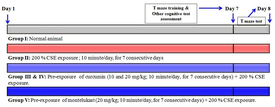 Figure 1.
Experimental design for the evaluation of curcumin in CSE induced changes of cognition.
Zebrafishes were engaged in different test apparatus to evaluate the neurocognitive functions after the CSE exposure. The cognitive functions were evaluated based on color recognition test; partition preference test; horizontal compartment tests; and T-Maze tests. All the animals were acclimatized for 5 minutes in all behavioral test apparatus to minimize the error. The animal movements were tracked by USB camera associated with the software controlled in-house computer (12 Megapixel USB Camera, Intex, India).
Color recognition test is one of the methods to evaluate the spatial memory function. This test was performed as per the descriptive method of Dubey et al. (42) with a minor modification of Rishitha and Muthuraman (43). Briefly, the test chamber (20L x 10W x 10H, cm; Length, Width, and Height respectively) was divided into two equal parts. One part of the chamber (10L x 10W x 10H, cm) was covered with red color glass and it was considered as the punishment chamber. Another half of the chamber was covered with green colored glass, and it was considered as the reward-chamber. The water level in the chamber was maintained up to 5 cm. The cognitive functions were evaluated by assessing the “Time spent in the green chamber (TSGC)” and “Number of entries into the red chamber (NERC)” by animals. When the animals were placed at the center of the chamber; if the animals preferred to stay at the green chamber, it indicated the improvement of memory function. If the animal did not prefer the green chamber, it indicated the poor memory function or lack of memory.
The partition preference test is another method for the assessment of working and spatial memory function. This test was performed as per the descriptive method of Dubey et al. (42) with a minor modification of Rishitha and Muthuraman (43). Briefly, the water level in chamber (20L x 10W x 10H; cm) was maintained up to the 3 cm; and the chamber was divided into two equal (10L x 10W x 10H; cm) parts with a vertical glass slide movement and the gap of 1 cm was maintained from the bottom of the inner glass chamber. The right side of the chamber was considered as home chamber; whereas the left side of the chamber was considered as a target chamber. The cognitive function was evaluated by the assessment of (i) number of the entries into the target chamber (ii) the time spent in the target chamber (TSTC). The number of entry was calculated as “Percentage entry into the target chamber” (% ETC). When the animals were placed in the home chamber; the animals preferred to reach the target chamber. If the animals were not entered into a home chamber within 60 seconds of the time period; the animals were guided to reach the target chamber and kept in same target chamber for a further 20 seconds by closing the entry gate with a glass slide. The preference of both chambers indicates the enhanced memory function. The preference of home chamber indicates the lack of memory function.
The horizontal compartment test is one of the methods for the assessment of neurocognitive function. It was performed as per the described method of Dubey et al. (42) with a minor modification of Rishitha and Muthuraman (43). Briefly, the water level in the chamber (20L x 10W x 24H; cm) was maintained up to the 21 cm; and the chamber was divided into three horizontal (7 x 7 x 7; cm height) compartments by marking a line in the outer chamber. One day before the experiment, a separate training was given (120 seconds) to all the fishes to swim in all the compartments; if the animals did not prefer all the compartments, the animals were guided with a guiding tool and food pellets. In the next day, the cognitive functions were evaluated by the assessment of “time spent in the upper segment” (TSUS); and “time spent in the lower segment” (TSLS). Generally, when the animals were placed in the test chamber; it preferred to swim in the upper segment of the chamber within 15 seconds. It indicated that the animals had a normal or improved memory function. If the animal did not prefer the upper segment or animals were swimming in the middle/lower segment of the chamber, it indicated the loss of memory.
The T-maze test is one of the well-established methods for the assessment of neurocognitive functions of rodents. Several research groups modified the T-maze test apparatus in order to assess the neurocognitive functions. In the present study, the T-maze test has shown reproducible result in zebrafishes. T-Maze test in zebrafishes was performed as per the described method of Colwill et al. (44) with a minor modification of Muthuraman and Rishitha (45). Briefly, the T maze apparatus consists of two short arms (10Lx 6W x 10H; cm) with the different colors (one arm with red color glass; and another end arm with green color glass). One long arm (20L x 10W x 10H; cm) was covered with the home chamber (5L x 6W x 10H; cm); and it was made-up with a normal non-transparent glass. The green color arm was employed as a favorable zone with the reward of a food pellet, and the red color arm was employed as a punishment zone by a string with a glass rod. One day before the test assessment; all the animals were allowed to learn the T-maze environment; and all the animals were allowed to enter the green chamber. If the animals did not learn, animals were guided to reach the green chamber. In the next day, the animals were placed in the corner of long arm i.e., starting point from home chamber and the target point was identified as an entry to any one of the short arm. The starting point and target point were separated by a vertical slide control panel. Each fish was exposed to 2 minutes for the assessment of learning and memory. The transfer latency (TL) and percentage target (green) chamber preference (% TCP) were noted for the assessment of neurocognitive function.
After the assessment of behavioral parameters, the zebrafish brain samples were isolated immediately by the microsurgical method and the brain samples were freeze-dried at -4 ˚C. In the next day, all the samples were homogenized with the solution of phosphate buffer. The supernatant of the samples was collected by a centrifugation at 1372 g-force for 15 minutes. The supernatant of zebrafish brain samples was used for the estimation of tissue biomarker changes, i.e., acetylcholinesterase (AChE) activity; lipid peroxidation (LPO); reduced glutathione (GSH); and total protein levels.
The AChE activity level in zebrafish brain samples was estimated by a spectrophotometric method, as described by Ellman et al. (46) with a minor modification of Rishitha and Muthuraman (43). Briefly, 500 µl of the supernatant of the zebrafish brain was mixed with 0.25 ml of DTNB (0.001 M) and incubated for 10 minutes. The formation of yellow color chromogen products and its intensity were assessed with the observation of changes in absorbance by using a spectrophotometer (DU 640B Spectrophotometer, Beckman Coulter Inc., CA, USA) at 420 nm wavelength. This absorbance changes with respect to the change in acetylthiocholine hydrolyzed product by AChE. Further, the AChE activity levels were calculated by using the following formula i.e., R = Δ O.D X Volume of the assay (3 ml) / ε X mg of protein. R represents the rate of enzyme activity in ‘n’ mole of acetylthiocholine iodide hydrolyzed per minute per mg of protein. The symbol of Δ O.D. represents a change of absorbance per minute. The symbol epsilon (ε) represents the extinction coefficient i.e., 13600 per mole per centimeter. The results were expressed as micromole (μM) of acetylthiocholine hydrolyzed per milligram of protein per minute.
The lipid peroxidation (LPO) level of zebrafish brain samples was estimated by a spectrophotometric method, as per the described method of Ohkawa et al. (47) with a minor modification of Rishitha and Muthuraman (43). Briefly, 0.2 ml of supernatant of zebrafish brain tissue was mixed with 0.2 ml of 8.1 % w/v of sodium dodecyl sulfate (SDS), 1.5. ml of 30 % v/v of acetic acid (pH 3.5.), 1.5 ml of 0.8 % w/v of thiobarbituric acid and the volume was made up to 4 ml of distilled water. The reaction mixture was incubated at 95˚C for 1 hour. Then, the reaction mixture was rapidly cooled with tap water. Further, 1 ml of distilled water and 5 ml of n-butanol-pyridine (15:1 v/v) was added. After 10 minutes, the tubes were centrifuged at 1372 g-force for 15 min. The formation of pink color products and its color intensity were assessed by using spectrophotometer (DU 640B Spectrophotometer, Beckman Coulter Inc., CA, USA) at 535 nm wavelength. These absorbance readings were used for the further calculation of LPO levels. The results were expressed as nanomole (nM) per mg of protein.
The GSH level of zebrafish brain samples was estimated by a spectrophotometric method as per the described method of Ellman (48) with a minor modification of Rishitha and Muthuraman (43). Briefly, 0.5 ml of supernatant was mixed with 2 ml of disodium hydrogen phosphate solution (0.3. M) and 2.5 ml of freshly prepared DTNB solution (0.001 M). The formation of yellow color chromogen products and its color intensity were assessed by using a spectrophotometer (DU 640B Spectrophotometer, Beckman Coulter Inc., CA, USA) at 412 nm wavelength. These readings were used for further calculation of GSH levels in tissue samples. The results were expressed as micromole (μM) of GSH per mg of protein.
The total protein level of zebrafish brain samples was estimated by a spectrophotometric method as per the described method of Lowry et al. (49) with a minor modification of Rishitha and Muthuraman (43). Briefly, 300 µl of zebrafish brain supernatant was diluted with distilled water up to 1 ml. Further, 5 ml of Lowry’s reagent was added and the mixture was allowed to stand for a further 15 minutes at room temperature (37 ˚C). Then, 0.5 ml of Folin-Ciocalteu reagent was added slowly and vortexed vigorously at room temperature for 30 min. The formation of purple color chromogen products and its color intensity were assessed by using spectrophotometer (DU 640B, UV-Spectrophotometer, Beckman Coulter Inc., CA, USA) at 750 nm wavelength. These readings were used for further calculation of total protein levels in tissue samples. The results were expressed as mg of protein per ml of supernatant.
All the results were expressed as the mean ± standard deviation (SD). Data obtained from all behavior tests and tissue biomarkers were statistically analyzed using one-way analysis of variance (ANOVA). Further, Tukey’s test was applied for Post-hoc analysis using Graph pad prism Version-5.0 software. A probability value of p < 0.05 was considered to be statistically significant.
The exposure of CSE produced the significant (p < 0.0.5.) decrement in TSGC and the significant increment in NERC values in comparison to a normal control group as an indication of cognitive impairment. The pre-exposure of curcumin (10 and 20 mg/kg) has shown to produce the neuroprotective action against CSE exposure induced cognitive impairment in a dose-dependent manner. Furthermore, the pre-exposure of the reference compound, i.e., montelukast (20 mg/kg) produced the significant ameliorative action against CSE induced cognitive impairment. It indicates that the pre-exposure of curcumin produces the neuroprotective action against CSE induced toxicity. The results are illustrated in Figure 2 (A and B).
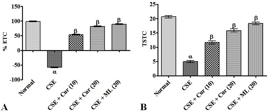 Figure 2.
Effect of curcumin in CSE induced changes of a color recognition test. Digits in parenthesis indicate dose mg/kg. Data were expressed as mean ± SD, n = 6 zebrafish per group. αp < 0.05 Vs normal group. βp < 0.05 Vs CSE exposure group. Abbreviation: CSE, cigarette smoke extract; Cur, curcumin; ML, montelukast; and TSGS, time spent in the green chamber; NERC, number of the entry to the red chamber.
The exposure of CSE produced the significant (p < 0.0.5.) decrement in the percentage of ETC and TSTC in comparison to a normal control group as an indication of cognitive impairment. The pre-exposure of curcumin (10 and 20 mg/kg) has shown to produce the neuroprotective action against CSE exposure induced cognitive impairment in a dose-dependent manner. Furthermore, the pre-exposure of the reference compound, i.e., montelukast (20 mg/kg) produced the significant ameliorative action against CSE induced cognitive impairment. It indicates that the pre-exposure of curcumin produces the neuroprotective action against CSE induced toxicity. The results are illustrated in Figure 3 (A and B).
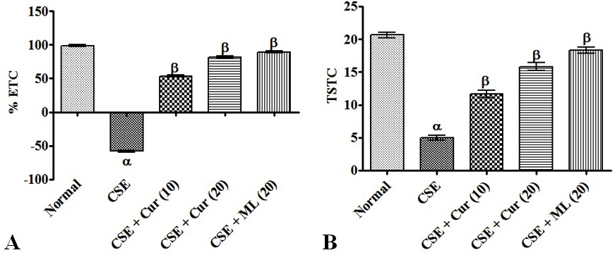 Figure 3.
Effect of curcumin in CSE induced changes of partition preference test. Digits in parenthesis indicate dose mg/kg. Data were expressed as mean ± SD, n = 6 zebrafish per group. αp < 0.05 Vs normal group. βp < 0.05 Vs CSE exposure group. Abbreviation: CSE, cigarette smoke extract; Cur, curcumin; ML, montelukast; % ETC, percentage entry to the target chamber; and TSTC, time spent in the target chamber.
The exposure of CSE produced the significant (p < 0.0.5.) decrement in TSUS and the significant increment in TSLS values in comparison to a normal control group as an indication of cognitive impairment. The pre-exposure of curcumin (10 and 20 mg/kg) has shown to produce the neuroprotective action against CSE exposure induced cognitive impairment in a dose-dependent manner. Furthermore, the pre-exposure of the reference compound, i.e., montelukast (20 mg/kg) produced the significant ameliorative action against CSE induced cognitive impairment. It indicates that the pre-exposure of curcumin produces the neuroprotective action against CSE induced toxicity. The results are illustrated in Figure 4 (A and B).
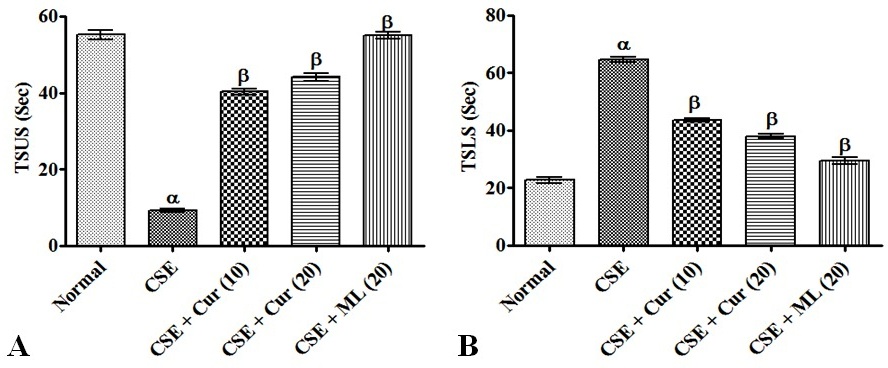 Figure 4.
Effect of curcumin in CSE induced changes of horizontal compartment test. Digits in parenthesis indicate dose mg/kg. Data were expressed as mean ± SD, n = 6 zebrafish per group. αp < 0.05 Vs normal group. βp < 0.05 Vs CSE exposure group. Abbreviation: CSE, cigarette smoke extract; Cur, curcumin; ML, montelukast; TSUS, time spent in the upper segment; and TSLS, time spent in the lower segment.
The exposure of CSE produced the significant (p < 0.0.5.) increment in TL values and the significant decrement in the percentage of TCP values in comparison to a normal control group as an indication of cognitive impairment. The pre-exposure of curcumin (10 and 20 mg/kg) has shown to produce the neuroprotective action against CSE exposure induced cognitive impairment in a dose-dependent manner. Furthermore, the pre-exposure of the reference compound, i.e., montelukast (20 mg/kg) produced the significant ameliorative action against CSE induced cognitive impairment. It indicates that the pre-exposure of curcumin produces the neuroprotective action against CSE induced toxicity. The results are illustrated in Figure 5 (A and B).
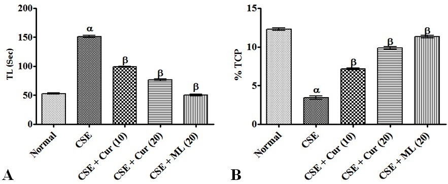 Figure 5.
Effect of curcumin in CSE induced changes of T-Maze tests. Digits in parenthesis indicate dose mg/kg. Data were expressed as mean ± SD, n = 6 zebrafish per group. αp < 0.05 Vs normal group. βp < 0.05 Vs CSE exposure group. Abbreviation: CSE, cigarette smoke extract; Cur, curcumin; ML, montelukast; TL, transfer latency; and % TCP, percentage target (green) chamber preference.
The exposure of CSE produced the significant (p < 0.0.5.) rise of brain AChE activity and LPO; and the significant decrement in the GSH level when compared to a normal control group. The pre-exposure of curcumin (10 and 20 mg/kg) has attenuated the CSE exposure induced biochemical changes in a dose-dependent manner. Further, the pre-exposure of the reference compound, i.e., montelukast (20 mg/kg) produced the significant ameliorative effect against CSE induced tissue biomarker changes in zebrafish brain samples. It indicates that the pre-exposure of curcumin produces the neuroprotective action against CSE induced cognitive dysfunction via free radical scavenging; anti-lipid peroxidative; and reduction of acetylcholinesterase activity. The results were presented in Table 1.
| Groups | AChE activity |
GSH |
LPO |
|---|---|---|---|
| Normal | 17.2 ± 1.72 | 16.6 ± 2.17 | 4.9 ± 1.02 |
| CSE | 51.3 ± 1.05 α | 5.8 ± 1.02 α | 13.8 ± 1.17 α |
| CSE + Cur (10) | 32.9 ± 0.98 β | 11.3 ± 1.52 β | 10.2 ± 0.69 β |
| CSE + Cur (20) | 23.4 ± 1.16 β | 13.5 ± 1.09 β | 6.8 ± 0.72 β |
| CSE + ML (10) | 18.9 ± 1.28 β | 15.6 ± 1.39 β | 5.2 ± 0.53 β |
| Digits in parenthesis indicate dose mg/kg. Data were expressed as mean ± SD, n = 6 zebrafish per group. αp < 0.0.5. Vs normal group. βp < 0.0.5. Vs CSE exposure group. Abbreviation: CSE, cigarette smoke extract; Cur, curcumin; ML, montelukast; AChE, acetylcholine esterase; GSH, reduced glutathione; and LPO, lipid peroxidation. | |||
The present study revealed the ameliorating effect of curcumin against CSE exposure induced cognitive impairment. The pre-exposure of curcumin indicated the various neurocognitive behavioral changes such as (i) decreased TSGC values and increased NERC values in color recognition test (Figure 2A and 2B) (ii) decreased percentage of ETC and TSTC values in partition preference test (fig 3A and 3B) (iii) decreased TSUS values and increased TSLS values in horizontal compartment test (Figure 4A and 4B) (v) increased TL and decreased percentage of TCP values in T-Maze tests (Figure 5A and 5B). Further, the pre-treatment of curcumin produces the potential attenuation of CSE induced biomarkers changes in the zebrafish brain. These results are statistically compared with the reference compound i.e., montelukast.
CSE consists of numerous toxic ingredients and it alters the physiological process of the body. The primary events of CSE have reported that it causes (i) the potent activation of free radicals and immune cells leading to initiating the oxidative stress; (ii) mitochondrial dysfunction and imbalance in cellular oxidant and anti-oxidant levels (32, 34). The endogenous anti-oxidant (reduced glutathione) enhances the free radical scavenging and reduces the free radical synthesis (48). Toxic materials reduce the glutathione synthesis; and enhance the high glutathione utilization via alteration of free radical synthesis and scavenging pathways. The present study also shows similar results, i.e., CSE exposure has shown to decrease the GSH levels. Further, the free radical accumulation and/or generation of radicals are responsible to modulate the cell membrane integrity via activation of lipid peroxidation process (50). Experimentally, it can be detected with thiobarbituric acid reaction mechanism. Because, the intermediate product of lipid peroxidation i.e., malondialdehyde (MDA) readily reacts with thiobarbituric acid and generates pink color chromogen (47). Lipid peroxidation products were detected by spectrophotometrically. The results have shown that CSE enhances the lipid peroxidation process (35). Later, it also alters the mitochondrial membrane peroxidation and produces the opening of mitochondrial permeability transition pore (MPTP) (51). In nuclear levels, it alters the nucleic acids mutation and produces cancer, tumor and other ailments (52).
CSE contents possess the higher affinity to various tissue proteins such as lung, kidney, liver, heart including nervous system (52). In the brain, it alters the neuroendocrine and neurotransmitter functions leading to change of the neurobehavioral pattern (34). In addition, the chronic exposure of CSE is known to cause the neurocognitive dysfunction via the alteration of cholinergic neurotransmitter i.e., acetylcholine (53). Acetylcholine is degraded by two enzymes i.e., acetylcholinesterase and butyrylcholinesterase. The degradation of acetylcholine leads to a reduction in the availability of acetylcholine content and enhances the cognitive dysfunction (54). In vitro acetylcholinesterase activity was estimated by using acetylthiocholine (as a substrate) and Ellman reagents (46). The results reveal that CSE challenge enhances the acetylcholinesterase activity in zebrafish brain (43, 45). These results produced the evidence for CSE challenge induced neurocognitive impairment. In addition, montelukast is also produced similar results and it is an established neuroprotective agent (38-39). The data in hand and other previous literature report and other research laboratory reports collectively support that the curcumin possesses the potential ameliorative effects in cognitive function against CSE exposure in zebrafishes. The neuroprotective mechanism of curcumin against CSE induced neurodegeneration and cognitive dysfunctions were illustrated in Figure 6.
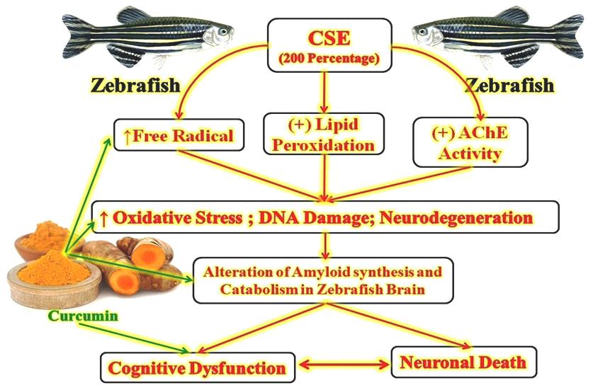 Figure 6.
The neuroprotective mechanism of curcumin in CSE induced neurodegeneration and cognitive dysfunction. Abbreviation: CSE, cigarette smoke extract; and AChE, acetylcholinesterase.
Hence, it may be concluded that curcumin may collectively act as a neuroprotective and nootropic agent against CSE associated neurocognitive dysfunction and neurotoxicity due to its potential pleiotropic action; i.e., free radical scavenging; anti-lipid peroxidative; anti-inflammatory and neurotransmitter regulatory actions.
The authors are thankful to the JSS Academy of Higher Education and Research, JSS College of Pharmacy, Mysuru -570 015, Karnataka (India) for their unconditional support and for providing technical facilities to carry out this research work.