[2]Abbas N, Kim TH. Cubic Cu2O-decorated B-doped g-C3N4 nanosheets for ultrasensitive electrochemical detection of metol. Journal of Environmental Chemical Engineering. 2025; 13: 118489. https://doi.org/10.1016/j.jece.2025.118489.
[4]Ostovar A, Tarkhani M, Mousavi SA, Asadollahi M. Surface modification of GO/PVDF composite membranes using MXene/protonated g-C3N4 nanosheet hybrids with enhanced dye separation and antifouling properties. Polymer Engineering & Science. 2024; 64: 565–576. https://doi.org/10.1002/pen.26566.
[5]Zhang R, Wang L, An C, Zhang S, Zhang Y, Wang Y, et al. Precursor-reforming strategy to hierarchical porous g-C3N4 with nitrogen defects for enhanced visible-light-driven hydrogen evolution. International Journal of Hydrogen Energy. 2024; 84: 731–738. https://doi.org/10.1016/j.ijhydene.2024.08.236.
[6]Lee JH, Jeong SY, Son YD, Lee SW. Facile Fabrication of TiO2 Quantum Dots-Anchored g-C3N4 Nanosheets as 0D/2D Heterojunction Nanocomposite for Accelerating Solar-Driven Photocatalysis. Nanomaterials. 2023; 13: 1565. https://doi.org/10.3390/nano13091565.
[7]Mathur N, Pachar K, Ankita A, Yadav A, Roy P, Gupta P. Interfacial electrochemistry of graphitic carbon nitride nanosheets and polymelamine nanofilms toward integrated sensing and energy platforms. Electrochimica Acta. 2025; 11: 147131. https://doi.org/10.1016/j.electacta.2025.147131.
[8]Amasegowda A, Alkanad K, Al-Zaqri N, Al-khawlani A, Kumar AU, Al-Maswari BM, et al. Enhanced photocatalytic H2 production over p–n S-scheme heterojunctions of Ni3V2O8 quantum dots decorated on ultrathin g-C3N4 nanosheets. Journal of Environmental Chemical Engineering. 2024; 12: 111841. https://doi.org/10.1016/j.jece.2023.111841.
[9]Morsy A, Toderas M, El‐marghany A, Rana D, Abdelaty M, Shokry F, et al. Oxygen/g-C3N4 smart nanocoatings for sustainable maintenance and sacrificial anode protection of mild steel. Journal of Chemical Technology & Biotechnology. 2025; 100: 187–201. https://doi.org/10.1002/jctb.7764.
[10]Iqbal S, Liu J. S-scheme synergies in Au nanorod-infused Fe2O3 quantum dots/g-C3N4 hybrids for visible-light-driven water splitting. Journal of Environmental Chemical Engineering. 2024; 12: 114866. https://doi.org/10.1016/j.jece.2024.114866.
[11]Hajizadeh-Oghaz M, Heydari G. Synthesis and characterization of sulfur-doped g-C3N4 nanosheets for enhanced photocatalytic removal of organic pollutants. Journal of Advanced Materials in Engineering. 2025; 45: 33–48. https://doi.org/10.47176/jame.45.1.1132.
[12]Chen M, Nie X, Gao W, Shang N, Zhou X, Gao S. Visible-light-driven selective C–H bond oxidation using lead-free Cs2AgBiBr6/g-C3N4 perovskite composites. Inorganic Chemistry Communications. 2025; 182: 115445. https://doi.org/10.1016/j.inoche.2025.115445.
[14]Nejat R, Zandi S. Visible-light-responsive La0.₇Sr0.3MnO3@TiO2/g-C3N4 nanocomposites for photocatalytic antibiotic degradation and bioactivity applications. Journal of Alloys and Compounds. 2025; 1036: 181866. https://doi.org/10.1016/j.jallcom.2025.181866.
[16]Wang Y, Wang X, Antonietti M. Polymeric graphitic carbon nitride as a heterogeneous organocatalyst: From photochemistry to multipurpose catalysis. Angewandte Chemie International Edition. 2012; 51: 68–89. https://doi.org/10.1002/anie.201101182.
[19]Zhang Y, Liu J, Wu G, Chen W. Effect of gamma irradiation on the structure and photocatalytic performance of g-C3N4. Radiation Physics and Chemistry. 2016; 122: 1–7. https://doi.org/10.1016/j.radphyschem.2016.01.021.
[20]Dong F, Zhao Z, Xiong T, Ni Z, Zhang W, Sun Y, et al. In situ construction of g-C₃N₄/g-C heterojunction for enhanced visible-light photocatalytic activity toward organic pollutants degradation. ACS Applied Materials & Interfaces. 2013; 5: 11392–11401. https://doi.org/10.1021/am403653a.
[21]Kralchevska R, Milanova M, Tsvetkov M, Dimitrov D, Todorovsky D. Influence of gamma irradiation on the photocatalytic activity of Degussa P25 TiO2. Journal of Materials Science. 2012; 47: 4936–4945. https://doi.org/10.1007/s10853-012-6368-4.
[23]Samet L, March K, Brun N, Hosni F, Stephan O, Chtourou R. Effect of gamma radiation on the photocatalytic properties of Cu-doped TiO2 nanoparticles. Materials Research Bulletin. 2018; 107: 1–7. https://doi.org/10.1016/j.materresbull.2018.07.004.
[24]Zhang Q, Jiang ZW, Wang MZ, Ge XW. Gamma-ray irradiation effects on Bi2WO6 photocatalysts. Chinese Journal of Chemical Physics. 2018; 31: 701–706. https://doi.org/10.1063/1674-0068/31/cjcp1805094.
[25]Bourezgui A, Kacem I, Daoudi M, Al-Hossainy AF. Influence of gamma irradiation on the structural, optical, and photocatalytic properties of TiO2 nanoparticles under controlled atmospheres. Journal of Electronic Materials. 2020; 49: 1904–1921. https://doi.org/10.1007/s11664-019-07887-z.
[26]Jeya P, Keerthana SP, Kungumadevi L, Yuvakkumar R, Ravi G, Kandasami A, et al. Gamma irradiation effect on photocatalytic properties of Cu and Sr ions codoped PbS. Environmental Research. 2023; 226: 115651. https://doi.org/10.1016/j.envres.2023.115651.
[27]Bisht R, Joshi GC, Singh JP, Joshi CS. Investigation of the effect of gamma-ray irradiation on SnO2 nanoparticles for photocatalysis application. Nuclear Instruments and Methods in Physics Research Section B: Beam Interactions with Materials and Atoms. 2025; 568: 165862. https://doi.org/10.1016/j.nimb.2025.165862.
[28]Katubi KM, Alsafari IA, Shaheen B, Boukhris I, Al Buriahi MS, Shakir I, et al. Gamma irradiated Co3O4 for outstanding photocatalytic activity toward removal of pharmaceutical contaminants present in wastewater. Journal of the Korean Physical Society. 2026; 88: 72–86. https://doi.org/10.1007/s40042-025-01487-3.
[30]Nefzi C, Yahmadi B, El Guesmi N, Kamoun-Turki N, Ahmed SA. A successful exploitation of gamma-radiation on chalcogenide Cu2InSnS4 towards clean water under photocatalysis approach. Journal of Molecular Structure. 2022; 1251: 131943. https://doi.org/10.1016/j.molstruc.2021.131943.
[32]Xu J, Zhang L, Shi R, Zhu Y. Chemical exfoliation of graphitic carbon nitride for efficient heterogeneous photocatalysis. Journal of Materials Chemistry A. 2013; 1: 14766–14772. https://doi.org/10.1039/C3TA13188B.
[33]El-Sayed ESR, Abdelhakim HK, Ahmed AS. Solid-state fermentation for enhanced production of selenium nanoparticles by gamma-irradiated Monascus purpureus and their biological evaluation and photocatalytic activities. Bioprocess and Biosystems Engineering. 2020; 43: 797–809. https://doi.org/10.1007/s00449-019-02275-7.
[34]Di Valentin C, Pacchioni G, Selloni A. Origin of the different photoactivity of N-doped anatase and rutile TiO₂. Physical Review B. 2004; 70: 085116. https://doi.org/10.1103/PhysRevB.70.085116.
[36]Bissenova M, Idrissov N, Kuspanov Z, Umirzakov A, Daulbayev C. Hybrid adsorption–photocatalysis composites: a sustainable route for efficient water purification. Materials for Renewable and Sustainable Energy. 2025; 14: 44. https://doi.org/10.1007/s40243-025-00319-5.
[37]Budimir M, Marković Z, Jovanović D, Vujisić M, Mičušík M, Danko M, et al. Gamma ray assisted modification of carbon quantum dot/polyurethane nanocomposites: structural, mechanical and photocatalytic study. RSC Advances. 2019; 9: 6278–6286. https://doi.org/10.1039/c9ra00500e.
[38]Saleh MR, El-Gendy RA, Bakier YM, El-Bery HM. Modulating g-C3N4 photocatalyst for H2 production via water splitting: The impact of Schiff base incorporation. Journal of Environmental Chemical Engineering. 2024; 12: 113866. https://doi.org/10.1016/j.jece.2024.113866.
[40]Fang Z, Zhou Y, Yang Z, Yang C, Zhang J, Hou Y. V2O5-assisted thermal oxidation strategy for synthesizing porous carbon nitride with enhanced photocatalytic NO removal performance. Surfaces and Interfaces. 2025; 60: 106023. https://doi.org/10.1016/j.surfin.2025.106023.
[41]González-Vázquez MY, Carrillo-Martínez CJ, Pinedo-Escobar JA, Escalante-García IL, Martínez AS, González-Rodríguez LM, et al. Photocatalytic Degradation of Methyl Orange and Metoprolol by Novel Ternary Photocatalyst TiO2-ZnO/g-C3N4 under UV and Visible Light. 2024. https://doi.org/10.20944/preprints202410.2481.v1. (preprint)
Graphitic carbon nitride (g-C3N4) has attracted sustained interest as a visible-light-driven photocatalyst; however, its practical efficiency is often constrained by limited surface accessibility and rapid recombination of photogenerated charge carriers. Herein, g-C3N4 nanosheets were prepared via an environmentally benign electrochemical exfoliation route and subsequently modified through controlled gamma irradiation (γ-irradiation) in the dose range of 0–50 kGy. Structural analysis revealed that the crystalline framework of g-C3N4 remained preserved after irradiation, while the average crystallite size decreased from ~24.6 to ~18.4 nm, accompanied by an increase in lattice microstrain from 2.05 × 10⁻³ to 2.68 × 10⁻³. Optical characterization demonstrated dose-dependent tuning of the band structure, with direct band gap energies shifting from ~3.0 to ~3.2 eV and indirect transition energies ranging from ~2.4 to ~2.7 eV. Photoluminescence (PL) and time-resolved measurements indicated a pronounced suppression of non-radiative recombination at moderate irradiation levels, with the carrier lifetime reaching a maximum of ~4.6 ns at ~25 kGy. In parallel, γ-irradiation induced a measurable enhancement in surface area and pore characteristics. These combined effects resulted in a marked improvement in visible-light photocatalytic degradation of methylene blue (MB), following pseudo-first-order kinetics. The results demonstrate that γ-irradiation provides an effective and contamination-free means of optimizing the structural and photophysical properties of g-C3N4 nanosheets for photocatalytic applications.
Keywords
g-C3N4 nanosheets
γ-irradiation
electrochemical exfoliation
photocatalysis
methylene blue degradation
charge-carrier dynamics
1. Introduction
Graphitic carbon nitride (g-C3N4) has emerged as a promising
metal-free photocatalyst owing to its suitable band gap, chemical stability, and
facile synthesis from earth-abundant precursors. Its ability to absorb visible
light renders it particularly attractive for photocatalytic environmental
remediation. Nevertheless, bulk g-C3N4 commonly exhibits strong
interlayer stacking, low specific surface area, and inefficient charge-carrier
separation, all of which substantially limit its photocatalytic performance
[1, 2, 3, 4].
Considerable efforts have therefore been devoted to overcoming these intrinsic
limitations through structural modification strategies such as nano-structuring,
defect engineering, and exfoliation into two-dimensional architectures [5, 6, 7].
Among these approaches, exfoliation into nanosheets is especially effective in
enhancing surface exposure and shortening charge-transport pathways. However,
exfoliation alone often offers limited control over defect type and density,
parameters that critically govern electronic structure and charge-carrier
dynamics [8, 9, 10].
High-energy gamma irradiation (-irradiation) has recently been recognized as a versatile
post-synthetic tool capable of inducing controlled lattice distortions and defect
states without introducing chemical impurities. Through precise dose regulation,
-irradiation enables fine modulation of optical and electronic
properties, potentially improving light absorption and suppressing charge
recombination [11, 12, 13]. Despite this potential, systematic investigations
combining electrochemical exfoliation with -irradiation for
g-C3N4 nanosheets remain limited, particularly with respect to
establishing clear correlations between irradiation dose, microstructural
evolution, and photocatalytic activity [14, 15].
Accordingly, the objective of the present work is to address this gap by
developing -irradiation-engineered g-C3N4 nanosheets via a
green electrochemical exfoliation approach and to elucidate, in a systematic
manner, the dose-dependent effects of -irradiation on their structural,
optical, textural, and photocatalytic properties. By integrating comprehensive
characterization with photocatalytic performance evaluation, this study aims to
provide deeper insight into structure–property–activity relationships in
irradiated g-C3N4 systems.
2. Materials and Methods
2.1 Materials
All experiments were performed using a standard electrochemical cell coupled
with a regulated direct current (DC) power supply (PHYWE DC Power Supply 0–12 V/5 A, PHYWE
Systeme GmbH & Co. KG, Göttingen, Germany). Sulfuric acid (H2SO4,
95%–98%, Merck, Cat. No. 100731 Darmstadt, Germany) was employed as the
electrolyte at various concentrations. Graphite electrodes prepared from recycled
carbon sources (spent batteries) and g-C3N4 electrodes synthesized from
analytical-grade urea (CO(NH2)2, 99%: Sigma-Aldrich, Cat. No. U5128, St.
Louis, MO, USA) were used as the working electrodes.
2.1.1 Novel Green Synthesis of g-C3N4 Nanosheets
g-C3N4 was synthesized using analytical-grade urea
(CO(NH2)2, 99% purity) as the precursor. In a typical
synthesis, 10 g of urea was placed in a covered alumina crucible and thermally
treated in a programmable muffle furnace (Nabertherm LHT 04/17, Nabertherm GmbH,
Lilienthal, Lower Saxony, Germany) at temperatures ranging from 400 °C to 650
°C for 3 h in ambient air, employing a heating rate of 5 °C
min-1. During the thermal polymerization process, urea decomposed into
gaseous by-products such as NH3 and CO2, resulting in the formation of
a yellow solid corresponding to bulk g-C3N4, as illustrated in Fig. 1.
The obtained material was collected, finely ground, and stored in airtight
containers for subsequent use. Exfoliated g-C3N4 nanosheets were
prepared via a simple and efficient electrochemical exfoliation approach. The
electrochemical cell comprised two electrodes: a graphite electrode fabricated
from recycled carbon sources (e.g., spent batteries) and a g-C3N4
electrode synthesized from urea. An acidic electrolyte, typically H2SO4
at various concentrations, was employed, and a DC power supply delivering 10–12
V was applied to promote interlayer expansion and exfoliation of
g-C3N4, as shown in Fig. 1. Upon completion of the exfoliation process,
the resulting suspension was allowed to stand until complete sedimentation of the
solid product. The precipitate was then collected, repeatedly washed with
deionized water, and dried in a laboratory oven (Memmert UN55, Memmert GmbH,
Schwabach, Bavaria, Germany) at 50–60 °C to obtain fine
g-C3N4 nanosheet powder. To evaluate the influence of post-thermal
treatment, the exfoliated nanosheets were subsequently annealed at temperatures
between 400 °C and 650 °C under controlled conditions [16, 17, 18]. This
annealing process enabled a systematic investigation of the effects of heat
treatment on the crystallinity, stacking order, and physicochemical properties of
the g-C3N4 nanosheets, as discussed in the Results and Discussion
section.
Fig. 1.
Schematic illustration of the synthesis of g-C3N4
nanosheets from Urea using a novel method. g-C3N4, Graphitic carbon
nitride.
2.1.2 -Irradiation Treatment of g-C3N4
Nanosheets
The g-C3N4 nanosheets were subjected to -irradiation using a
60Co source at the National Center for Radiation Research and Technology
(NCRRT), Cairo, Egypt [19, 20]. Irradiation was performed at room temperature
under ambient atmospheric conditions, with absorbed doses ranging from 5 to 50
kGy. The dose rate was accurately calibrated prior to each irradiation run to
ensure uniform energy delivery to the samples. During exposure, the nanosheets
were sealed in quartz vials to prevent contamination and to preserve their
structural integrity. This controlled -irradiation process was designed
to induce defect states, modify surface characteristics, and promote structural
rearrangements within the g-C3N4 framework. Systematic variation of the
irradiation dose enabled a reliable evaluation of dose-dependent changes in
crystallinity, electronic structure, optical properties, and photocatalytic
performance, as discussed in the Results and Discussion section.
2.2 Characterization Tools
2.2.1 XRD Technique
X-ray diffraction (XRD) measurements were employed to investigate the
crystallographic structure and irradiation-induced microstructural changes in
bulk g-C3N4 and exfoliated g-C3N4 nanosheets. The diffraction
patterns were recorded using (D8 Advance, Bruker AXS GmbH, Karlsruhe, Germany, Cu
K radiation, = 1.5406 Å) over a 2 range of
5°–90°. Structural parameters were quantitatively evaluated using the
most intense (002) reflection located at approximately 2 27.4°, which is associated with the interlayer stacking of the
conjugated g-C3N4 framework. The crystallite size (D) was estimated
using the Scherrer Eqn. 1 [21, 22]:
(1)
where is the wavelength, is the full width at half maximum
(FWHM) of the (002) peak expressed in radians, and is the Bragg angle.
The lattice microstrain () was calculated following the
Williamson–Hall approximation [6, 22]:
(2)
The dislocation density (), which reflects the defect concentration
within the crystalline domains, was determined using the relation [22, 23]:
(3)
The derived parameters should be regarded as approximate values, as instrumental
broadening effects were not explicitly subtracted. This approach is widely
adopted for comparative studies aimed at elucidating relative structural changes
induced by external treatments such as -irradiation. In addition, the
XRD data were subjected to Rietveld refinement to extract detailed
microstructural parameters using the MAUD software package (version 2.94,
developed by University of Trento, Trento, Italy). Complementary peak analysis
and phase identification were performed with X’Pert HighScore Plus software
(version 4.9, Malvern Panalytical B.V., Almelo, The Netherlands) where required
[9, 10].
2.2.2 Microscopic Characterization of Bulk g-C3N4 and
Nanosheets
The morphology and size of the synthesized nanostructures were examined using a
high-resolution scanning electron microscope (SEM, JEOL JSM-IT200, Tokyo, Japan)
operated at an accelerating voltage of 25 kV. The structural features and
dimensional characteristics of bulk g-C3N4 and its exfoliated
nanosheets were further investigated by high-resolution transmission electron
microscopy (HRTEM, JEOL 3010, JEOL Ltd., Tokyo, Japan) operated at 200 kV [11].
Additional morphological analysis and uniformity assessment were performed using
a ZEISS EVO-MA10 scanning electron microscope equipped with an energy-dispersive
X-ray spectroscopy (EDX) detector (Carl Zeiss Microscopy GmbH, Oberkochen,
Baden-Württemberg, Germany), enabling simultaneous evaluation of surface
morphology and elemental composition. SEM images were processed using ImageJ
software (version 1.53, National Institutes of Health, Bethesda, MD,
USA) to determine particle size distributions based on statistical analysis of
more than 100 particles per sample. Elemental composition obtained from EDX
measurements was analyzed using the instrument-integrated AZtecEnergy EDX
analysis software (version 3.3, Oxford Instruments NanoAnalysis Ltd., High
Wycombe, Buckinghamshire, UK) [11, 12].
2.2.3 Raman Spectroscopy Test
Raman spectra were recorded using an inVia Raman spectrometer (Renishaw plc,
Gloucestershire, UK) equipped with a thermoelectrically cooled charge-coupled device (CCD)
detector maintained at –80 °C. A He–Ne laser with an excitation
wavelength of 532 nm was employed, with the laser power restricted to 1 mW to
prevent sample heating. The integration time for each measurement was set to 100
s.
2.2.4 UV–Vis Diffuse Reflectance Spectroscopy
The optical absorption properties of the prepared g-C3N4 nanosheets
were examined by ultraviolet–visible diffuse reflectance spectroscopy (UV–Vis
DRS) using a Shimadzu UV-2600 UV–Vis spectrophotometer (Shimadzu Corporation,
Kyoto, Japan) equipped with an integrating sphere accessory (ISR-2600Plus). The
measurements were carried out at room temperature over the wavelength range of
200–800 nm. Barium sulfate (BaSO4) was employed as a non-absorbing
reflectance standard, and all spectra were collected under identical conditions
to ensure reliable comparison among samples subjected to different
-irradiation doses. The obtained diffuse reflectance data were
converted to the corresponding absorption spectra using the Kubelka–Munk
function (Eqn. 4) [12, 13]:
(4)
where R is the measured reflectance. The optical band gap energies were
estimated by constructing Tauc plots based on the Kubelka–Munk-transformed data,
assuming both direct and indirect electronic transitions for g-C3N4.
Linear extrapolation of the absorption edge to the energy axis was used to
determine the band gap values. All DRS measurements were repeated to confirm
reproducibility. This characterization provides essential information on
irradiation-induced changes in light absorption behavior and electronic structure
of the g-C3N4 nanosheets.
2.2.5 BET Surface Area and Porosity Analysis
The specific surface area and pore structure of the prepared g-C3N4
nanosheets were evaluated by nitrogen adsorption–desorption measurements using a
Micromeritics ASAP 2020 surface area and porosity analyzer (Micromeritics
Instrument Corporation, Norcross, GA, USA). Prior to analysis, all samples were
degassed under vacuum at 150 °C for 12 h to remove physically adsorbed
moisture and residual gases [12, 13, 14]. Nitrogen adsorption–desorption isotherms
were recorded at 77 K over a relative pressure (P/P0) range of 0.01–0.99. The
specific surface area was calculated using the Brunauer–Emmett–Teller (BET)
method within the relative pressure range of 0.05–0.30, where a linear BET plot
was obtained. The total pore volume was estimated from the amount of nitrogen
adsorbed at a relative pressure close to unity (P/P0 0.99). Pore size
distribution curves were derived from the adsorption branch of the isotherms
using the Barrett–Joyner–Halenda (BJH) method, assuming cylindrical pore
geometry [11, 13, 15]. All measurements were conducted under identical experimental
conditions to ensure meaningful comparison among samples subjected to different
-irradiation doses. The BET analysis provides insight into
irradiation-induced changes in surface area, pore volume, and pore size
distribution, which are key parameters influencing adsorption capacity and
photocatalytic performance.
2.2.6 Photoluminescence Characterization
Photoluminescence (PL) spectroscopy was employed to investigate the electronic
structure, defect-related states, and charge-carrier recombination behavior of
the prepared g-C3N4 nanosheets before and after -irradiation.
Steady-state PL measurements were carried out at room temperature using an
Edinburgh Instruments FLS1000 fluorescence spectrometer (Edinburgh Instruments
Ltd., Livingston, Scotland, UK) equipped with a continuous-wave xenon lamp as the
excitation source. The excitation wavelength was fixed at 350 nm, corresponding
to the intrinsic absorption of g-C3N4, while the emission spectra were
recorded over the wavelength range of 380–650 nm [12, 16]. All spectra were
collected under identical experimental conditions to ensure reliable comparison
among samples subjected to different -irradiation doses. Time-resolved
photoluminescence (TRPL) measurements were performed using the same system
operated in time-correlated single-photon counting (TCSPC) mode. A pulsed diode
laser with an excitation wavelength of 375 nm was employed, and the emission
decay profiles were monitored at the dominant PL emission wavelength of each
sample. The obtained decay curves were fitted using a multi-exponential decay
model to extract the average lifetime and individual decay components, which
provide insight into the relative contributions of radiative recombination and
defect-assisted non-radiative processes [17, 18]. All PL and TRPL measurements
were repeated to confirm reproducibility. The extracted lifetimes and emission
intensities were analyzed comparatively to evaluate the influence of
-irradiation dose on charge-carrier recombination dynamics. This PL
characterization approach enables a reliable assessment of irradiation-induced
defect states and their role in modulating the photophysical properties of
g-C3N4 nanosheets.
2.2.7 Photocatalytic Degradation Analysis
The photocatalytic activity of the synthesized g-C3N4 nanosheets was
evaluated through the degradation of methylene blue (MB) under visible-light
irradiation. A 300 W xenon lamp equipped with a cutoff filter (420
nm) was employed as the light source to simulate visible solar irradiation. In a
typical photocatalytic experiment, 50 mg of the photocatalyst was dispersed in
100 mL of an aqueous MB solution with an initial concentration of 10 mg L-1
under continuous magnetic stirring to ensure homogeneous suspension.
Prior to light exposure, the reaction mixture was stirred in the dark for 30 min
to establish adsorption–desorption equilibrium between MB molecules and the
catalyst surface. Upon visible-light irradiation, aliquots of 3 mL were withdrawn
at regular intervals of 20 min, centrifuged to remove suspended catalyst
particles, and subsequently analyzed using a UV–Vis spectrophotometer. The
residual concentration of MB was determined by monitoring the characteristic
absorption maximum at 664 nm [19, 20, 21]. The photocatalytic degradation efficiency
(D%) was calculated using the following Eqn. 5:
(5)
where C0 and Ct represent the MB concentrations at the initial time
and at irradiation time t, respectively. The degradation kinetics were
analyzed using a pseudo-first-order kinetic model, expressed as Eqn. 6 [16]:
(6)
where k (min-1) denotes the apparent first-order rate constant. This
kinetic approach enables a quantitative comparison of the photocatalytic
performances of g-C3N4 samples prepared under different synthesis
temperatures and -irradiation doses, while providing a consistent
framework for evaluating their relative activity, stability, and reusability
under visible-light irradiation.
To gain insight into the reactive species involved in the photocatalytic
degradation process, scavenger experiments were conducted under otherwise
identical experimental conditions. Isopropyl alcohol (IPA, 10 mM, 99.5%,
Cat. No. 278475, Sigma-Aldrich, St. Louis, MO, USA), p-benzoquinone (BQ, 1 mM,
98%, Cat. No. B10358, Sigma-Aldrich, St. Louis, MO, USA), and
ethylenediaminetetraacetic acid (EDTA, 2 mM, 99%, Cat. No. E5134,
Sigma-Aldrich, St. Louis, MO, USA) were employed as selective quenchers for
hydroxyl radicals (•OH), superoxide radicals (•O2–), and
photogenerated holes (h+), respectively. The scavengers were added to the
reaction system after the adsorption–desorption equilibrium was reached and
before visible-light irradiation. The resulting changes in photocatalytic
degradation efficiency were analyzed to elucidate the relative contributions of
the different reactive species to the MB degradation process [13, 16, 17].
3. Results
3.1 XRD Analysis of Bulk and Exfoliated g-C3N4 Under
-Irradiation
Fig. 2a and Fig. 2b present the XRD patterns of bulk g-C3N4 and
exfoliated g-C3N4 nanosheets synthesized at 550 °C and
subjected to -irradiation doses ranging from 0 to 50 kGy. In both
materials, the characteristic diffraction peaks located at approximately
13.1° and 27.4° are retained, corresponding to the (100)
in-plane structural periodicity of tri-s-triazine units and the (002) interlayer
stacking of the conjugated aromatic framework, respectively. Quantitative
evaluation of the diffraction data was performed using the (002) reflection to
estimate the crystallite size, lattice microstrain, and dislocation density
according to Eqns. 1,2,3, with the calculated values summarized in Table 1 for
bulk g-C3N4 and Table 2 for exfoliated g-C3N4 nanosheets.
Fig. 2.
XRD patterns of g-C3N4 materials under -irradiation.
(a) XRD patterns of bulk g-C3N4 prepared at 550 °C under
different -irradiation doses. (b) XRD patterns of g-C3N4
nanosheet prepared at 550 °C under different -irradiation
doses. XRD, X-ray diffraction. -irradiation, gamma irradiation.
Table 1.
Crystallite size, microstrain, and dislocation density of bulk
g-C3N4 derived from XRD analysis at different -irradiation
doses.
Dose (kGy)
D (nm)
(10–3)
(1015 m–2)
0
~39.8
1.21
0.63
10
~36.5
1.33
0.75
20
~33.9
1.46
0.87
30
~31.6
1.58
1.00
40
~30.2
1.65
1.10
50
~28.9
1.72
1.20
Table 2.
Crystallite size, microstrain, and dislocation density of
exfoliated g-C3N4 derived from XRD analysis at different
-irradiation doses.
Dose (kGy)
D (nm)
(10–3)
(1015 m–2)
0
~24.6
2.05
1.65
10
~22.9
2.18
1.91
20
~21.4
2.31
2.18
30
~20.1
2.45
2.47
40
~19.2
2.57
2.71
50
~18.4
2.68
2.95
As shown in Table 1, the apparent crystallite size of bulk g-C3N4
decreases gradually with increasing irradiation dose, while the corresponding
microstrain and dislocation density increase systematically. This trend indicates
progressive accumulation of irradiation-induced lattice distortions and defect
sites, without evidence of phase transformation or long-range structural
disruption, as further supported by the absence of peak shifts or secondary
diffraction features in Fig. 2a. In the case of exfoliated g-C3N4
nanosheets (Fig. 2b and Table 2), the calculated crystallite sizes are
consistently smaller and the microstrain values higher than those of the bulk
material at all irradiation doses, reflecting reduced interlayer coherence
associated with the exfoliation process.
With increasing -irradiation dose, both microstrain and dislocation
density show a further incremental increase, while the characteristic
g-C3N4 diffraction peaks remain preserved, confirming that the
fundamental crystalline framework is maintained up to 50 kGy [3, 15, 22]. Overall,
the trends observed in Tables 1,2 are in good agreement with the peak-broadening
behavior evident in Fig. 2a,b. The combined analysis based on Eqns. 1,2,3
demonstrates that -irradiation primarily affects microstructural
parameters such as crystallite size, lattice strain, and defect density, while
preserving the long-range crystallographic structure of both bulk and exfoliated
g-C3N4.
3.2 Vibrational Features of g-C3N4 Nanosheets Under
-Irradiation
Fig. 3 illustrates the Fourier-transform infrared spectroscopy (FTIR) spectra of
exfoliated g-C3N4 nanosheets synthesized at 550 °C and
subjected to -irradiation doses ranging from 0 to 50 kGy. All spectra
exhibit the characteristic vibrational features of graphitic carbon nitride,
confirming that the fundamental chemical framework of the material is preserved
following irradiation.
Fig. 3.
FTIR spectra of g-C3N4 nanosheets prepared at 550
°C under different -irradiation doses. FTIR,
Fourier-transform infrared spectroscopy.
The broad absorption band observed in the region of 3000–3400 cm-1 is
attributed to stretching vibrations of N–H and O–H groups, which may originate
from terminal amine functionalities and adsorbed moisture on the nanosheet
surface [23, 24]. The group of intense bands located between approximately 1200
and 1650 cm-1 corresponds to the stretching modes of C–N and C=N bonds
within the heterocyclic tri-s-triazine units, which constitute the backbone of
the g-C3N4 structure. In addition, the distinct band centered near 810
cm-1 is assigned to the breathing mode of the tri-s-triazine ring, serving
as a fingerprint vibration of polymeric g-C3N4. With increasing
-irradiation dose, no new absorption bands are detected, and no
disappearance of existing peaks is observed, indicating the absence of chemical
decomposition or phase transformation. However, subtle variations in band
intensity and slight broadening of selected vibrational modes can be discerned at
higher doses, suggesting localized structural disorder or modification of surface
functional groups induced by irradiation. These changes are consistent with
irradiation-induced defect formation and bond distortion rather than alterations
to the primary chemical structure. Overall, the FTIR results demonstrate that
-irradiation up to 50 kGy does not disrupt the intrinsic molecular
architecture of exfoliated g-C3N4 nanosheets, while inducing minor
modifications in local bonding environments.
3.3 Effect of -Irradiation on the Raman Spectra of
g-C3N4 Nanosheets
Fig. 4 illustrates the Raman spectra of g-C3N4 nanosheets synthesized
at 550 °C and subjected to -irradiation doses ranging from 0
to 50 kGy. All spectra exhibit the characteristic vibrational features of
graphitic carbon nitride, confirming that the fundamental molecular framework is
preserved irrespective of irradiation dose. The prominent bands observed in the
region between ~700 and 1700 cm-1 are attributed to the
breathing modes of tri-s-triazine units and the stretching vibrations of
conjugated C–N heterocycles, which are hallmarks of the g-C3N4
structure. Notably, the overall spectral profiles remain largely unchanged with
increasing irradiation dose, indicating that gamma exposure does not induce
significant chemical transformation or phase alteration within the detection
limits of Raman spectroscopy [19, 20]. However, a gradual variation in band
intensity and slight peak broadening can be discerned at higher doses,
particularly above 30 kGy. These changes may be indicative of irradiation-induced
lattice disorder, localized bond distortion, or an increased defect population
within the polymeric network. Such effects are consistent with microstructural
perturbations rather than long-range structural reorganization. Importantly, no
new Raman bands or peak shifts associated with secondary phases or bond cleavage
are detected, underscoring the structural robustness of g-C3N4
nanosheets under -irradiation up to 50 kGy. It should be emphasized
that Raman spectroscopy primarily provides qualitative insight into vibrational
and bonding environments; therefore, the observed spectral variations are
interpreted as indicative trends rather than definitive evidence of defect
formation. Overall, the Raman results support the conclusion that
-irradiation predominantly introduces subtle microstructural
modifications while preserving the intrinsic framework of g-C3N4
nanosheets [25].
Fig. 4.
Raman spectra of g-C3N4 nanosheets prepared
at 550 °C under different -irradiation doses.
3.4 Elemental Composition Analysis of Bulk and Exfoliated
g-C3N4
EDX elemental mapping was employed to examine the bulk compositional uniformity
of g-C3N4 before and after exfoliation, as shown in Fig. 5a,b. It
should be noted that EDX provides semi-quantitative elemental information from a
near-surface bulk region and is therefore used here to assess overall elemental
distribution rather than surface-specific chemistry. The EDX maps of bulk
g-C3N4 (Fig. 5a) reveal a homogeneous and continuous distribution of
carbon and nitrogen across the analyzed area, with no evidence of elemental
segregation. Quantitative analysis indicates atomic percentages of approximately
43.0 at.% C, 55.0 at.% N, and 2.0 at.% O, consistent with the expected
stoichiometry of polymeric g-C3N4 and indicative of a chemically stable
framework. In comparison, the exfoliated g-C3N4 nanosheets (Fig. 5b)
exhibit spatially overlapping C and N signals localized within thinner,
flake-like regions, reflecting the nanosheet morphology produced by the
exfoliation process [6, 9].
Fig. 5.
SEM images and EDX elemental mapping of (a) bulk
g-C3N4 and (b) exfoliated g-C3N4 nanosheets, showing the
distribution of C (K), N (K), and O (K) and their
corresponding atomic percentages. Both samples exhibit uniform bulk elemental
distribution and preserved stoichiometry, with no detectable impurities after
exfoliation. SEM, scanning electron microscope; EDX, energy-dispersive X-ray
spectroscopy. Scale bar = 100 nm.
The corresponding elemental composition comprises 48.0 at.% C, 49.0 at.% N,
and a slightly increased oxygen content of 3.0 at.%. The modest increase in
oxygen is plausibly attributed to surface-exposed edge sites or mild oxidation
during exfoliation, rather than the incorporation of extraneous phases.
Importantly, no additional elemental signals are detected in either sample,
confirming the high purity of the materials.
Overall, the comparative EDX results demonstrate that exfoliation preserves the
intrinsic C–N framework and bulk stoichiometry of g-C3N4, while
inducing morphological thinning without compromising compositional integrity—an
essential prerequisite for maintaining reliable physicochemical and
photocatalytic performance.
3.5 SEM Micrographs of Bulk g-C3N4 and Exfoliated
g-C3N4 Nanosheets
The morphological characteristics of bulk and exfoliated g-C3N4 were
examined by field-emission scanning electron microscopy (FESEM), as shown in Fig. 6a,c present FESEM images of bulk g-C3N4 recorded at low and high
magnifications, respectively, revealing densely packed, irregular agglomerates
composed of thick, stacked plate-like structures with lateral dimensions
predominantly in the sub-micron range. This compact morphology indicates strong
interlayer interactions and limited exposure of accessible surface sites. In
contrast, Fig. 6b,d display FESEM images of exfoliated g-C3N4
nanosheets at corresponding magnifications. As observed in Fig. 6b, the bulk
agglomerates are transformed into loosely stacked, sheet-like assemblies with
reduced lateral dimensions, while the higher-magnification image in Fig. 6d
clearly reveals thinner, delaminated layers with smoother surfaces. Although the
lateral sizes of the exfoliated sheets remain within the sub-micron regime, the
evident delamination and structural thinning relative to the bulk material
confirm the effectiveness of the exfoliation process. Such morphological
evolution is expected to enhance surface accessibility and improve interfacial
contact with reactant molecules, which is advantageous for photocatalytic
applications, without invoking strict size-based classification as nanomaterials
[26].
Fig. 6.
FESEM images of g-C3N4 showing (a,c) bulk material
with stacked, dense morphology and (b,d) exfoliated nanosheets with thin, layered
structures at low and high magnifications. FESEM, field-emission scanning
electron microscopy. Fig. 6a scale bar = 1 µm; Fig. 6b scale bar = 300 nm; Fig. 6c scale bar
= 500 nm; Fig. 6d scale bar = 100 nm.
3.6 Structural Evolution of Bulk g-C3N4 Into Exfoliated
Nanosheets
Fig. 7 presents transmission electron microscopy (TEM) images illustrating the
morphological differences between bulk g-C3N4 and exfoliated
g-C3N4 nanosheets at various magnifications. In Fig. 7a, the bulk
g-C3N4 appears as a compact, nearly spherical agglomerate composed of
densely packed layers, indicating strong interlayer interactions and significant
material thickness. Fig. 7c further reveals the internal texture of the bulk
sample, where the dark contrast and limited transparency confirm the presence of
thick, highly stacked domains with restricted accessibility of surface sites. In
contrast, Fig. 7b shows the exfoliated g-C3N4 in the form of extended,
sheet-like structures with markedly enhanced transparency, reflecting a
pronounced reduction in thickness. The layered and partially overlapping nature
of the nanosheets is clearly visible, demonstrating successful delamination of
the bulk precursor. This observation is further supported by Fig. 7d, which
highlights ultrathin, flexible nanosheets with smooth surfaces and folded edges,
characteristic of two-dimensional architectures. Collectively, the TEM images in
Fig. 7 confirm the effective transformation of bulk g-C3N4 into
ultrathin nanosheets, accompanied by increased structural openness and surface
exposure, which are advantageous for charge transport and surface-dominated
processes [27].
Fig. 7.
TEM images of g-C3N4. (a,c) Bulk g-C3N4
showing dense and aggregated morphology at different magnifications. (b,d)
Exfoliated g-C3N4 nanosheets exhibiting thin, transparent, and layered
sheet-like structures. TEM, transmission electron microscopy. Fig. 7a scale
bar = 500 nm.; Fig. 7b scale bar = 300 nm; Fig. 7c scale bar = 200 nm; Fig. 7d scale bar = 100 nm.
3.7 Optical Properties and Band Gap Analysis of
-Irradiated g-C3N4 Nanosheets
The optical properties of g-C3N4 nanosheets synthesized at 550
°C and exposed to -irradiation doses ranging from 0 to 50 kGy
were systematically investigated using UV–Vis DRS, absorption analysis,
Kubelka–Munk transformation, and Tauc plot evaluation. The combined results
demonstrate that -irradiation induces subtle yet measurable
modifications in the optical band structure of g-C3N4 while preserving
its intrinsic semiconducting framework [28]. Diffuse reflectance and absorption
spectra reveal slight shifts in the absorption edge with increasing irradiation
dose, suggesting defect-assisted modulation of electronic transitions. Further
analysis using the Kubelka–Munk function confirms irradiation-induced variations
in optical response, which can be attributed to localized structural distortions
and enhanced exfoliation effects within the nanosheet architecture. Band gap
values derived from Tauc plots indicate the presence of both direct (3.0–3.2 eV)
and indirect (2.4–2.8 eV) optical transitions, with irradiated samples
exhibiting modest upward shifts relative to the pristine material. These
controlled changes in band gap energy suggest that -irradiation
provides an effective means of tuning the light-harvesting characteristics of
g-C3N4 nanosheets without compromising their structural integrity.
Overall, the results establish -irradiation as a versatile strategy for
engineering the optical and electronic properties of g-C3N4, which is
particularly advantageous for applications in photocatalysis, solar energy
conversion, and optoelectronic devices where optimized band alignment is critical
for performance [29].
3.7.1 Optical Reflection Behavior of -Irradiated
g-C3N4 Nanosheets
Fig. 8 presents the UV–Vis DRS of g-C3N4 nanosheets synthesized at
550 °C and subjected to varying -irradiation doses (0–50
kGy). All samples exhibit a strong absorption response in the visible region,
characteristic of the intrinsic electronic structure of g-C3N4. The
absorption edge is located around the blue-green region of the spectrum,
confirming the suitability of the material for visible-light-driven applications.
Fig. 8.
DRS of g-C3N4 nanosheets prepared at 550
°C under different -irradiation doses. DRS, diffuse
reflectance spectroscopy.
With increasing -irradiation dose, a subtle but systematic shift in
the absorption edge is observed, accompanied by minor variations in reflectance
intensity. These changes suggest irradiation-induced modifications in the
electronic structure, which may originate from defect formation, lattice
distortion, or altered charge distribution within the g-C3N4 framework.
Importantly, the overall spectral profile remains well preserved across all
irradiation doses, indicating that the fundamental optical characteristics of
g-C3N4 nanosheets are retained despite irradiation treatment. The
controlled tuning of optical absorption through -irradiation highlights
an effective strategy for tailoring the electronic and light-harvesting
properties of g-C3N4 nanosheets without compromising their structural
integrity, which is advantageous for photocatalytic and optoelectronic
applications.
3.7.2 Kubelka–Munk Function Analysis of -Irradiated
g-C3N4 Nanosheets
The optical band structure of -irradiated g-C3N4 nanosheets
was evaluated using the Kubelka–Munk function derived from the UV–Vis DRS. The
reflectance data were transformed according to the Kubelka–Munk formalism,
enabling a reliable assessment of the absorption behavior and band gap evolution
of the material. As shown in Fig. 9, all samples exhibit a well-defined
absorption edge in the visible region, confirming the semiconducting nature of
g-C3N4 nanosheets.
Fig. 9.
Effect of -irradiation on the Kubelka–Munk
function of g-C3N4 nanosheets.
With increasing -irradiation dose, a gradual shift of the absorption
edge toward longer wavelengths is observed, accompanied by subtle variations in
reflectance intensity. This behavior indicates irradiation-induced modification
of the electronic structure, which can be reasonably attributed to the formation
of localized defect states and lattice perturbations within the g-C3N4
framework. Importantly, the overall spectral profile remains largely unchanged
across the investigated dose range, suggesting that -irradiation
induces controlled electronic tuning without causing significant structural
degradation. Such moderate band structure modulation is advantageous for
enhancing visible-light absorption and improving charge carrier utilization,
which are critical factors governing the photocatalytic performance of
g-C3N4-based materials. The optical band structure of
-irradiated g-C3N4 nanosheets was further evaluated using the
Kubelka–Munk function derived from UV–Vis DRS. The reflectance data were
converted to the corresponding absorption coefficient using the Kubelka–Munk
equation (Eqn. 4), which allows reliable estimation of the optical band gap for
semiconducting materials [20, 30].
3.7.3 Effect of -Irradiation on the Optical Absorption
of g-C3N4 Nanosheets
The UV–Vis absorption spectra of -irradiated g-C3N4
nanosheets, derived from diffuse reflectance measurements, are presented in Fig. 10. All samples exhibit strong absorption in the near-UV and visible regions,
with a well-defined absorption edge characteristic of the semiconducting nature
of g-C3N4. As the -irradiation dose increases, a gradual red
shift of the absorption edge is observed, together with slight changes in
absorbance intensity.
Fig. 10.
UV–Vis absorption spectra of g-C3N4 nanosheets
prepared at 550 °C under different -irradiation doses. UV–Vis, ultraviolet–visible.
These spectral variations indicate irradiation-induced modification of the
electronic structure, which can be attributed to the formation of localized
defect states and subtle lattice distortions within the g-C3N4
framework. Importantly, the overall absorption profile remains largely preserved
across the investigated dose range, suggesting that -irradiation
enables controlled tuning of the optical properties without causing significant
structural damage. Such moderate enhancement of visible-light absorption is
beneficial for improving photoexcited charge generation and is expected to
contribute positively to the photocatalytic performance of g-C3N4
nanosheets [15, 31].
3.7.4 Direct Band Gap Analysis of g-C3N4 Nanosheets
Under -Irradiation
The optical band gap of g-C3N4 nanosheets synthesized at 550
°C was evaluated using Tauc plots derived from DRS, as shown in Fig. 11,
for samples subjected to -irradiation doses ranging from 0 to 50 kGy.
The well-defined linear regions of the Tauc plots indicate that the optical
absorption is governed predominantly by direct electronic transitions, from which
band gap energies in the range of approximately 3.0–3.2 eV were determined. The
pristine g-C3N4 nanosheets exhibit a band gap of about 3.0 eV, whereas
a gradual increase to nearly 3.2 eV is observed with increasing
-irradiation dose, evidencing a subtle blue shift in the absorption
edge.
Fig. 11.
Tauc plot analysis for direct band gap determination of
-irradiated g-C3N4 nanosheets.
This slight widening of the band gap can be ascribed to irradiation-induced
structural distortions and the formation of defect states, which alter the local
electronic environment and influence optical transition energies. Importantly,
despite these irradiation-induced modifications, the intrinsic semiconducting
framework of g-C3N4 remains largely preserved, highlighting its
structural and electronic robustness under high-dose -irradiation. The
controlled modulation of the band gap via -irradiation thus represents
a viable strategy for fine-tuning the optical properties of g-C3N4
nanosheets for advanced photocatalytic and optoelectronic applications [28].
3.7.5 Indirect Band Gap Analysis of g-C3N4 Nanosheets
Under -Irradiation
Fig. 12 presents the Tauc plots used to evaluate the indirect optical band gap
of g-C3N4 nanosheets synthesized at 550 °C and exposed to
-irradiation doses ranging from 0 to 50 kGy. The indirect band gap
energies were determined by extrapolating the linear regions of the
(h)1/2 versus photon energy plots. The pristine
g-C3N4 nanosheets exhibit an indirect band gap of approximately 2.45
eV, while the irradiated samples show a gradual increase to values in the range
of 2.4–2.7 eV with increasing irradiation dose. These modest upward shifts in
band gap energy suggest that -irradiation induces localized structural
distortions and defect states that subtly influence the electronic transition
pathways without significantly altering the intrinsic band structure of the
material. Importantly, the consistent position of the absorption edge across all
irradiation doses confirms the robust semiconducting nature and structural
stability of g-C3N4 under high-dose -irradiation. The
observed fine-tuning of the indirect band gap highlights -irradiation
as an effective and controllable strategy for tailoring the optical and
electronic properties of g-C3N4 nanosheets, which is particularly
beneficial for photocatalytic and energy conversion applications where optimized
band alignment is critical for performance [29].
Fig. 12.
Indirect band gap estimation of g-C3N4 nanosheets at
550 °C derived from Tauc plot analysis under varying
-irradiation doses.
3.8 Textural Properties of -Irradiated g-C3N4
Nanosheets
The textural characteristics of g-C3N4 nanosheets synthesized at 550
°C were systematically investigated as a function of
-irradiation dose using nitrogen adsorption–desorption measurements.
Key parameters, including specific surface area, total pore volume, and average
pore diameter, were analyzed to elucidate the influence of -irradiation
on the porous architecture of the nanosheets. -irradiation is known to
induce microstructural modifications through defect generation, partial
exfoliation, and disruption of interlayer interactions, which can collectively
alter the accessibility and distribution of surface and pore features. The
present results demonstrate that controlled -irradiation effectively
modulates the textural properties of g-C3N4 without compromising its
structural integrity. The irradiation-induced enhancement of surface area and
pore-related parameters is expected to facilitate improved mass transport and
increased exposure of catalytically active sites, thereby providing favorable
conditions for enhanced photocatalytic performance. Detailed discussions of the
individual contributions of surface area, pore volume, and pore diameter as
functions of irradiation dose are presented in the following subsections
(Sections 3.8.1–3.8.3).
3.8.1 Effect of -Irradiation on the Surface Area of
g-C3N4 Nanosheets
The variation in the BET specific surface area of g-C3N4 nanosheets
synthesized at 550 °C as a function of -irradiation dose is
presented in Fig. 13. The pristine sample exhibits a moderate surface area
characteristic of layered g-C3N4 materials. Upon
-irradiation, a gradual increase in surface area is observed with
increasing dose, reaching a maximum at intermediate irradiation levels before
showing a tendency toward saturation at higher doses. This behavior can be
attributed to irradiation-induced exfoliation and the generation of
microstructural defects, which promote partial delamination of stacked nanosheets
and increase the availability of accessible surface sites [30, 31]. At higher
irradiation doses, the stabilization of surface area suggests a balance between
exfoliation and defect recombination or partial structural relaxation.
Importantly, no abrupt loss in surface area is detected across the investigated
dose range, indicating that the overall framework of g-C3N4 remains
structurally robust under -irradiation. The irradiation-driven
enhancement in surface area is expected to be beneficial for photocatalytic
applications, as increased surface exposure facilitates improved adsorption of
reactant species and more efficient utilization of active sites during
photocatalytic reactions.
Fig. 13.
BET surface area of g-C3N4 nanosheets as a function
of -irradiation dose. BET, Brunauer–Emmett–Teller.
3.8.2 -Irradiation-Induced Modulation of Pore Volume in
g-C3N4 Nanosheets
Fig. 14 illustrates the variation in total pore volume of g-C3N4
nanosheets synthesized at 550 °C as a function of -irradiation
dose. The pristine sample exhibits a relatively low pore volume, consistent with
the dense stacking typically observed in bulk g-C3N4. Upon
-irradiation, a gradual increase in total pore volume is observed with
increasing dose, indicating irradiation-induced microstructural modification.
This enhancement is primarily attributed to partial exfoliation, defect
formation, and the creation of interlayer voids arising from the disruption of
van der Waals interactions between adjacent nanosheets. At higher irradiation
doses, the pore volume tends to stabilize, suggesting that a dynamic equilibrium
is reached between defect generation and structural relaxation. Importantly, no
abrupt collapse or degradation of the porous framework is detected, confirming
the structural resilience of g-C3N4 under -irradiation. The
observed increase in pore volume, together with the enhanced surface area, is
expected to facilitate improved mass transport and greater accessibility of
active sites, which are critical factors for enhancing photocatalytic efficiency
and reaction kinetics [29, 30].
Fig. 14.
Total pore volume of g-C3N4 nanosheets as a function
of -irradiation dose.
3.8.3 Impact of -Irradiation on the Average Pore
Diameter of g-C3N4 Nanosheets
Fig. 15 presents the variation in average pore diameter of g-C3N4
nanosheets synthesized at 550 °C as a function of -irradiation
dose. The pristine sample exhibits a relatively small average pore diameter,
reflecting the compact stacking and limited interlayer spacing characteristic of
non-irradiated g-C3N4. With increasing -irradiation dose, a
gradual enlargement of the average pore diameter is observed, indicating
progressive modification of the porous architecture. This behavior can be
attributed to irradiation-induced exfoliation and defect formation, which promote
partial delamination of the nanosheets and the expansion of interlayer voids. At
higher doses, the increase in pore diameter becomes less pronounced, suggesting
the establishment of a structural balance between defect generation and framework
stabilization [31]. Importantly, the absence of abrupt pore collapse or excessive
pore coalescence confirms the robustness of the g-C3N4 framework under
-irradiation. The moderate enlargement of pore diameter, combined with
enhanced surface area and pore volume, is expected to improve reactant diffusion
and active-site accessibility, thereby favorably influencing photocatalytic
reaction kinetics and overall performance.
Fig. 15.
Average pore diameter of g-C3N4 nanosheets as a
function of -irradiation dose.
3.9 PL Characteristics and Charge-Carrier Dynamics of
-Irradiated g-C3N4 Nanosheets
PL spectroscopy and TRPL measurements were employed to elucidate the influence
of -irradiation on the electronic structure, defect states, and
charge-carrier recombination dynamics of g-C3N4 nanosheets synthesized
at 550 °C. PL-based analyses provide critical insight into excitonic
behavior, radiative and non-radiative recombination pathways, and the role of
irradiation-induced defects in governing optical performance.
-irradiation is known to introduce lattice distortions and defect
states of varying depths, which can either enhance or suppress luminescence
depending on their nature and concentration. The present results reveal a strong
dose-dependent modulation of PL intensity, emission wavelength, and carrier
lifetime, highlighting a delicate balance between beneficial shallow defect
formation and detrimental deep-level trap generation [31, 32, 33]. Moderate
-irradiation effectively suppresses fast non-radiative recombination,
prolongs exciton lifetime, and enhances radiative emission, whereas excessive
irradiation induces defect saturation and structural disorder that accelerate
carrier quenching. These findings demonstrate that controlled
-irradiation offers a powerful strategy for tuning the photophysical
properties and charge-carrier dynamics of g-C3N4 nanosheets, with
direct implications for optimizing their performance in photocatalysis,
light-harvesting systems, and optoelectronic applications. Detailed discussions
of PL intensity evolution, lifetime behavior, decay kinetics, emission wavelength
shifts, PL quantum yield variation, Relative PL efficiency and recombination
suppression efficiency are provided in the following subsections (Sections
3.9.1–3.9.8).
3.9.1 Photoluminescence Response of g-C3N4 Nanosheets
Under Different Gamma Irradiation Doses
The PL spectra of g-C3N4 nanosheets exhibit a strong dependence on
-irradiation dose, as illustrated in Fig. 16. The pristine sample (0
kGy) shows a characteristic emission band centered at approximately 455 nm, which
gradually intensifies with increasing irradiation dose and reaches a maximum at
around 20–25 kGy. This enhancement is mainly attributed to the formation of
irradiation-induced shallow defect states, such as nitrogen vacancies and
localized lattice distortions, which act as temporary charge-carrier trapping
centers.
Fig. 16.
Normalized PL spectra of g-C3N4 nanosheets at varying
gamma irradiation doses. PL, Photoluminescence.
These shallow traps promote exciton localization and suppress rapid
non-radiative recombination, thereby increasing the probability of radiative
transitions. When the -irradiation dose exceeds 25 kGy, a pronounced
decrease in PL intensity is observed. This behavior indicates the onset of defect
saturation and the generation of deep trap states that function as efficient
non-radiative recombination centers [32, 33, 34]. Excessive irradiation may also
induce structural disorder within the conjugated g-C3N4 framework,
further accelerating carrier quenching and reducing emission efficiency. Overall,
these results demonstrate that controlled -irradiation provides an
effective means to tailor the electronic structure and excitonic recombination
behavior of g-C3N4 nanosheets, where an optimal balance between
beneficial shallow defect formation and structural preservation is essential for
maximizing optical performance.
3.9.2 Influence of Gamma Irradiation Dose on the Average Lifetime
of g-C3N4 Nanosheets
Fig. 17 presents the variation in the average PL lifetime of g-C3N4
nanosheets synthesized at 550 °C as a function of -irradiation
dose. The lifetime displays a clear non-monotonic behavior, initially increasing
at low irradiation doses and reaching a pronounced maximum of approximately 4.6
ns at 25 kGy. This enhancement indicates that moderate -irradiation
introduces a suitable density of shallow defect states, which act as temporary
charge-carrier traps and effectively suppress fast non-radiative recombination,
thereby prolonging carrier lifetime.
Fig. 17.
Variation of average lifetime of g-C3N4
nanosheets as a function of gamma irradiation dose.
In contrast, further increasing the irradiation dose beyond 25 kGy results in a
noticeable reduction in the average PL lifetime. This decrease is associated with
defect saturation and irradiation-induced structural disorder, leading to the
formation of deep trap states that facilitate rapid non-radiative recombination.
These observations highlight that controlled -irradiation plays a
crucial role in optimizing charge-carrier dynamics in g-C3N4
nanosheets, whereas excessive irradiation adversely affects their optical
performance and recombination behavior [32, 33].
3.9.3 Dose-Dependent Photoluminescence Intensity of
g-C3N4 Nanosheets
Fig. 18 illustrates the variation in PL peak intensity of g-C3N4
nanosheets synthesized at 550 °C as a function of -irradiation
dose. The PL intensity initially increases with increasing dose and reaches a
maximum at approximately 15–20 kGy. This improvement arises from
irradiation-induced shallow defect states, such as nitrogen vacancies and
localized lattice distortions, which enhance exciton localization and favor
radiative recombination by temporarily trapping charge carriers and suppressing
rapid non-radiative processes. In this dose range, defect generation remains well
controlled and does not significantly disrupt the conjugated heptazine framework,
resulting in improved electronic interactions and enhanced emission intensity.
With further increase in -irradiation dose beyond 20 kGy, the PL
intensity progressively decreases. This decline indicates the onset of defect
saturation and the formation of deep trap states that act as efficient
non-radiative recombination centers. Excessive irradiation may also induce
structural distortions within the g-C3N4 network, accelerating carrier
quenching and reducing luminescence efficiency. Overall, the observed
dose-dependent PL behavior reflects a critical balance between beneficial defect
generation and irradiation-induced structural damage, demonstrating that moderate
-irradiation optimizes the optical performance of g-C3N4
nanosheets, whereas higher doses lead to deterioration of their luminescence
properties.
Fig. 18.
Variation of PL peak intensity of g-C3N4
nanosheets with gamma irradiation dose.
3.9.4 Time-Resolved Photoluminescence Decay of g-C3N4
Nanosheets Under Gamma Irradiation
Fig. 19 presents the TRPL decay profiles of g-C3N4 nanosheets
synthesized at 550 °C and subjected to different -irradiation
doses [33]. All decay curves display a rapid initial drop in PL intensity
followed by a slower decay tail, which is characteristic of multi-exponential
recombination behavior involving both radiative and non-radiative processes. This
behavior reflects the coexistence of free exciton recombination and
defect-assisted carrier trapping within the g-C3N4 framework. At low to
moderate irradiation doses (5–25 kGy), the decay profiles become slightly
prolonged, indicating suppression of fast non-radiative recombination pathways.
This effect is attributed to the formation of irradiation-induced shallow defect
states that temporarily trap charge carriers, thereby extending exciton lifetime
and enhancing radiative recombination probability. In contrast, at higher
irradiation doses (30 kGy), the decay curves converge toward shorter
lifetimes, suggesting the emergence of deep-level trap states and increased
structural disorder. These deep traps act as efficient non-radiative
recombination centers, accelerating carrier quenching and diminishing
luminescence efficiency. Overall, the TRPL results confirm that moderate
-irradiation effectively optimizes charge carrier dynamics in
g-C3N4 nanosheets, whereas excessive irradiation induces unfavorable
defect states and structural distortions that compromise recombination behavior
and optical performance.
Fig. 19.
Normalized PL decay curves of g-C3N4
nanosheets at different gamma irradiation doses.
3.9.5 Gamma Irradiation-Dependent Evolution of 2
Lifetime in g-C3N4 Nanosheets
Fig. 20 shows the dependence of the longer decay component (2) of
g-C3N4 nanosheets synthesized at 550 °C on
-irradiation dose. The 2 lifetime increases gradually from
approximately 3.5 ns for the pristine sample to a maximum value of about 4.4 ns
at 25–30 kGy. This prolongation indicates effective suppression of non-radiative
recombination pathways and enhanced stabilization of photogenerated charge
carriers, which can be attributed to the controlled formation of
irradiation-induced shallow defect states that temporarily trap carriers and
delay recombination [34]. When the irradiation dose exceeds 30 kGy, the
2 lifetime decreases progressively. This reduction reflects the
accumulation of irradiation-induced structural disorder and the emergence of
deep-level trap states that act as efficient non-radiative recombination centers,
thereby accelerating carrier recombination. These results clearly demonstrate the
dual role of -irradiation in tailoring the photophysical properties of
g-C3N4 nanosheets and hence moderate irradiation improves exciton
lifetime and charge carrier dynamics, while excessive exposure leads to
deterioration of optical performance due to defect saturation and structural
damage.
Fig. 20.
Variation of 2 model lifetime of g-C3N4
nanosheets with gamma irradiation dose.
3.9.6 -Irradiation-Induced Shifts in Emission
Wavelength of g-C3N4 Nanosheets
Fig. 21 illustrates the evolution of the PL emission peak of g-C3N4
nanosheets synthesized at 550 °C as a function of -irradiation
dose. The pristine sample exhibits an emission maximum at approximately 469 nm,
whereas the irradiated nanosheets show a pronounced blue-shift to about 456 nm at
an irradiation dose of 30 kGy. This blue-shift originates from quantum
confinement effects induced by -irradiation, arising from nanosheet
thinning and localized lattice distortions, which modify the spatial confinement
of charge carriers. In addition, irradiation reduces the effective conjugation
length and alters defect-related energy levels through the formation of shallow
defect states, collectively leading to a widening of the effective bandgap and
emission at shorter wavelengths. At higher irradiation doses, a slight red-shift
is observed at 40 kGy, followed by a minor blue-shift at 50 kGy, indicating the
coexistence of competing effects related to partial defect passivation and
increasing structural disorder. Overall, these systematic variations in emission
wavelength demonstrate the strong sensitivity of the electronic structure of
g-C3N4 nanosheets to -irradiation and confirm that controlled
radiation exposure provides an effective approach for tuning their optical
properties [35].
Fig. 21.
Variation of PL peak wavelength of g-C3N4 nanosheets
with gamma irradiation dose.
3.9.7 Effect of -Irradiation on PL Quantum Yield and
Charge Carrier Recombination Behavior of g-C3N4 Nanosheets
The influence of -irradiation on the radiative recombination behavior
of g-C3N4 nanosheets was further evaluated through PL quantum yield
analysis (Fig. 22). The pristine sample exhibits the highest PL quantum yield,
approximately 0.32, indicating a relatively high probability of radiative
electron–hole recombination. Upon -irradiation, the PL quantum yield
decreases progressively with increasing dose, declining to about 0.29–0.26 at
intermediate doses and reaching a minimum value of approximately 0.18 at 50 kGy
[35, 36]. This monotonic reduction signifies an effective suppression of radiative
recombination pathways, which can be attributed to irradiation-induced structural
or electronic modifications, such as the formation of defect states or trap sites
that favor non-radiative charge transfer. At higher doses, the pronounced
decrease in PL quantum yield implies substantial inhibition of electron–hole
recombination, a feature generally considered beneficial for photocatalytic
performance. It should be emphasized that the reported PL quantum yield values
are discussed in a comparative sense to illustrate irradiation-induced trends
rather than absolute quantum efficiencies. Nonetheless, the clear dose-dependent
behavior demonstrates that controlled -irradiation provides an
effective strategy for tuning charge carrier dynamics in g-C3N4
nanosheets.
Fig. 22.
Variation of PL quantum yield of g-C3N4
nanosheets prepared at 550 °C as a function of -irradiation
dose.
3.9.8 Dose-Dependent PL Efficiency and Recombination Suppression
of -Irradiated g-C3N4 Nanosheets
Fig. 23 illustrates the effect of -irradiation on the relative PL
efficiency and PL suppression behavior of g-C3N4 nanosheets prepared at
550 °C. The relative PL efficiency, normalized to the pristine sample,
decreases gradually with increasing irradiation dose, indicating a progressive
reduction in radiative electron–hole recombination. This trend is accompanied by
a corresponding increase in PL suppression, which becomes particularly pronounced
at higher doses. Such behavior suggests that -irradiation induces
modifications in the electronic structure of g-C3N4 that favor charge
carrier separation, likely through the introduction of defect states or trapping
centers that facilitate non-radiative pathways. Importantly, these results are
discussed comparatively, reflecting relative changes in emission behavior rather
than absolute quantum efficiencies [37]. Nevertheless, the inverse relationship
between PL efficiency and PL suppression provides clear evidence that controlled
irradiation effectively modulates charge carrier recombination dynamics, a
feature that is generally advantageous for enhancing photocatalytic performance.
Fig. 23.
Relative PL efficiency and PL suppression of g-C3N4
nanosheets prepared at 550 °C as a function of -irradiation
dose.
3.10 Photocatalytic Degradation of MB Using g-C3N4
Nanosheets Under -Irradiation
This section examines the photocatalytic degradation behavior of methylene blue
(initial concentration of 10 mg L-1) over g-C3N4 nanosheets
synthesized at different calcination temperatures and subjected to varying
-irradiation doses. A systematic evaluation was conducted by monitoring
the time-dependent UV–Vis absorption spectra of MB, assessing the influence of
irradiation dose and synthesis temperature on degradation efficiency, analyzing
the associated photocatalytic kinetics, and examining both catalytic activity and
recyclability. The primary objective was to clarify how the combined effects of
synthesis conditions and -irradiation regulate the photocatalytic
performance of the prepared g-C3N4 nanosheets. The results demonstrate
that moderate -irradiation doses in the range of 10–25 kGy markedly
enhance MB degradation efficiency, which is attributed to the formation of
favorable defect states that promote charge separation and prolong the lifetime
of photogenerated charge carriers [36]. In contrast, exposure to higher
irradiation doses (40 kGy) leads to a noticeable deterioration in
photocatalytic performance, likely resulting from excessive structural disruption
and defect saturation. Synthesis temperature was also found to be a decisive
factor, with nanosheets prepared at 550–600 °C exhibiting significantly
higher degradation efficiencies than those synthesized at either lower or higher
temperatures. Furthermore, stability and recyclability tests reveal that
appropriately controlled -irradiation not only improves the initial
photocatalytic activity but also contributes to maintaining performance over
successive reaction cycles. Collectively, these findings underscore the
synergistic role of synthesis temperature and -irradiation in tailoring
the photocatalytic properties of g-C3N4 nanosheets for efficient and
durable MB degradation.
3.10.1 Time-Resolved UV–Vis Absorption of MB Using
g-C3N4 Nanosheets Without -Irradiation
Fig. 24 presents the time-dependent evolution of the UV–Vis absorption spectra
of MB during its photocatalytic degradation in the presence of g-C3N4
nanosheets synthesized at 550 °C under non-irradiated conditions. The
initial MB solution displays a strong absorption band centered at approximately
664 nm, along with a weaker shoulder around 610 nm, both of which are
characteristic of the electronic transitions of MB molecules. Upon visible-light
irradiation, a progressive decrease in the intensity of these absorption features
is observed as the reaction time increases from 0 to 120 minutes, indicating the
gradual degradation of the dye. The continuous attenuation of absorbance reflects
the effective photocatalytic activity of the g-C3N4 nanosheets. During
the process, photogenerated charge carriers participate in successive oxidation
and reduction reactions, leading to the breakdown of MB into smaller intermediate
species and, ultimately, to mineralized products. These results demonstrate the
intrinsic photocatalytic capability of non-irradiated g-C3N4 nanosheets
and provide a reliable reference point for evaluating the performance enhancement
achieved through gamma irradiation [36, 37].
Fig. 24.
UV–Vis absorption spectra of MB solution
recorded at different irradiation times in the presence of g-C3N4 nanosheets
prepared at 550 °C under different -irradiation doses. MB, methylene blue.
3.10.2 Effect of -Irradiation Dose on the
Photocatalytic Degradation of MB Over g-C3N4 Nanosheets
Fig. 25 illustrates the UV–Vis absorption spectra of the MB solution after 120
min of photocatalytic treatment over g-C3N4 nanosheets synthesized at
550 °C and exposed to different -irradiation doses. The
pristine MB solution exhibits its characteristic main absorption band centered at
approximately 664 nm, accompanied by a weaker shoulder around 610 nm, which are
typical signatures of the chromophoric structure of MB [36]. Upon photocatalytic
treatment in the presence of g-C3N4, a progressive attenuation of these
absorption features is observed, with the extent of intensity reduction strongly
dependent on the applied irradiation dose. In particular, nanosheets subjected to
moderate -irradiation doses in the range of 10–25 kGy show the most
pronounced decrease in MB absorbance, indicating enhanced photocatalytic
degradation efficiency. This improvement can be reasonably attributed to
irradiation-induced defect states that facilitate charge separation and increase
the availability of active sites without severely disrupting the material
framework. In contrast, samples exposed to higher irradiation doses (40
kGy) retain relatively stronger absorption bands after irradiation, suggesting a
decline in degradation efficiency, which is likely associated with defect
saturation and partial structural deterioration of the nanosheets. Overall, these
results demonstrate that controlled -irradiation plays a beneficial
role in promoting the photocatalytic degradation of MB, whereas excessive
irradiation adversely affects performance.
Fig. 25.
UV–Vis absorption spectra of MB degradation over
g-C3N4 nanosheets (550 °C) under different
-Irradiation at 120 min.
3.10.3 Effect of Preparation Temperature on the Photocatalytic
Degradation of MB in the Absence of -Irradiation
Fig. 26 depicts the UV–Vis absorption spectra of the MB solution after 120 min
of photocatalytic treatment using g-C3N4 nanosheets synthesized at
different preparation temperatures in the absence of -irradiation. The
residual MB solution exhibits a characteristic absorption band centered at
approximately 664 nm, accompanied by a weaker shoulder at lower wavelengths,
reflecting incomplete dye degradation under suboptimal catalytic conditions. A
clear dependence on synthesis temperature is evident from the variation in
absorbance intensity [22, 37]. Nanosheets prepared at 550 °C display the
lowest residual absorbance, indicating the highest photocatalytic degradation
efficiency among the examined samples. Samples synthesized at 600 °C
also show relatively strong degradation performance, albeit slightly inferior to
that of the 550 °C sample. In contrast, materials prepared at lower
temperatures (400–450 °C) retain higher absorbance intensities,
suggesting reduced photocatalytic activity, which can be reasonably attributed to
incomplete condensation and limited structural development of the
g-C3N4 framework. Meanwhile, the sample synthesized at 650 °C
exhibits diminished degradation efficiency, likely resulting from partial
structural deterioration and loss of active sites at excessive calcination
temperatures. Overall, these results demonstrate that synthesis temperature plays
a critical role in determining the photocatalytic performance of g-C3N4
nanosheets, with an optimal preparation window centered around 550 °C
under non-irradiated conditions.
Fig. 26.
UV–Vis absorption spectra of MB degradation over
g-C3N4 nanosheets synthesized at different temperatures without
-irradiation after 120 min.
3.10.4 Effect of Synthesis Temperature on the Photocatalytic
Degradation of MB Under 20 kGy -Irradiation After 120 min
Fig. 27 displays the UV–Vis absorption spectra of the MB solution after 120 min
of photocatalytic treatment over g-C3N4 nanosheets synthesized at
different temperatures and subjected to -irradiation at a dose of 20
kGy. For all samples, the characteristic MB absorption band centered near 664 nm
is markedly attenuated compared with the non-irradiated condition, confirming
that -irradiation enhances the overall photocatalytic degradation
process. A pronounced dependence on synthesis temperature is clearly observed. In
particular, nanosheets prepared at 550 °C exhibit the lowest residual
absorbance intensity, indicating the most efficient MB degradation under
irradiated conditions [37, 38]. Samples synthesized at 600 °C also
demonstrate relatively high photocatalytic activity, although slightly inferior
to that of the 550 °C sample. By contrast, materials prepared at lower
temperatures (400–450 °C) retain higher absorbance levels, reflecting
weaker degradation efficiency, which can be attributed to incomplete
polymerization and suboptimal structural development of the g-C3N4
framework. Similarly, the sample synthesized at 650 °C shows reduced
performance, likely due to partial structural degradation and excessive defect
formation induced at elevated calcination temperatures. Overall, these results
demonstrate that, under moderate -irradiation, synthesis temperature
plays a decisive role in governing photocatalytic efficiency, with an optimal
preparation window centered around 550–600 °C [38].
Fig. 27.
UV–Vis absorption spectra of MB degradation over
g-C3N4 nanosheets synthesized at different temperatures after 120 min
and under 20 kGy - irradiation.
3.10.5 Photocatalytic Degradation Kinetics of MB Over
g-C3N4 Nanosheets Under Different -Irradiation Doses
Fig. 28 illustrates the photocatalytic degradation kinetics of MB over
g-C3N4 nanosheets synthesized at 550 °C and subjected to
different -irradiation doses under visible-light illumination. For all
investigated samples, the normalized concentration ratio (Ct/C0) decreases
progressively with irradiation time, confirming continuous and effective
photocatalytic degradation of MB throughout the reaction period. A pronounced
dependence on -irradiation dose is evident. Nanosheets treated with
moderate irradiation doses (10–25 kGy) exhibit a more rapid decline in Ct/C0,
indicating enhanced degradation kinetics and improved photocatalytic efficiency.
This behavior can be reasonably attributed to the introduction of
irradiation-induced defect states that facilitate charge carrier separation and
migration while preserving the structural integrity of the g-C3N4
framework. In contrast, samples exposed to higher irradiation doses (40
kGy) show a comparatively slight decrease in Ct/C0, suggesting suppressed
photocatalytic activity, which is likely associated with excessive defect
formation and the emergence of charge-recombination centers. The non-irradiated
sample displays intermediate kinetic behavior, highlighting the beneficial yet
dose-dependent role of -irradiation. Overall, these results demonstrate
that controlled -irradiation at moderate doses effectively optimizes
the photocatalytic degradation kinetics of MB, whereas excessive irradiation
adversely affects the activity of g-C3N4 nanosheets [39].
Fig. 28.
Degradation profiles of MB (C𝐭/C0) over
g-C3N4 nanosheets synthesized at 550 °C under varying
-irradiation.
3.10.6 Degradation Efficiency of MB Over g-C3N4
Nanosheets Under Different -Irradiation
Fig. 29 illustrates the photocatalytic degradation efficiency of MB as a
function of irradiation time over g-C3N4 nanosheets synthesized at 550
°C and exposed to different -irradiation doses under
visible-light illumination. For all samples, the degradation efficiency increases
steadily with irradiation time, confirming the continuous and effective
photocatalytic removal of MB during the reaction process. A pronounced dependence
on -irradiation dose is observed. Nanosheets treated with moderate
irradiation doses (10–25 kGy) exhibit the highest degradation efficiencies
throughout the reaction period, reaching markedly higher removal percentages
after 120 min compared with the pristine sample. This enhancement can be
reasonably attributed to irradiation-induced defect states that improve charge
carrier separation and utilization while maintaining the structural integrity of
the g-C3N4 framework. In contrast, samples exposed to higher
irradiation doses (40 kGy) display noticeably lower degradation
efficiencies, indicating that excessive defect formation and partial structural
disruption adversely affect photocatalytic performance [38]. The non-irradiated
sample shows intermediate behavior, highlighting the beneficial yet
dose-dependent influence of -irradiation. Overall, these results
demonstrate that controlled -irradiation at moderate doses
significantly enhances the photocatalytic degradation efficiency of MB, whereas
excessive irradiation diminishes the activity of g-C3N4 nanosheets.
Fig. 29.
Degradation efficiency of MB over g-C3N4 nanosheets
under different gamma irradiation doses.
3.10.7 Identification of Dominant Reactive Species During
Photocatalytic Degradation
To clarify the contribution of the main reactive species involved in the
photocatalytic degradation of MB, scavenger experiments were carried out under
visible-light irradiation, and the results are summarized in Fig. 30. In the
absence of scavengers, the -irradiated g-C3N4 nanosheets
exhibit a clear dependence of photocatalytic performance on irradiation dose,
with the degradation efficiency increasing up to an intermediate dose and
subsequently declining at higher doses. This behavior suggests that
-irradiation modifies the photocatalytic activity without fundamentally
altering the reaction pathway. Upon the addition of p-benzoquinone (BQ), a
pronounced suppression of degradation efficiency is observed across the entire
irradiation dose range, indicating that superoxide radicals
(•O2–) play a dominant role in the photocatalytic
degradation process. The introduction of PA also leads to a noticeable decrease
in photocatalytic activity, though less severe than that induced by BQ, implying
that hydroxyl radicals (•OH) contribute as secondary oxidative
species. In contrast, the presence of EDTA results in a comparatively moderate
reduction in degradation efficiency, suggesting a less dominant but still
measurable involvement of photogenerated holes (h+).
Fig. 30.
Effect of reactive species scavengers on the photocatalytic
degradation of MB over -irradiated g-C3N4 nanosheets.
Importantly, the relative suppression trends induced by the different scavengers
remain consistent across all -irradiation doses, indicating that
-irradiation primarily influences charge separation efficiency and
surface reactivity rather than introducing a new photocatalytic mechanism. The
predominance of •O2– radicals is consistent with
the electronic structure of g-C3N4, whose conduction band position
favors the reduction of dissolved oxygen under visible-light excitation. Overall,
the scavenger results provide experimental support for the proposed
photocatalytic pathway and demonstrate that -irradiation acts as an
effective strategy to tune the photocatalytic efficiency of g-C3N4
nanosheets without altering the fundamental degradation mechanism.
3.10.8 Stability and Recyclability of g-C3N4 Nanosheets
in MB Photodegradation Under -Irradiation
Fig. 31 presents the variation of the apparent pseudo-first-order rate constant
(kapp) for MB degradation over -irradiated g-C3N4
nanosheets as a function of successive photocatalytic cycles. For all samples, a
gradual decline in the rate constant is observed with increasing cycle number,
indicating partial deactivation of the photocatalyst during repeated use.
Nevertheless, nanosheets exposed to moderate -irradiation doses (10–25
kGy) consistently exhibit higher rate constants throughout the cycling tests
compared with the pristine material, confirming their superior and more stable
photocatalytic activity [39, 40]. The enhanced performance at these doses can be
attributed to an optimized defect structure that promotes efficient charge
carrier separation while preserving the integrity of the g-C3N4
framework. In contrast, samples irradiated at higher doses (40 kGy)
display significantly lower rate constants and a more pronounced decline upon
reuse, suggesting that excessive irradiation induces structural disorder and
non-productive recombination centers. Despite the observable decrease in activity
over repeated cycles, the overall trends demonstrate that controlled
-irradiation at appropriate doses improves both the photocatalytic
efficiency and the reusability of g-C3N4 nanosheets, whereas
over-irradiation is detrimental to long-term performance [41].
Fig. 31.
Apparent rate constants of MB degradation over g-C3N4
nanosheets at different -irradiation doses across successive cycles.
3.10.9 Cycling Stability and Retention of Photocatalytic Kinetics
in -Irradiated g-C3N4 Nanosheets
The cycling stability of the prepared g-C3N4 nanosheets was
systematically assessed by tracking the retention of the apparent first-order
rate constant (kn/k1) over successive photocatalytic runs, as illustrated in Fig. 32. Across all samples, a slight but progressive reduction in kn/k1 is observed
with increasing cycle number, which can reasonably be attributed to minor
catalyst loss during recovery, partial surface coverage by residual
intermediates, and gradual attenuation of accessible active sites. Importantly,
the overall decrease remains limited, indicating that the structural integrity
and intrinsic photocatalytic functionality of g-C3N4 are largely
maintained throughout repeated use. In comparison with the pristine material, the
-irradiated samples—particularly those treated at moderate
doses—exhibit noticeably higher retention of the apparent rate constant,
reflecting improved resistance to deactivation under cyclic operation. This
enhanced stability is plausibly linked to irradiation-induced modifications of
the electronic structure and surface states, which promote more robust charge
separation while mitigating irreversible surface deterioration. Collectively,
these findings are regarding catalyst durability and confirm that controlled
-irradiation offers a viable strategy for improving both the activity
and long-term reusability of g-C3N4-based photocatalysts [41].
Fig. 32.
Retention of apparent first-order rate constant
(k𝐧/k1) of -irradiated g-C3N4 nanosheets over successive
photocatalytic cycles for MB degradation.
4. Discussion
4.1 Interpretation of Structural Changes Induced by
-Irradiation
As shown in Table 1, the apparent crystallite size of bulk g-C3N4 decreases
gradually with increasing irradiation dose, while the corresponding microstrain
and dislocation density increase systematically. This trend indicates progressive
accumulation of irradiation-induced lattice distortions and defect sites, without
evidence of phase transformation or long-range structural disruption, as further
supported by the absence of peak shifts or secondary diffraction features in Fig. 2a. In the case of exfoliated g-C3N4 nanosheets (Fig. 2b and Table 2), the
calculated crystallite sizes are consistently smaller and the microstrain values
higher than those of the bulk material at all irradiation doses, reflecting
reduced interlayer coherence associated with the exfoliation process. With
increasing -irradiation dose, both microstrain and dislocation density
show a further incremental increase, while the characteristic g-C3N4 diffraction
peaks remain preserved, confirming that the fundamental crystalline framework is
maintained up to 50 kGy.
4.2 Discussion of Photocatalytic Activity
In contrast, samples exposed to higher irradiation doses (40 kGy)
display noticeably lower degradation efficiencies, indicating that excessive
defect formation and partial structural disruption adversely affect
photocatalytic performance. The non-irradiated sample shows intermediate
behavior, highlighting the beneficial yet dose-dependent influence of
-irradiation. Overall, these results demonstrate that controlled
-irradiation at moderate doses significantly enhances the
photocatalytic degradation efficiency of MB, whereas excessive irradiation
diminishes the activity of g-CN4 nanosheets.
4.3 Discussion of Reactive Species Mechanism
To clarify the contribution of the main reactive species involved in the
photocatalytic degradation of MB, scavenger experiments were carried out under
visible-light irradiation. Upon the addition of p-benzoquinone, a pronounced
suppression of degradation efficiency is observed, indicating that superoxide
radicals (•O2–) play a dominant role in the photocatalytic
degradation process. The introduction of isopropyl alcohol also leads to a
noticeable decrease in photocatalytic activity, implying that hydroxyl radicals
(•OH) contribute as secondary oxidative species, while EDTA indicates
participation of photogenerated holes (h+).
4.4 Discussion of Catalyst Stability
Across all samples, a slight but progressive reduction in the apparent rate
constant is observed during successive photocatalytic cycles. Nevertheless,
nanosheets exposed to moderate -irradiation doses exhibit higher rate
constants and improved resistance to deactivation, confirming enhanced
photocatalytic durability.
4.5 Limitations
The scope of the present study is confined to controlled laboratory conditions
and batch-scale experiments, which may not fully represent the behavior of the
synthesized g-C3N4 nanosheets under practical environmental or
industrial wastewater treatment scenarios. Photocatalytic performance was
assessed exclusively using MB as a single model contaminant; consequently, the
applicability of the findings to more complex or multi-component pollutant
systems has not yet been established. In addition, although
-irradiation and thermal treatments were systematically implemented,
the lack of in situ or operando characterization restricts direct insight into
dynamic structural, electronic and surface-state evolution during irradiation and
photocatalytic processes. Furthermore, long-term durability, reusability under
continuous operating conditions, and performance stability over extended cycles
were not thoroughly evaluated. Accordingly, future studies should focus on
investigations involving real wastewater matrices, continuous-flow systems,
extended stability testing, and advanced in situ analytical techniques to more
comprehensively validate the practical applicability of the proposed
photocatalytic system.
5. Conclusions
In this study, g-C3N4 nanosheets were successfully synthesized through
electrochemical exfoliation and subsequently tailored by controlled
-irradiation over a wide dose range (0–50 kGy). Structural
investigations confirmed that -irradiation did not disrupt the
fundamental g-C3N4 framework, while inducing progressive
microstructural refinement, evidenced by a reduction in crystallite size from
~24.6 to ~18.4 nm and a corresponding increase in
lattice microstrain from 2.05 10-3 to 2.68
10-3. Optical analyses revealed dose-dependent band gap modulation, with
direct transitions shifting from ~3.0 to ~3.2 eV
and indirect band gaps spanning from ~2.4 to ~2.7 eV.
Photoluminescence and time-resolved measurements demonstrated that moderate
-irradiation effectively suppresses non-radiative recombination,
yielding a maximum carrier lifetime of approximately 4.6 ns at an optimal dose of
~25 kGy. In addition, -irradiation enhanced surface
area and pore characteristics, thereby improving reactant accessibility and mass
transport. These synergistic structural and photophysical modifications led to a
pronounced enhancement in the visible-light photocatalytic degradation of
methylene blue, which followed pseudo-first-order reaction kinetics.
Overall, the findings establish -irradiation as a robust and
contamination-free strategy for fine-tuning the properties of g-C3N4
nanosheets. The combined exfoliation–irradiation approach presented here offers
a viable pathway for designing efficient metal-free photocatalysts with improved
charge-carrier utilization and photocatalytic performance.
Availability of Data and Materials
The datasets generated and analyzed in the present study can be made available
by the corresponding author upon reasonable request.
Author Contributions
AM conceived and designed the study, conducted the principal experimental work, performed data analysis and interpretation, and prepared the original manuscript. MI and HY contributed to the experimental work, data analysis, and discussion of the results. AA contributed to the interpretation of the results and revision of the manuscript. All authors contributed to critical revision of the manuscript for important intellectual content. All authors reviewed and approved the final version of the manuscript. All authors have participated sufficiently in the work and agreed to be accountable for all aspects of the work.
Ethics Approval and Consent to Participate
Not applicable.
Acknowledgment
The authors acknowledge the Physics Department, Faculty of Science, Suez University, P.O. Box 43221, Suez, Egypt, for providing the facilities and technical support necessary to carry out this research. The authors also gratefully acknowledge E. R. Shaaban for valuable scientific discussions and support.
Funding
This research received no external funding.
Conflict of Interest
The authors declare no conflict of interest.
Declaration of AI and AI-Assisted Technologies in the Writing Process
During the preparation of this work, the authors used ChatGPT-3.5 to improve language clarity and readability. The authors take full responsibility for the content of the publication.
References
[1] de Quadro Carvalho FD, Gomes-da-Silva NC, de Souza JI, Baracho PH, Freire TM, Xavier LN, et al. Pharmacokinetic analysis of ultrathin g-C3N4 nanosheets via acid exfoliation. Surfaces and Interfaces. 2025; 72: 107223. https://doi.org/10.1016/j.surfin.2025.107223.
[2] Abbas N, Kim TH. Cubic Cu2O-decorated B-doped g-C3N4 nanosheets for ultrasensitive electrochemical detection of metol. Journal of Environmental Chemical Engineering. 2025; 13: 118489. https://doi.org/10.1016/j.jece.2025.118489.
[4] Ostovar A, Tarkhani M, Mousavi SA, Asadollahi M. Surface modification of GO/PVDF composite membranes using MXene/protonated g-C3N4 nanosheet hybrids with enhanced dye separation and antifouling properties. Polymer Engineering & Science. 2024; 64: 565–576. https://doi.org/10.1002/pen.26566.
[5] Zhang R, Wang L, An C, Zhang S, Zhang Y, Wang Y, et al. Precursor-reforming strategy to hierarchical porous g-C3N4 with nitrogen defects for enhanced visible-light-driven hydrogen evolution. International Journal of Hydrogen Energy. 2024; 84: 731–738. https://doi.org/10.1016/j.ijhydene.2024.08.236.
[6] Lee JH, Jeong SY, Son YD, Lee SW. Facile Fabrication of TiO2 Quantum Dots-Anchored g-C3N4 Nanosheets as 0D/2D Heterojunction Nanocomposite for Accelerating Solar-Driven Photocatalysis. Nanomaterials. 2023; 13: 1565. https://doi.org/10.3390/nano13091565.
[7] Mathur N, Pachar K, Ankita A, Yadav A, Roy P, Gupta P. Interfacial electrochemistry of graphitic carbon nitride nanosheets and polymelamine nanofilms toward integrated sensing and energy platforms. Electrochimica Acta. 2025; 11: 147131. https://doi.org/10.1016/j.electacta.2025.147131.
[8] Amasegowda A, Alkanad K, Al-Zaqri N, Al-khawlani A, Kumar AU, Al-Maswari BM, et al. Enhanced photocatalytic H2 production over p–n S-scheme heterojunctions of Ni3V2O8 quantum dots decorated on ultrathin g-C3N4 nanosheets. Journal of Environmental Chemical Engineering. 2024; 12: 111841. https://doi.org/10.1016/j.jece.2023.111841.
[9] Morsy A, Toderas M, El‐marghany A, Rana D, Abdelaty M, Shokry F, et al. Oxygen/g-C3N4 smart nanocoatings for sustainable maintenance and sacrificial anode protection of mild steel. Journal of Chemical Technology & Biotechnology. 2025; 100: 187–201. https://doi.org/10.1002/jctb.7764.
[10] Iqbal S, Liu J. S-scheme synergies in Au nanorod-infused Fe2O3 quantum dots/g-C3N4 hybrids for visible-light-driven water splitting. Journal of Environmental Chemical Engineering. 2024; 12: 114866. https://doi.org/10.1016/j.jece.2024.114866.
[11] Hajizadeh-Oghaz M, Heydari G. Synthesis and characterization of sulfur-doped g-C3N4 nanosheets for enhanced photocatalytic removal of organic pollutants. Journal of Advanced Materials in Engineering. 2025; 45: 33–48. https://doi.org/10.47176/jame.45.1.1132.
[12] Chen M, Nie X, Gao W, Shang N, Zhou X, Gao S. Visible-light-driven selective C–H bond oxidation using lead-free Cs2AgBiBr6/g-C3N4 perovskite composites. Inorganic Chemistry Communications. 2025; 182: 115445. https://doi.org/10.1016/j.inoche.2025.115445.
[13] Pandi K, Kangeyan KP, Lakhera SK, Kiong TS, Bernaurdshaw N. Melem hydrate-derived g-C3N4 microrods coordinated with Cu²⁺: Understanding the superior cocatalytic role of copper. Materials Today Sustainability. 2024; 27: 100942. https://doi.org/10.1016/j.mtsust.2024.100942.
[14] Nejat R, Zandi S. Visible-light-responsive La0.₇Sr0.3MnO3@TiO2/g-C3N4 nanocomposites for photocatalytic antibiotic degradation and bioactivity applications. Journal of Alloys and Compounds. 2025; 1036: 181866. https://doi.org/10.1016/j.jallcom.2025.181866.
[16] Wang Y, Wang X, Antonietti M. Polymeric graphitic carbon nitride as a heterogeneous organocatalyst: From photochemistry to multipurpose catalysis. Angewandte Chemie International Edition. 2012; 51: 68–89. https://doi.org/10.1002/anie.201101182.
[19] Zhang Y, Liu J, Wu G, Chen W. Effect of gamma irradiation on the structure and photocatalytic performance of g-C3N4. Radiation Physics and Chemistry. 2016; 122: 1–7. https://doi.org/10.1016/j.radphyschem.2016.01.021.
[20] Dong F, Zhao Z, Xiong T, Ni Z, Zhang W, Sun Y, et al. In situ construction of g-C₃N₄/g-C heterojunction for enhanced visible-light photocatalytic activity toward organic pollutants degradation. ACS Applied Materials & Interfaces. 2013; 5: 11392–11401. https://doi.org/10.1021/am403653a.
[21] Kralchevska R, Milanova M, Tsvetkov M, Dimitrov D, Todorovsky D. Influence of gamma irradiation on the photocatalytic activity of Degussa P25 TiO2. Journal of Materials Science. 2012; 47: 4936–4945. https://doi.org/10.1007/s10853-012-6368-4.
[23] Samet L, March K, Brun N, Hosni F, Stephan O, Chtourou R. Effect of gamma radiation on the photocatalytic properties of Cu-doped TiO2 nanoparticles. Materials Research Bulletin. 2018; 107: 1–7. https://doi.org/10.1016/j.materresbull.2018.07.004.
[24] Zhang Q, Jiang ZW, Wang MZ, Ge XW. Gamma-ray irradiation effects on Bi2WO6 photocatalysts. Chinese Journal of Chemical Physics. 2018; 31: 701–706. https://doi.org/10.1063/1674-0068/31/cjcp1805094.
[25] Bourezgui A, Kacem I, Daoudi M, Al-Hossainy AF. Influence of gamma irradiation on the structural, optical, and photocatalytic properties of TiO2 nanoparticles under controlled atmospheres. Journal of Electronic Materials. 2020; 49: 1904–1921. https://doi.org/10.1007/s11664-019-07887-z.
[26] Jeya P, Keerthana SP, Kungumadevi L, Yuvakkumar R, Ravi G, Kandasami A, et al. Gamma irradiation effect on photocatalytic properties of Cu and Sr ions codoped PbS. Environmental Research. 2023; 226: 115651. https://doi.org/10.1016/j.envres.2023.115651.
[27] Bisht R, Joshi GC, Singh JP, Joshi CS. Investigation of the effect of gamma-ray irradiation on SnO2 nanoparticles for photocatalysis application. Nuclear Instruments and Methods in Physics Research Section B: Beam Interactions with Materials and Atoms. 2025; 568: 165862. https://doi.org/10.1016/j.nimb.2025.165862.
[28] Katubi KM, Alsafari IA, Shaheen B, Boukhris I, Al Buriahi MS, Shakir I, et al. Gamma irradiated Co3O4 for outstanding photocatalytic activity toward removal of pharmaceutical contaminants present in wastewater. Journal of the Korean Physical Society. 2026; 88: 72–86. https://doi.org/10.1007/s40042-025-01487-3.
[30] Nefzi C, Yahmadi B, El Guesmi N, Kamoun-Turki N, Ahmed SA. A successful exploitation of gamma-radiation on chalcogenide Cu2InSnS4 towards clean water under photocatalysis approach. Journal of Molecular Structure. 2022; 1251: 131943. https://doi.org/10.1016/j.molstruc.2021.131943.
[32] Xu J, Zhang L, Shi R, Zhu Y. Chemical exfoliation of graphitic carbon nitride for efficient heterogeneous photocatalysis. Journal of Materials Chemistry A. 2013; 1: 14766–14772. https://doi.org/10.1039/C3TA13188B.
[33] El-Sayed ESR, Abdelhakim HK, Ahmed AS. Solid-state fermentation for enhanced production of selenium nanoparticles by gamma-irradiated Monascus purpureus and their biological evaluation and photocatalytic activities. Bioprocess and Biosystems Engineering. 2020; 43: 797–809. https://doi.org/10.1007/s00449-019-02275-7.
[34] Di Valentin C, Pacchioni G, Selloni A. Origin of the different photoactivity of N-doped anatase and rutile TiO₂. Physical Review B. 2004; 70: 085116. https://doi.org/10.1103/PhysRevB.70.085116.
[36] Bissenova M, Idrissov N, Kuspanov Z, Umirzakov A, Daulbayev C. Hybrid adsorption–photocatalysis composites: a sustainable route for efficient water purification. Materials for Renewable and Sustainable Energy. 2025; 14: 44. https://doi.org/10.1007/s40243-025-00319-5.
[37] Budimir M, Marković Z, Jovanović D, Vujisić M, Mičušík M, Danko M, et al. Gamma ray assisted modification of carbon quantum dot/polyurethane nanocomposites: structural, mechanical and photocatalytic study. RSC Advances. 2019; 9: 6278–6286. https://doi.org/10.1039/c9ra00500e.
[38] Saleh MR, El-Gendy RA, Bakier YM, El-Bery HM. Modulating g-C3N4 photocatalyst for H2 production via water splitting: The impact of Schiff base incorporation. Journal of Environmental Chemical Engineering. 2024; 12: 113866. https://doi.org/10.1016/j.jece.2024.113866.
[40] Fang Z, Zhou Y, Yang Z, Yang C, Zhang J, Hou Y. V2O5-assisted thermal oxidation strategy for synthesizing porous carbon nitride with enhanced photocatalytic NO removal performance. Surfaces and Interfaces. 2025; 60: 106023. https://doi.org/10.1016/j.surfin.2025.106023.
[41] González-Vázquez MY, Carrillo-Martínez CJ, Pinedo-Escobar JA, Escalante-García IL, Martínez AS, González-Rodríguez LM, et al. Photocatalytic Degradation of Methyl Orange and Metoprolol by Novel Ternary Photocatalyst TiO2-ZnO/g-C3N4 under UV and Visible Light. 2024. https://doi.org/10.20944/preprints202410.2481.v1. (preprint)


 , Mahmoud A. Ibrahim 1
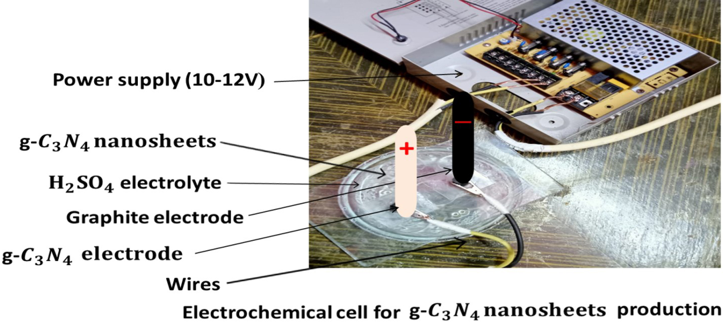 Fig. 1.
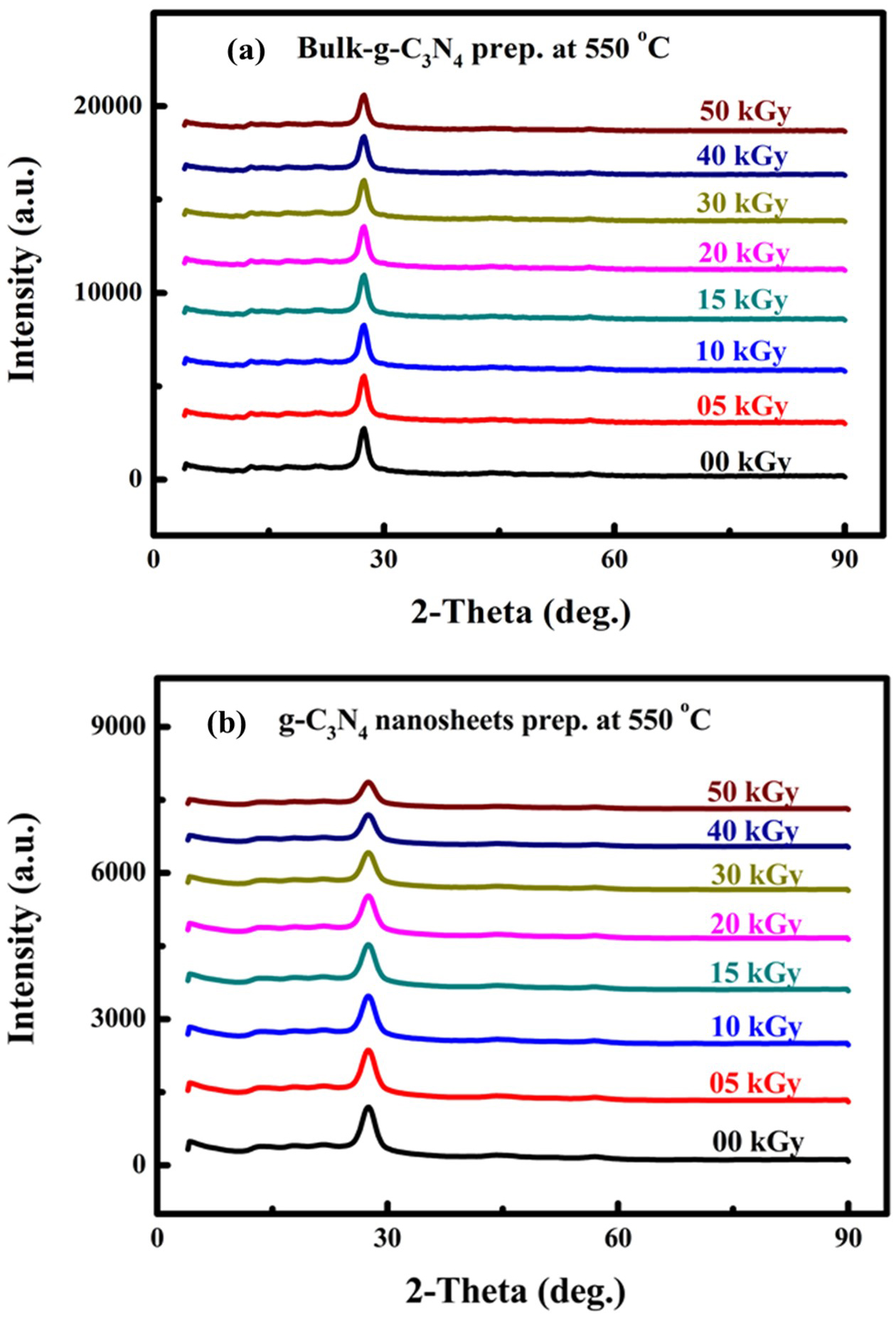 Fig. 2.
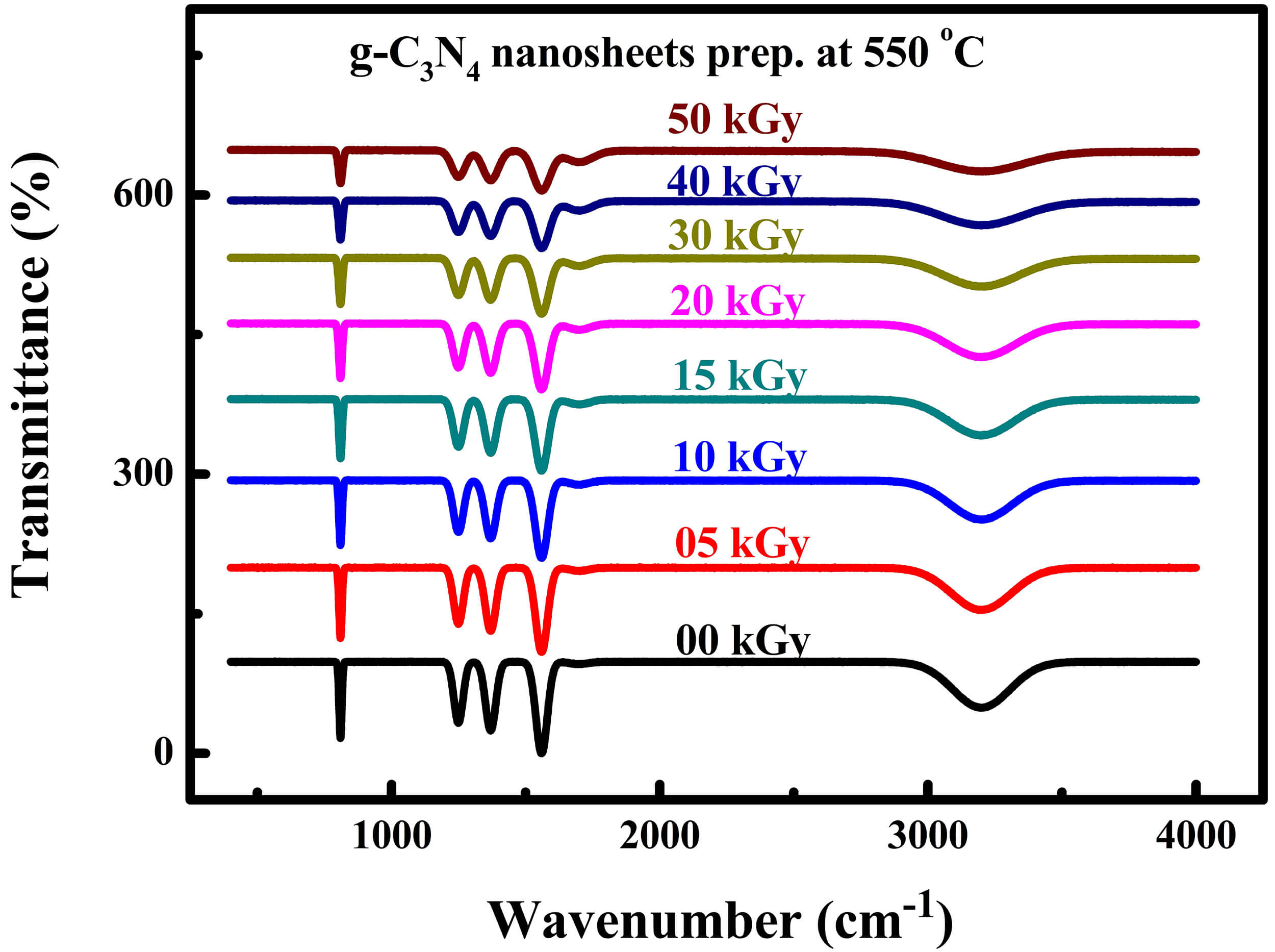 Fig. 3.
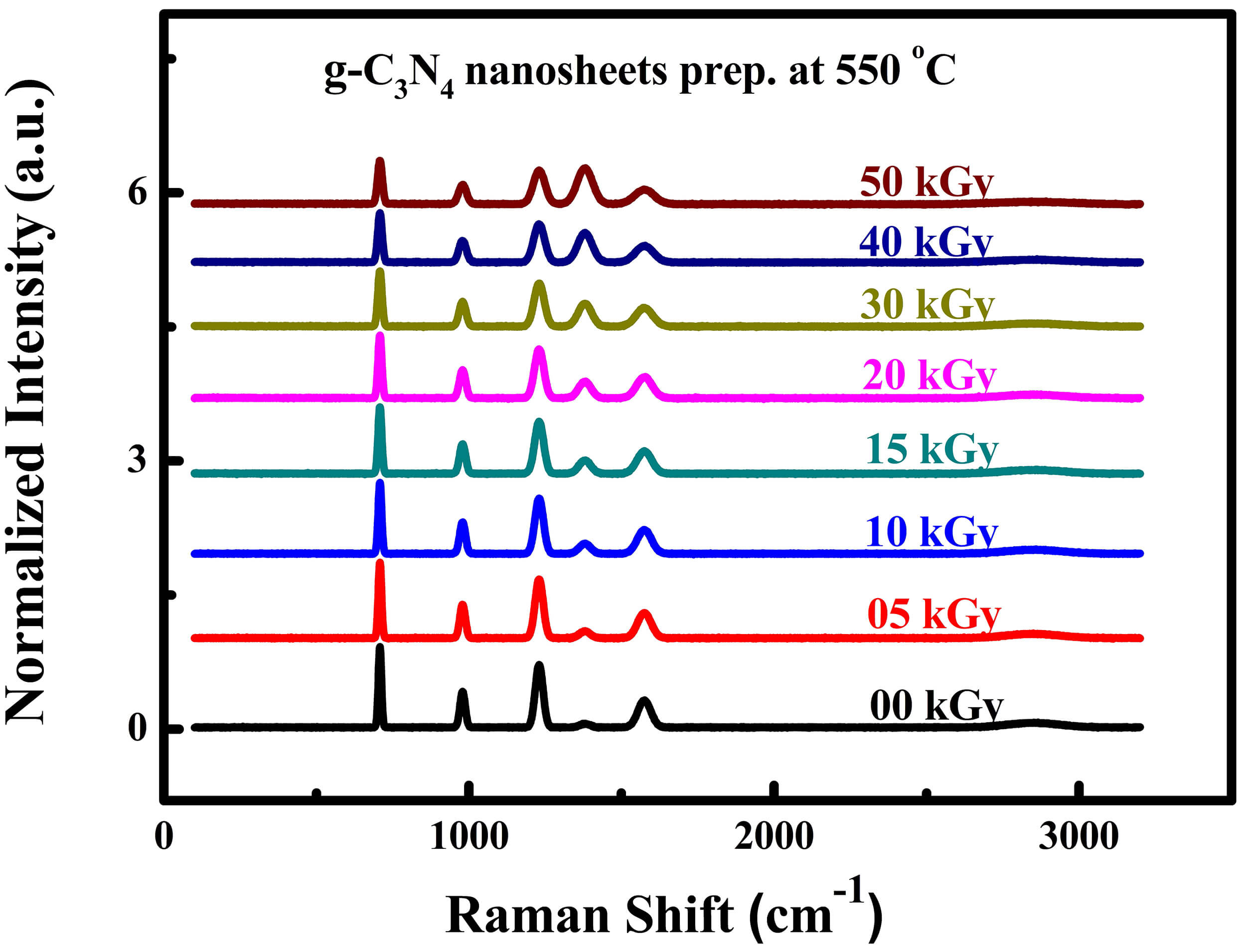 Fig. 4.
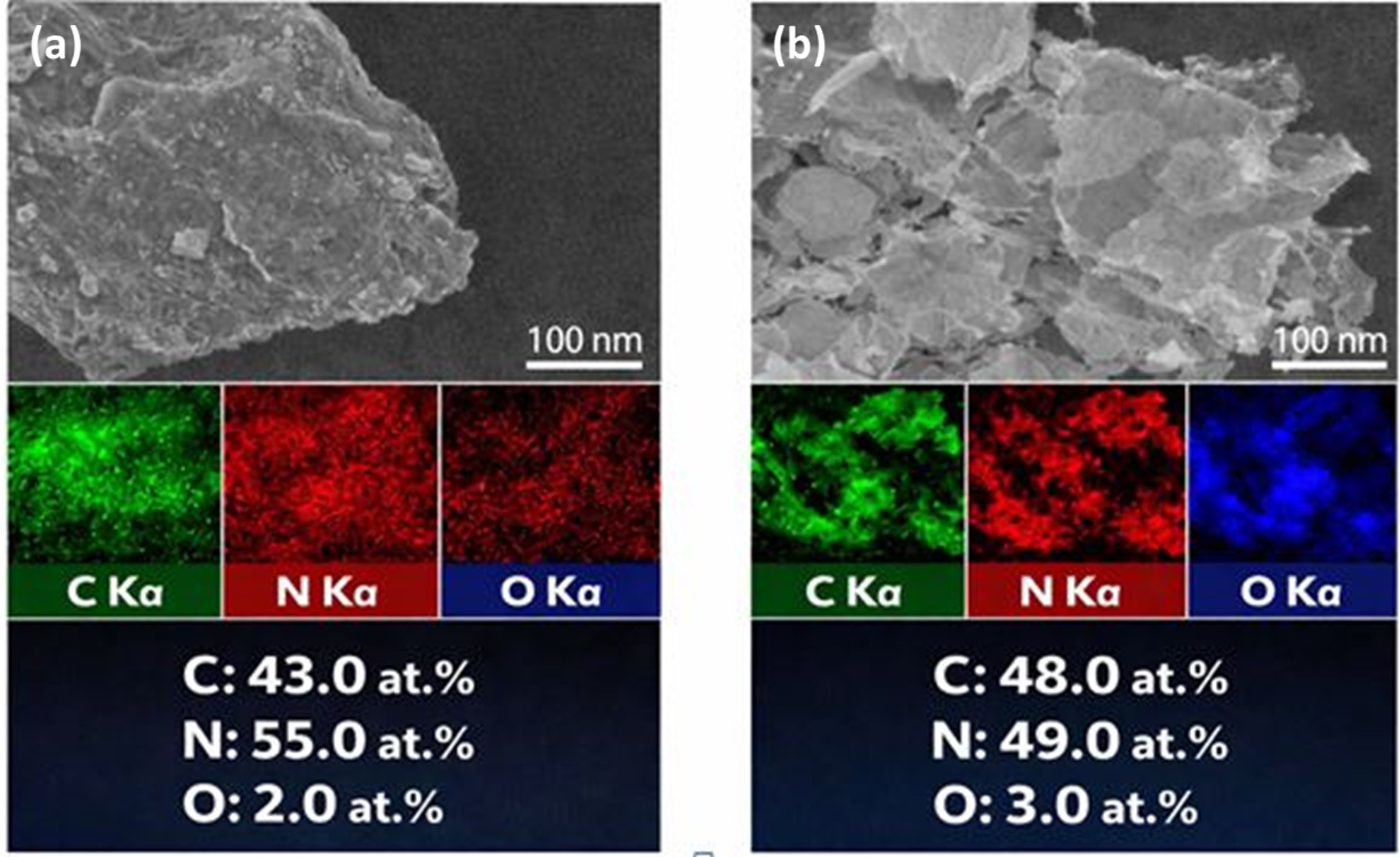 Fig. 5.
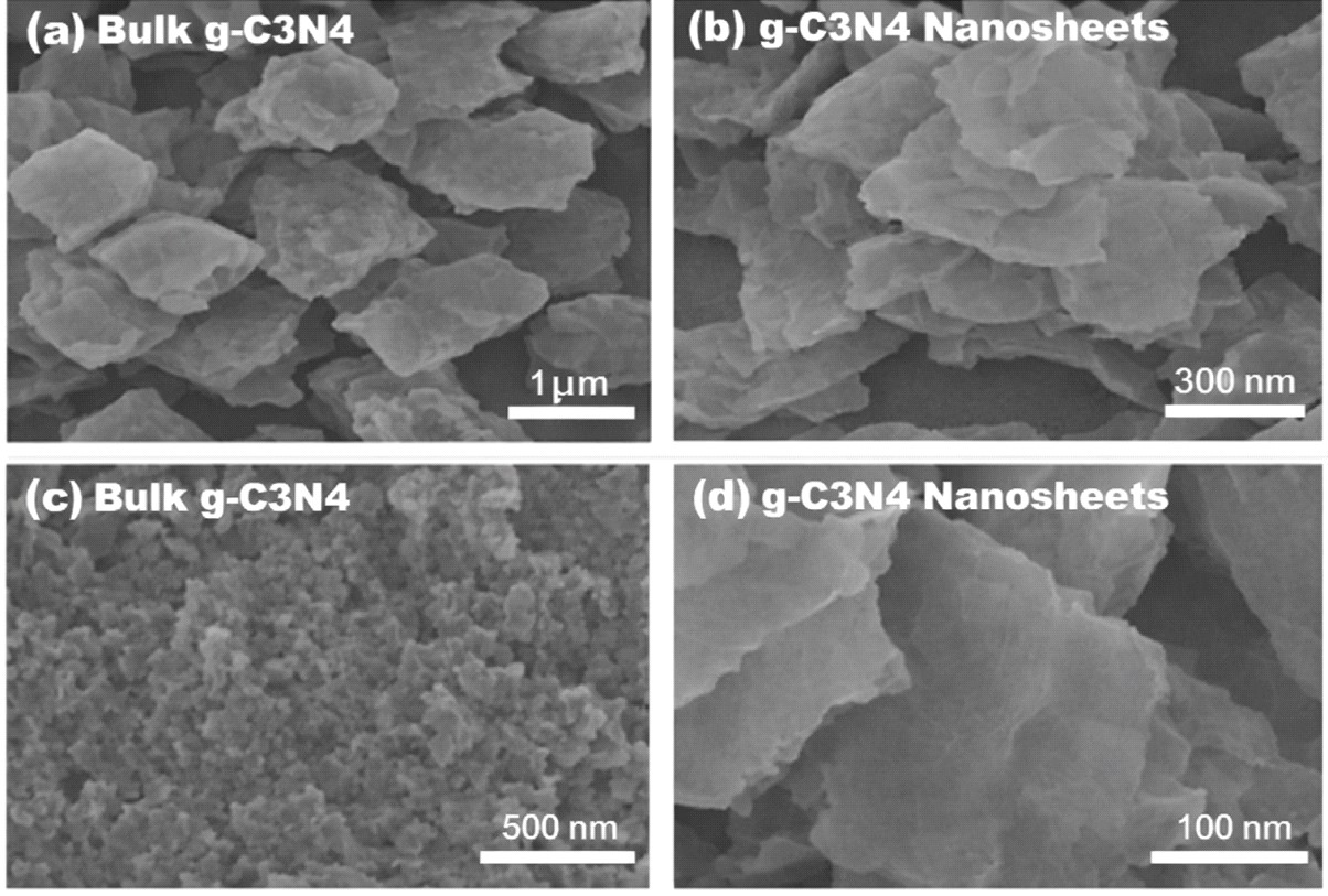 Fig. 6.
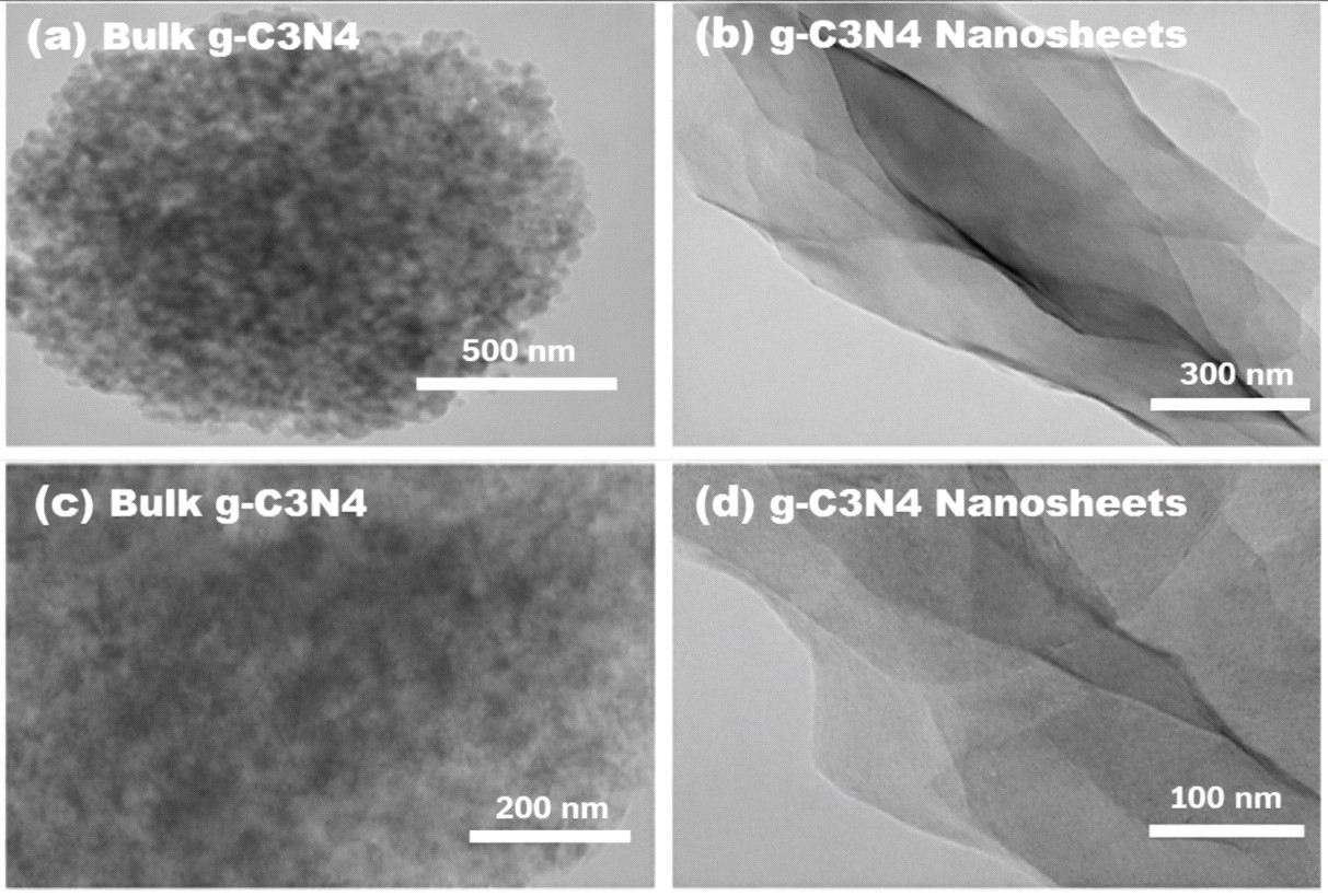 Fig. 7.
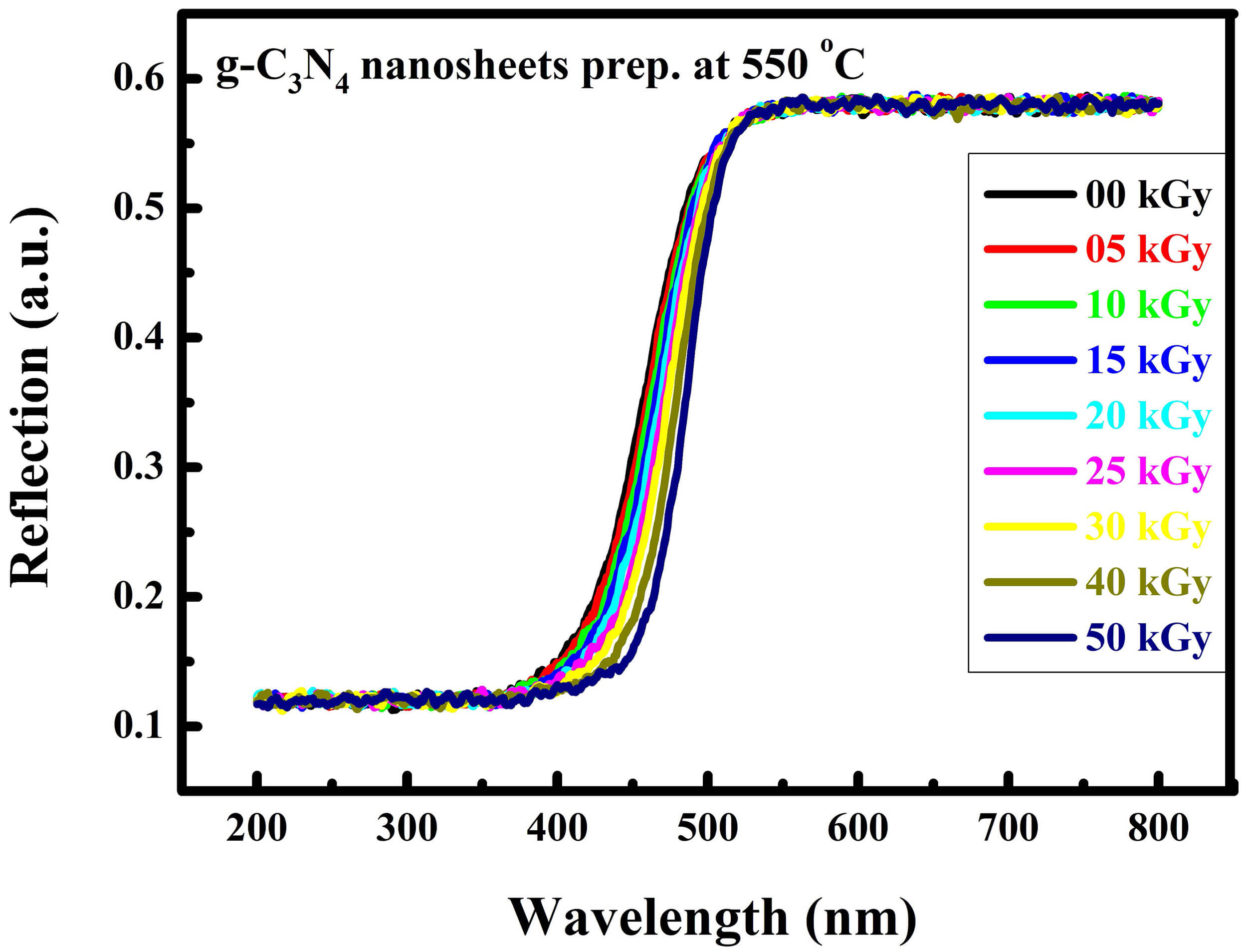 Fig. 8.
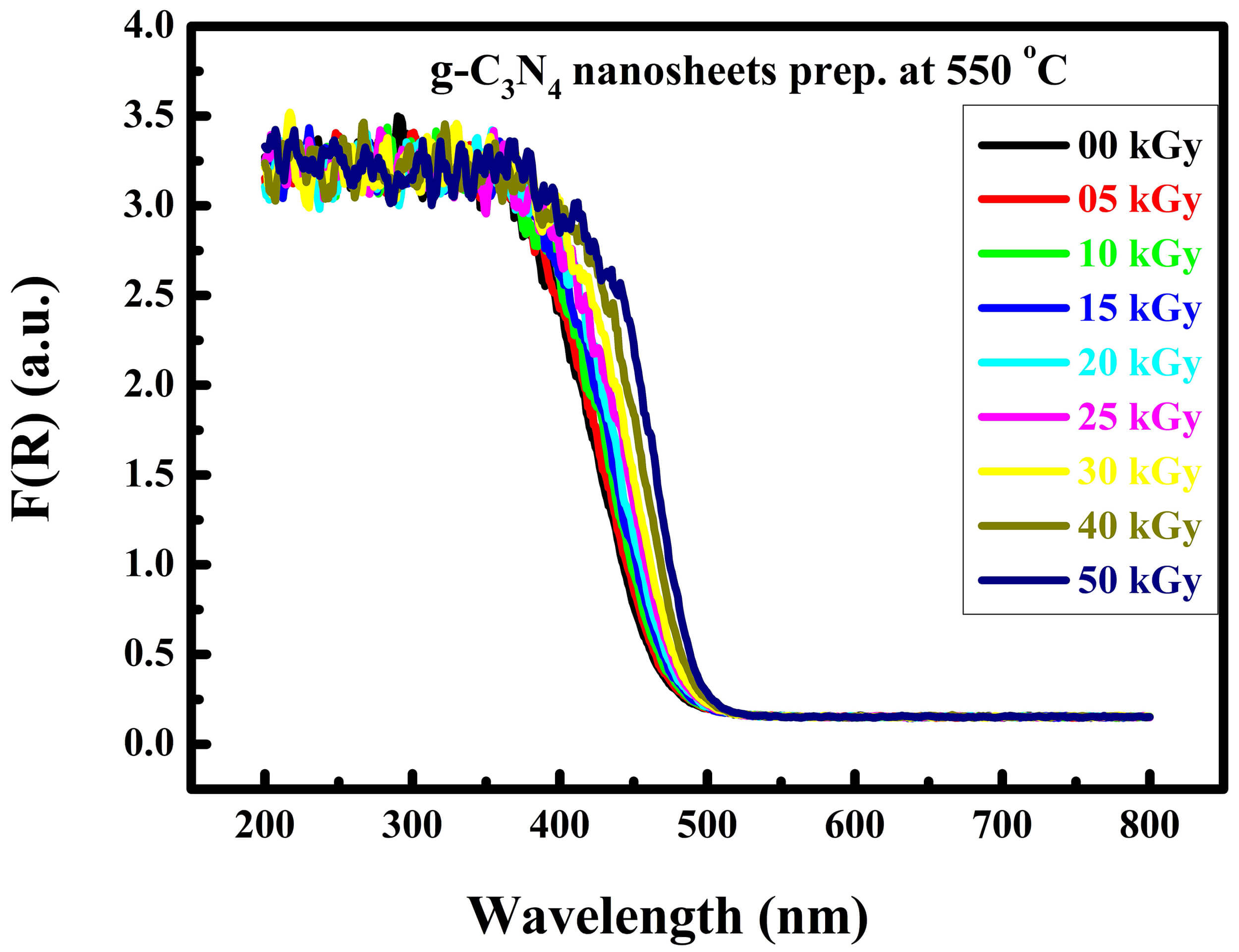 Fig. 9.
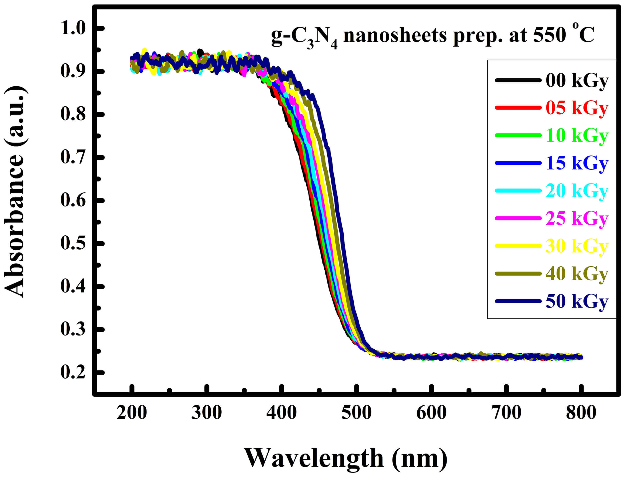 Fig. 10.
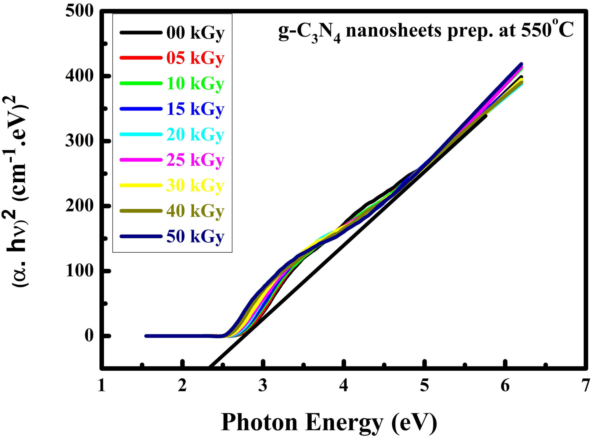 Fig. 11.
 Fig. 12.
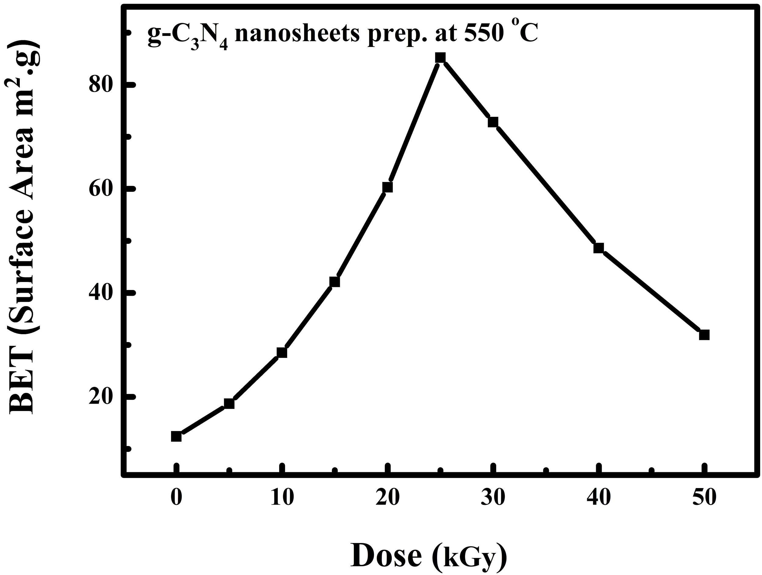 Fig. 13.
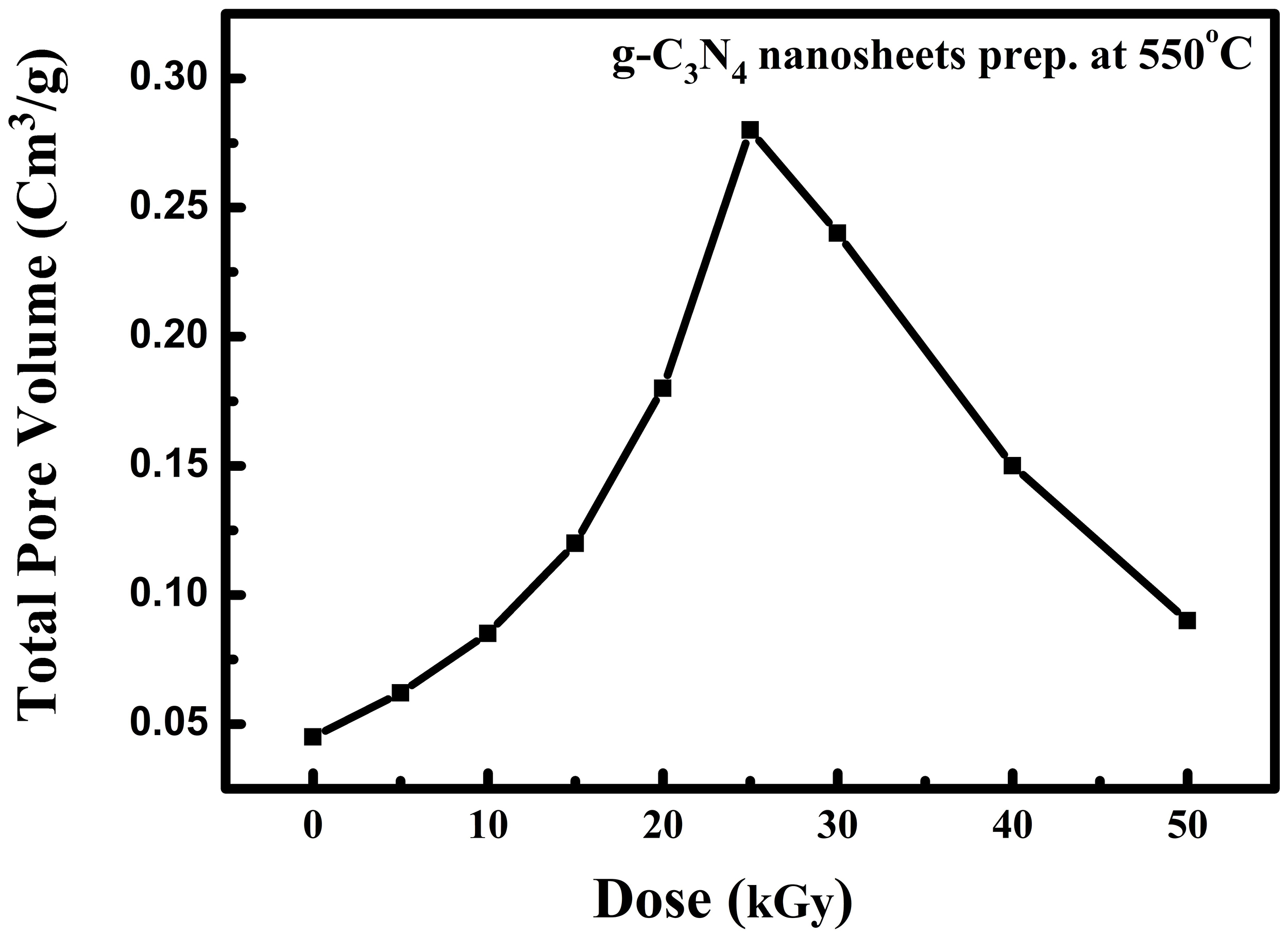 Fig. 14.
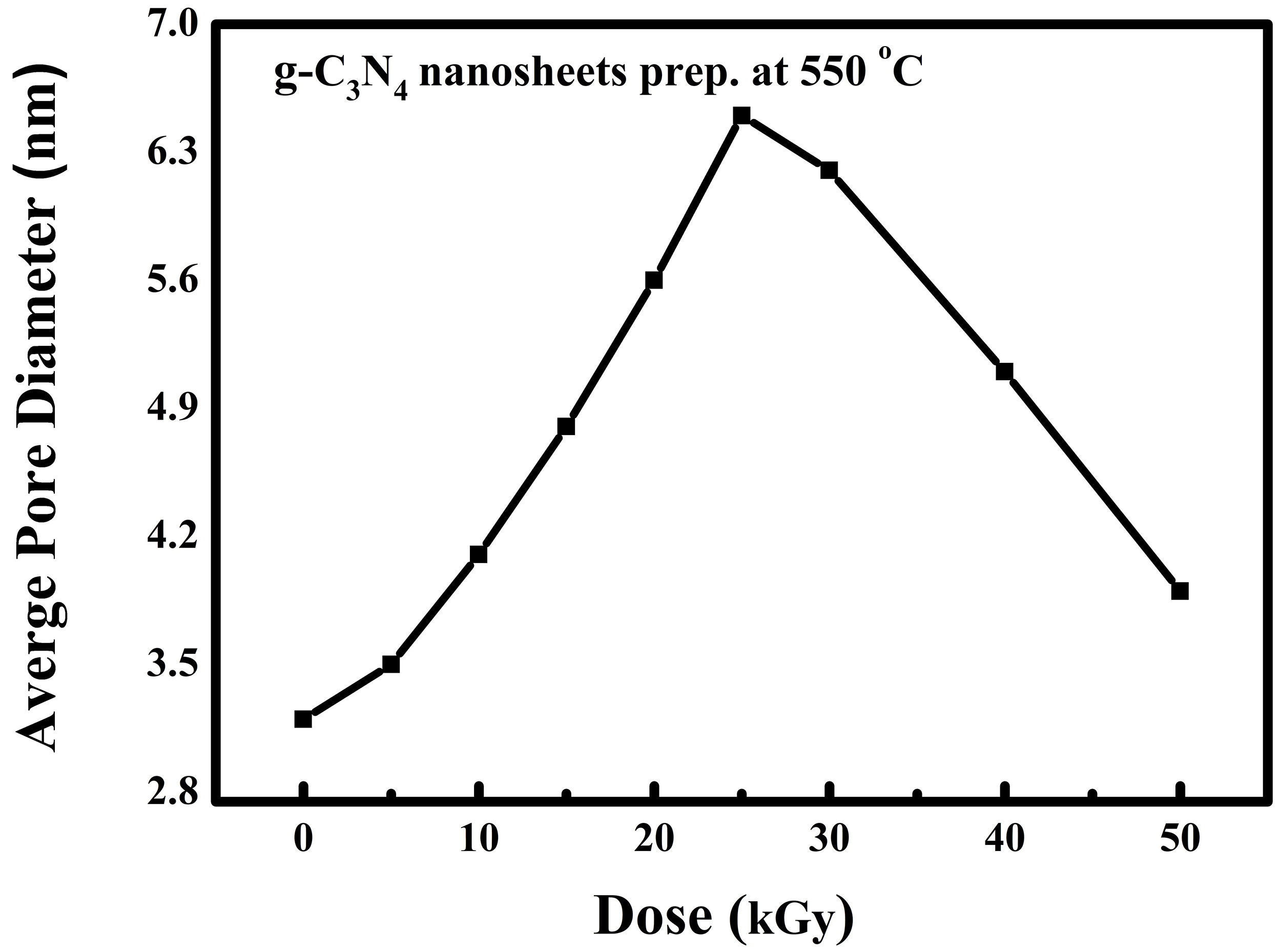 Fig. 15.
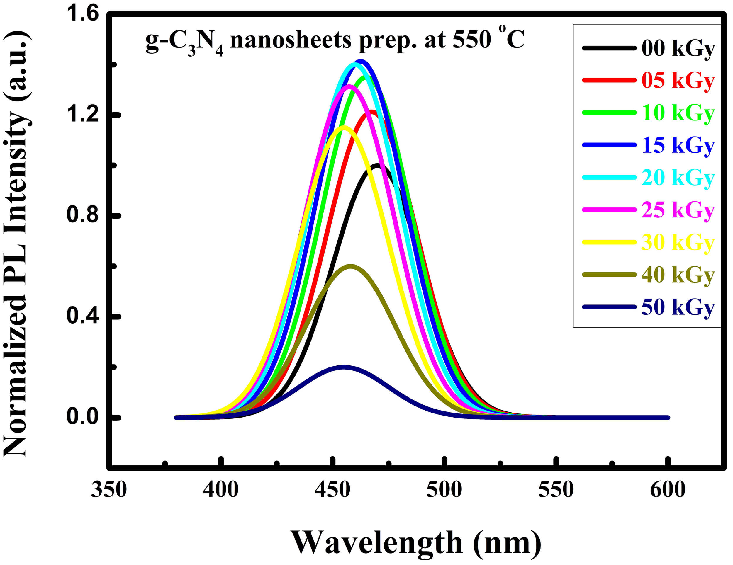 Fig. 16.
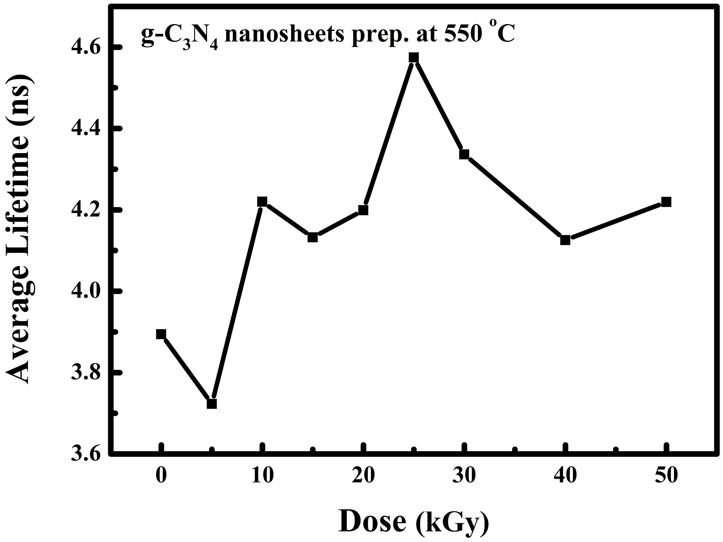 Fig. 17.
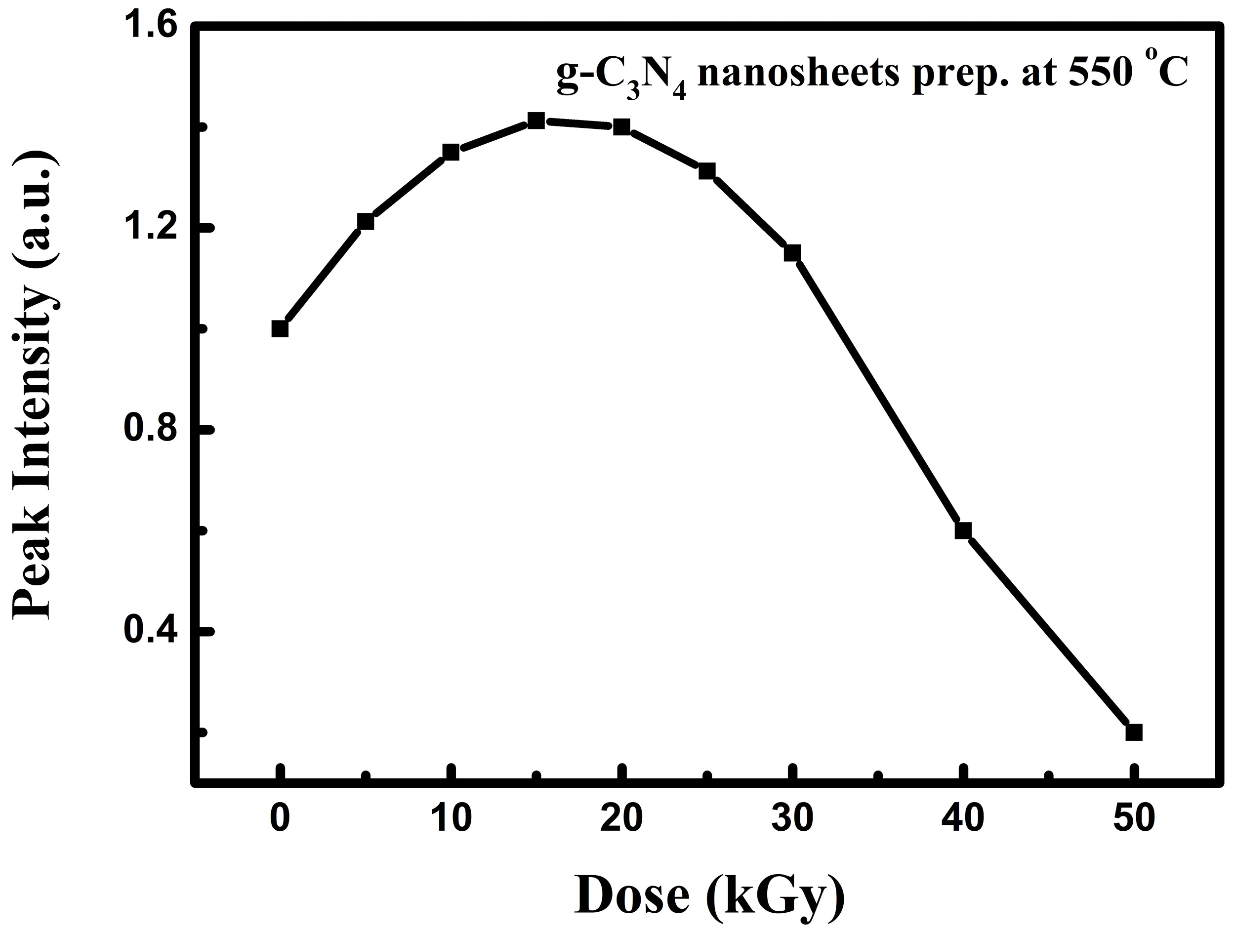 Fig. 18.
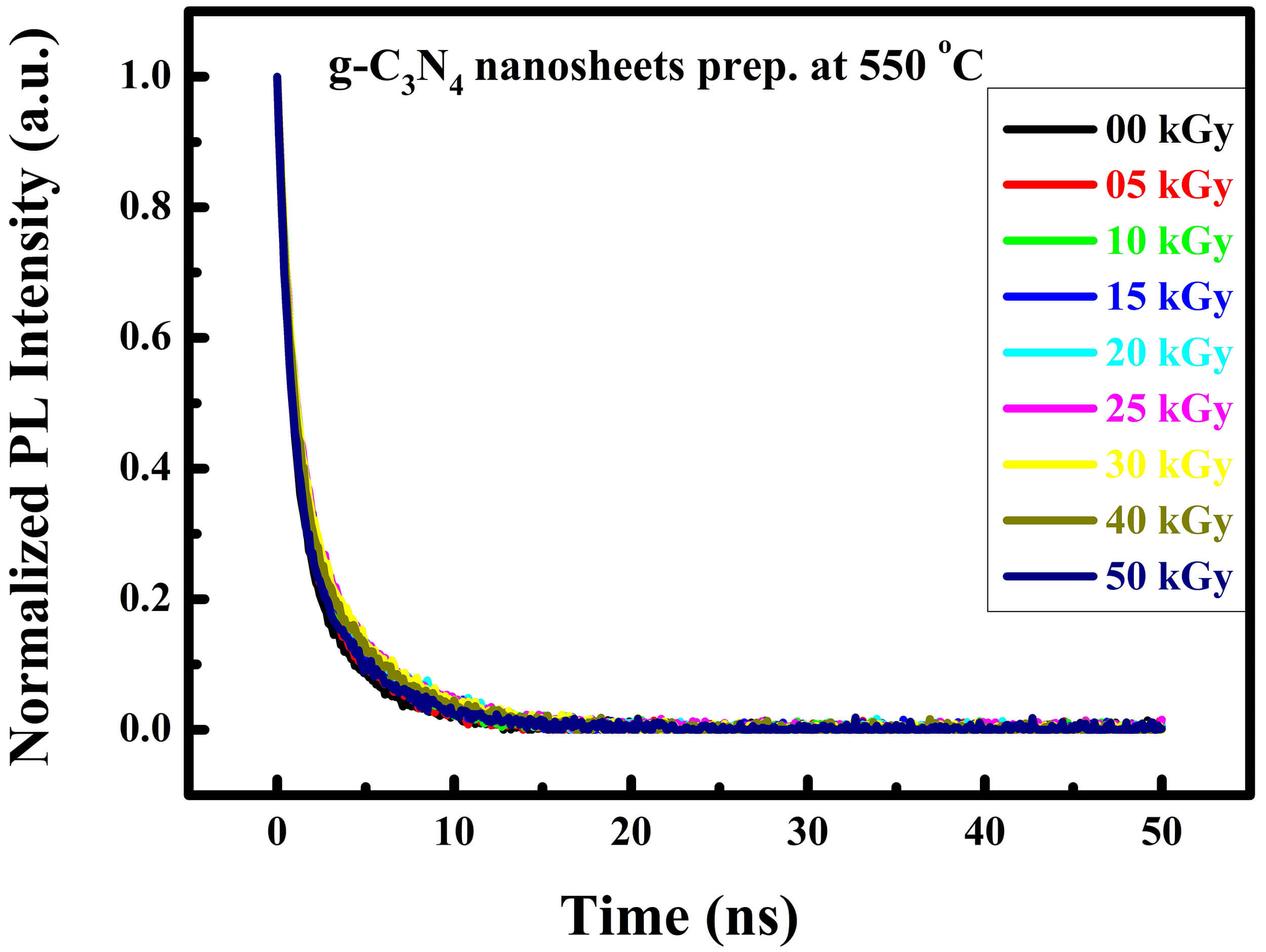 Fig. 19.
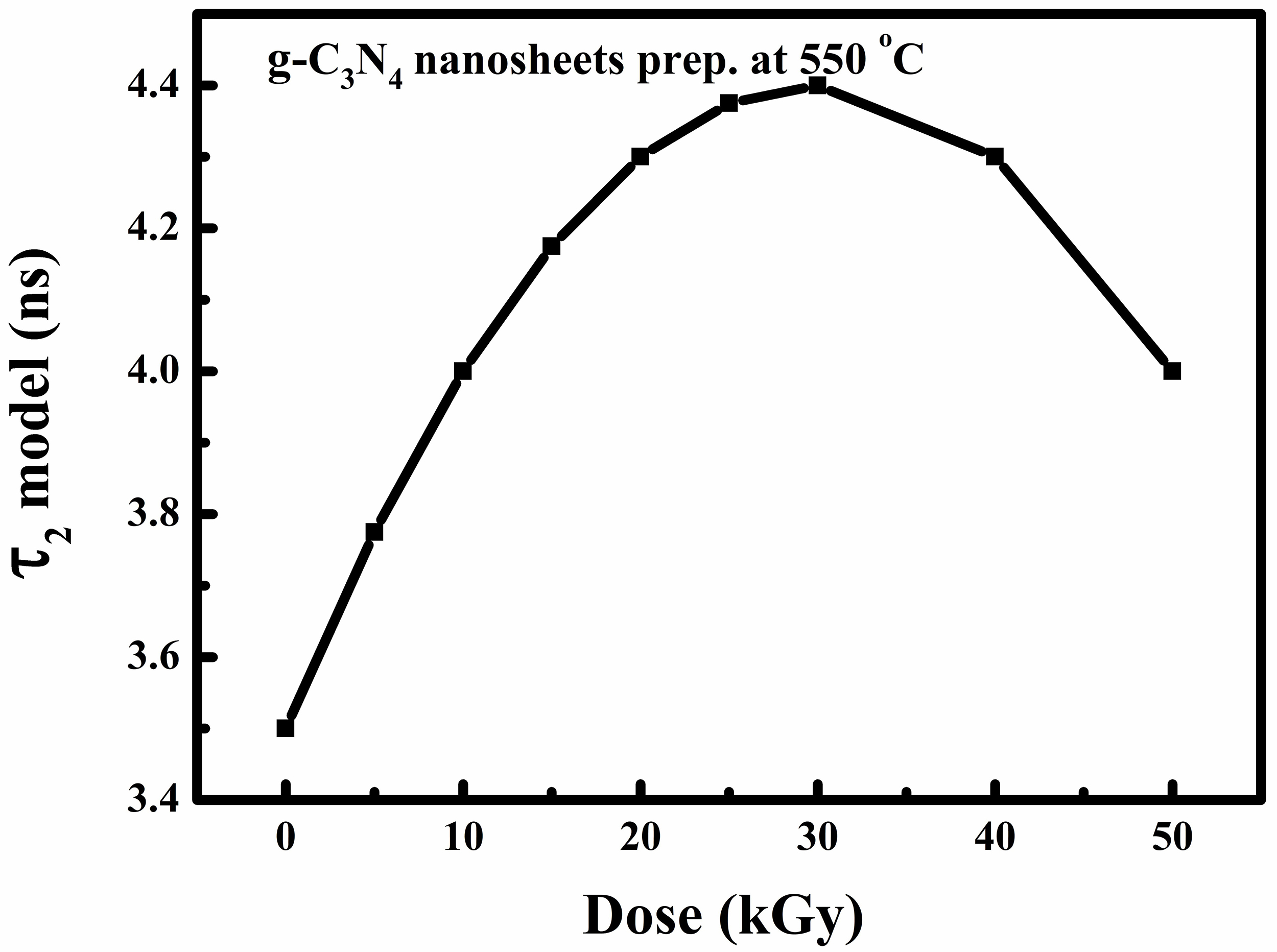 Fig. 20.
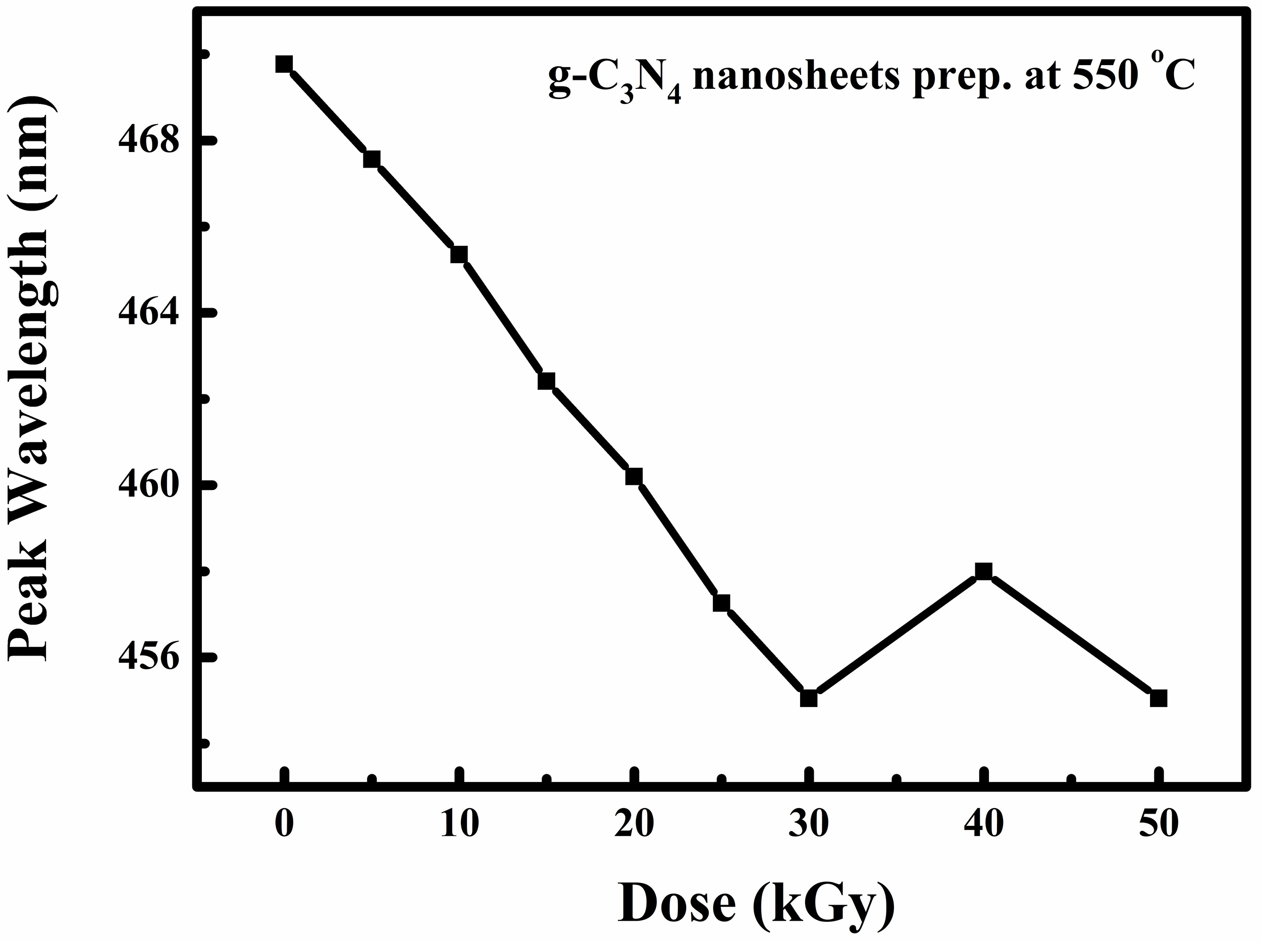 Fig. 21.
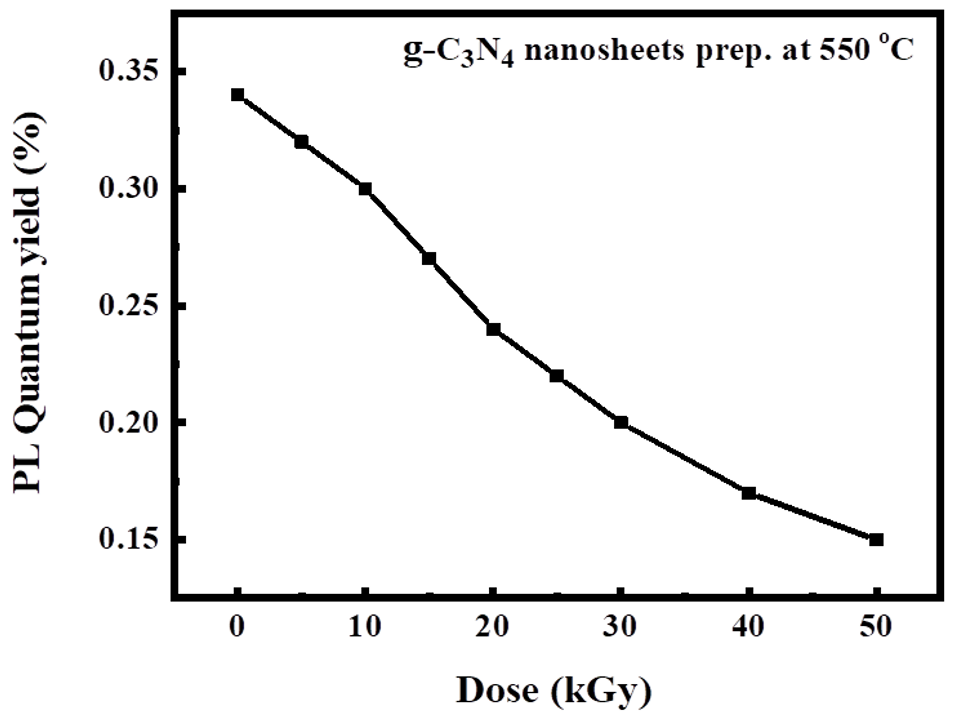 Fig. 22.
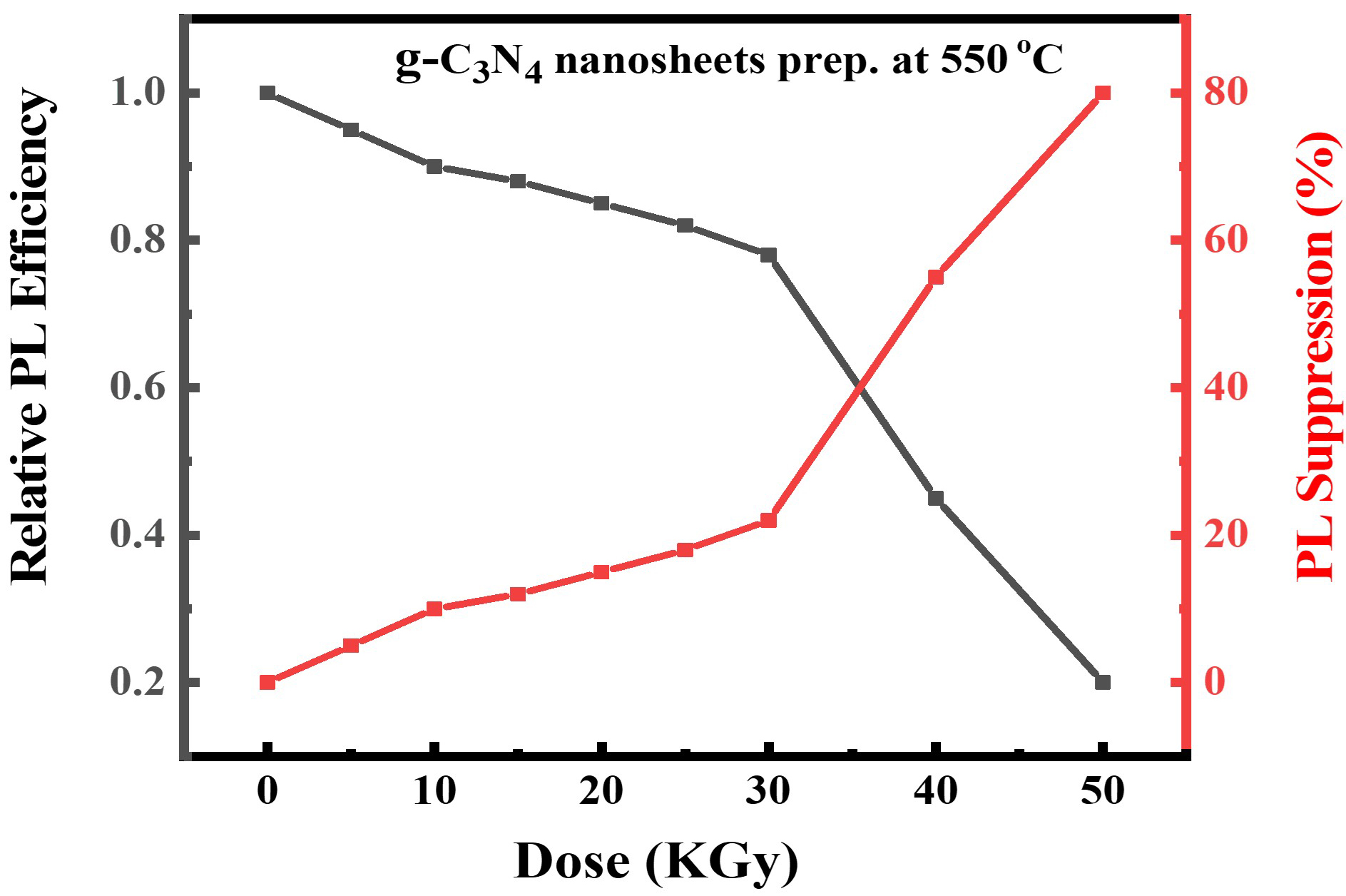 Fig. 23.
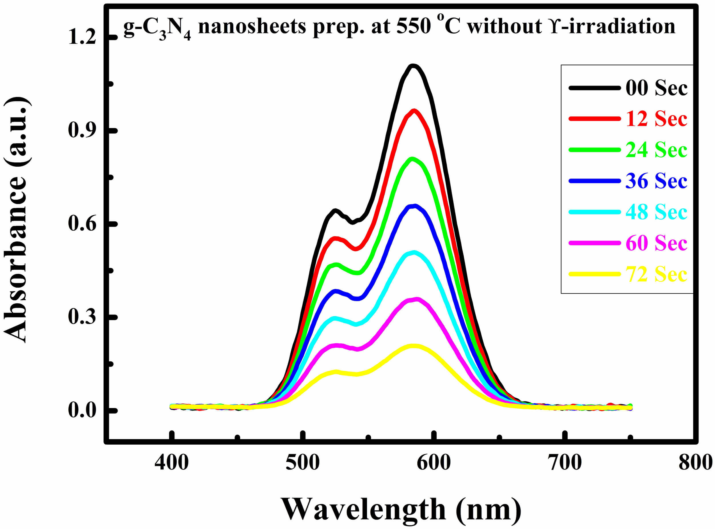 Fig. 24.
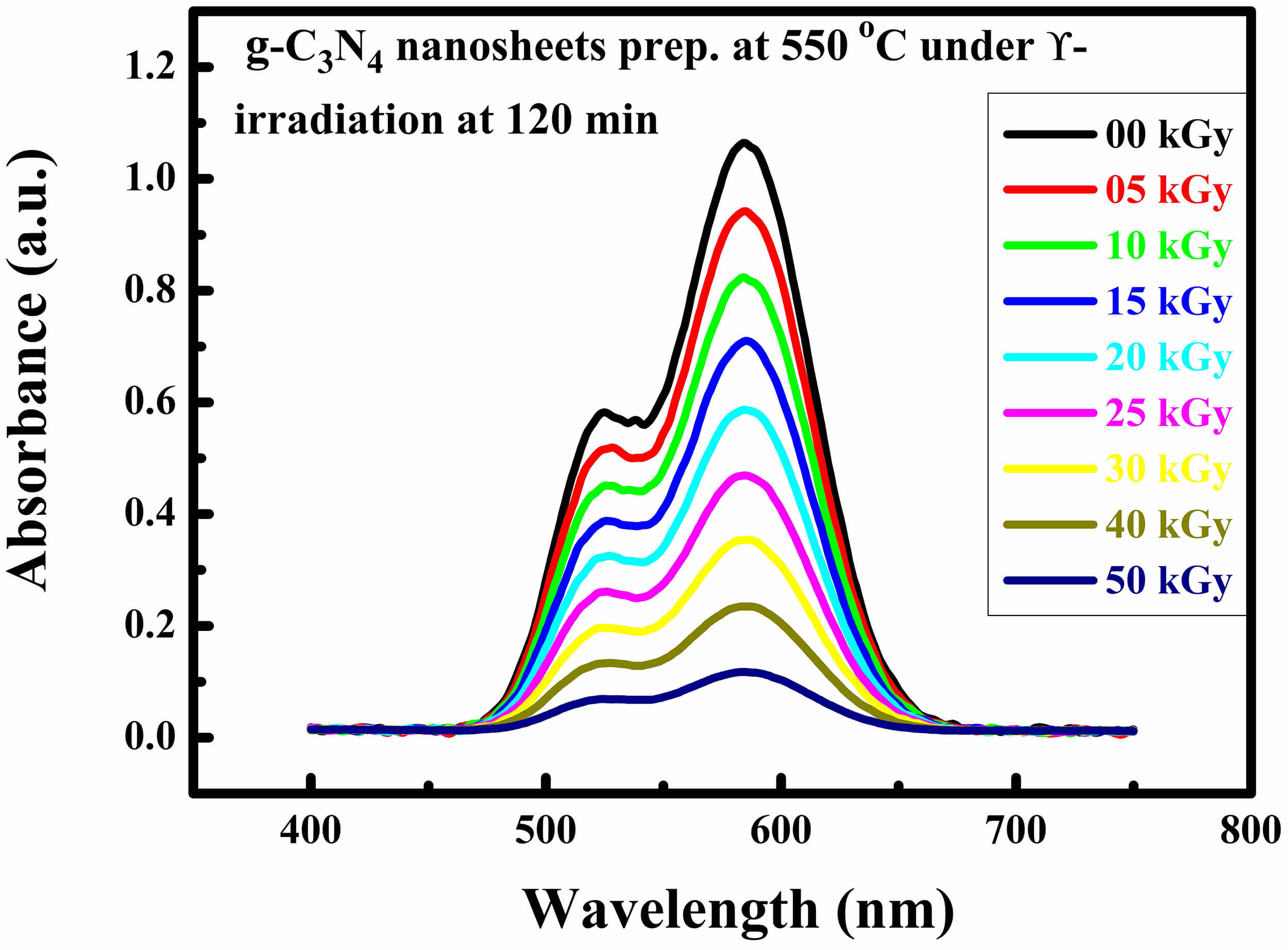 Fig. 25.
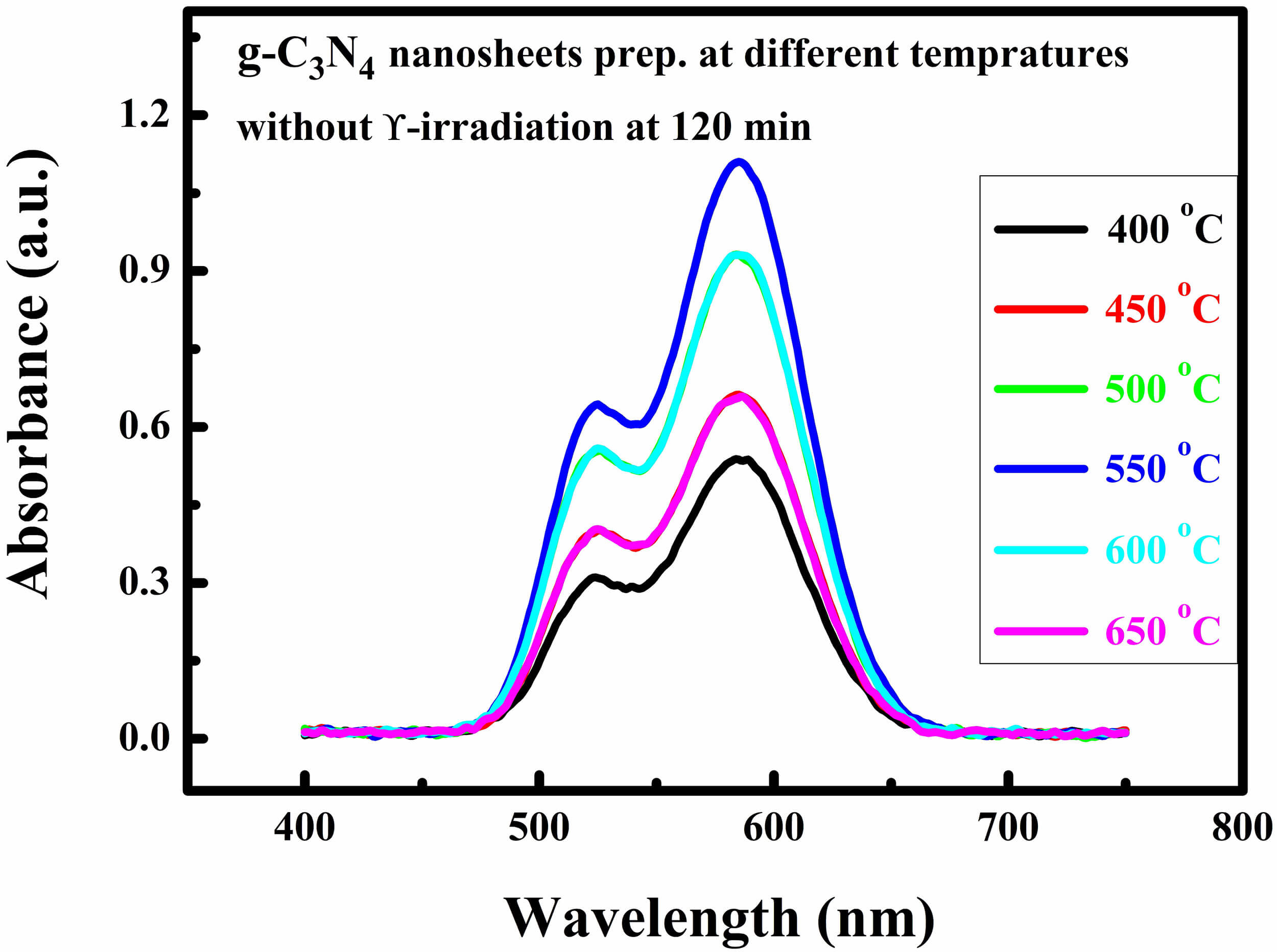 Fig. 26.
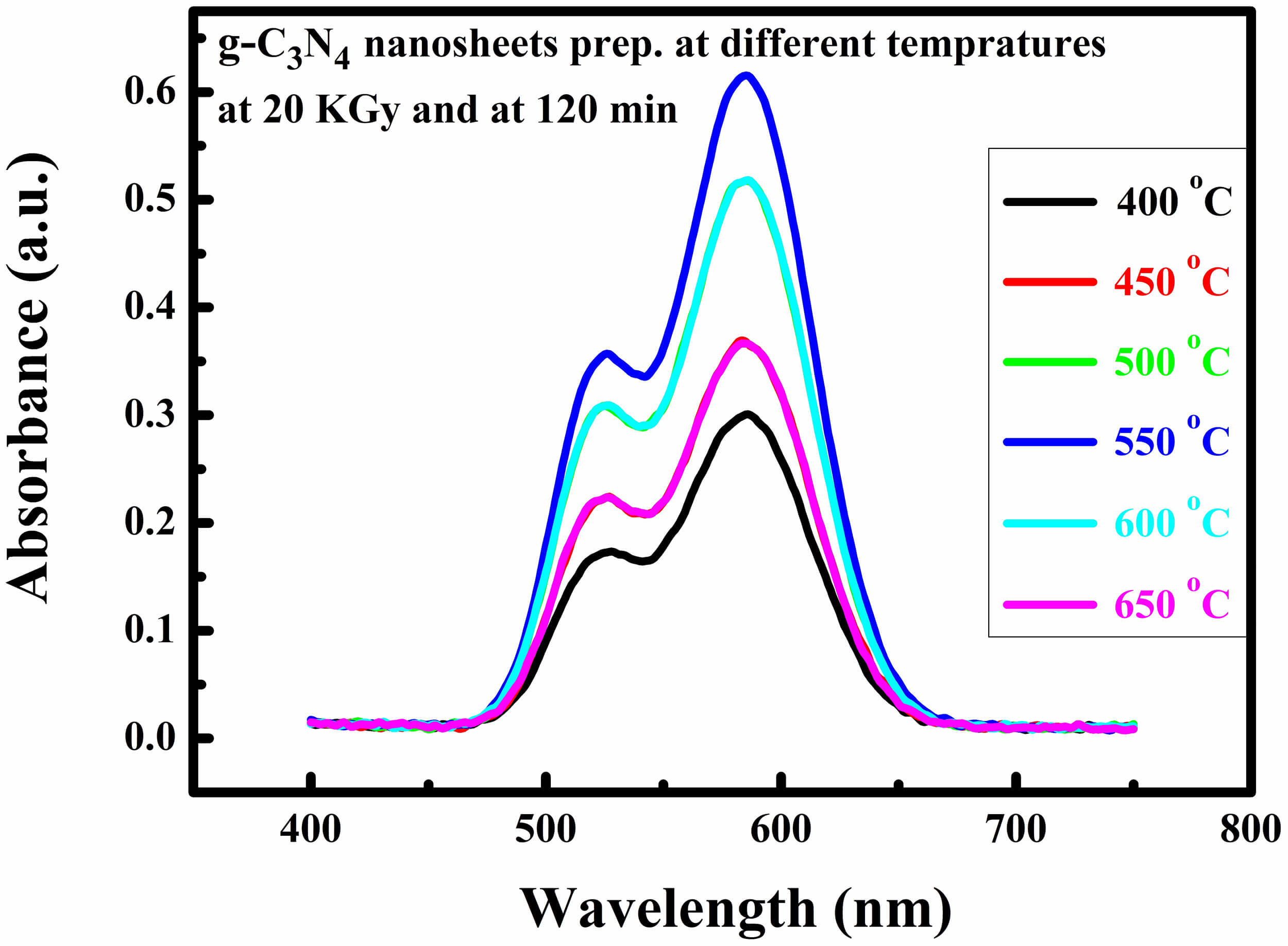 Fig. 27.
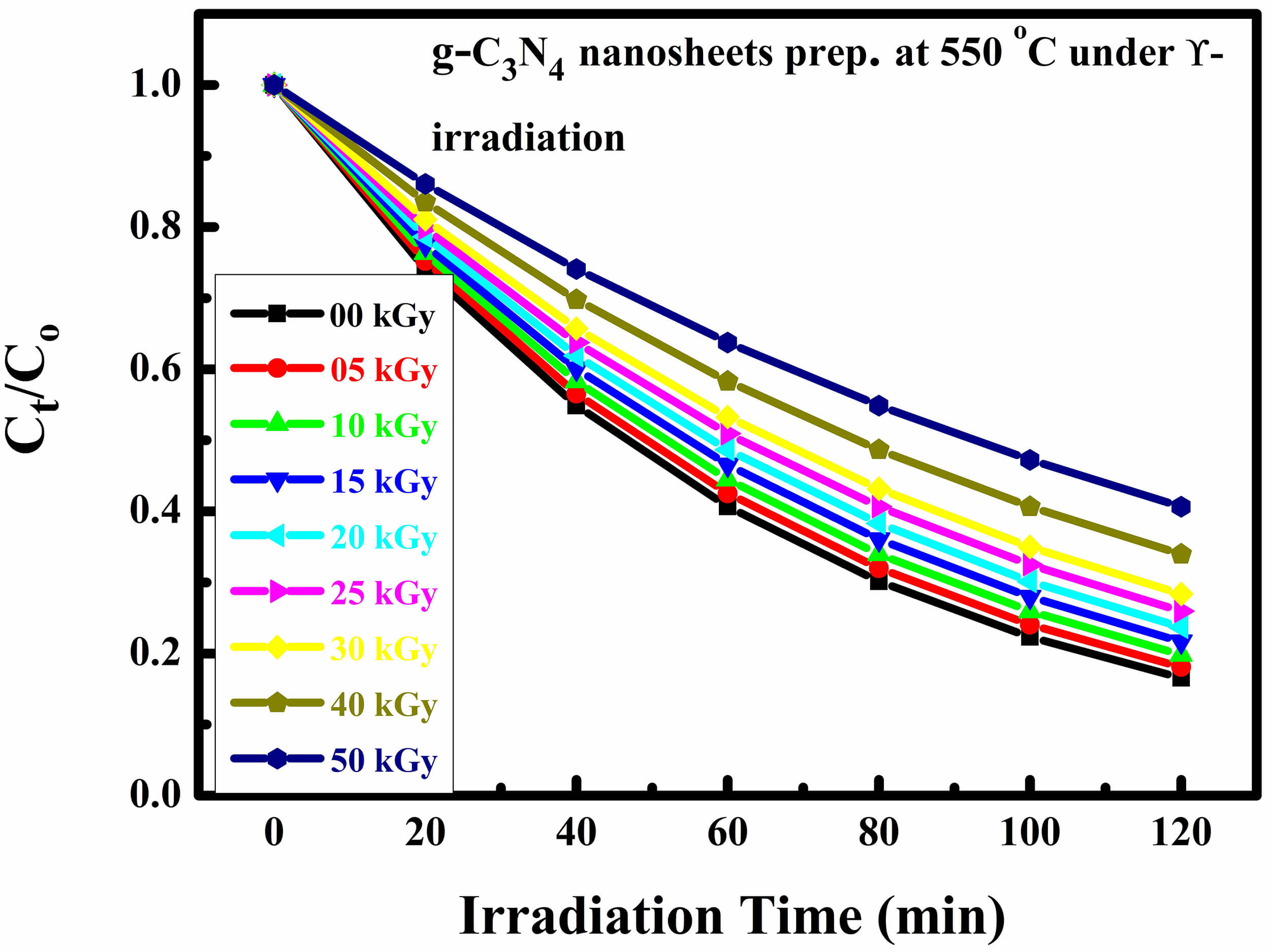 Fig. 28.
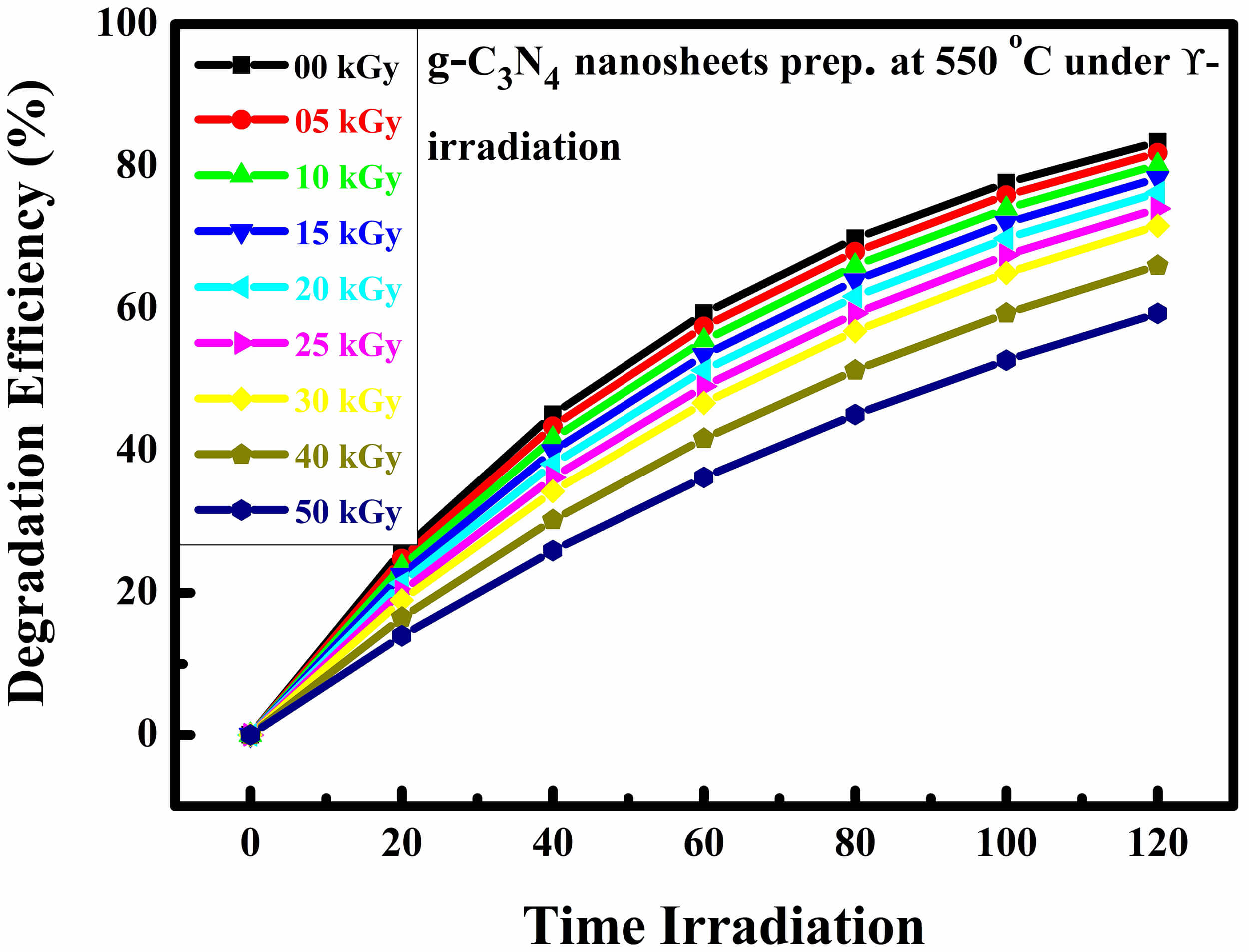 Fig. 29.
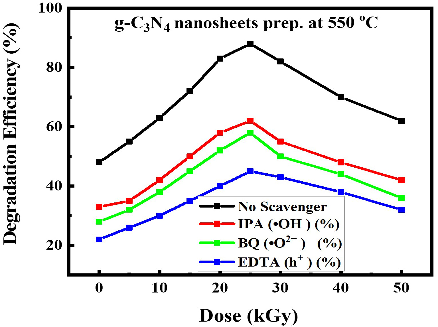 Fig. 30.
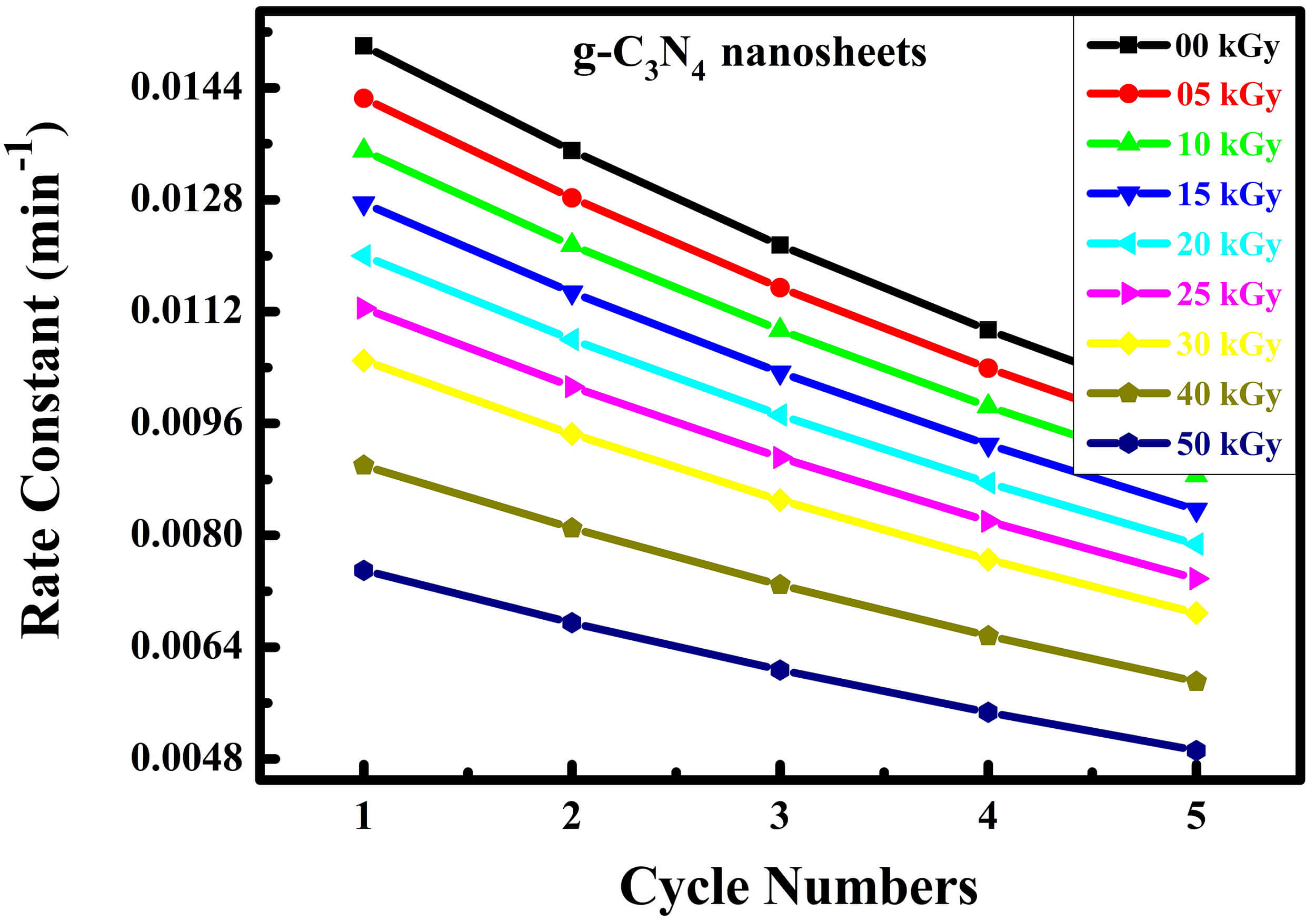 Fig. 31.
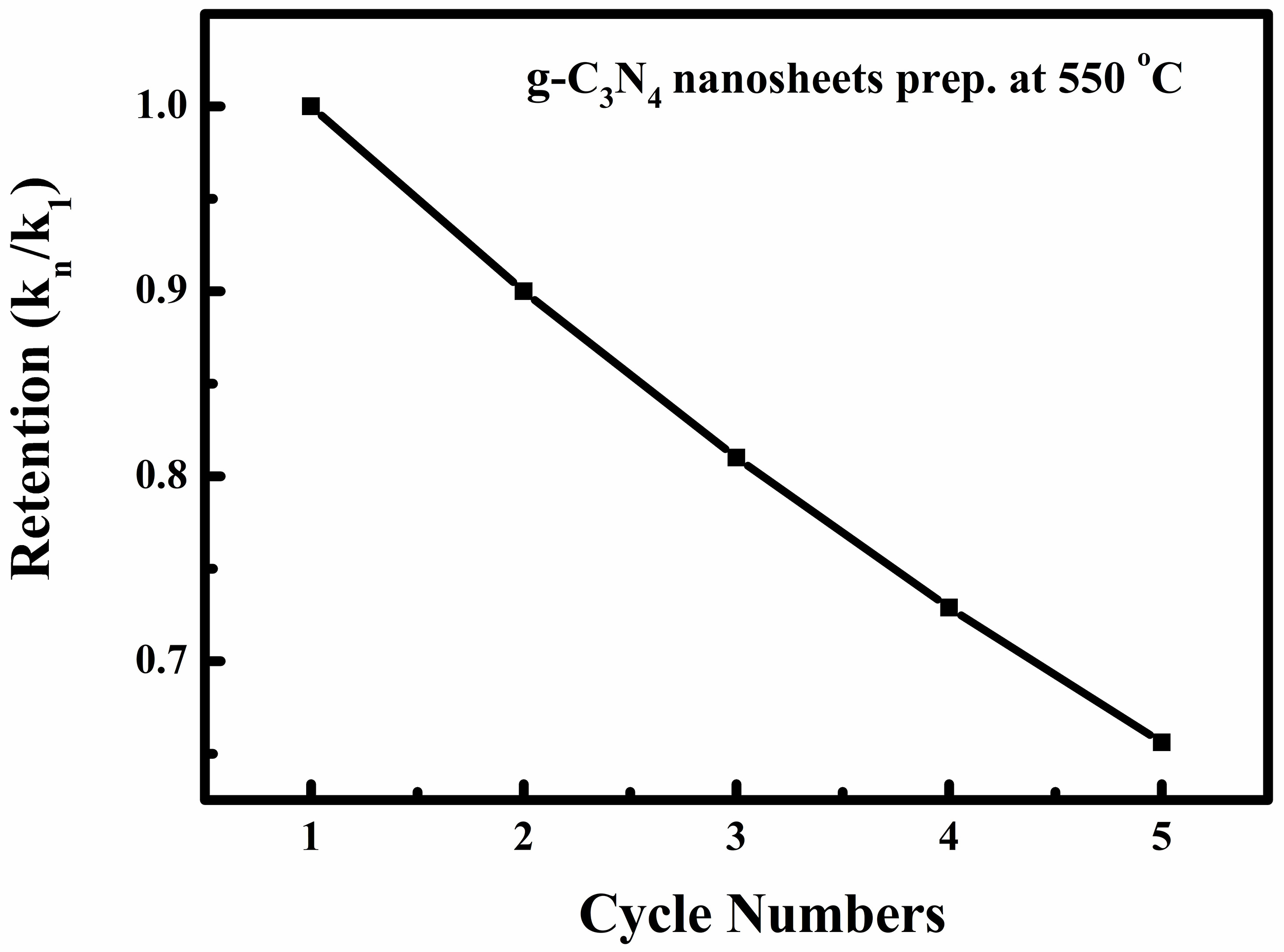 Fig. 32.