

 , Fani Gkrozou 2, Nabila Iram 1, Georgios Ntritsos 3,4, Evangelos Dimitriou 5, Alexandros Tzallas 3, Angelos Daniilidis 6, Alexandra Papoudou-Bai 7, Vassiliki Siafaka 8, Thomas Vrekoussis 9, Iordanis Navrozoglou 2, Minas Paschopoulos 2
1 Reproductive Medicine and Assisted Conception, Assisted Conception Unit, Guy’s and St. Thomas' NHS Foundation Trust, SE1 9RT London, UK
2 Department of Obstetrics and Gynaecology, University Hospital of Ioannina, 45500 Ioannina, Greece
3 Department of Informatics & Telecommunications, School of Informatics & Telecommunications, University of Ioannina, 45500 Ioannina, Greece
4 Department of Hygiene and Epidemiology, University of Ioannina Medical School, 45500 Ioannina, Greece
5 Medical School, Department of Mathematics, National Kapodistrian University of Athens, 11527 Athens, Greece
6 2nd Department of Obstetrics and Gynaecology, Hippokration General Hospital, Aristotle University of Thessaloniki, 54942 Thessaloniki, Greece
7 Department of Pathology, Faculty of Medicine, School of Health Sciences, University of Ioannina, 45500 Ioannina Greece
8 School of Health Sciences, University of Ioannina, 45500 Ioannina, Greece
9 Department of Obstetrics and Gynaecology, University Hospital of Heraklion, 71409 Heraklion, Greece
Abstract
Background: Office hysteroscopy is a widely-accepted and useful tool in the every-day practice of gynaecologists. Methods: In this 20-year-retrospective study, data originating from 2675 patients who underwent vaginoscopic office hysteroscopy are presented. The Endoscopic Unit is located in the Department of Gynaecology, University Hospital of Ioannina and it is considered one of the busiest public institutions providing care for a large amount of Greek population in Epirus, North-western Greece. Our findings presented in this very first and nationally largest retrospective study, will contribute to the overall scientific knowledge by providing substantial data with regards to hysteroscopy and to epidemiology of endometrial pathology. Results: Common hysteroscopic indication across all age groups was Abnormal Uterine Bleeding (AUB). Predictive characteristics of hysteroscopy in the diagnosis of various conditions were evaluated for these patients in comparison with their histologic report as the gold standard. Comparing hysteroscopic findings with the respective histology reports revealed that in cases of normal endometrium, sensitivity of 60.9%, specificity of 92.1%, Positive Predictive Value (PPV) of 79.07% and Negative Predictive Value (NPV) 82.8% were estimated. Hysteroscopic detection of endometrial polyps demonstrated sensitivity of 92.04%, specificity of 89.1%, PPV of 73.5% and NPV 97.1%. For fibroids, sensitivity and specificity were calculated at 98.5% and 100% respectively, while PPV and NPV at 100% and 99.9%, respectively. For endometrial cancer, the predictive characteristics were estimated at 87.5% and 99.7% with regards to sensitivity and specificity, and 63.6% and 99.9% for PPV and NPV, respectively. Finally, for cases of hyperplasia, hysteroscopy showed sensitivity of 75.0%, specificity of 91.03%, PPV of 11.7% and NPV of 99.5%. Conclusions: To date, this is the largest retrospective study on office hysteroscopy with the use of vaginoscopic approach technique in Greece. This study has been conducted in one of the busiest public gynecologic endoscopic units across Greece. Our findings are consistent with the international scientific evidence, which has proven that hysteroscopy is an efficient and safe method to investigate pathologies within the uterine cavity and in general is accompanied by satisfactory patient acceptance.
Keywords
- office hysteroscopy
- sensitivity
- specificity
- hysteroscopic indication
- abnormal uterine bleeding
- vaginoscopic approach
- distension medium
- normal saline
- uterine cavity
- endometrial pathology
Office Hysteroscopy is a leading endoscopic tool in cases of abnormal uterine
bleeding, sonographic evidence of submucous myomas and endometrial polyps, as
well as, in detecting subfertility-associated underlying endometrial pathology
[1, 2, 3]. Advancements with regards to hysteroscopic equipment led to the
establishment of hysteroscopy as a Gold Standard technique when assessing
endometrial pathologies. According to the literature, hysteroscopy demonstrated
an overall sensitivity of 98% [4], when compared with histological results. On
the contrary, scientific evidence showed that dilatation and curettage (D&C)
offers a moderate sensitivity of 65% [5]. Nowadays the traditional method
requiring general anaesthesia is largely abandoned, since technological
developments led to finer hysteroscopes with high resolution imaging allowing
accurate assessment of the uterine cavity. These novel hysteroscopes can better
visualise the uterine cavity by utilizing light properties [6]. With regards to
the distension medium, normal saline is widely used and compared to CO
Italians were the first to introduce the technique in 1997 [18], and over the past twenty years, office hysteroscopy has emerged as a gold standard technique in detecting endometrial and endocervical pathology over traditional methods. Vaginoscopic hysteroscopy is a simple technique which requires a short learning curve [10]. The hysteroscope is introduced slowly along the posterior vaginal wall and as the vagina distends, advancement of the hysteroscope until the visualization of external cervical os follows. In order to locate the cervix, which at times poses the greatest obstacle of the technique, literature suggests initially introduction of the hysteroscope to the posterior vaginal wall, known as cul-de-sac, and slow withdrawal of the equipment until the external cervical os is identified [19]. Vaginoscopic hysteroscopy requires rigid hysteroscopes, since it is very likely to experience difficulties when using flexible equipment [20, 21].
Moreover, the outdated concept of ‘diagnostic hysteroscopy’ has changed to ‘office hysteroscopy’, in which ‘see and treat’ of any pathological findings at the time of the procedure can be achieved, where applicable [22]. Hysteroscopic approach seems to be more efficient than that of sonography regarding the detection of endometrial pathology as it permits direct visualisation of the endocervix and the endometrial cavity [23]. Recent advancements resulted in reduced diameters of hysteroscopes and allowed the use of different types of electrosurgery offering the option for the majority of the procedures to be performed in an outpatient/office setting. Compared to “outpatient” hysteroscopy which may suggest the use of some type of analgesia, the term “office” hysteroscopy suggests vocal-local analgesia [24, 25]. Combined with a vaginoscopic approach when introducing the hysteroscope, patients can be diagnosed and treated in one visit with high levels of acceptance due to minimal discomfort or pain experienced during the procedure [12].
In this study, data from 2675 patients who underwent office hysteroscopy, in the past 20 years, was extracted in order to detect the feasibility of the method, as well as, determine the role of office hysteroscopy in various pathological intrauterine conditions and its relevance to available published international scientific evidence. In addition, the patient’s acceptance and overall experience were also studied.
This is a retrospective study on office hysteroscopy performed at the Endoscopic Unit in the Gynaecology Department of the University Hospital of Ioannina, Epirus, Greece. In this endoscopic unit, hysteroscopies are performed since its foundation in 1989, and it is considered one of the busiest national public institutions. In 1997, office hysteroscopy was introduced in this department and since then the service is offered, in the majority of cases, in an office setting, when medically indicated. The endoscopic unit provides care, in terms of diagnosis and treatment of intrauterine pathology, for a large number of patients coming from all around Ioannina and Epirus, North Western and Central Greece, Northwest Macedonia, Ionian Islands and Southern Albania. In addition, the Endoscopic Unit also treats patients from all over Greece and abroad. To our best of knowledge, this is the first large-series study on office hysteroscopy in Greece to date.
This study presents data from 2675 patients from January 1997 to September 2021. All these cases underwent office hysteroscopy in the Endoscopic Unit at the University Hospital of Ioannina, Epirus, Greece. All cases with suspected endometrial pathology due to symptomatology, sonographic evidence, or common clinical indications (Hormonal Replacement Treatment (HRT), surveillance prior and after tamoxifen use, recurrent miscarriage to name a few) underwent office hysteroscopy, although specific criteria for patient selection based on parity, menopausal status and patient’s compliance did not apply. Exclusion criteria for this study were considered the following: menstruation at the time of examination, suspicion of reproductive tract infection, positive pregnancy test, acute uterine bleeding. No records of the number of patients excluded from office hysteroscopy were retrieved due to the retrospective nature of this study. All patients, if applicable, were scheduled at the proliferative phase of their cycle, since according to the literature the features of the uterine cavity can be better visualized [26]. Even though hysteroscopy can also be performed in cases of active uterine bleeding, we have decided not to use the data from such population, but to study the endometrial cavity during the same cyclical phase in order to minimize risk of bias. Based on local protocol, patients with active bleeding were referred to inpatient hysteroscopy if medically indicated. In cases of hysteroscopic failure mainly due to closed cervical os, the procedure was abandoned, and patients were rescheduled for either hysteroscopy under general anaesthesia or any other form of blind endometrial biopsy. All hysteroscopies were supervised by the same highly skilled and experienced hysteroscopist. A total of six hysteroscopists performed these procedures and the interobserver agreement was perfect reaching a kappa value of 0.82 [27]. The procedure was explained in detail to all patients, who signed an informed consent prior to the procedure and answered a non-standardised “patient’s satisfaction questionnaire” designed to assess their overall experience according to the local protocol. All patients were categorised in two different groups, group A (n: 2323) and group B (n: 442). Patients in group A received written information leaflet prior to their appointment for office hysteroscopy. On the contrary, group B did not receive any information leaflet prior office Hysteroscopy. Regardless of group, all patients received a fully detailed explanation of the procedure at the time of the appointment just prior to the actual procedure. Informed consents were approved by the Ethical Committee of the University Hospital of Ioannina at the time of the intervention. For this retrospective study, further Institutional Review Board approval was obtained.
No medication was administered prior to the procedure, and all patients were
advised to be escorted by a family member after the procedure for safety reasons.
All hysteroscopies performed adopting a vaginoscopic approach as described above
[9, 18, 28]. During the procedure, no analgesia was administered, apart from the
“vocal analgesia”, where a specialised nurse tries to talk the patient through
the process in order to achieve distraction and overall positive experience
during the procedure [22]. All data was stored, filed, and then processed by the
endoscopic team. Initially, the Hamou office Hysteroscope (Karl Storz,
Tuttlingen, Germany) consisting of a small diameter rigid scope (2.9 ram) along
with an operative sheath offering a total diameter of 5 mm (3.7
Percentage agreement was calculated comparing patient’s indication and hysteroscopic finding. In addition, subgroup analysis was performed based on reproductive/postmenopausal status. Sensitivity, specificity, negative predictive value (NPV) and positive predictive value (PPV) of office hysteroscopy in diagnosing normal and pathologic findings were also calculated. Feasibility of the technique was calculated and a qualitative analysis of the questionnaire given to patients was analyzed and presented. Statistical analysis was performed using the statistical package Stata 14 (StataCorp. 2015. Stata Statistical Software: Release 14. College Station, TX, USA).
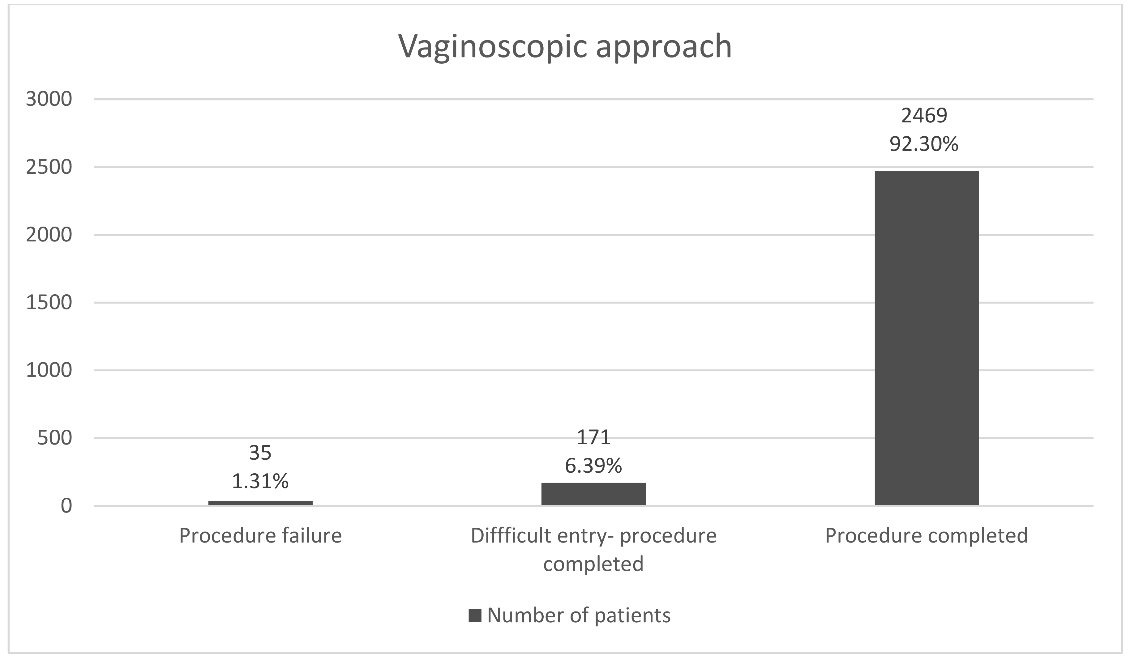
In this 20-year-period, 2675 women underwent office hysteroscopy with a mean age of 41.2 years (range 18 to 83 years of age). In this population sample, 18.8% were menopausal (no menstrual periods for at least 12 consecutive months), while the rest were of reproductive age. The procedure was successful in the majority of cases (2645 out of 2675) resulting to approximately 1% of failure mainly due to inability to introduce the hysteroscope through a stenosed cervical os.
In 171 out of 2675 cases (6.39%) the cervical canal was stenosed. The hysteroscopist managed though, to surpass the closed cervical os by taking advantage of normal saline flow and its distension properties as well as its force by the cuff-pressure bag facilitating effectively smoother entrance through the cervical canal. Although these cases were initially characterised by a closed cervical os, the procedure was completed, after applying mechanical pressure and by temporally increasing distension medium pressure.
In 35 out of all cases (1.31%) failure of the technique was noted. Out of 35 patients, where vaginoscopic approach failed, four (11.4%) were menopausal, two cases (5.7%) presented with a history of at least one unremarkable vaginal delivery and the remaining 29 (82.9%) were nulliparous or had an obstetric history of C-section as mode of delivery. All failures were due to closed external cervical os. No harm of the vaginal outlet or inlet was reported (Fig. 1).
 Fig. 1.
Vaginoscopic approach feasibility.
All patients received a questionnaire addressing their experience at the end of the procedure. The overall pain perception after vaginoscopic office hysteroscopy showed a mean score of 3.6466, on a 0 to 10 pain scale, where 10 indicated the worst pain experience. All patients were categorised in two different groups, group A (n: 2323) and group B (n: 442). The mean score regarding the pain for group A was 3.6376, while mean score for group B was estimated at 6.0826. Distribution of pain’s score is demonstrated at Fig. 2.
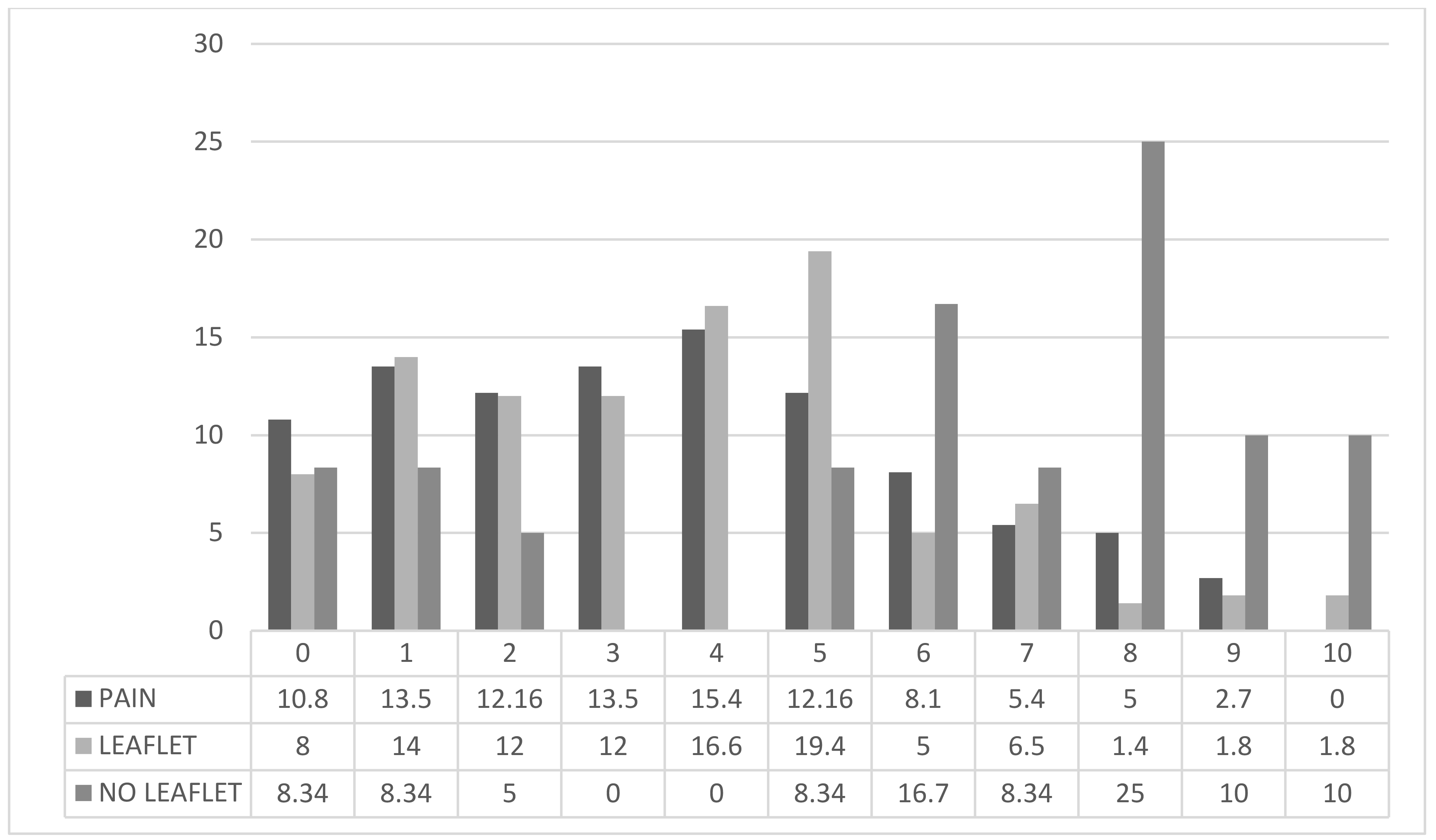 Fig. 2.
Pain perception using a 0 to 10 pain scale. Left histogram “PAIN”: overall pain perception during the procedure; Middle histogram “LEAFLET”: pain perception during the procedure in patients who received written information prior to hysteroscopy; Right histogram “NO LEAFLET”: pain perception during the procedure in patients who received no written information prior to hysteroscopy (Data is presented as percentage (%)).
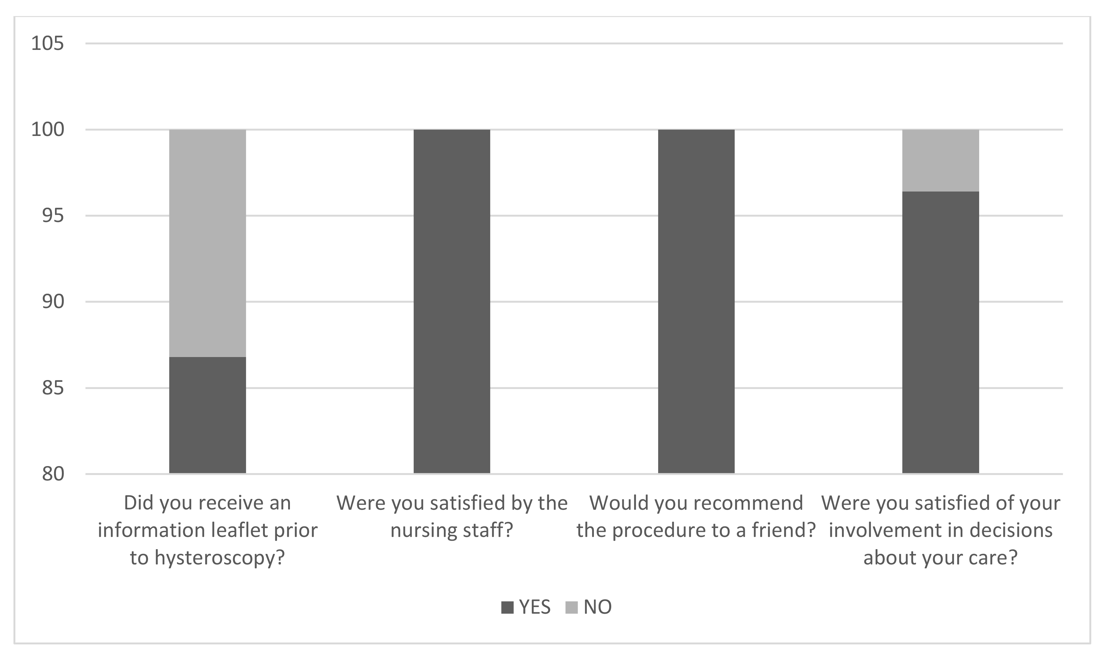
In general, all patients reported high levels of satisfaction as it is illustrated in Fig. 3. Regarding the benefits of vaginoscopic office hysteroscopy, 60.80% of the patients responded, “quick recovery” and 32.43% of the patients were satisfied for receiving feedback straight away. One out of three patients reported minimal disruption from their everyday life by choosing the method, while 1.35% reported no benefit of the procedure.
 Fig. 3.
Overall patient’s satisfaction.
In this study the term AUB (Abnormal Uterine Bleeding) refers to all women experiencing bleeding with no restriction to reproductive/menopausal status, however further subgroup analysis will be presented. The term “follow-up” refers to all women who underwent operative hysteroscopy prior to office hysteroscopy. Table 1 comprises of the main indications for office hysteroscopy in the sense of initial presenting complaint regardless of the hysteroscopic findings. Increased endometrial thickness [29], as well as the presence of fibroids and/or polyps, were set as an indication after sonographic evaluation of the patients through Transvaginal Sonography (TVS)—to avoid any misinterpretation these patients presented as asymptomatic at the time of sonographic assessment. With regards to increased endometrial thickness, for postmenopausal women presenting with AUB a cut-off of 4 mm was applied whereas 7 mm was set as the limit for asymptomatic postmenopausal women. For women of reproductive age during their proliferative phase of menstrual cycle an endometrial thickness of more than 10 mm was considered a clinical indication for further hysteroscopic assessment as per Trust’s guidance. For patients with suspected endometrial polyp, the feeding vessel was visualized using Doppler. All hysteroscopic indications were compared to hysteroscopic results at the end of the procedure (Table 2).
| Indications | Number of patients, n (%) | |
|---|---|---|
| AUB | 819 (30.6) | |
| Removal of foreign body | 14 (0.5) | |
| Subfertility | 304 (11.4) | |
| Adhesions | 19 (0.7) | |
| Polyps* | 322 (12) | |
| Fibroids* | 227 (8.5) | |
| Congenital Malformations | 98 (3.7) | |
| Tamoxifen use | 100 (3.7) | |
| Follow up | 306 (11.4) | |
| Recurrent miscarriages | 202 (7.6) | |
| Secondary infertility | 111 (4.2) | |
| Adenomyosis | 12 (0.4) | |
| Endometritis | 20 (0.7) | |
| Menstrual cycle imbalance | 154 (5.8) | |
| Increased Endometrial thickness | 151 (5.6) | |
| Fluid* | 18 (0.7) | |
| Cancer | 7 (0.3) | |
| Menopause | 11 (0.4) | |
| Abortion | 30 (1.1) | |
| Pelvic Pain | 8 (0.3) | |
| DUB | 31 (1.2) | |
| Pathologic colposcopy/Test Pap | 44 (1.6) | |
| HT use | 13 (0.5) | |
| Endometriosis | 5 (0.2) | |
AUB, abnormal uterine bleeding; DUB, dysfunctional uterine bleeding; HT, hormonal treatment.
*Indication after sonographic evidence.
| Indications | Hysteroscopy results | Hysteroscopy results | ||||||||||||||||||||||||||||||||||||||||||||||
|---|---|---|---|---|---|---|---|---|---|---|---|---|---|---|---|---|---|---|---|---|---|---|---|---|---|---|---|---|---|---|---|---|---|---|---|---|---|---|---|---|---|---|---|---|---|---|---|---|
| Normal | Endometrial Polyp | Submucous Fibroid T0 | Submucous Fibroid T1 | Submucous Fibroid T2 | Adhesions | Asherman | Hyperplasia | Cancer | Adenomyosis | Tamoxifen use | Uncategorized Path | Endometritis | POM | Blood Retain | Subtle Endometrial Lesions | Cervical Pathology | Fallopian Tube Obstruction | Micropolyps | Closed Cervical OS | Atrophy | Invisible Fall. Tubes | Total | ||||||||||||||||||||||||||
| AUB | 80 (9.77) | 285 (34.8) | 51 (6.23) | 13 (1.59) | 21 (2.56) | 26 (3.17) | 2 (0.24) | 178 (21.73) | 10 (1.22) | 38 (4.64) | 9 (1.10) | 72 (8.79) | 20 (2.44) | 6 (0.73) | 9 (1.10) | 3 (0.37) | 114 (13.92) | 5 (0.61) | 159 (19.4) | 61 (7.45) | 100 (12.21) | 41 (5.01) | 819 | |||||||||||||||||||||||||
| Sonographic evidence of endometrial Polyps | 43 (13.35) | 170 (52.8) | 4 (1.24) | 4 (1.24) | 1 (0.31) | 1 (0.31) | 0 (0) | 45 (13.98) | 0 (0) | 5 (1.55) | 1 (0.31) | 39 (12.11) | 5 (1.55) | 0 (0) | 1 (0.31) | 1 (0.31) | 49 (15.22) | 2 (0.62) | 24 (7.45) | 9 (2.8) | 34 (10.56) | 8 (2.48) | 322 | |||||||||||||||||||||||||
| Subfertility | 113 (37.17) | 53 (17.43) | 4 (1.32) | 3 (0.99) | 5 (1.64) | 9 (2.96) | 0 (0) | 3 (0.99) | 1 (0.33) | 6 (1.97) | 0 (0) | 12 (3.95) | 39 (12.8) | 2 (0.66) | 0 (0) | 0 (0) | 23 (7.57) | 3 (0.99) | 85 (27.9) | 20 (6.58) | 1 (0.33) | 4 (1.32) | 304 | |||||||||||||||||||||||||
| Sonographic evidence of submucous Fibroids | 27 (11.89) | 62 (27.31) | 42 (18.5) | 16 (7.05) | 18 (7.93) | 7 (3.08) | 0 (0) | 21 (9.25) | 3 (1.32) | 5 (2.2) | 1 (0.44) | 23 (10.13) | 4 (1.76) | 0 (0) | 0 (0) | 1 (0.44) | 27 (11.89) | 2 (0.88) | 34 (14.9) | 7 (3.08) | 31 (13.66) | 11 (4.85) | 227 | |||||||||||||||||||||||||
| Recurrent miscarriages | 81 (40.1) | 8 (3.96) | 1 (0.5) | 0 (0) | 2 (0.99) | 28 (13.8) | 1 (0.5) | 0 (0) | 0 (0) | 5 (2.48) | 0 (0) | 8 (3.96) | 39 (19.31) | 3 (1.49) | 0 (0) | 1 (0.5) | 17 (8.42) | 0 (0) | 59 (29.21) | 8 (3.96) | 2 (0.99) | 4 (1.98) | 202 | |||||||||||||||||||||||||
| AUB, abnormal uterine bleeding; POM, products of miscarriage. Data are presented as n (%). | ||||||||||||||||||||||||||||||||||||||||||||||||
Common indication was Abnormal Uterine Bleeding (AUB) (n = 819), followed by sonographic evidence of endometrial polyps (n = 322), “follow-up” (n = 306), subfertility (n = 304), sonographic evidence of submucous fibroids (n = 227), recurrent miscarriages (n = 202), menstrual cycle imbalance (n = 154) and increased endometrial thickness (n = 151).
Subgroup analysis regarding the menopausal status of the patients showed minor but significantly different results regarding the frequency of indications that were observed, compared to the main analysis. In detail, for women of reproductive age (n = 2052), common indications were AUB (n = 595), subfertility (n = 298), sonographic evidence of endometrial polyps (n = 269), “follow-up” (n = 227), recurrent miscarriages (n = 198) and sonographic evidence of endometrial fibroids (n = 180). For menopausal women (n = 494), common indications were AUB (n = 188), increased endometrial thickness on TVS (n = 77), tamoxifen use (n = 76), “follow-up” (n = 68) and polyps (n = 50) (Table 3 provides data as both absolute numbers and percentages).
| Indications | Women in reproductive age | Postmenopausal women |
|---|---|---|
| Number of patients, n (%) | ||
| AUB | 595 (28.9) | 188 (38) |
| Removal of foreign body | 13 (0.6) | 1 (0.2) |
| Subfertility | 298 (14.5) | |
| Adhesions | 18 (0.8) | |
| Polyps* | 269 (13.1) | 50 (10.1) |
| Fibroids* | 180 (8.7) | 34 (6.9) |
| Cong Malformations | 92 (4.4) | 4 (0.8) |
| Tamoxifen use | 18 (0.8) | 76 (15.4) |
| Follow up | 227 (11) | 68 (13.8) |
| Recurrent miscarriages | 198 (9.6) | 1 (0.2) |
| Secondary infertility | 110 (5.3) | 1 (0.2) |
| Adenomyosis | 9 (0.4) | 2 (0.4) |
| Endometritis | 20 (0.9) | |
| Menstrual cycle imbalance | 117 (5.7) | 30 (6.1) |
| Increased Endometrial thickness | 67 (3.2) | 77 (15.6) |
| Fluid* | 7 (0.3) | 10 (2) |
| Cancer | 3 (0.1) | 4 (0.8) |
| Menopause | 4 (0.1) | 4 (0.8) |
| Abortion | 28 (1.3) | |
| Pelvic Pain | 8 (0.3) | |
| DUB | 24 (1.1) | 3 (0.6) |
| Pathologic colposcopy/Test Pap | 29 (1.4) | 14 (2.8) |
| HT use | 1 (0.04) | 7 (1.4) |
| Endometriosis | 5 (0.2) | |
AUB, abnormal uterine bleeding; DUB, dysfunctional uterine bleeding; HT, hormonal treatment.
* indication after sonographic evidence.
For patients presenting with AUB, common hysteroscopic findings were endometrial polyps, hyperplasia, micropolyposis, cervical pathology and atrophy. The term “micropolyps” refers to endometrial polypoid protrusions less than 0.1cm according to the international literature [30]. For patients with an indication of sonographic evidence of endometrial polyps, hysteroscopy results agreed in almost 53% of cases, suspected by preoperative assessment with 2D TVS. The hysteroscopic findings in patients suffering from subfertility revealed normal endometrium, micropolyps and endometrial polyps whereas, for patients with an indication of recurrent miscarriages, the main hysteroscopic findings were normal endometrium, micropolyps and endometritis (Table 2). Endometrial oedema, focal or diffuse hyperaemia and haemorrhagic spots were considered suggestive of endometritis. Table 3 comprises hysteroscopic indications in women of reproductive aged compared to women in menopause.
For women of reproductive age with clinical indication of AUB, common hysteroscopic findings were endometrial polyps (n = 193), hyperplasia (n = 144) and micropolyps (n = 144). In this group patients suffering from subfertility presented with hysteroscopic findings: normal endometrium (n = 111), micropolyps (n = 85) and endometrial polyps (n = 52). For women in reproductive age with an indication of sonographic evidence of endometrial polyps, hysteroscopy findings agreed in almost 53% of the cases (Table 4).
| Indications | Hysteroscopy results | Hysteroscopy results | ||||||||||||||||||||||||||||||||||||||||||||||
|---|---|---|---|---|---|---|---|---|---|---|---|---|---|---|---|---|---|---|---|---|---|---|---|---|---|---|---|---|---|---|---|---|---|---|---|---|---|---|---|---|---|---|---|---|---|---|---|---|
| Normal | Polyp | Submucous Fibroid T0 | Submucous Fibroid T1 | Submucous Fibroid T2 | Adhesions | Asherman | Hyperplasia | Cancer | Adenomyosis | Tamoxifen use | Uncategorized Path | Endometritis | POM | Blood Retain | Subtle Endometrial Lesions | Cervical Pathology | Fallopian Tube Obstruction | Micropolyps | Closed Cervical OS | Atrophy | Invisible Fallopian Tubes | Total | ||||||||||||||||||||||||||
| Reproductive age (n = 2052) | ||||||||||||||||||||||||||||||||||||||||||||||||
| AUB | 73 (12.27) | 193 (32.44) | 42 (7.06) | 10 (1.68) | 17 (2.86) | 19 (3.19) | 1 (0.17) | 144 (24.2) | 0 (0) | 28 (4.71) | 2 (0.34) | 48 (8.07) | 17 (2.86) | 5 (0.84) | 8 (1.34) | 3 (0.5) | 78 (13.11) | 4 (0.67) | 144 (24.2) | 31 (5.21) | 19 (3.19) | 22 (3.7) | 595 | |||||||||||||||||||||||||
| Subfertility | 111 (37.25) | 52 (17.45) | 3 (1.01) | 3 (1.01) | 5 (1.68) | 8 (2.68) | 0 (0) | 3 (1.01) | 1 (0.34) | 6 (2.01) | 0 (0) | 12 (4.03) | 39 (13.09) | 2 (0.67) | 0 (0) | 0 (0) | 22 (7.38) | 3 (1.01) | 85 (28.52) | 19 (6.38) | 1 (0.34) | 4 (1.34) | 298 | |||||||||||||||||||||||||
| Sonographic evidence of endometrial Polyps | 41 (15.24) | 143 (53.16) | 3 (1.12) | 3 (1.12) | 1 (0.37) | 0 (0) | 0 (0) | 41 (15.24) | 0 (0) | 2 (0.74) | 0 (0) | 34 (12.64) | 5 (1.86) | 0 (0) | 1 (0.37) | 1 (0.37) | 37 (13.75) | 1 (0.37) | 22 (8.18) | 6 (2.23) | 6 (2.23) | 4 (1.49) | 269 | |||||||||||||||||||||||||
| Menopausal (n = 494) | ||||||||||||||||||||||||||||||||||||||||||||||||
| AUB | 3 (1.60) | 84 (44.68) | 7 (3.72) | 1 (0.53) | 3 (1.6) | 7 (3.72) | 1 (0.53) | 24 (12.77) | 9 (4.79) | 6 (3.19) | 7 (3.72) | 22 (11.7) | 1 (0.53) | 1 (0.53) | 0 (0) | 26 (13.83) | 1 (0.53) | 9 (4.79) | 26 (13.83) | 74 (39.36) | 17 (9.04) | 2 (1.06) | 188 | |||||||||||||||||||||||||
| Increased Endometrial Thickness | 1 (1.30) | 39 (50.65) | 1 (1.30) | 0 (0) | 0 (0) | 2 (2.6) | 0 (0) | 6 (7.79) | 1 (1.30) | 0 (0) | 4 (5.19) | 7 (9.09) | 0 (0) | 2 (2.6) | 3 (3.9) | 17 (22.08) | 1 (1.3) | 1 (1.3) | 15 (19.48) | 43 (55.84) | 4 (5.19) | 2 (2.6) | 77 | |||||||||||||||||||||||||
| Tamoxifen use | 0 (0) | 19 (25) | 0 (0) | 0 (0) | 1 (1.32) | 2 (2.63) | 0 (0) | 5 (6.58) | 0 (0) | 0 (0) | 43 (56.58) | 5 (6.58) | 0 (0) | 0 (0) | 0 (0) | 6 (7.89) | 2 (2.63) | 0 (0) | 17 (22.37) | 31 (27.63) | 13 (17.11) | 0 (0) | 76 | |||||||||||||||||||||||||
| AUB, abnormal uterine bleeding; POM, products of miscarriage. Data are presented as n (%). | ||||||||||||||||||||||||||||||||||||||||||||||||
For menopausal women with an indication of AUB, most common hysteroscopic findings were endometrial polyps (n = 84). In this group presenting with an indication of increased endometrial thickness, the most common hysteroscopic findings were also endometrial polyps (n = 39). Finally, an indication of tamoxifen use revealed hysteroscopic findings of endometrial changes affected by the use of this agent agreed in almost 56.5% of the cases (Table 4).
Through the years we were able to collect histologic data for the majority of cases (n = 2089 out of 2675) for this study. Predictive characteristics of hysteroscopy in the diagnosis of various conditions were performed for those patients and by comparing hysteroscopic findings with the histology results, for the assessment of a normal endometrium hysteroscopy showed a sensitivity of 60.9%, specificity of 92.1%, PPV of 79.07% and NPV 82.8%. In cases of endometrial polyps, hysteroscopy had a sensitivity of 92.04%, specificity of 89.1%, PPV of 73.5% and NPV 97.1%. For fibroids, sensitivity and specificity were calculated at 98.5% and 100 % respectively, while PPV and NPV at 100% and 99.9%, respectively. For endometrial cancer, the predictive characteristics of hysteroscopy were 87.5% and 99.7% with regards to sensitivity and specificity, and 63.6% and 99.9% for PPV and NPV, respectively. Finally, in cases of hyperplasia, hysteroscopy shows a sensitivity of 75.0%, specificity of 91.03%, PPV of 11.7% and NPV of 99.5% (Table 5).
| Sensitivity | Specificity | PPV | NPV | Diagnostic accuracy | |
|---|---|---|---|---|---|
| Polyps | 92.04% | 89.14% | 73.52% | 97.16% | 89.86% |
| Submucous Myoma | 98.55% | 100.0% | 100.0% | 99.93% | 99.93% |
| Cancer | 87.5% | 99.74% | 63.64% | 99.93% | 99.67% |
| Hyperplasia | 75.0% | 91.03% | 11.76% | 99.56% | 90.78% |
| Normal | 60.9% | 92.1% | 79.07% | 82.84% | 81.88% |
PPV, positive predictive value; NPV, negative predictive value.
To our best of knowledge, this is the first retrospective study presenting data from a large population on office hysteroscopy in Greece to date and one of the largest internationally. The procedure was efficient and safe. All women were discharged after the intervention with no complications. No analgesia or anesthesia was needed. This retrospective study of 2765 cases of vaginoscopic office hysteroscopy demonstrates high feasibility of the technique.
The failure rate of the technique was approximately 1%, in line with the
international literature [15, 31]. However, many studies indicate great
discrepancies with regards to failure rates spanning from 0.5% to more than 50%
[32]. Hysteroscopy remains an operator-dependent technique. Thus, decreased rates
of failure correspond with the experience and personal style of each
hysteroscopist [33]. The level of expertise of the members of our endoscopic unit
was already advanced and this possibly explains our results regarding the
reported minimal failure rate of office hysteroscopy. There have been studies
suggesting that the failure rate of the technique is estimated at approximately
5% of cases when employing vaginoscopic approach [20]. It seems that apart from
the dimensions of the hysteroscopes, the distension medium also plays an
assistive role at increasing the feasibility of the technique. With regards to
distension medium, normal saline found to be superior of CO
Vaginoscopic hysteroscopy is feasible, simple and well tolerated technique that can be safely performed in an outpatient/office setting [9, 14]. Vaginoscopic approach offers benefits for both specialists and patients [41]. The absence of speculum or tenaculum results in less pain, discomfort and stress experienced by patients as it is noted in our study [42]. Moreover, analgesic premedication is not indicated when performing office hysteroscopy through vaginoscopy [43, 44]. Also, a higher degree of free movements and manoeuvres from the perspective of the operator and less equipment in use simplify the procedure as experienced by the specialist [45]. Limitations of this approach are vulvar, vaginal or endometrial infection, in case of which, the procedure ought to be postponed and treatment of the underlying disease should be initiated [46].
Management of anxiety experienced by patients prior to the procedure cannot be overemphasized. As reported also in published evidence, patients that receive clear information before proceeding with an intervention are more likely to report less anxiety and stress [47]. Our findings are in keeping with this trend, since patients who had the chance to familiarise themselves with the procedure in the form of written information leaflets, felt more supported and prepared experiencing at the same time less pain. Anxiety is a common experience for many patients undergoing medical procedures such as office hysteroscopy [48]. The impact of anxiety on the procedure is well documented, with studies showing that high levels of anxiety can lead to increased pain perception, higher rates of complications, and reduced satisfaction with the procedure [49]. In addition, anxiety can also have long-lasting effects, with some patients reporting ongoing fear and avoidance of medical procedures in the future. Therefore, it is important for healthcare providers to take steps to minimize anxiety and provide support to patients undergoing office hysteroscopy, in order to ensure the best possible outcomes and patient experience [50]. This may include providing information and education about the procedure which can help to alleviate fears and uncertainties about the procedure.
On a systematic review by Cooper et al. [45], vaginoscopic approach demonstrated less pain and better overall experience by patients compared to the traditional technique. In fact, vaginoscopic approach is ideal in specific groups, such as patients with restricted movement due to an orthopaedic condition of lower extremities, or women with no history of penetrative sexual intercourse, nulliparous patients or women suffering from atrophy, in which vaginal contact often results in further distress and contributes to negative experience [45, 51]. In our study, the overall satisfaction rates were almost perfect, however mild pain sensation during the procedure was still reported. It is worth mentioning that patients who received additional written information about the procedure, were more likely to experience less pain than those who did not. It is likely that written information helped patients to understand better the procedure, minimizing at the same time stress and anxiety in advance.
In 1996, Nagele et al. [52], reported large-series scientific evidence regarding outpatient diagnostic hysteroscopies. Bettocchi et al. [26] was one of the first researchers to present the concept of office hysteroscopy combining diagnostic and operative hysteroscopy in an outpatient setting without the use of analgesia or anesthesia, establishing the concept of office hysteroscopy. Later on, almost a decade ago, Di Spiezio et al. [53] provided more information on the subject, when data arising from 5000 office hysteroscopies was published. Taken into consideration our relatively small country, it seems that our data could potentially be used as a solid representative of Greek population in terms of epidemiologic data regarding endometrial and endocervical pathologies that could significantly add up to the existing European and International scientific evidence.
The main hysteroscopic indication in our sample was AUB, but as expected the respective hysteroscopic findings were dependent on reproductive/menopausal status of the patients [53]. For women of reproductive age subfertility issues were a common hysteroscopic indication, while in menopausal women, increased endometrial thickness after sonographic evaluation indicated the need for further hysteroscopic assessment. Our findings are in keeping with the existing international scientific evidence [54, 55]. Hysteroscopy has an important role in women with postmenopausal bleeding (PMB) and decreased endometrial thickness, since identifying different endometrial pathologies under direct vision is achieved in real time [56]. In addition, hysteroscopy offers biopsies or excision of endometrial lesions under direct vision, allowing accurate diagnosis.
The efficacy of the technique is well demonstrated by offering high sensitivity and specificity in four major endometrial pathologies as a metanalysis by Gkrozou et al. [57] indicates. In case of normal endometrium, although sensitivity of the technique is not quite high, irreversibly high negative predictive value shows that in the presence of normal hysteroscopic traits, endometrial pathology should be excluded [58]. In the available literature, there are endometrial features that do not correspond to any traditional category of endometrial pathology [59]. Our study shows that in cases where the hysteroscopist fails to identify specific endometrial pathology, endometrial biopsy facilitates the diagnosis, especially in cases where suspicious endometrial patterns are recognized.
Strength of this study is mainly the large number of cases attending a single Endoscopic unit, as well as patient’s feedback addressing their overall satisfaction and pain perception by this method. This is the largest retrospective study of vaginoscopic office hysteroscopy in Greece to date. Unfortunately, no learning curve data could be obtained by this study, since only highly experienced hysteroscopists performed all hysteroscopies. However, the international literature suggests that initial adaptation of the approach tallies temporary with an increase in the duration of the procedure. More specifically, Bettocchi et al. [18] concluded that after the initial ten cases of vaginoscopic hysteroscopy, the duration of the technique is similar to the traditional method. To conclude, vaginoscopic approach technique is known as a non-traumatic technique, which is safe, efficient and improves the overall patients’ experience [60]. Our study has confirmed the importance of this technique in the everyday clinical practice. The main limitation of this study is the retrospective data analysis which could potentially increase the risk of bias. The hysteroscopists performing all hysteroscopies over the years were quite experienced and our data cannot provide information regarding hysteroscopic dexterity since no recordings on the development of their skills was retrieved. Hysteroscopy remains a subjective technique and its efficacy depends on the experience of the operator.
This retrospective study presents large data regarding vaginoscopic office hysteroscopy in a busy public endoscopic unit in Ioannina, Greece. Our findings are in keeping with current scientific evidence and provide substantial data with regards to epidemiology.
Vaginoscopic office hysteroscopy offers great diagnostic accuracy in patients with hyperplasia, endometrial cancer, fibroids, and endometrial polyps. The technique is proven to be simple, safe, and feasible.
The datasets used and/or analyzed during the current study are available from the corresponding author on reasonable request.
OT—first authorship, extraction and drafting of the manuscript, data collection, performed procedures; FG edited and revised manuscript initial draft of the manuscript, performed procedure; IN, TV, AD, NI contributed to the interpretation of the data of this project, reviewed this project critically for important intellectual content, revised the final form of this manuscript and edited discussion; VS collected the questionnaires, edited patient experience assessment; APB collected histologic reports, edited histologic findings on the initial data; MP supervised and conceived the project, performed procedures; ED, GN, AT performed statistical analysis of the data collected. All authors read and approved the final version of this manuscript. All authors contributed to editorial changes in the manuscript. All authors have participated sufficiently in the work and agreed to be accountable for all aspects of the work.
All subjects gave their informed consent for inclusion before they participated in the study. The study was conducted in accordance with the Declaration of Helsinki. For this study Institutional Review Board Approval was obtained by the University Hospital of Ioannina (IRB number 207/15-03-2020).
The authors of this manuscript would like to thank Mr. Michael Thompson for editing the final form of this manuscript as a native English speaker. His contribution is much appreciated.
This research received no external funding.
The authors declare no conflict of interest.
References
Publisher’s Note: IMR Press stays neutral with regard to jurisdictional claims in published maps and institutional affiliations.