


1 Department of Neurosurgery, The First Affiliated Hospital of Xiamen University, 361005 Xiamen, Fujian, China
2 Department of Neurosurgery, Fuzhou 900th Hospital, Fujian Medical University Fuzong Clinical College, 350025 Fuzhou, Fujian, China
†These authors contributed equally.
Abstract
Background: Magnetic resonance imaging (MRI) has become the most important radiological procedure for diagnosing and following pituitary tumors. But previous MRI studies on pituitary adenomas are mainly focused on the posterior pituitary. Few research has been done on residual normal pituitary tissue before and after transsphenoidal surgery. This retrospective cohort study investigates the pre- and postoperative magnetic resonance imaging characteristics of normal pituitary tissues regarding transsphenoidal resection of pituitary macroadenomas. Methods: Pre- and postoperative magnetic resonance imaging scanning of 112 consecutive pituitary macroadenoma patients who underwent tumor resection via transsphenoidal approach was performed, and their medical records were studied. Results: On preoperative MRI, 66 cases of pituitary stalks were identifiable, 9 of them were roughly in the middle, and 57 cases showed left or right deviation, with the angle between pituitary stalks and the sagittal plane was 5.32°–64.05° (average 21.65°). Among the 57 patients with preoperative pituitary stalk deviation, 55 of the pituitary stalk deviations improved in 1 week after surgery, and 30 cases were almost in the middle in 4–6 months after operation, with the other cases get better in varying degrees. The diameter of pituitary stalk was 1.08–3.89 mm (mean 2.36 mm) in pre-operation, and 1.29–3.43 mm (mean 2.30 mm) in 4–6 months after operation. The length of pituitary stalk was 1.41–11.74 mm (mean 6.12 mm) preoperatively, 3.61–11.63 mm (mean 6.93 mm) early postoperatively, and 5.37–17.57 mm (mean 8.83 mm) in 4–6 months after operation. Pituitary stalk was thickened or compressed on preoperative MR images, and gradually recovered to normal during postoperative period. It tended to be in the middle position and its length increased gradually until 4–6 months after operation. On preoperative MRI, 69 out of 112 patients showed residual pituitary tissues (RPT)(+) on enhanced MRI. RPT were likely located above the adenomas in somatotroph adenoma patients. Morphological restitution of postoperative normal pituitary tissues was better in lateral displacement than in superior or superolateral patterns on preoperative magnetic resonance imaging. Postoperative normal pituitary tissues usually subsided directly in superior displacement pattern on preoperative MRI, while were likely to be confined in the lateral side in lateral and superolateral displacement patients. Postoperative morphologic remodeling grade of RPT was positively correlated with the maximum diameter of pituitary adenoma (p = 0.000), but not with age. Conclusions:The larger the tumor diameter, the worse the pituitary morphological recovery after tumor resection. Relative locations of normal pituitary and adenoma tissues may be related to adenoma type and may affect postoperative reconstruction of residual normal pituitary tissues. These findings enable surgeons to distinguish pituitary tissue from residual or recurring tumor tissue on postoperative magnetic resonance imaging.
Keywords
- MRI
- pituitary macroadenomas
- residual pituitary tissues
- posterior pituitary bright spot
- pituitary stalk
- transsphenoidal approach
Pituitary adenomas (PAs) account for 10%–25% of all intracranial neoplasms, have different morphology and expansion directions, and about 35% are invasive [1, 2]. PAs usually originate from the anterior pituitary gland (adenohypophysis). The adenohypophyseal tumors are recently renamed pituitary neuroendocrine tumors (PitNET), which may present various behaviors, such as invasive, aggressive and malignant with metastases. They are classified into seven morphofunctional types and three lineages: lactotroph, somatotroph and thyrotroph (PIT-1 lineage), corticotroph (T-PIT lineage) or gonadotroph (SF-1 lineage), null cell or immunonegative tumor and plurihormonal tumors [3]. The World Health Organization (WHO) 2017 classification of PitNET is mostly based on immunohistochemistry for pituitary hormones, pituitary-specific transcription factors, and other immunohistochemical markers commonly used in pathology practice [4, 5].
Transsphenoidal surgery remains the first-line approach for most PAs (except for
prolactinoma) resection because of its minimal invasiveness and low morbidity
[6]. The most frequent complications associated with transsphenoidal pituitary
surgery are normal pituitary tissue damage and cerebrospinal fluid leakage,
leading to partial or complete insufficiency of one or more hormonal axes
(hypopituitarism) [7]. PAs originate from the adenohypophysis and combine with
the normal pituitary tissue by deformation and displacement. The average adult
gland measures 6 mm superior-inferior
However, few reports have investigated the pre-and post-operative MRI appearance of the normal pituitary tissues in patients with pituitary macroadenomas, including size, shape, location, and its clinical correlation [12]. The normal pituitary tissues, including adenohypophysis and neurohypophysis, can be identified easily on MRI T1-weighted images (T1WI) in healthy adults or microadenoma patients [13]. Previous studies are mainly focused on the posterior pituitary [14, 15]. Our previous studies suggest that neurohypophysis exhibits high signal intensity on MRI T1WI, related to the neuro-secretory granules containing antidiuretic hormone (ADH). As central diabetes insipidus (DI) is characterized by reducing ADH release, this entity may be reflected by reducing the posterior pituitary bright spot (PBBS) signal [16, 17].
The purpose of this work was to investigate the pre-and postoperative magnetic resonance imaging of normal pituitary tissues in the context of transsphenoidal resection of pituitary macroadenomas. The morphology and remodeling of residual normal pituitary tissues following transsphenoidal resection were observed on contrast-enhanced MRI, and their relationship with PAs was analyzed. The appearance of pituitary stalk was also analyzed as well as its clinical significance.
A total of 112 patients (58 males and 54 females; age range 23–75 years; mean
age at surgery 45 years) between September 2013 and September 2018 were
retrospectively studied. Their data regarding PA characteristics are shown in
Table 1. During this period, 335 patients diagnosed with PA were treated. Still,
only those who met the following criteria were included in the study: (i) surgery
via a transsphenoidal approach for pituitary macroadenomas (diameter
| No. (n) | Percentage (%) | ||
| Gender | |||
| Male | 58 | 51.8% | |
| Female | 54 | 48.2% | |
| Tumor immunohistochemistry | |||
| Gonadotroph adenoma | 35 | 31.3% | |
| Lactotroph adenoma | 18 | 7.1% | |
| Somatotroph adenoma | 29 | 25.9% | |
| Thyrotroph adenoma | 5 | 4.5% | |
| Null-cell adenoma | 9 | 8.0% | |
| Corticotroph adenoma | 7 | 6.2% | |
| Plurihormonal adenomas | 9 | 8.0% | |
| Tumor size | |||
| 46 | 41.1% | ||
| 66 | 58.9% | ||
| Cavernous sinus invasion (Knosp grades) | |||
| 0 grade | 4 | 3.6% | |
| 1 grade | 26 | 23.2% | |
| 2 grade | 34 | 30.4% | |
| 48 | 42.8% | ||
All patients underwent a standard microsurgical pituitary adenoma resection via
a transsphenoidal approach by the corresponding author. The sellar floor was
opened bilaterally, form a bone window of about 1.0 cm
In addition to surgery, each patient was evaluated and treated
endocrinologically. The diagnosis of DI was made according to international
guidelines [18]. In detail, postoperative DI was defined as polyuria (urine
production
The Ethics Committee approved all procedures of Fujian Medical University. Written informed consent was obtained from every patient.
The MRI scanning was performed with a 3.0T magnetic resonance scanner (Tim Trio,
Siemens Medical Solutions, Erlangen, Germany). T1-weighted images (400-500/8-15,
TR/TE) were obtained before and after the Gadolinium diethylenetriaminepenta-acetic acid (Gd-DTPA) injection. The parameters used
were as follows: scanning field, 180 mm
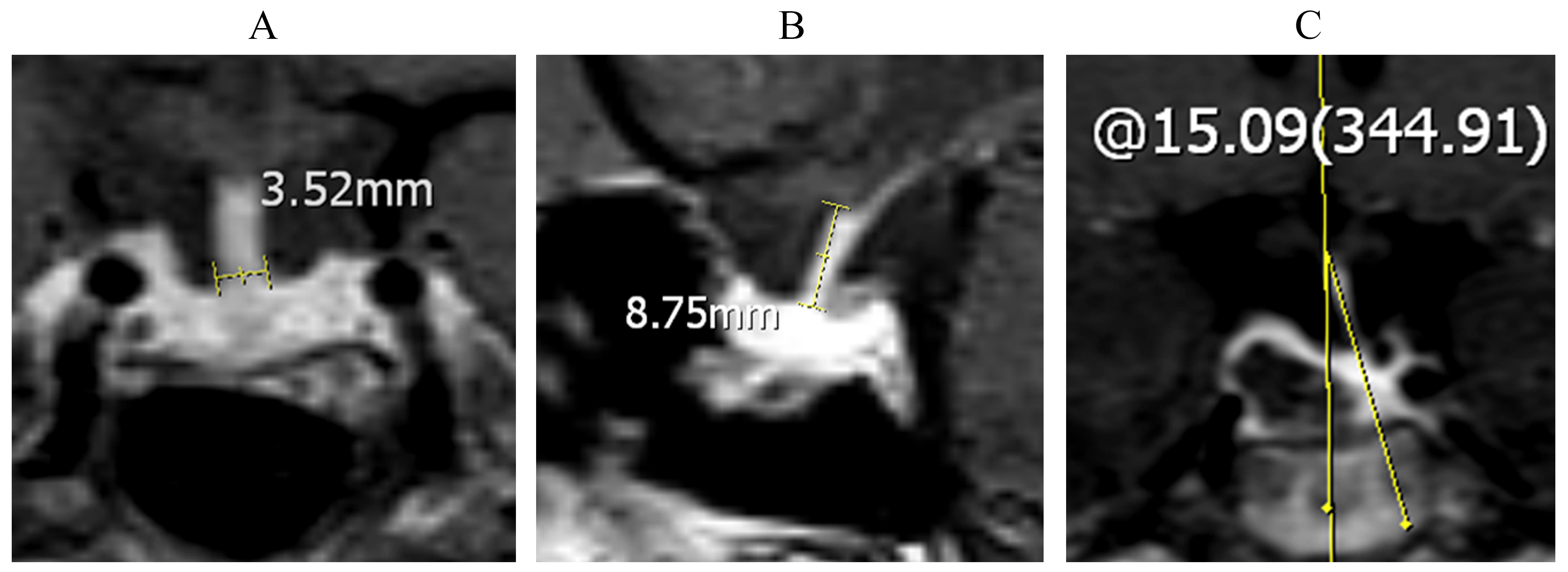 Fig. 1.
The width, length and deviation angle of pituitary stalk were measured with the picture archiving and communication system (PACS) tools on enhanced magnetic resonance imaging (MRI). (A) Diameter of pituitary stalk at the superior edge of pituitary gland on coronal enhanced T1WI was recorded as its width. (B) Distance from the optic chiasm plane to the upper edge of pituitary gland on sagittal enhanced T1WI was recorded as the length of pituitary stalk. (C) The angle between pituitary stalks and the sagittal plane was recorded as its deviation angle.
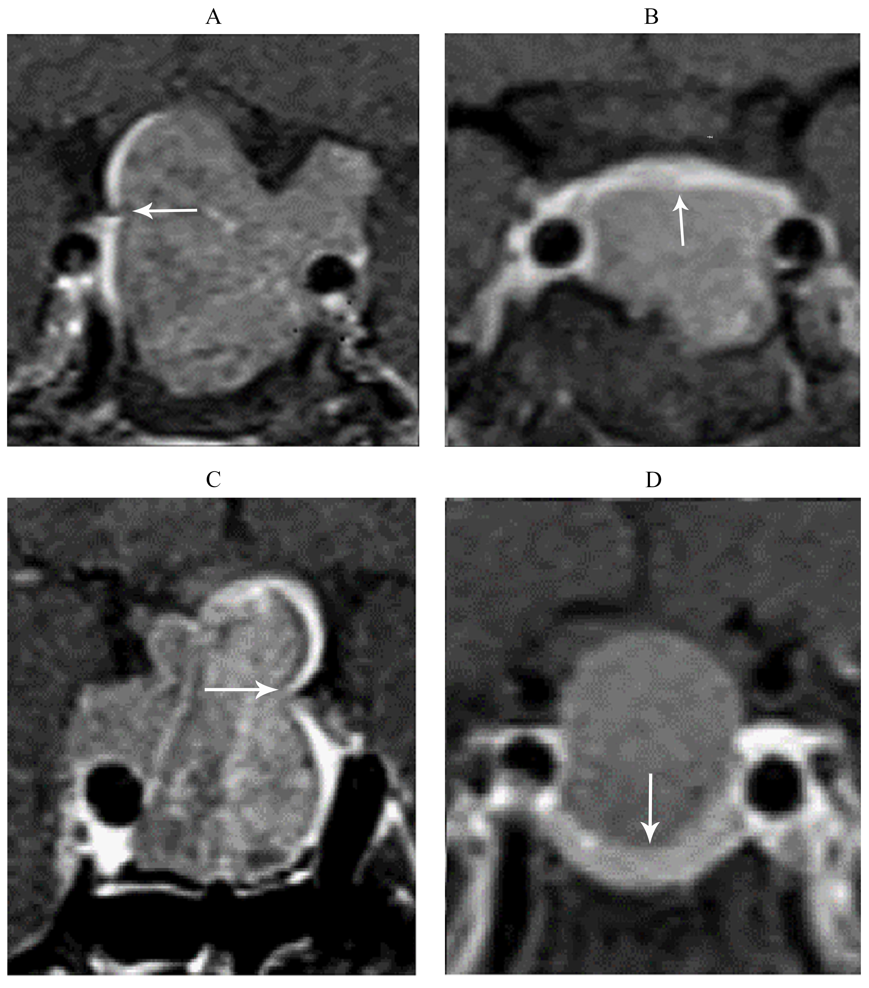 Fig. 2.
Distortion location of the residual normal pituitary gland in pituitary macroadenoma patient on preoperative coronal contrast-enhanced Magnetic resonance imaging (MRI). (A) Lateral displacement. (B) Superior displacement. (C) Superolateral displacement. (D) Inferior displacement. The white arrow indicates residual normal pituitary tissues.
According to the extent of postoperative morphological restitution, Di Maio S
subdivided the recovery degree of normal residual pituitary tissue into the
following four groups: Group 1—normal residual gland or almost normal, Group
2—more than 50% recovery, Group 3—less than 50% of the normal residual gland,
Group 4—little or no residual gland [20]. In the present paper, on the
postoperative (4th–6th month) MRI, the residual normal pituitary glands were
also divided into four groups: G1, measured residual volume = 75–100% of adult
pituitary volume; G2, measured volume = 50–75% of adult pituitary volume; G3,
measured volume = 20–50% of adult pituitary volume; G4, measured volume
Histological analysis of the surgical specimens, including HE staining and immunohistochemistry, was performed. Their data regarding histological variations are shown in Table 2. According to the criteria published by the World Health Organization in 2017 [4, 5], the specimens were classified into the following types: 18 cases of lactotroph, 29 cases of somatotroph, 5 cases of thyrotroph, 7 cases of corticotroph, 35 cases of gonadotroph, 9 cases of null-cell, and 9 cases of plurihormonal adenomas.
| Adenoma type | No. (n) | Pituitary hormones and other immunomarkers | Transcription factors |
| Gonadotroph adenoma | 35 | b-FSH, b-LH, |
SF-1, ERa |
| Lactotroph adenoma | 18 | PRL | PIT-1, ERa |
| Somatotroph adenoma | 29 | GH, |
PIT-1 |
| Thyrotroph adenoma | 5 | b-TSH, |
PIT-1, GATA2 |
| Corticotroph adenoma | 7 | ACTH | T-PIT |
| Null-cell adenoma | 9 | No markers | None |
| Plurihormonal adenomas | 9 | GH,PRL, b-TSH, |
PIT-1 |
Note: FSH, Follicle-stimulating Hormone; LH, Luteinizing Hormone; PRL, Prolactin; GH, Growth Hormone; TSH, Thyrotropin-stimulating Hormone; ACTH, Adrenocorticotropic Hormone; SF-1, Steroidogenic Factor 1; ER, Estrogen Receptor; PIT-1, Pituitary Transcription Factor 1; GATA, GATA transcription factor.
All statistical analyses were performed using SPSS 19.0 (IBM, Armonk, NY, USA)
and p value less than 0.05 was considered statistically significant.
Quantitative data were expressed as mean
On preoperative MRI T1-weighted imaging, pituitary stalk can be identified in 66 out of 112 patients. Preoperative pituitary stalks were nearly centered in 9 cases, left deviation in 25 cases, right deviation in 32 cases, with the angle between pituitary stalk and the sagittal plane was 5.32°–64.05° (average 21.65°). Some pituitary stalks were squeezed in “S” or “C” shape (Fig. 3). On the 1st week postoperative MRI, the 57 patients with preoperative pituitary stalk deviation showed deviation relieved in 55 cases, and the angle between the pituitary stalk and the median sagittal plane was reduced to 6.66°–36.10° (average 16.58°). Moreover, on the 4th–6th month postoperative MRI, the angle was further reduced to 4.34°–25.70° (average 12.11°), and 30 pituitary stalks were almost in the middle position. The pituitary stalks with “S” or “C” distortion gradually returned to normal after adenoma resection (Fig. 3). These results suggest that pituitary stalks were thickened or compressed on preoperative MR images, and tended to be in the middle position after operation.
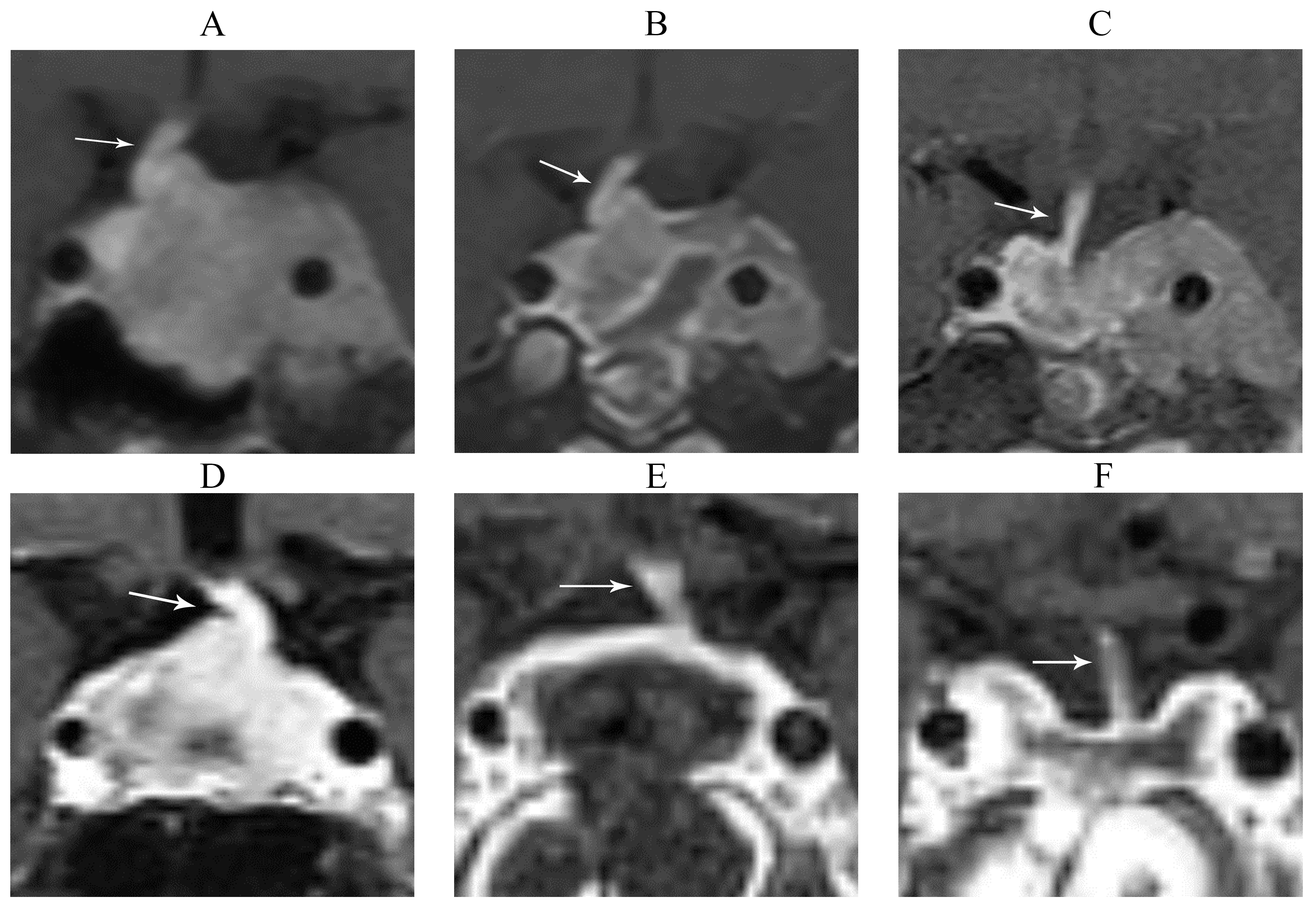 Fig. 3.
Pituitary stalks were squeezed in “S” or “C” shape preoperatively and showed deviation relieved after transsphenoidal pituitary adenoma resection. (A) Preoperatively and (B) 1st week postoperatively and (C) 6th month postoperatively, Coronal contrast-enhanced T1WI of gonadotroph adenoma with the pituitary stalk squeezed in “S” shape. (D) Preoperatively and (E) 1st week postoperatively and (F) 6th month postoperatively, Coronal contrast-enhanced T1WI of somatotroph adenoma with the pituitary stalk squeezed in “C” shape. The white arrow indicates pituitary stalks.
According to the 66 cases of recognizable pituitary stalk on enhanced MRI, the diameter of pituitary stalk was 1.08–3.89 mm (mean 2.36 mm) in pre-operation, and 1.29–3.43 mm (mean 2.30 mm, n = 66) in 4–6 months after operation. The length of pituitary stalk was 1.41–11.74 mm (mean 6.12 mm) preoperatively, 3.61–11.63 mm (mean 6.93 mm) early postoperatively, and 5.37–17.57 mm (mean 8.83 mm, n = 66) in 4–6 months after operation. While in the 46 cases of pituitary stalk cannot being clearly displayed before operation, 26 cases could be identified early after operation, and 40 cases could be identified on the 4th–6th month postoperative MRI, with the length of pituitary stalk from 2.73–11.64 mm (mean 6.69 mm, n = 26) to 5.13–12.79 mm (mean 9.14 mm, n = 40). These results showed that there was no statistically significant difference in the diameter of the pituitary stalk between preoperative and 4-6 months after operation (p = 0.616), while the length of pituitary stalk was longer in the 4–6 months after operation period (p = 0.000) (Table 3). The pituitary stalks presented no obvious change in their diameters, while their length extended gradually during postoperative period.
| Diameter (mm) | Length (mm) | |
| Pre-operation | 2.36 |
6.12 |
| Post-operation | 2.30 |
8.83 |
| t | 0.503 | 6.778 |
| p | 0.616 | 0.000 |
Note: The diameter of pituitary stalk was not different pre- and 4–6 months after pituitary adenoma surgery (Independent Samples t Test, t = 0.503, p = 0.616), while the length of pituitary stalk was significantly different statistically (Independent Samples t Test, t = 6.778, p = 0.000).
On preoperative MRI, 88 out of 112 patients showed PPBS(+) on T1WI (78.6%).
Nine of these PPBS(+) patients had PPBS(–) at one week postoperatively, and
still 65 cases presented PPBS(+) in 4–6 months after operation. In these cases,
some PPBS are ectopic to the lower part of the pituitary stalk due to tumor
influence (Fig. 4). While on the postoperative MRI, PPBS can be enlarged or
disappeared (Fig. 4). These results indicate that PPBS(+) may persisted
preoperatively and postoperatively in most cases, and only disappeared in a few
cases. In all cases, 24 patients (21.43%) developed postoperative diabetes
insipidus in one week after operation, all of them were transient diabetes
insipidus and improved by deammoniacal vasopressin treatment, no permanent
diabetes insipidus. The incidence of postoperative diabetes insipidus was higher
in PPBS(–) group (29.2%) than in PPBS(+) group (13.6%) in the early
postoperative period, but the difference was not statistically significant
(
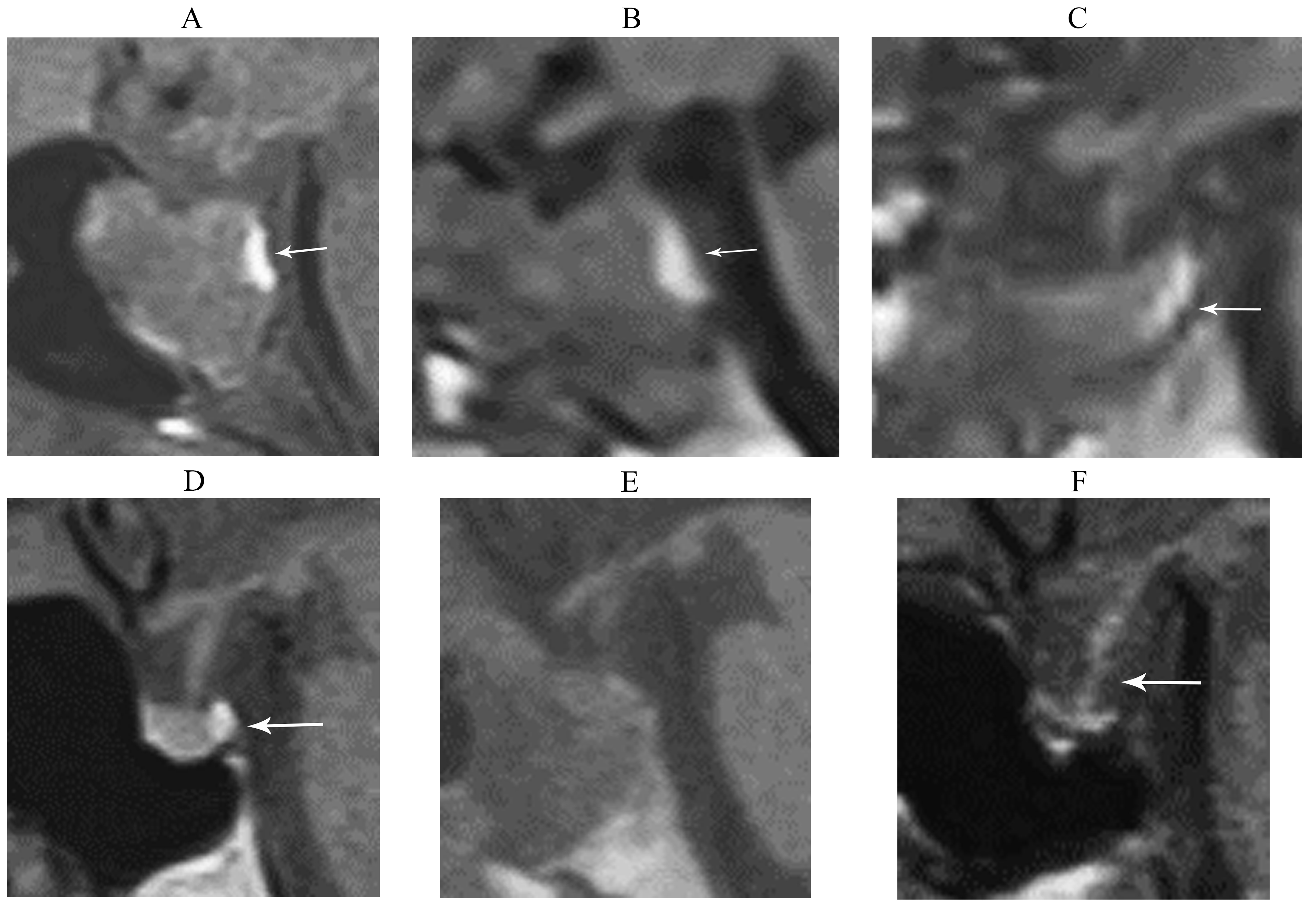 Fig. 4.
Changes of Posterior pituitary bright spots (PPBS) after transsphenoidal tumor resection in patients with pituitary adenoma on sagittal MRI-T1WI. (A) Preoperatively and (B) 3-day postoperatively and (C) 4th month postoperatively, male, 47 years old, PPBS persisted and its signal intensity decreased slightly after operation. Posterior lobe of pituitary gland descended to the sellar bottom, lying in the posterior part of sellar bottom. No high signal was found in the pituitary stalk. The patient had no postoperative diabetes insipidus. The white arrow indicates PPBS. (D) Preoperatively and (E) 2-day postoperatively and (F) 5th month postoperatively, Female, 34 years old, PPBS(+) was obvious preoperatively, and disappeared after operation. The signal of pituitary stalk was higher than normal, which show that the antidiuretic hormone might storage in pituitary stalk replaced posterior pituitary. The patient had postoperative transient diabetes insipidus for 3 days and urine output for 4000 mL/days (The white arrow indicates PPBS and pituitary stalk).
| PPBS(+) | PPBS(–) | p | ||
| Postoperative diabetes insipidus | ||||
| (+) | 12 | 7 | 3.229 | 0.072 |
| (–) | 76 | 17 |
Note: The incidence of postoperative diabetes insipidus was not different statistically between PPBS(+) group and PPBS(–) group. Chi Square Test,
On preoperative MRI, T1WI and contrast-enhanced MRI was employed to identify the
RPT, and 69 out of 112 patients showed RPT(+) on enhanced MRI. As documented on
MRI, the position of RPT relative to PA was found superior in 29 cases, lateral
in 30 cases, and superolateral in 10 cases. To examine the relationship between
RPT location and immunohistochemical types, the 69 patients with RPT(+) on
preoperative MRI were classified into gonadotroph adenoma (26 cases), somatotroph
adenoma (21 cases), lactotroph adenoma (12 cases), corticotroph adenoma (5
cases), and null-cell adenoma (5 cases). The data showed that RPT tended to be
above the tumor in somatotroph adenoma (Fig. 3). While in gonadotroph adenoma
patients, RPT was more likely to be located on one side of the tumor (Fig. 3). It
was found that 16 out of the 21 somatotroph adenomas had RPT superior and
superolateral displacement (76.2%), which was significantly different from that
among the 26 gonadotroph adenoma cases (12 cases; 46.1%) (
| Location of normal pituitary | Somatotroph | Lactotroph | Corticotroph | Gonadotroph | Null-cell adenoma | Total |
| Lateral | 5 | 6 | 3 | 14 | 2 | 30 |
| Superior | 13 | 5 | 2 | 8 | 1 | 29 |
| Superio-lateral | 3 | 1 | 0 | 4 | 2 | 10 |
To explore the relationship between RPT location on preoperative MRI and postoperative pituitary recovery of the 69 patients who showed RPT(+) on preoperative enhanced MRI, normal pituitary tissue recovery status after surgery was followed. All 69 patients showed visible normal pituitary tissues in T1WI before and after surgery. Among the 69 patients, postoperative pituitary volume was recovered in 24 cases of G1, 26 cases of G2, 12 cases of G3, and 7 cases of G4. For G1 and G2, 26 cases were of lateral type (52%), 18 cases were of superior type (36%), and 6 cases were of superolateral type (12%), with significant differences among the 3 types (p = 0. 032) (Table 6). Moreover, morphological restitution of postoperative normal pituitary tissues was better in lateral displacement than in superior or superolateral patterns on preoperative magnetic resonance imaging. Postoperative RPT usually subsided directly in superior displacement pattern on preoperative MRI, while were mostly confined in the lateral side in lateral and superolateral displacement patients. These results indicate that the lateral type of pituitary usually has the best recovery on MRI.
| Pituitary location | G1 | G2 | G3 | G4 | Total |
| Lateral | 13 | 13 | 3 | 1 | 30 |
| Superior | 7 | 11 | 6 | 4 | 28 |
| Superio-lateral | 4 | 2 | 3 | 2 | 11 |
Note: G1, measured residual volume = 75–100% of adult pituitary volume; G2,
measured volume = 50–75% of adult pituitary volume; G3, measured volume = 20–50% of adult pituitary volume; G4, measured volume
To determine how RPT postoperative recovery is related to adenoma diameter, tumor resection, we examined the adenomas from all 112 patients. As documented in the Table 7, postoperative pituitary volume was good recovered in 50 cases (G1 and G2), poor recovered in 62 cases (G3 and G4). Postoperative morphologic remodeling grade of RPT was positively correlated with the maximum diameter of pituitary adenoma (p = 0.000), but not with age or gender. The larger the diameter of pituitary adenoma, the worse the postoperative RPT morphological remodeling grade, that is, the worse the postoperative pituitary morphological recovery (Fig. 5). All subjects were followed up for 4 months to 5 years. MRI showed that total resection was achieved in 71 cases (63.4%), 41 cases (36.6%) had residual tumors, including 29 cases of cavernous sinus, 10 cases of suprasellar sinus and 2 cases of cavernous sinus and suprasellar sinus. The inter-sellar tumor was removed in all cases operatively. Complete resection was achieved in all cases without suprasellar extension or cavernous sinus invasion. The primary cause of tumor remains was invasion of the cavernous sinus, followed by suprasellar expansion. The patients with residual tumor were treated with stereotactic radiotherapy and drug after operation. After four months and ~3 years follow-up, the volume of residual tumor decreased gradually or had no obvious change. Furthermore, there was significant correlation between the degree of postoperative RPT recovery and the total tumor resection rate.
| G1 and G2 | G3 and G4 | p | |||
| Gender | |||||
| Male | 28 | 30 | |||
| Female | 22 | 32 | 0.374 | 0.541 | |
| Age (years) | |||||
| 17 | 19 | ||||
| 33 | 43 | 0.143 | 0.706 | ||
| Tumor diameter (cm) | |||||
| 1.0~2.0 | 34 | 5 | |||
| 2.0~3.0 | 13 | 24 | |||
| 3 | 33 | 49.112 | 0.000 | ||
| Tumor total resection on MRI | |||||
| (+) | 37 | 24 | |||
| (–) | 13 | 38 | 13.899 | 0.002 | |
Note: Between group G1+G2 and group G3+G4, postoperative morphologic remodeling grade of RPT was positively correlated with the maximum diameter of pituitary adenoma (p = 0.000) and the tumor total resection on MRI (p = 0.002), but not with age or gender (p
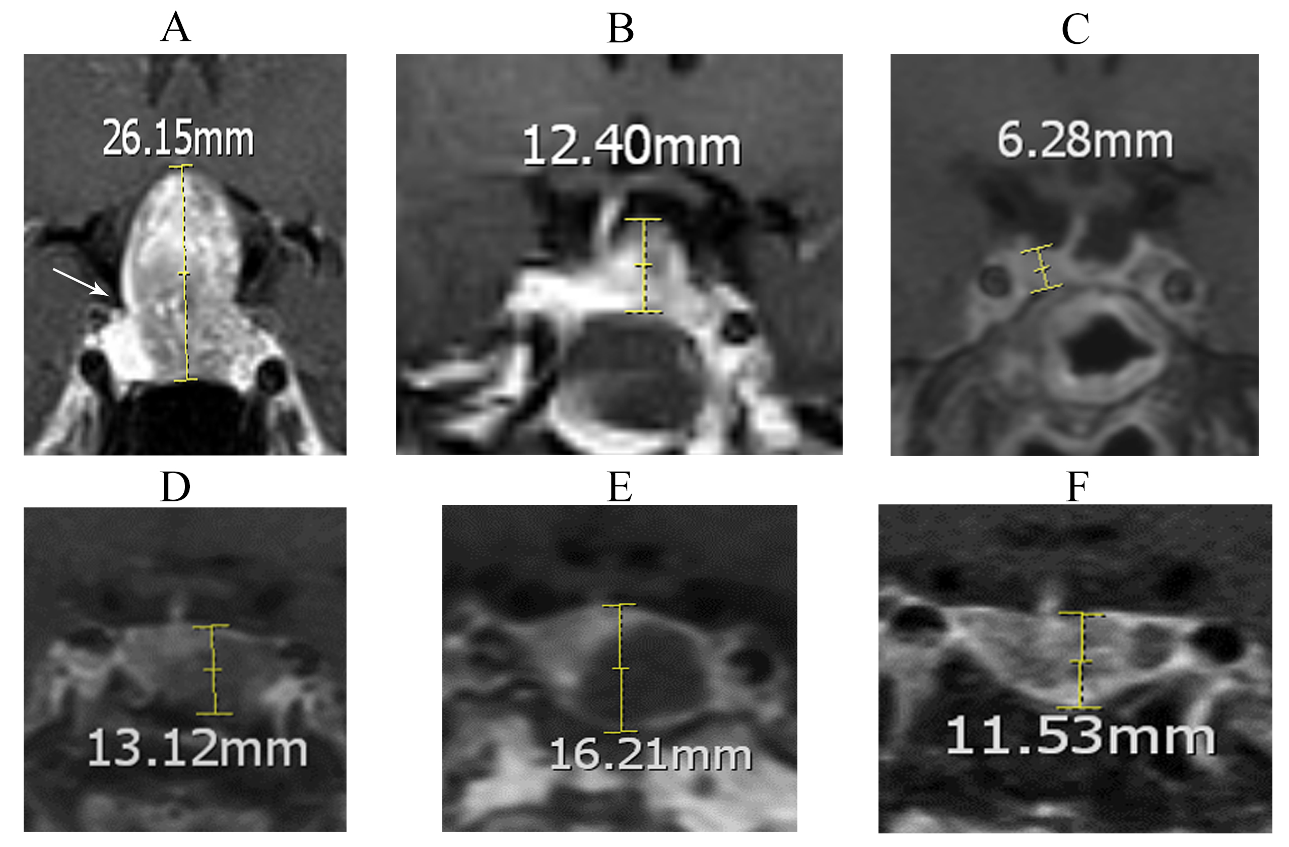 Fig. 5.
The postoperative recovery of normal pituitary tissues in patients with pituitary adenoma on coronal contrast-enhanced images of T1WI. (A) Preoperatively and (B) 1-week postoperatively and (C) 4th month postoperatively, Female, 43 years old, RPT was attached to the medial wall of the right cavernous sinus before operation. Pituitary morphology recovered obviously in the early post-operation, with the grade G3 of pituitary volume recovery in 4th month after operation (The white arrow indicates RPT). (D) Preoperatively and (E) 1-week postoperatively and (F) 5th month postoperatively, male, 47 years old, the pituitary gland was located on the right side of pituitary fossa in inverted triangle before operation. Pituitary morphology got a good recovery with grade G1 type in 5th month after operation.
Until now, MRI studies of pituitary adenomas have mainly focused on the preoperative diagnosis and evaluation of the extent of tumor resection, while few studies of normal structures such as pituitary stalks and residual pituitary tissues have been done [20, 21, 22]. In this study, MRI features of pituitary stalk and residual pituitary tissue before and after transsphenoidal surgery were analyzed, and some significant results were observed. As we all know, pituitary stalk connects hypothalamus to the pituitary gland and plays an important role in maintaining the normal physiological function of the pituitary gland [23, 24]. In pituitary adenoma patients, pituitary stalk maybe relatively thickened or compressed by the pituitary adenomas, and even be curled up into “C” or “S” type, some of which are indistinguishable from the saddle diaphragm. The pituitary stalk is thinned, deflected and pulled by the tumor, which may affect the blood supply of the pituitary portal system and thus change the pituitary function. Furthermore, surgery or tumor resection can also cause traction or change in position of the pituitary stalk, thus affecting the function of pituitary stalk, resulting in postoperative complications, such as postoperative diabetes insipidus.
As documented in this paper, pituitary stalk was thickened or compressed on preoperative MR images, and gradually recovered to normal during postoperative period. It tended to be in the middle position and its length increased gradually until 4–6 months after operation. Among the 57 patients with preoperative pituitary stalk deviation, 55 of the pituitary stalk deviations recovered in 1 week after surgery, and 30 cases were almost in the middle in 4–6 months after operation, with the other cases get better in varying degrees. These results showed that the pituitary stalk tended to recover to the middle position after pituitary adenoma operation. After operation, the sellar septum descended, the pituitary stalk was relieved of compression, the diameter of pituitary stalk returned to a relatively normal state, and the pituitary stalk curled up and bent out gradually. Of course, excessive tamponade of the intrasellar lumen during the operation may result in more pronounced ascending displacement of the pituitary stalk in the early postoperative period. But generally, 4–6 months after the operation, after the absorption of gelatin sponge and effusion, the sellar septum was further decreased, and the pituitary stalk was gradually prolonged and recovered to be in the middle.
The posterior pituitary is usually hyperintense on T1WI, commonly referred to as the “posterior pituitary bright spot, PPBS”, which is currently considered to be a short T1 signal of arginine vasopressin stored in the posterior pituitary [14, 15]. As tumor compression releases after operation, some cases of PPBS(–) on preoperative MRI may show postoperative PPBS(+). It reflects the rebuilding of the neurohypophysis pathway and recovery of posterior pituitary function. To some extent, the signal intensity of neurohypophysis is related to the compression that it receives. The signal intensity of PPBS may also be associated with the amount of ADH stored in the posterior pituitary. Higher compression may reduce the transportation and storage of ADH in neurohypophysis [25].
Preoperative diabetes insipidus is rare in patients with PA [26]. The incidence of postoperative DI is 21.4%, consistent with the reported incidence of 0.5–25% in PAs [26, 27]. As reported in the literature, the absence of hyperintensity in the posterior pituitary may be correlated with postoperative DI [28]. However, some patients with PPBS(–) do not develop DI, some patients with idiopathic DI still have PPBS(+), which leads to doubts about using the absence of PPBS as the diagnostic criterion for central DI [29]. Patients without pre-operative PPBS had a higher incidence of postoperative DI, suggesting preexisting posterior pituitary dysfunction. In general, because postoperative diabetes insipidus may be related to the disorder of antidiuretic hormone secretion and storage, we should pay more attention to the protection of the pituitary stalk and posterior lobe during surgery. As we all know, pituitary stalk is composed of the blood vessels and the axons of the large neurons in the hypothalamic nucleus. The axons have no perineuria and adventitia, and may be damaged or even broken during the pull-down process accompanied by diaphragm subsidence, resulting in the failure of ADH to pass down through the pituitary stalk, showed with diabetes insipidus and water-electrolyte disturbance.
Zada et al. [30] showed that growth hormone-secreting adenomas tend to grow below the sella, and nonfunctioning PAs tend to extend above the sella. Di Maio S et al. [20] studied 79 cases of MRI of PAs before surgery and classified the displacement of residual normal pituitary tissue as superior, superolateral or lateral in location. However, these authors did not find any statistically significant difference between these preoperative normal pituitary tissue locations and tumor types. We found that the location of normal pituitary tissue on preoperative MRI was significantly different between patients with different immunohistochemical types. Normal pituitary tissues were likely located above the adenomas in somatotroph adenoma patients, while in gonadotroph adenoma patients (almost clinically nonfunctioning adenoma), RPT was more likely to be located on one side of the tumor (parasellar). This observation is consistent with the report by Zada et al. [30]. The downward growth expansion pattern of growth hormone (GH) is associated with the presence of GH cells in the anterior flank of the anterior pituitary gland, which may explain why GH adenomas often first appear in this region on MRI. Additionally, patients with gonadotroph adenoma accounted for 31.2%, similar to previous findings [31, 32].
In our findings, 61.6% of the 112 patients showed normal pituitary tissue on preoperative enhanced MRI, while 95% on the postoperative enhanced MRI images. This change may be due to the re-expansion and repositioning of normal pituitary tissues after surgery. The re-expansion degrees are related to the size and location of adenomas. Meyrignac et al. [33] showed that nearly all normal pituitary tissues in patients with PAs of diameter more than 20 mm were pushed out of the sella. Adenomas that expand superiorly tend to push normal pituitary tissues above the sella. The normal pituitary tissues in about 49% of nonfunctioning adenomas are pushed outside the sella due to the large size of the tumor, and 76% of secreting adenomas push the normal pituitary tissues aside outside the sella [34]. Preoperative evaluation of cavernous sinus invasion has an important effect on surgery and prognosis. However, the medial wall of the cavernous sinus cannot generate its image in T1WI at all times [35]. Pituitary tissues with lateral displacement can indirectly indicate that ipsilateral cavernous sinus is not affected by tumor invasion [36] because it is unlikely that adenomas across normal pituitary tissues invade cavernous sinus. Consistent with this, we showed that patients with lateral normal pituitary tissues before surgery did not have ipsilateral cavernous sinus invasion by tumors.
Postoperative dynamic MR imaging was used not only for early detection of residual or recurrent pituitary adenomas, but also for evaluation of postoperative RPT recovery. Early postoperative MRI (1 week after operation) can roughly judge the degree of tumor resection and provide imaging reference for subsequent follow-up. On postoperative MRI, RPT showed obvious “re-tensioning”, and on T1 enhanced imaging, the pituitary gland was obviously enlarged. While in the 4–6 months after operation, with the absorption of gelatin sponge, the septum sellae decreased gradually, RPT was reshaped, and some patients could appear partial empty sellae on MRI. The residual tumor is usually located in suprasellar or bilateral cavernous sinus, showing low signal intensity on T1WI, high signal intensity on T2 weighted imaging (T2WI) and weak enhancement on T1WI. In this study, 71 patients (63.4%) underwent total tumor resection and 41 patients (36.6%) underwent residual resection, which was approximately the same as that reported previously [30]. The primary cause of tumor remnant was the invasion of cavernous sinus, followed by the obvious suprasellar extension of the tumor.
Understanding postoperative pituitary remodeling is helpful to evaluate postoperative pituitary recovery. RPT was often compressed by the tumor and was usually located in the periphery of the tumor (continuous or discontinuous) in a flaky manner, which can generally be distinguished on enhanced MRI [34]. Preoperative evaluation of the RPT location was helpful to identify and protect the normal pituitary tissue during the operation and reduce the incidence of postoperative hypopituitarism. The pituitary stalk was also used to help locate pituitary tissue, which has a high signal intensity on enhanced MRI.
Our research showed that the postoperative pituitary volume recovery was observed in most cases, and flaky RPT may present volume enlargement in some cases even in the early postoperative period. During postoperative follow-up, attention should be paid to distinguish pituitary volume recovery from residual tumors or organized tissues. On MRI T1WI enhanced scan, normal pituitary was usually enhanced significantly, while residual tumors and organized tissues showed weak enhancement due to poor blood supply. Di Maio S et al. [20] suggest that the period between the 4th–6th month after surgery is ideal for evaluating pituitary MRI, preventing the influence by postoperative tumor residuals or hemostatic materials. At the early stage after surgery, residual hemostatic materials may cover the remaining pituitary tissues [13, 37]. Our study also found that the postoperative RPT volume recovery is not related to the patient’s age, but negatively related to the size of the tumor. The larger the tumor, the more difficult the postoperative pituitary volume to recover, possibly because the larger the pituitary tumor, the more severe the pituitary tissue compression, the longer the compression time, resulting in the postoperative pituitary morphology more difficult to recover. Pituitary tissues with lateral location have better recovery compared with other types. The results suggest that suprasellar extension of PA may cause normal pituitary tissue (adenohypophysis and neurohypophysis) to receive more compression. Di Maio S et al. [20] showed that the degree of pituitary morphological remodeling was related to pituitary function after pituitary adenoma surgery.
In conclusion, our findings demonstrate that for the first time that pituitary stalk tends to be in the middle position and its length increased gradually until 4–6 months after operation. Somatotroph adenoma patients tend to have normal pituitary tissues with superior and superolateral displacement before surgery, while gonadotropin adenoma patients tend to have normal pituitary tissue with lateral displacement. Postoperative pituitary tissue remodeling and volume recovery were related to tumor size. The larger the tumor, the worse the pituitary tissue recovery.
PPBS, posterior pituitary bright spot; MRI, Magnetic resonance imaging; T1WI, T1-weighted images; ADH, antidiuretic hormone; DI, diabetes insipidus; RPT, residual pituitary tissues; PGV, Pituitary gland volume.
The datasets used and/or analyzed during the current study are available from the corresponding author on reasonable request.
SW conceived and designed the research study; SW, DX, SZ, KL, LZ and LW performed the research; SZ and DX analyzed the data; DX and SW wrote the paper. All authors contributed to editorial changes in the manuscript. All authors read and approved the final manuscript. All authors have participated sufficiently in the work and agreed to be accountable for all aspects of the work.
All procedures were approved by the Ethics Committee of Fujian Medical University. The Ethics approval number is EC of FGH 2012-017. Written informed consent was obtained from every patient.
Not applicable.
This work was supported by Fujian Provincial Natural Science Foundation of China [grant number 2015D014] and Fujian Provincial Key Project of Science and Technology Plan of China [grant number 2018Y0067].
The authors declare no conflict of interest.
References
Publisher’s Note: IMR Press stays neutral with regard to jurisdictional claims in published maps and institutional affiliations.