

 , Young Eun Kim 1,2,*
1 Department of Neurology, Hallym University Sacred Heart Hospital, Hallym University College of Medicine, 14068 Anyang, Republic of Korea
2 Hallym Neurological Institute, Hallym University, 24263 Chuncheon, Republic of Korea
3 Department of Nuclear Medicine, Severance Hospital, Yonsei University College of Medicine, 03722 Seoul, Republic of Korea
4 Department of Neurology, Yongin Severance Hospital, Yonsei University College of Medicine, 16995 Yongin, Republic of Korea
5 Department of Nuclear Medicine, Hallym University Sacred Heart Hospital, Hallym University College of Medicine, 14068 Anyang, Republic of Korea
†These authors contributed equally.
Academic Editor: Gernot Riedel
Abstract
Background and Purpose: REM sleep behavior disorder (RBD) in
Parkinson’s disease (PD) is associated with characteristic clinical subtypes and
prognosis. In addition, nigrostriatal pathway, the most vulnerable anatomical
area in PD, formed neuronal network interplaying with cortical and subcortical
structures, and which may cause PD clinical phenotype. We evaluated the regional
selectivity of presynaptic striatal dopaminergic denervation associated with RBD
in PD. Methods: We compared two groups (n = 16) of PD patients with and
without RBD in terms of specific binding ratios (SBR) in subregions of the
striatum, which were measured using positron emission tomography with 18F-FP-CIT.
SBRs of the anterior and posterior caudate, ventral striatum, and posterior and
ventral putamen regions were measured in more or less affected side, and right or
left side, or bilateral sum of the striatum. Results: Age, disease
duration, and severity of parkinsonism were not significantly different between
groups. Although group differences in all areas were not significant with
multiple comparison corrections, SBR of the ventral striatum and anterior caudate
in sum of both sides was significantly less in the RBD than in the non-RBD group
without correction (p
Keywords
- REM sleep behavior disorder
- Parkinson's disease
- striatal binding ratio
- attention
REM sleep behavior disorder (RBD) is commonly observed in several neurodegenerative diseases, especially in synucleinopathy, which includes Parkinson’s Disease (PD). A total of 30%–60% of patients with PD present with RBD during their disease course; patients with RBD seem to be a distinct clinical entity compared with patients with PD without RBD [1, 2]. Previous cross-sectional studies have reported poorer motor features, cognitive dysfunction, and hallucination in PD with RBD compared with PD without RBD, and a longitudinal study has reported poorer clinical outcomes with RBD [3, 4, 5]. However, it is unknown why PD with RBD showed distinct clinical phenotype.
Neuronal damage causing parkinsonism is mainly on the degeneration of nigrostriatal pathway in PD. In addition, striatum has wide connection with various anatomical structures including cortex and other subcortical areas, hence, subregional difference in striatal degeneration can contribute to clinical phenotype of PD. Considering PD with RBD have a distinct clinical entity compared to PD without RBD, there is a possibility of subregional difference in the striatal degeneration associated with RBD. Nonetheless, the denervation pattern in the striatal subregions has never been evaluated according to the existence of RBD in PD. In this study, we aimed to investigate whether the nigrostriatal degeneration pattern differs between patients with RBD and those without RBD (non-RBD) with early-stage PD who are naïve to drug therapy.
Thirty-two consecutive patients with PD who visited our neurology clinic for the first time and met following inclusion and exclusion criteria were enrolled in the study. PD was diagnosed using the Movement Disorder Society clinical diagnostic criteria by movement specialists [6]. We included consecutive patients with PD who were older than 30 years, drug-naïve, and had a disease duration of less than 3 years at enrolment. Clinical dementia was excluded. Brain magnetic resonance imaging (MRI) was performed to exclude patients with structural lesions in subcortical structure including the nigrostriatal area, cortical area, and brain stem. For the diagnosis of PD, positron emission tomography using 18F-FP-CIT was performed in all participants. Participants with a clinical diagnosis of dementia, stroke, other neurological disorders were excluded. And polysomnography was done in all participants to reveal the existence of RBD. Use of offending drugs causing RBD and severe obstructive sleep apnea to disturb the diagnosis of RBD were excluded. This study was approved by our institution’s ethical committee (IRB 2019-06-008).
Dopamine transporter imaging via 18F-FP-CIT (Philips GEMINI TF-64, 7146,
Philips, USA) obtained images with three-dimensional resolution of 2.3 mm full
width at half maximum. All subjects did not take medication which can disturb
ligand bining. PET emission acquisition was performed at 3 hours for 15 minutes
in the three-dimensional mode after 5 mCi injection of 18F-FP-CIT injection after
brain CT scan, which was performed in the axial helix at 120 Kvp and 200 mAs. And
PET image was reconstructed from CT data by all-pass filter with a 512
The diagnosis of RBD was based on history of dream enactment behavior and via polysomnographic confirmation of excessive electromyography activity in REM sleep proposed by the International Classification of Sleep Disorders (ICSD)-2 [7]. We recruited patients without RBD who had no clinical history confirmed by polysomnography.
To assess the motor and nonmotor status of parkinsonism, the Movement Disorder Society Unified Parkinson Disease Rating Scale and Hoehn and Yahr’s scale were measured at baseline. Initial clinical features were characterized as tremor dominant, Akineto-rigidity and gait disturbance by a chief complaint. Attention, language, visuospatial memory, and frontal functions were assessed using the Seoul Neuropsychological Screening Battery, a neuropsychological battery consisting of standardized, validated tests, including the Montreal Cognitive Assessment. The Seoul Neuropsychological Screening Battery in this study included the following: (1) forward and backward digit span, (2) the Korean version of the Boston Naming test, (3) the Rey–Osterrieth Complex Figure Test, (4) the Seoul Verbal Learning Test, (5) contrasting program/go-no-go test, (6) the Controlled Oral Word Association Test, and (7) the Stroop test (color reading). For each test, specific norms for comparisons based on age, sex, and education, which were based on assessments of 447 normal Korean participants, were used to transform the data into z-scores [8].
SBR in six striatal areas was compared between the RBD and non-RBD group in the
left and right sides of the striatum, the more and less affected sides, or
bilateral sum. The more affected side was defined as that with the lower SBR
between the right and left striatum. The Mann–Whitney U test or the Chi-squared
test was used to compare demographical features, other clinical variables, and
SBR between the two groups. The Benjamini-Hochberg multiple comparison correction
was calculated because multiple tests for each striatal areas were done. A
p value of
Table 1 shows the different demographic and clinical features of the RBD and
non-RBD groups. Age, gender, and clinical features related to parkinsonism
(disease duration, akineto-rigid phenotype, Hoehn and Yahr’s scale, and Unified
Parkinson Disease Rating Scale) were not significantly different between the two
groups (p
| Non-RBD (n = 16) | RBD (n = 16) | p | |
| Age | 57.00 |
62.44 |
0.094 |
| Gender (M/F) | 7/9 | 10/6 | 0.479 |
| Disease duration | 1.63 |
0.83 |
0.539 |
| Initial clinical manifestation (tremor/AR/GD) | 10/3/3 | 9/5/2 | 0.686 |
| Hoehn & Yahr (1/2/3) | 5/10/1 | 1/12/3 | 0.146 |
| UPDRS part I | 1.44 |
1.63 |
0.809 |
| UPDRS part II | 5.93 |
7.00 |
0.533 |
| UPDRS part III | 22.13 |
25.13 |
0.361 |
| MOCA | 25.06 |
25.00 |
0.254 |
| MMSE | 28.31 |
27.38 |
0.163 |
| Abbreviations: AR, Akineto-rigidity; GD, gait disturbance. | |||
Among the six striatal areas, the PP area showed the lowest SBR values in both
RBD and non-RBD groups and in all participants (p
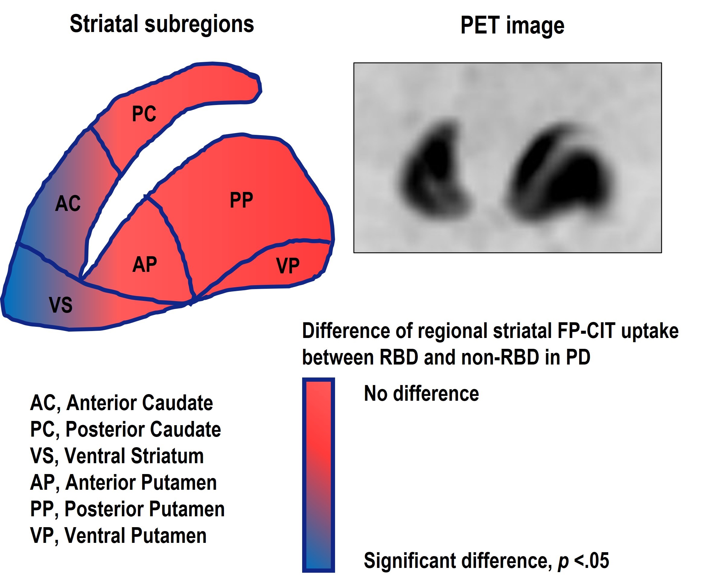 Fig. 1.
Tophography of 6 striatal areas and the areas showing different value according to the existence of RBD in PD. AC and VS area showed lower striatal binding in the RBD group compared to non-RBD group.
| Side | Region | Non-RBD (n = 16) | RBD (n = 16) | p |
|---|---|---|---|---|
| More affected side | Whole striatum | 12.93 |
11.12 |
0.195 |
| AC | 3.03 |
2.41 |
0.160 | |
| PC | 1.89 |
1.43 |
0.239 | |
| VS | 2.90 |
2.38 |
0.032 | |
| AP | 2.43 |
2.26 |
0.838 | |
| PP | 0.90 |
0.97 |
0.305 | |
| VP | 1.79 |
1.68 |
0.724 | |
| Less affected side | Whole striatum | 15.92 |
12.81 |
0.053 |
| AC | 3.47 |
2.63 |
0.073 | |
| PC | 2.09 |
1.58 |
0.160 | |
| VS | 3.26 |
2.61 |
0.026 | |
| AP | 3.17 |
2.60 |
0.224 | |
| PP | 1.47 |
1.30 |
0.423 | |
| VP | 2.45 |
2.09 |
0.196 | |
| Sum of both side | Whole striatum | 28.85 |
23.93 |
0.098 |
| AC | 6.50 |
5.04 |
0.046 | |
| PC | 3.98 |
3.01 |
0.136 | |
| VS | 6.16 |
4.98 |
0.020 | |
| AP | 5.60 |
4.86 |
0.222 | |
| PP | 2.37 |
2.26 |
0.751 | |
| VP | 4.23 |
3.77 |
0.244 | |
| Values indicate mean Abbreviations: AC, Anterior Caudate; PC, Posterior Caudate; VS, Ventral Striatum; AP, Anterior Putamen; PP, Posterior Putamen; VP, Ventral Putamen; Both side means sum of more and less affected sides. All p value was uncorrected for multiple comparisons. And p values using Benjamini-Hochberg multiple comparison correction were not significant (all p value | ||||
Comparing the left and right sides of the striatum, the left VS had
significantly lower SBR in the RBD group than in the non-RBD group (p =
0.017); the right VS showed a moderate difference (p = 0.051). In
addition, the right AC had lower SBR in the RBD group than in the non-RBD group
(p = 0.047) (Supplementary Table 1). However, this statistical
significance was not definite after multiple comparison correction (p
In cognitive assessment, the forward digit span in attention domain and the
Controlled Oral Word Association Test (animal) assessment of frontal/executive
function were decreased in the RBD group compared with the non-RBD group
(p = 0.35 and p = 0.32, respectively). Across the five
cognitive domains, the RBD group had a significantly decreased attention function
compared with the non-RBD group (p = 0.022). There were no statistically
significant differences between the groups in the other cognitive domains
(language, visuospatial memory, and frontal function) (Table 3). Dysfunction in
attention domain was well correlated with SBR in VS (r = 0.358, p
| Neuropsychological domain | Neuropsychological test (max score) | Non-RBD (n = 16) | RBD (n = 16) | p |
| Attention | Forward digit span (9) | 0.29 |
–0.35 |
0.035 |
| Backward digit span (8) | –0.40 |
–0.48 |
0.445 | |
| Language | K-BNT (60) | 0.33 |
0.21 |
0.669 |
| Visuospatial | RCFT (copying) (36) | 0.05 |
0.33 |
0.642 |
| Memory | SVLT (immediate recall) (36) | –0.63 |
–0.73 |
0.799 |
| SVLT (delayed recall) (12) | –0.27 |
–0.60 |
0.341 | |
| SVLT (recognition) (24) | –0.18 |
–0.12 |
0.809 | |
| True positive + false negative | ||||
| RCFT(immediate recall) (36) | –0.28 |
–0.34 |
0.926 | |
| RCFT(delayed recall) (36) | –0.16 |
–0.41 |
0.564 | |
| RCFT (recognition) (24) | –0.03 |
0.18 |
0.381 | |
| True positive + false negative | ||||
| Frontal/Executive function | Contrasting program (20) | 20.00 |
20.00 |
1.000 |
| Go-no-go test (20) | 19.88 |
18.75 |
0.985 | |
| COWAT (animal) | 0.23 |
–0.36 |
0.032 | |
| COWAT (supermarket items) | 0.06 |
–0.09 |
0.724 | |
| COWAT (phonemic fluency) | –0.17 |
–0.27 |
0.985 | |
| Stroop test: color reading (112) | 0.18 |
0.14 |
0.897 | |
| SNSB_II_Domain_Attention | –0.02 |
–0.56 |
0.022 | |
| SNSB_II_Domain_Language | 0.50 |
0.30 |
0.619 | |
| SNSB_II_Domain_Visuospatial | 0.20 |
0.01 |
0.531 | |
| SNSB_II_Domain_Memory | –0.24 |
–0.47 |
0.449 | |
| SNSB_II_Domain_frontal | 0.44 |
–0.11 |
0.215 | |
| All p value was uncorrected for multiple comparisons. And p
values using Benjamini-Hochberg multiple comparison correction were not
significant (all p value | ||||
We evaluated subregional differences in presynaptic dopaminergic neurodegeneration of the striatum with regard to RBD in drug-naïve patients with early PD. Overall, striatal SBR was significantly decreased in the PP area, as expected, irrespective of RBD; however, in the AC and VS areas, which are relatively well preserved in early PD, the RBD group had a significantly lower uptake than the non-RBD group in this study.
Striatal degeneration in PD shows a distinctive spatial and temporal pattern during the course of the disease. In PD, a degeneration pattern with an anteroposterior gradient has been reported in previous literature [9]. This pattern is quite different from the Parkinson Plus syndrome [9]. From the early stage of PD, the PP area is the most affected; this corresponds well with the pattern of neurodegeneration in the substantia nigra, i.e., early degeneration of the ventrolateral substantia nigra, which was demonstrated in autopsy studies [10, 11]. This pattern was common in all our study patients irrespective of RBD. The severity of degeneration in the PP area did not differ between the RBD and non-RBD groups. However, among the relatively preserved areas of the striatum, uptake in the AC and VS differed significantly between the groups according to the presence or absence of RBD.
The PP area has a primary connection with the motor cortical area, which may explain why motor dysfunction is prominent in early PD [12]. The VS area may have strong connectivity with the ventromedial prefrontal cortex and orbitofrontal cortex, which may imply that it has limbic and attentional functions [12, 13]. Similarly, the AC area is connected to the prefrontal cortex and the major parts of the orbitofrontal cortex or dorsal anterior cingulate cortex [13]. From these connection, the VS and AC areas are associated with emotional and cognitive function. Although we did not evaluate reward or emotional aspects sufficiently, our cognitive assessments indicated that attentional deficit was prominent in the RBD group; this dysfunction may be associated with the different neurodegeneration levels observed in the AC or VS area or both.
There have been few positron emission tomography imaging studies comparing the
patterns of nigrostriatal dopaminergic degeneration in PD between individuals
with and without RBD. Arnaldi et al. [14, 15] used
I
Although our study has a limitation in that multiple test corrections by Bonferroni did not find any significant statistical differences, this study is an exploratory study design and has a small sample size. However, this study did not demonstrate the statistical difference in multiple comparisons, it should be evaluated in larger scale or longitudinal design. Therefore, based on the current study, further study is required for the interpretation of these findings. Nevertheless, we used polysomnography to confirm RBD, unlike previous studies that used clinical diagnoses to establish RBD. Although we included only a small number of patients, this study has methodological merits because RBD confirmed by polysomnography increased sensitivity for determining RBD-related characteristics.
The regional selectivity of nigrostriatal degeneration may be observed in PD with RBD compared with PD without RBD implies that there are different clinical phenotypic presentations. It remains unknown why this differential degeneration pattern occurs in PD. Thus, further investigation using large scale is required for statistical confirmation.
IK, investigation and data curation, original draft preparation. YKL, investigation and data curation, original draft preparation. HM, and YJK, investigation and data curation. SL, investigation and data curation, review and editing of manuscript. MY, investigation and data curation, review and editing of manuscript. HSH, Investigation, review and final preparation of review and editing of manuscript. YEK, investigation, data curation, original draft preparation, review and final preparation of review and editing of manuscript.
This study was approved by Hallym University Sacred Heart Hospital Institution’s ethical committee (IRB 2019-06-008). Informed consent was waived by IRB because medical records were used only for this study.
The authors thank Jaeseol Park for advice on neuropsychological test.
This research was supported by Hallym University Research Fund and the National Research Foundation of Korea (NRF) grant funded by the Korean government (MSIT) (2020R1F1A1076697).
The authors declare no conflict of interest.
References
Publisher’s Note: IMR Press stays neutral with regard to jurisdictional claims in published maps and institutional affiliations.