IMR Press / JIN / Volume 21 / Issue 6 / DOI: 10.31083/j.jin2106165
Open Access Original Research
Differences in CSF Biomarkers Profile of Patients with Parkinson's Disease Treated with MAO-B Inhibitors in Add-On
Show Less
1 Unit of Neurology, Department of Systems Medicine, University of Roma Tor Vergata, 00133 Rome, Italy
2 Department of Experimental Medicine, University of Roma La Sapienza, 00185 Rome, Italy
3 IRCCS Fondazione Santa Lucia, 00179 Rome, Italy
4 Institute of Translational Pharmacology (IFT), Consiglio Nazionale delle Ricerche (CNR), 00185 Rome, Italy
5 Experimental Neuroscience Unit, IRCCS Fondazione Santa Lucia, 00179 Rome, Italy
*Correspondence: t.schirinzi@yahoo.it; tommaso.schirinzi@uniroma2.it (Tommaso Schirinzi)
Academic Editor: Ho-Sung Ryu
J. Integr. Neurosci. 2022, 21(6), 165; https://doi.org/10.31083/j.jin2106165
Submitted: 16 June 2022 | Revised: 23 July 2022 | Accepted: 5 August 2022 | Published: 8 October 2022
(This article belongs to the Special Issue Advances in Parkinson's Disease)
Copyright: © 2022 The Author(s). Published by IMR Press.
This is an open access article under the CC BY 4.0 license.
Abstract

Background: Monoamine oxidase type B inhibitors (iMAO-Bs) are a class of largely-used antiparkinsonian agents that, based on experimental evidence, are supposed to exert different degrees of neuroprotection in Parkinson’s disease (PD). However, clinical proofs on this regard are very scarce. Since cerebrospinal fluid (CSF) reflects pathological changes occurring at brain level, we examined the neurodegeneration-related CSF biomarkers profile of PD patients under chronic treatment with different iMAO-Bs to identify biochemical signatures suggestive for differential neurobiological effects. Methods: Thirty-five PD patients under chronic treatment with different iMAO-Bs in add-on to levodopa were enrolled and grouped in rasagiline (n = 13), selegiline (n = 9), safinamide (n = 13). Respective standard clinical scores for motor and non-motor disturbances, together with CSF biomarkers of neurodegeneration levels (amyloid- β -42, amyloid- β -40, total and 181-phosphorylated tau, and lactate) were collected and compared among the three iMAO-B groups. Results: No significant clinical differences emerged among the iMAO-B groups. CSF levels of tau proteins and lactate were instead different, resulting higher in patients under selegiline than in those under rasagiline and safinamide. Conclusions: Although preliminary and limited, this study indicates that patients under different iMAO-Bs may present distinct profiles of CSF neurodegeneration-related biomarkers, probably because of the differential neurobiological effects of the drugs. Larger studies are now needed to confirm and extend these initial observations.

Keywords
Parkinson's disease
CSF biomarkers
neurodegeneration
neuroprotection
MAO inhibitors
selegiline
rasagiline
safinamide
1. Introduction

Parkinson’s disease (PD) is a common neurodegenerative disorder responsible for a disabling syndrome, including motor and non-motor disturbances. While neuropathology and main pathogenic pathways have been established over time, PD still remains an incurable disorder. Indeed, effective symptomatic therapies exist, which mostly replace deficient dopamine, but, to date, no disease-modifying treatments are available [1].

Monoamine Oxidase type B Inhibitors (iMAO-Bs) are a class of antiparkinsonian drugs, used in mono or add-on therapy along the entire disease course, that potentiate dopaminergic transmission at striatal level by reducing dopamine degradation [2]. The three main iMAO-Bs are selegiline, rasagiline, and safinamide, which have individual peculiarities in the spectrum of molecular effects and in clinical indications [3]. Beside symptomatic relief in motor and non-motor disturbances, iMAO-Bs have been considered as neuroprotective agents, based on a body of in vitro and in vivo experimental evidence. Specifically, iMAO-Bs may operate neural defence against mitochondrial toxins (MPTP, rotenone and paraquat) [4, 5, 6, 7], monoamine-depleting neurotoxins (N-(2-chloroethyl)-N-ethyl-2-bromobenzylamine hydrochloride (DSP-4)) [8], and hypoxic conditions [9, 10], and exert antioxidant properties [4, 11, 12, 13], antiapoptotic [5, 14, 15, 16] and neurotrophic effects [17, 18]. Nevertheless, human-based proofs of iMAO-Bs-associated neuroprotection are very limited and not completely univocal; moreover, no comparison studies among the single iMAO-Bs exist.

Cerebrospinal fluid (CSF) reflects pathological changes of the brain. Accordingly, the CSF measurement of neurodegeneration-related peptides, such as amyloid- β fragments and tau proteins, or metabolites, such as lactate, allows tracking in vivo molecular events underlying neurodegenerative diseases [19]. In particular, in PD, CSF amyloid- β -42 may inform on concurrent brain amyloidopathy and predict the risk of cognitive decline; tau levels and lactate, instead, mostly mirror neuronal loss and oxidative stress respectively, providing readout of global brain pathology [20].

Here we thus conducted a study on the CSF biomarkers profile of PD patients chronically treated with both L-dopa and different iMAO-Bs in add-on, aiming to disclose biochemical signatures suggestive of differential neurobiological effects.

2. Methods
2.1 Study Population and Clinical-Biochemical Assessement

The study involved a total of 35 PD patients afferent to Neurology Unit of Tor Vergata University Hospital (Rome, Italy). PD was diagnosed following the 2015 MDS criteria. All patients were receiving iMAO-Bs add-on therapy for one year at least (n = 13 rasagiline 1 mg/die, n = 9 selegiline 10 mg/die, n = 13 safinamide 100 mg/die); the remaining dopaminergic therapy was stable for at least 6 months. For each participant, demographics and medical history were collected, together with MDS-UPDRS pars III [21] and Montreal Cognitive Assessment (MoCA) [22] scores; the levodopa equivalent daily dose (LEDD) was calculated. They all underwent lumbar puncture (LP) according to standard procedures [23]. Briefly, CSF was collected in polypropylene tubes, carried on ice to local laboratory, centrifuged at 2000 rpm for 10 min at 4°, aliquoted in polypropylene vials (0.5 mL) and stored at –80°, pending analysis. A routine blood sample was obtained contemporary to LP, to allow a comparative chemical analysis of CSF. In addition, CSF sample was excluded if microscopic observation revealed > 5 cells/ μ L. Chemistry assays were performed by using commercially available kits following the manufacturer’s specifications (Flex reagent cartridge, Dimension Vista System, Siemens Healthcare Diagnostics GmbH, Munich, Germany). CSF assay of amyloid- β -42 (A β 42), amyloid- β -40 (A β 40), total and 181-phosphorilated tau (t-tau and p-tau), was performed by using commercially available sandwich enzyme-linked immunosorbent assays, as previously described [19, 24]. The A β 42/A β 40 ratio was also calculated.

2.2 Statistical Analysis

Variables distribution was preliminarily examined by Shapiro-Wilk test. Clinical-demographic data were compared among the groups by Mann–Whitney U test. Categorical variables were compared by chi-square test. Considering the small sample size, although normally distributed, CSF neuorodegenerative biomarkers and lactate levels were compared among groups by Kruskal-Wallis test. Pairwise comparisons after Kruskal-Wallis test was also assessed. A p 0.05 was considered significant. Analysis was conducted blindly with IBM-SPSS-22 (IBM Corp., Chicago, IL, USA).

3. Results

No differences resulted in demographics and clinical parameters among the groups. Regarding CSF biomarkers, t-tau (p = 0.005), p-tau (p = 0.017) and lactate (p = 0.037) levels significantly differed among the groups (Table 1). Pairwise comparisons showed significant differences of t-tau (selegiline vs. rasagiline, p = 0.027; selegiline vs. safinamide, p = 0.006), p-tau (selegiline vs. safinamide, p = 0.05; selegiline vs. safinamide, p = 0.021) and lactate (selegiline vs. safinamide, p = 0.041). Graphical data for t-tau, p-tau and lactate are represented in Fig. 1A–C. CSF biomarkers did not differ between females and males.
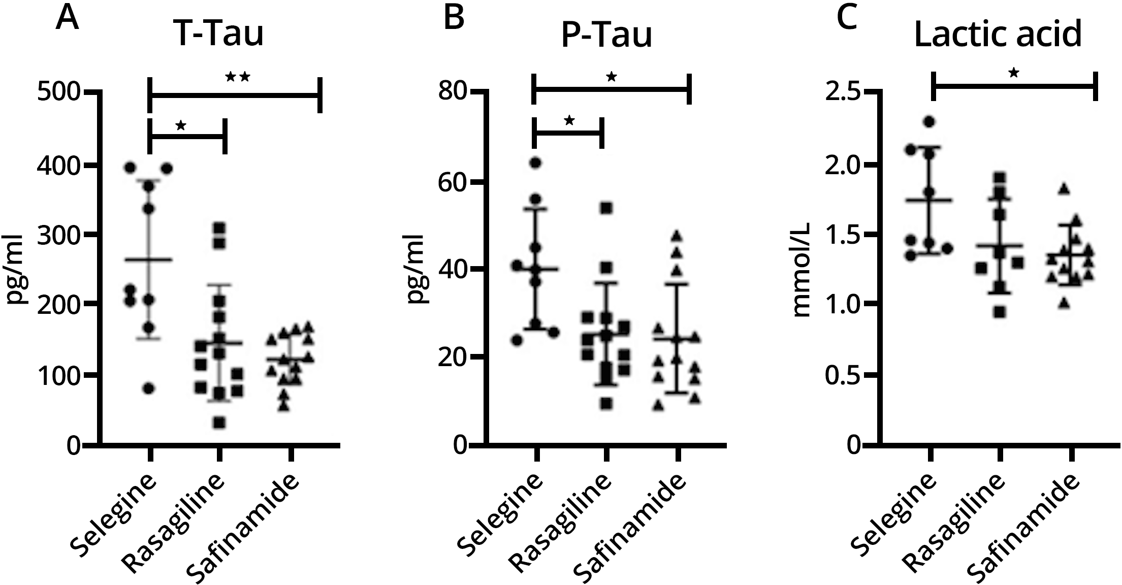
Fig. 1.

T-Tau (Fig. 1A), P-Tau (Fig. 1B) and lactic acid (Fig. 1C) levels are expressed as mean ± SD. Significant differences between groups * at p < 0.05 and ** at p < 0.01 by pairwise comparisons.

Table 1.Demographic, clinical and biochemical variables of the study population. Unit of measure are expressed by the following abbrevations pmol/mL (picomoles/milliliter) and mmol/mL (millimoles/milliliter).
Variable Selegiline Rasagiline Safinamide Significants
n = 9 n = 13 n = 13
(f: 55.6%; m: 44.4%) (f: 30.8%; m: 69.2%) (f: 69.2%; m: 30.8%) ns
Clinical variable Mean S.D. Mean S.D. Mean S.D.
Age (y) 54.7 7.6 52.1 13.1 54.3 9.3 ns
Duration (y) 3.9 2.5 6.1 1.4 6.9 1.0 ns
MOCA 26.0 1.2 27.5 1.3 24.2 2.0 ns
UPDRS III 25.7 11.2 26.7 6.6 35.2 6.1 ns
LEDD 543.9 152.1 567.5 139.6 718.8.3 74.1 ns
CSF biomarker
t-tau (pmol/mL) 264.5 112.3 146.2 82.3 122.8 36.1 p = 0.005
p-tau (pmol/mL) 40.1 13.6 25.4 11.5 24.4 12.3 p = 0.017
A β 42 (pmol/mL) 775.0 249.7 837.2 316.5 912.3 264.8 Ns
A β 40 (pmol/mL) 7553.3 1663.7 5239.7 1043.7 5401.0 1737.3 Ns
A β 42/A β 40 0.131 0.063 0.167 0.057 0.172 0.025 Ns
Lactate (mmol/mL) 1.74 0.38 1.41 0.33 1.35 0.21 p = 0.037
4. Discussion

This pilot study examined the profile of neurodegeneration-related CSF biomarkers of three clinically-homogenous PD groups treated with different iMAO-Bs (namely, selegiline, rasagiline and safinamide) in add-on, looking for biochemical signatures suggestive for differential neurobiological effects. The higher levels of t-tau, p-tau, and lactate measured in patients under selegiline, compared to those under rasagiline and safinamide, indicate distinct molecular or neuropathological substrates associated with different iMAO-Bs.

Tau proteins in CSF are basically considered as marker of neuronal damage, and tauopathy overall. While the CSF levels proportionally reflect neurofibrillary tangles deposition in Alzheimer’s disease (AD) [25], the neuropathological correlate in PD is not completely clear yet. Molecular interactions between tau and α -synuclein have been reported in Lewy body pathology characterizing PD and other synucleinopathies [26]; however, higher CSF levels of tau proteins in PD have been associated with major brain networks disruption [27, 28], age-related or environmental-related neuropathological changes [20, 29, 30], and, in general, with greater clinical severity and poorer outcome, either in motor or cognitive aspects [31, 32, 33]. Accordingly, we could accept the higher CSF tau proteins levels, the wider neurodegeneration, even in PD. CSF lactate, instead, could be considered a readout of brain mitochondrial activity and redox balance, whose levels increase in parallel with oxidative stress and metabolic impairment [19].

The clinical homogeneity of our study population allows us to refer, at least in part, differences in CSF biomarkers profile of the three iMAO-Bs groups (in particular, the higher levels of tau proteins and lactate in selegiline group compared to rasagiline and safinamide groups) to the distinct action mechanisms and metabolism of the drugs. Indeed, selegiline is metabolized to 1-amphetamine and to 1-methamphetamine [34], having amphetamine-like properties [35], whereas rasagiline is metabolized to 1-aminoindan, not displaying amphetamine-like effects [36]. It is well known that amphetamine may be neurotoxic [37]. Conversely, experimental data from cellular models disclose neuroprotective effects of 1-aminoindan [38, 39]. Moreover, selegiline and rasagiline differently modulate synaptic activity at corticostriatal level [2], which is consistent with distinct pathways of molecular activation in neurons that, in turn, may translate in long-term differences in proteinopathy and degeneration shaping. Safinamide, instead, has a prominent antiglutamatergic effect, either by antagonizing NMDA-mediated signalling or by blocking voltage-dependent sodium and N-type calcium channels [40, 41, 42]. The inhibition of glutamate transmission, the subsequent attenuation of associated excitotoxicity, and the containment of oxidative stress [43], might thus account for the milder profile of neurodegeneration observed in patients taking safinamide, compared to those taking selegiline.

We only observed differences in tau proteins, but not in amyloid- β peptides. Increasing body of evidence, even from humans, is indicating substantial interactions between iMAO-Bs and tau protein pathway [44, 45], which may account for this preferential effects. Conversely, regarding amyloid- β pathway, recent studies demonstrated the involvement of MAO-B into the amyloidogenic cascade [46, 47]; however, they mostly relied on AD models and did not find confirmation in our cohort of PD patients.

5. Conclusions

This study is limited by the sample size, the retrospective design, the relative exiguity of the biomarkers panel, and the absence of a control group without iMAO-Bs (which indeed could have presented with substantial differences in clinical severity or disease duration). Despite limitations, we provided preliminary evidence on the distinct biochemical endophenotype of PD patients under various iMAO-Bs. With the due caution, we could refer differences in CSF neurodegeneration-related biomarkers to respective pharmacodynamic peculiarities of selegiline, rasagiline, and safinamide. In particular, the lower levels of tau proteins and lactate may indicate a differential effect on neurodegenerative cascade operated by rasagiline and safinamide that, differently from selegiline, do not display an amphetamine-like behaviour. This preliminary work also highlighted the need for prospective comparative trials to examine neuroprotection activity associated with different iMAO-Bs, which should include CSF biomarkers of neurodegeneration among the outcomes, as more recent studies do [48, 49].

Author Contributions

HZ collected and analyzed data, wrote the manuscript. NC analyzed data and wrote the manuscript. AF, CV and NBM contributed to data interpretation and edited the final manuscript. PG, RB, and CS collected and analyzed data. TS analyzed and interpreted data, edited the final manuscript.

Ethics Approval and Consent to Participate

The study agreed the ethical principles of Helsinki declaration, and received approval of the local committee (0026092/2017). Informed written consent was signed by all the participants.

Acknowledgment

Not applicable.

Funding

This research received no external funding.

Conflict of Interest

The authors declare no conflict of interest.

References
[1]
Schirinzi T, Sancesario GM, Di Lazzaro G, Biticchi B, Colona VL, Mercuri NB, et al. CSF α -synuclein inversely correlates with non-motor symptoms in a cohort of PD patients. Parkinsonism and Related Disorders. 2019; 61: 203–206.
[2]
Madeo G, Schirinzi T, Martella G, Latagliata EC, Puglisi F, Shen J, et al. PINK1 heterozygous mutations induce subtle alterations in dopamine-dependent synaptic plasticity. Movement Disorders. 2014; 29: 41–53.
[3]
Alborghetti M, Nicoletti F. Different Generations of Type-B Monoamine Oxidase Inhibitors in Parkinson’s Disease: from Bench to Bedside. Current Neuropharmacology. 2019; 17: 861–873.
[4]
Saravanan KS, Sindhu KM, Senthilkumar KS, Mohanakumar KP. L-deprenyl protects against rotenone-induced, oxidative stress-mediated dopaminergic neurodegeneration in rats. Neurochemistry International. 2006; 49: 28–40.
[5]
Chau KY, Cooper JM, Schapira AHV. Rasagiline protects against alpha-synuclein induced sensitivity to oxidative stress in dopaminergic cells. Neurochemistry International. 2010; 57: 525–529.
[6]
Kupsch A, Sautter J, Götz ME, Breithaupt W, Schwarz J, Youdim MB, et al. Monoamine oxidase-inhibition and MPTP-induced neurotoxicity in the non-human primate: comparison of rasagiline (TVP 1012) with selegiline. Journal of Neural Transmission. 2001; 108: 985–1009.
[7]
Caccia C, Maj R, Calabresi M, Maestroni S, Faravelli L, Curatolo L, et al. Safinamide: from molecular targets to a new anti-Parkinson drug. Neurology. 2006; 67: S18–S23.
[8]
Gibson CJ. Inhibition of MAO B, but not MAO a, blocks DSP-4 toxicity on central NE neurons. European Journal of Pharmacology. 1987; 141: 135–138.
[9]
Speiser Z, Mayk A, Eliash S, Cohen S. Studies with rasagiline, a MAO-B inhibitor, in experimental focal ischemia in the rat. Journal of Neural Transmission. 1999; 106: 593–606.
[10]
Speiser Z, Mayk A, Litinetsky L, Fine T, Nyska A, Blaugrund E, et al. Rasagiline is neuroprotective in an experimental model of brain ischemia in the rat. Journal of Neural Transmission. 2007; 114: 595–605.
[11]
Carrillo MC, Kanai S, Nokubo M, Kitani K. (−) deprenyl induces activities of both superoxide dismutase and catalase but not of glutathione peroxidase in the striatum of young male rats. Life Sciences. 1991; 48: 517–521.
[12]
Carrillo MC, Minami C, Kitani K, Maruyama W, Ohashi K, Yamamoto T, et al. Enhancing effect of rasagiline on superoxide dismutase and catalase activities in the dopaminergic system in the rat. Life Sciences. 2000; 67: 577–585.
[13]
Budni P, de Lima MNM, Polydoro M, Moreira JCF, Schroder N, Dal-Pizzol F. Antioxidant Effects of SelegilIne in Oxidative Stress Induced by Iron Neonatal Treatment in Rats. Neurochemical Research. 2007; 32: 965–972.
[14]
Chetsawang B, Kooncumchoo P, Govitrapong P, Ebadi M. 1-Methyl-4-phenyl-pyridinium ion-induced oxidative stress, c-Jun phosphorylation and DNA fragmentation factor-45 cleavage in SK-N-SH cells are averted by selegiline. Neurochemistry International. 2008; 53: 283–288.
[15]
Zhao Q, Cai D, Bai Y. Selegiline rescues gait deficits and the loss of dopaminergic neurons in a subacute MPTP mouse model of Parkinson’s disease. International Journal of Molecular Medicine. 2013; 32: 883–891.
[16]
Inaba-Hasegawa K, Akao Y, Maruyama W, Naoi M. Type a monoamine oxidase is associated with induction of neuroprotective Bcl-2 by rasagiline, an inhibitor of type B monoamine oxidase. Journal of Neural Transmission. 2012; 119: 405–414.
[17]
Mizuta I, Ohta M, Ohta K, Nishimura M, Mizuta E, Hayashi K, et al. Selegiline and Desmethylselegiline Stimulate NGF, BDNF, and GDNF Synthesis in Cultured Mouse Astrocytes. Biochemical and Biophysical Research Communications. 2000; 279: 751–755.
[18]
Maruyama W, Nitta A, Shamoto-Nagai M, Hirata Y, Akao Y, Yodim M, et al. N-Propargyl-1 (R)-aminoindan, rasagiline, increases glial cell line-derived neurotrophic factor (GDNF) in neuroblastoma SH-SY5Y cells through activation of NF-kappaB transcription factor. Neurochemistry International. 2004; 44: 393–400.
[19]
Zenuni H, Grillo P, Sancesario GM, Bernardini S, Mercuri NB, Schirinzi T. How Comorbidity Reflects on Cerebrospinal Fluid Biomarkers of Neurodegeneration in Aging. Journal of Alzheimer’s Disease Reports. 2021; 5: 87–92.
[20]
Schirinzi T, Di Lazzaro G, Sancesario GM, Summa S, Petrucci S, Colona VL, et al. Young-onset and late-onset Parkinson’s disease exhibit a different profile of fluid biomarkers and clinical features. Neurobiology of Aging. 2020; 90: 119–124.
[21]
Goetz CG, Fahn S, Martinez-Martin P, Poewe W, Sampaio C, Stebbins GT, et al. Movement Disorder Society-sponsored revision of the Unified Parkinson’s Disease Rating Scale (MDS-UPDRS): Process, format, and clinimetric testing plan. Movement Disorders. 2007; 22: 41–47.
[22]
Nasreddine ZS, Phillips NA, Bédirian V, Charbonneau S, Whitehead V, Collin I, et al. The Montreal Cognitive Assessment, MoCA: a Brief Screening Tool for Mild Cognitive Impairment. Journal of the American Geriatrics Society. 2005; 53: 695–699.
[23]
Schirinzi T, Sancesario GM, Ialongo C, Imbriani P, Madeo G, Toniolo S, et al. A clinical and biochemical analysis in the differential diagnosis of idiopathic normal pressure hydrocephalus. Frontiers in Neurology. 2015; 6: 86.
[24]
Sancesario GM, Di Lazzaro G, Alwardat M, Biticchi B, Basile V, Salimei C, et al. Amyloid- β 42/Neurogranin Ratio as a Potential Index for Cognitive Impairment in Parkinson’s Disease. Journal of Alzheimer’s Disease. 2020; 76: 1171–1178.
[25]
Tapiola T, Alafuzoff I, Herukka S, Parkkinen L, Hartikainen P, Soininen H, et al. Cerebrospinal Fluid β -Amyloid 42 and Tau Proteins as Biomarkers of Alzheimer-Type Pathologic Changes in the Brain. Archives of Neurology. 2009; 66: 382–389.
[26]
Irwin DJ, Hurtig HI. The Contribution of Tau, Amyloid-Beta and Alpha-Synuclein Pathology to Dementia in Lewy Body Disorders. Journal of Alzheimer’s Disease & Parkinsonism. 2018; 8: 444.
[27]
Zhang Y, Wu IW, Tosun D, Foster E, Schuff N. Progression of Regional Microstructural Degeneration in Parkinson’s Disease: A Multicenter Diffusion Tensor Imaging Study. PLoS ONE. 2016; 11: e0165540.
[28]
Abbasi N, Mohajer B, Abbasi S, Hasanabadi P, Abdolalizadeh A, Rajimehr R. Relationship between cerebrospinal fluid biomarkers and structural brain network properties in Parkinson’s disease. Movement Disorders. 2018; 33: 431–439.
[29]
Taghdiri F, Multani N, Tarazi A, Naeimi SA, Khodadadi M, Esopenko C, et al. Elevated cerebrospinal fluid total tau in former professional athletes with multiple concussions. Neurology. 2019; 92: e2717–e2726.
[30]
Schirinzi T, Grillo P, Di Lazzaro G, Zenuni H, Salimei C, Dams-O’Connor K, et al. Effects of head trauma and sport participation in young-onset Parkinson’s disease. Journal of Neural Transmission. 2021; 128: 1185–1193.
[31]
Hall S, Surova Y, Ohrfelt A, Zetterberg H, Lindqvist D, Hansson O. CSF biomarkers and clinical progression of Parkinson disease. Neurology. 2015; 84: 57–63.
[32]
Kang JH, Irwin DJ, Chen-Plotkin AS, Siderowf A, Caspell C, Coffey CS, et al. Association of Cerebrospinal Fluid β -Amyloid 1-42, T-tau, P-tau181, and α -Synuclein Levels With Clinical Features of Drug-Naive Patients With Early Parkinson Disease. JAMA Neurology. 2013; 70: 1277–1287.
[33]
Schirinzi T, Zenuni H, Grillo P, Bovenzi R, Guerrera G, Gargano F, et al. Tau and Amyloid- β Peptides in Serum of Patients With Parkinson’s Disease: Correlations With CSF Levels and Clinical Parameters. Frontiers in Neurology. 2022; 13: 748599.
[34]
Reynolds G, Elsworth J, Blau K, Sandler M, Lees A, Stern G. Deprenyl is metabolized to methamphetamine and amphetamine in man. British Journal of Clinical Pharmacology. 1978; 6: 542–544.
[35]
Simpson LL. Evidence that deprenyl, a type B monoamine oxidase inhibitor, is an indirectly acting sympathomimetic amine. Biochemical Pharmacology. 1978; 27: 1591–1595.
[36]
Youdim MBH, Gross A, Finberg JPM. Rasagiline [N-propargyl-1R(+)-aminoindan], a selective and potent inhibitor of mitochondrial monoamine oxidase B. British Journal of Pharmacology. 2001; 132: 500–506.
[37]
Moratalla R, Khairnar A, Simola N, Granado N, García-Montes JR, Porceddu PF, et al. Amphetamine-related drugs neurotoxicity in humans and in experimental animals: Main mechanisms. Progress in Neurobiology. 2017; 155: 149–170.
[38]
Tazik S, Johnson S, Lu D, Johnson C, Youdim MBH, Stockmeier CA, et al. Comparative Neuroprotective Effects of Rasagiline and Aminoindan with Selegiline on Dexamethasone-Induced Brain Cell Apoptosis. Neurotoxicity Research. 2009; 15: 284–290.
[39]
Am OB, Amit T, Youdim MBH. Contrasting neuroprotective and neurotoxic actions of respective metabolites of anti-Parkinson drugs rasagiline and selegiline. Neuroscience Letters. 2004; 355: 169–172.
[40]
Müller T, Foley P. Clinical Pharmacokinetics and Pharmacodynamics of Safinamide. Clinical Pharmacokinetics. 2017; 56: 251–261.
[41]
deSouza RM, Schapira A. Safinamide for the treatment of Parkinson’s disease. Expert Opinion on Pharmacotherapy. 2017; 18: 937–943.
[42]
Pisanò CA, Brugnoli A, Novello S, Caccia C, Keywood C, Melloni E, et al. Safinamide inhibits in vivo glutamate release in a rat model of Parkinson’s disease. Neuropharmacology. 2020; 167: 108006.
[43]
Melov S, Adlard PA, Morten K, Johnson F, Golden TR, Hinerfeld D, et al. Mitochondrial Oxidative Stress Causes Hyperphosphorylation of Tau. PLoS ONE. 2007; 2: e536.
[44]
Murugan NA, Chiotis K, Rodriguez-Vieitez E, Lemoine L, Ågren H, Nordberg A. Cross-interaction of tau PET tracers with monoamine oxidase B: evidence from in silico modelling and in vivo imaging. European Journal of Nuclear Medicine and Molecular Imaging. 2019; 46: 1369–1382.
[45]
Ng KP, Pascoal TA, Mathotaarachchi S, Therriault J, Kang MS, Shin M, et al. Monoamine oxidase B inhibitor, selegiline, reduces 18F-THK5351 uptake in the human brain. Alzheimer’s Research and Therapy. 2017; 9: 25.
[46]
Schedin-Weiss S, Inoue M, Hromadkova L, Teranishi Y, Yamamoto NG, Wiehager B, et al. Monoamine oxidase B is elevated in Alzheimer disease neurons, is associated with γ -secretase and regulates neuronal amyloid β -peptide levels. Alzheimer’s Research and Therapy. 2017; 9: 57.
[47]
Borroni E, Bohrmann B, Grueninger F, Prinssen E, Nave S, Loetscher H, et al. Sembragiline: a Novel, Selective Monoamine Oxidase Type B Inhibitor for the Treatment of Alzheimer’s Disease. Journal of Pharmacology and Experimental Therapeutics. 2017; 362: 413–423.
[48]
Pagan F, Hebron M, Valadez EH, Torres-Yaghi Y, Huang X, Mills RR, et al. Nilotinib Effects in Parkinson’s disease and Dementia with Lewy bodies. Journal of Parkinson’s Disease. 2016; 6: 503–517.
[49]
Paoletti FP, Gaetani L, Parnetti L. The Challenge of Disease-Modifying Therapies in Parkinson’s Disease: Role of CSF Biomarkers. Biomolecules. 2020; 10: 335.
Share
Back to top