IMR Press / JIN / Volume 21 / Issue 5 / DOI: 10.31083/j.jin2105148
Open Access Review
A Multidisciplinary Hypothesis about Serotonergic Psychedelics. Is it Possible that a Portion of Brain Serotonin Comes From the Gut?
Show Less
1 National University of Public Services, H-1083 Budapest, Hungary
2 Psychosomatic Outpatient Clinics, H-1037 Budapest, Hungary
3 Center for Neuropsychiatric Research of Traumatic Stress, Department of Psychiatry & UHSL, First Faculty of Medicine, and Department of Psychiatry, Faculty of Medicine Pilsen, Charles University, CZ-12108 Prague, Czechia
4 Neuroscience and Consciousness Research Department, Vision Research Institute, Lowell, MA 01854 USA
*Correspondence: bokkoni@yahoo.com (István Bókkon)
Academic Editor: Parisa Gazerani
J. Integr. Neurosci. 2022, 21(5), 148; https://doi.org/10.31083/j.jin2105148
Submitted: 22 June 2022 | Revised: 15 July 2022 | Accepted: 1 August 2022 | Published: 31 August 2022
Copyright: © 2022 The Author(s). Published by IMR Press.
This is an open access article under the CC BY 4.0 license.
Abstract

Here we present a complex hypothesis about the psychosomatic mechanism of serotonergic psychedelics. Serotonergic psychedelics affect gut microbes that produce a temporary increase of 5-HT by their host enterochromaffin cells (ECs). This increased 5-HT production—which is taken up and distributed by platelets—may work as a hormone-like regulatory signal that could influence membrane permeability in the host organs and tissues and in the brain. Increased plasma 5-HT levels could enhance permeability of the blood-brain barrier (BBB). Transiently increased permeability of the BBB allows for plasma 5-HT to enter the central nervous system (CNS) and be distributed by the volume transmission. Next, this gut-derived 5-HT could modulate excitatory and inhibitory neurotransmission and produce special network disintegration in the CNS. This transient perturbation of the normal neural hierarchy allows patients access to suppressed fear information and perform an emotional reset, in which the amygdale may have a key role.

Keywords
classic psychedelics
serotonin
gut-brain axis
platelets
membrane permeability
volume transmission
amygdala
1. Introduction

Serotonergic psychedelics (serotonergic hallucinogens) are a subclass of psychedelic drugs that bind and activate serotonin 5-HT2A receptors. Various studies have suggested that activation of the 5-HT2A receptor (a G-protein coupled receptor, coded by the HTR2A gene) has a key role in the effects of hallucinogenic drugs. 5-HT2A receptors are widely expressed in the human body, including in the gastrointestinal tract, platelets, and the nervous system [1]. There is growing evidence that serotonergic psychedelics can have important therapeutic effects on psychiatric disorders, but the underlying neurobiology and pharmacology of serotonergic psychedelics are currently not well understood [2, 3, 4, 5, 6].

Recently, we suggested that intestinal 5-HT, produced by enterochromaffin cells (ECs) and picked up by circulating platelets, may work as a hormone-like regulator and has a significant function in the regulation of membrane permeability in the intestine, brain, and other organs [7]. This notion may in part elucidate how gut dysbiosis could be linked to mental disorders.

After a brief review of the relevant literature about serotonin, classic psychedelics, gut-brain axis, platelets, blood-brain barrier (BBB) and volume transmission, and based on our previous articles’ conception [7, 8], we present a hypothesis about a possible mechanism for serotonergic psychedelics. We also point out briefly that the psychedelic-induced state bears similarities to the sleep-like state that may also contribute to a better understanding of the neuromolecular processes of psychedelics [9, 10].

2. Serotonin as a Versatile Signal Molecule with Diverse Functions

Serotonin is an evolutionarily ancient molecule that can be found from nematodes to humans [11]. Serotonin (5-hydroxytryptamine, 5-HT) is a monoamine synthesized from tryptophan in two steps by tryptophan hydroxylase (TPH) and aromatic L-amino acid decarboxylase (AADC). TPH is a rate-limiting enzyme of 5-HT synthesis. There are two isoforms of TPH in vertebrates. TPH1 is primarily expressed in peripheral tissues mainly in intestinal enterochromaffin cells (ECs) and the pineal gland. TPH2 is expressed in peripheral myenteric neurons in the gut and in the neurons of raphe nuclei in the brainstem [12].

5-HT is not only known as a neurotransmitter, but it can also regulate nerve activity and various neuropsychological processes [13]. 5-HT also has hormonal, autocrine, and paracrine actions and can act as a growth factor [13]. About 95% of the body’s 5-HT is produced in the gut. In the gastrointestinal (GI) tract, 5-HT is produced by endocrine cells, called enterochromaffin cells (ECs), as well as mucosal mast cells and myenteric neurons [13, 14]. However, the most of the body’s 5-HT is produced by ECs in the gut.

5-HT, released in the gut from ECs, can regulate several gastrointestinal functions such as vasodilation, secretion, peristalsis, pain perception, nausea, etc. by activating 5-HT receptors on intrinsic and extrinsic afferent nerve fibers [15]. Much of the serotonin from the ECs is taken up by platelets and stored in their dense bodies (together with ATP, ADP, and Ca2+) and distributed throughout the body, similar to a hormone and released upon their activation, which is involved in diverse physiological functions [16, 17, 18].

5-HT has key roles in diverse normal physiological conditions and in pathological processes such as aggression; depression; schizophrenia and anxiety; sexual behavior and mood; cognitive functions; memory and learning; sleep; appetite; body temperature regulation; gastrointestinal regulations; glucose homeostasis and lipid metabolism; bone metabolism; vascular and immune processes including chemotaxis, leukocyte activation, proliferation, and cytokine secretion; hematological diseases; energy and apoptosis; age-related processes; among others [12, 17, 19, 20, 21, 22, 23, 24, 25, 26, 27].

5-HT has a surprisingly strong lipid membrane affinity [28]. Recently, Dey et al. [29] found that when 5-HT binds to the membrane, it can directly change membrane properties and cellular function, which is independent of 5-HT receptors. Other studies have also shown that 5-HT can modulate vascular permeability [30, 31]. In addition, Engberg et al. [32] found that small membrane-binding molecules as neurotransmitters can influence essential membrane properties. The authors found that 5-HT can bind to biologically relevant membrane models that induce a significant structural change in its domain size. As a result, the conformation of membrane proteins can be changed by altered membrane properties that modify membrane signaling processes (for example, via modification of the secondary structure of G protein-coupled receptors).

3. Platelets

There are approximately one trillion anucleated platelet cells derived from megakaryocytes in the blood. Platelets are not only mediators of homostasis and thrombin generation, but also have essential roles in various physiological and pathophysiological processes such as homeostasis, inflammation, innate and adaptive immunity, angiogenesis and tissue repair, cardiovascular disease, and cancer, among others [33, 34]. It is currently accepted that platelets work as immune cells with an incredible number of functional roles [35, 36].

The major granule types of platelets are α-granules, dense (or δ-granules) granules, and lysosomes [28, 37, 38]. α-granules include various proteins, chemokines (CXCL1 and interleukin-8), cytokines, and growth factors like fibroblast growth factor (FGF). Dense δ-granules contain Adenosine diphosphate (ADP), polyphosphates, pyrophosphates, glutamate, serotonin, histamine, and calcium. Platelet lysosomes contain acid enzymes such as glycohydrolases and enzymes that degrade glycoproteins, glycolipids, and glycosaminoglycans.

Several ATP-binding cassette (ABC) transporters and solute carriers (SLCs) have been found in platelets [39]. The platelet agonists, like thrombin, ADP, PAF, epinephrine, and 5-HT, interact with their 5-HT2A receptors on platelets [40]. On the platelet surface, there are serotonin transporters (SERT) and 5-HT2A receptors [41, 42, 43, 44]. SERT is an important mechanism that regulates plasma 5HT levels [45]. Platelet SERTs can quickly re-uptake secreted 5-HT from dense granules and contribute to 5-HT release during platelet activation [46].

Platelets express various CXC chemokine receptors and pattern recognition receptors (PRRs) such as Toll-like receptors (TLRs) that detect pathogen associated molecular patterns (PAMPs) from pathogens. TLRs on circulating platelets permit platelets to bind pathogens [47]. This allows platelets to efficaciously kill pathogens or present them to cells of the immune system. Platelet-derived CXC chemokines are important mediators of inflammation, immune defense, and repair processes following vascular or tissue damage [48, 49].

Platelets can communicate with other cells in different ways, such as direct cell-cell interactions by membrane receptors or indirectly through the release of various soluble factors from their granules, and the release of microparticles. Microparticles are enriched in non-coding microRNAs (miRNAs) that suppress mRNA translation through multiple mechanisms and are potent regulators of gene expression [50, 51].

Activated platelets can also release some of their mitochondria, and these extracellular mitochondria can induce paracrine or endocrine responses, interact with neutrophils, and trigger neutrophil adhesion to the endothelial wall [52, 53].

Activated platelets can mediate T-cell functions (by platelet factor 4 (PF4, CXCL4)-RANTES or serotonin.) and can also activate peripheral blood B cells and increase production of immunoglobulins [33, 54, 55].

4. Platelets as Peripheral Models for Neuronal 5-HT Dynamics

Platelets also serve as peripheral models for neuronal 5-HT dynamics [56, 57]. Several proteins have been identified in both neurons and platelets, such as serotonin transporters and receptors, reelin, amyloid-beta precursor protein (APP), and brain-derived neurotrophic factor (BDNF), among others. Platelets also contain chemokines and cytokines, and neurotransmitters (5-HT, dopamine, epinephrine, histamine, and gamma-aminobutyric acid (GABA)) [58, 59]. Platelets and neurons have similar secretory vesicles that store gamma-aminobutyric acid (GABA), glutamate, dopamine, 5-HT, epinephrine, calcium, adenosine 5-diphosphate (ADP), and adenosine 5-triphosphate (ATP). These are released during platelet activation or from neurons subsequent to an action potential [59].

Activated platelets can synthesize pro-inflammatory mediators such as platelet-activating factor (PAF), prostaglandins (PGs), and thromboxanes. Activated platelets release 5-HT and PAF that have important functions in the regulation of neuroinflammation, hemorrhage, and neuronal plasticity after traumatic brain injury [60].

In platelets and in the brain cortex, significant similarities were revealed between the characteristics of the 5-HT2A receptor or SERT structure [61, 62, 63]. Since selective serotonin reuptake inhibitors (SSRIs) block the reuptake of 5-HT into neurons as well as the uptake of 5-HT into platelets, various researchers use platelets as peripheral models for neuronal 5-HT dynamics, mainly in depression and schizophrenia, and for monitoring the effect of antidepressants [59, 64, 65, 66].

5. Gut-Brain Axis

In recent decades, it has become increasingly evident that the gastrointestinal microbiota and the genome of the gut microbiota (microbiome) play essential roles in maintaining health (homeostasis). The gut-brain axis (GBA) provides, as a continuous bi-directional communication system, the flow of information between the gastrointestinal tract and the brain. Bidirectional communication within the GBA can be mediated through the vagus nerve, via the systemic nervous system by the release of hormones, microbial metabolites (short-chain fatty acids, amino acids), and neurotransmitters, and through the immune system by the production of cytokines [67, 68].

Recently, Spencer et al. [69] found that large populations of excitatory and inhibitory neurons in the enteric nervous system (ENS) produce temporally synchronized bursts of activity that are independent of the central nervous system. Nowadays, the ENS is considered as the “second brain” because it can work independently of the brain. In a new research, Morarach et al. [70] have defined 12 different kinds of neurons in the ENS of mice. Wei et al. [71] demonstrated that indole, a tryptophan metabolite, produced by tryptophanase-expressing intestinal microbes, induced neurogenic effects in the adult mouse hippocampus. This result provides a possible elucidation of how gut-brain communication is translated into brain cell renewal through molecules produced by gut microbes that stimulate neurogenesis in the adult brain.

Diverse psychological and environmental stresses can perturb the healthy functioning of the gastrointestinal microbiota system that causes dysbiosis. Dysbiosis has been associated with various diseases such as irritable bowel syndrome (IBS), cardiovascular diseases, obesity, arthritis, diabetes, kidney disease, allergy, asthma, metabolic syndrome, among others [72, 73, 74, 75, 76, 77, 78]. Antidepressants can also disturb the gut microbiome and intestinal microbial 5-HT synthesis and cause dysbiosis [15, 79, 80, 81, 82, 83, 84, 85, 86]. Relatively rarely, antidepressants can also induce visual and auditory hallucinations [87, 88, 89, 90].

Dysbiosis also has a key role in the development of neuropsychiatric, neurodevelopmental, and neurodegenerative disorders such as schizophrenia, depression, autism, attention deficit-hyperactivity disorder (ADHD), and Parkinson’s disease, among others [91, 92, 93, 94, 95]. In mice, commensal microbiota can influence the levels of brain-derived neurotrophic factor expression in the cortex and hippocampus, as well as the postnatal development of the hypothalamic-pituitary-adrenal (HPA) stress response [96]. In addition, perturbed gut microbiota can change neurotransmitter modulation in the brain and, as a consequence, can also alter mood, cognition, behavior, and memory mechanisms [97, 98, 99, 100].

We should consider that in evolution, the ENS evolved before the central nervous system (CNS) and is considered a “second brain” that can operate independently of the brain and spinal cord [101]. ENS may perform implicit learning and memorization, so it may work like a little brain in the gut [101]. Gut microbes are part of our non-conscious system that can regulate behavior [102]. Post-natal gut microbial colonization takes place in parallel with cognitive development that lasts throughout our entire lives [102].

6. The Gut Microbiota Regulate 5-HT Production of Enterochromaffin Cells

There is a continuous dynamic interaction between the microbiome and the GI system in which 5-HT is a key signaling molecule [103]. The microbiota is required for the maturation and modulation of the ENS in a 5-HT-dependent manner [104, 105]. Various studies suggested a key role of intestinal bacteria in the regulation of 5-HT within the gut [18, 106, 107, 108]. Wikoff et al. [109] revealed that the plasma 5-HT levels were 2.8-fold higher in conventionalized mice compared with their germ-free (GF) counterparts. Mandić et al. [107] found that Clostridium ramosum promoted 5-HT secretion from ECs. In experiments by Reigstad et al. [110], short-chain fatty acids produced by gut microbes promoted colonic 5-HT production through an effect on ECs. Yano et al. [18] found that the gut microbiota has a central function in promoting levels of colon and blood 5-HT, basically by increasing the 5-HT synthesis of host ECs. Explicitly, the authors found that the microbiota can modulate high levels of peripheral 5-HT, 64% of colonic and 49% of serum concentrations. These results support the concept that the microbiota can regulate 5-HT metabolism mostly by affecting host colonic ECs. The synthesis of gut-derived 5-HT in ECs is controlled by indigenous spore-forming bacteria, mainly Turicibacter sanguinis and Clostridial species that are present in the human microbiota [18]. The indigenous microbiota modulates hippocampal levels of 5-HT, supporting the role of the microbiota in regulation of the brain’s serotonergic system [111].

7. Temporarily and Reversibly Increases of the BBB Permeability: 5-HT could be Transported from the Bloodstream to the CNS by Various Proposed Mechanisms

The BBB is a complicated network of vasculature comprising microvessel endothelial cells with various enzymes, efflux pumps, and transporters [112]. Although the BBB is referred to as a “barrier”, the term is misleading because it allows for bidirectional cell and substance exchange [113].

Platelet activating factor (PAF) is a G-protein coupled receptor that is a ubiquitous phospholipid inflammatory mediator that performs autocrine and paracrine functions [114]. PAF is released by various cells, like platelets, monocytes/macrophages, neutrophils, and endothelial cells. PAF can modulate the function of the brain microvascular endothelial cells and temporarily and reversibly increase the permeability of the BBB [115].

Under physiological conditions, the concentration of PAF is very low in the circulation. In contrast, under inflammation, the concentration of circulating PAF essentially increases, which contributes to inflammation-mediated tissue injury, including BBB breakdown [116]. PAF binds to its receptor that mobilizes Ca2+ that activates cellular signaling pathways such as phospholipase-C-mediated signaling and induces platelet arachidonic-acid release and 5-HT secretion, which transiently increases the permeability of the BBB [116, 117, 118, 119].

In experiments by Bulat and Supek [120, 121], rats were injected intravenously with 5-HT that produced a conspicuous increase in 5-HT in the brain. Nakatani et al. [122] demonstrated that augmented brain serotonin could cross the BBB through the 5-HT transporter from the brain to the circulating blood. Recently, Young et al. [123] reported that SERT is present in the BBB of the rat, suggesting that 5-HT can enter into the CNS. Young et al. [123] emphasized that because the SERT can function bidirectionally, we must consider whether 5-HT could be transported from the bloodstream to the CNS.

Extracellular vesicles (EVs) are lipid bilayer enclosed microvesicles or exosomes of cells that are released by all types of cells into the extracellular space [124]. EVs serve cell-to-cell communication and facilitate the exchange of a wide variety of molecules between adjacent or distant cells. Platelet-derived EVs (pEVs) can release numerous soluble mediators that are in general stored in platelet granules, such as vascular endothelial growth factor (VEGF), platelet factor 4 (PF4), Willebrand factor (vWF), serotonin, among others [125, 126]. Although platelets cannot pass through tissue barriers, their EVs can enter lymph, bone marrow, and synovial fluid. Puhm et al. [124] proposed that pEVs may be transferred across tissue barriers such as the BBB, especially under inflammatory conditions.

Latest experiments found that platelets can enter the CNS parenchyma and directly interact with neuronal cells [127]. When platelets are activated in the CNS, they release various pro-inflammatory mediators, neurotrophic factors, and neurotransmitters, which stimulate neuronal electrical and synaptic activity and promote the development of new synapses and axonal reconstruction near the site of damage [127]. Kopeikina and Ponomarev emphasize [127]: “Platelets and their secreted factors could affect many cell types involved in the regulation of BBB integrity including endothelial cells, astroglia, and pericytes”.

It can be seen that 5-HT could be transported from the bloodstream to the CNS by various proposed mechanisms. Namely, by the help of pEVs [126]; by the help of SERT that is present in the BBB [123]; by means of increased plasma 5-HT level that can increase permeability of microvessel endothelial cells as well as the BBB [118, 128, 129, 130]; by PAF that modulates the function of the brain microvascular endothelial cells—that are the major component of the BBB—and increases the permeability of the BBB [115, 128]; or platelets may enter CNS parenchyma and directly interact with neuronal cells [127].

Based on the above, the view that serotonin cannot cross the BBB may be re-examined.

8. Classic Psychedelics

Classical serotonergic psychedelics create altered states of consciousness that involve a change in sensory perception, mood and, thinking, including the perception of reality and self-esteem. In the last few decades, various studies have suggested that serotonergic psychedelics can have beneficial effects on psychiatric disorders like mood and depressive symptoms, anxiety, post-traumatic stress disorder (PTSD), alcoholism, or inflammatory diseases [131, 132, 133, 134, 135, 136].

Hallucinogens are naturally occurring chemicals, including mescaline (3,4,5-trimethoxyphenethylamine), psilocybin (magic mushrooms), and DMT (N,N-dimethyltryptamine) or synthetic compounds, such as lysergic acid diethylamide (LSD), which can induce alterations in human consciousness, emotion, and cognition.

Studies agree that serotonergic psychedelics are mediated primarily by activation of the brain’s serotonin 5-HT2A receptors and that 5-HT2A receptor activation is necessary for most of the psychoactive effects [137, 138, 139, 140, 141]. However, there is evidence that interactions with other receptor sites also play a role in the psychopharmacological and behavioral effects of serotonergic hallucinogens [142, 143, 144]. Serotonergic psychedelics have a significant effect on the major neuronal populations that regulate excitatory and inhibitory neurotransmission [145]. Psychedelic agents directly activate some of the excitatory neurons expressing 5-HT2A and then other cell types, which include subpopulations of inhibitory somatostatin and parvalbumin GABAergic interneurons and astrocytes that create distinct and regional responses [146]. Nevertheless, the effects of serotonergic psychedelic drugs on neurotransmission and cortical networks have been partially elucidated to date [147].

9. Classic Psychedelics: The Default Mode Network and the Amygdala

Although serotonergic hallucinogens can produce numerous alterations in the brain networks [148], here we focus on the amygdala and the default mode network (DMN), since the amygdala gets input from all sensory modalities and is a key structure in the emotional brain, and the DMN is considered as the backbone of cortical integration [149, 150].

The DMN is a group of anatomically separated areas in the brain. DMN’s typical electrophysiological manifestations are strong low-frequency oscillations, coherent during the resting state, that are mainly activated when individuals are focused on their internal mentalstates, such as self-referential processing, interoception, autobiographical memory retrieval, or imagining the future [150, 151].

The classical regions of the DMN are the posterior cingulate cortex and retrosplenial cortex; ventromedial, anteromedial, and dorsal prefrontal cortex; temporal pole; middle temporal gyrus; hippocampus and parahippocampal cortex; amygdala and the posterior parietal cortex.

There are several studies about the function of the amygdala as related to affective dysfunctions in many psychiatric disorders [152, 153, 154, 155, 156, 157]. The human amygdala gets input from all sensory modalities, and the visual modality is the most significant for emotional aspects of social interactions. The amygdala is a key structure in the emotional brain, with diverse affective processes [158]. It seems that the amygdala may be a core brain network, and signals from the amygdala reach around 90% of the prefrontal cortex (PFC) [159, 160]. Furthermore, emotional stimulation can be processed without awareness that activates the amygdala, which has an impact on human behavior [161].

A recent review by De Gregorio et al. [162] concluded that psilocybin and LSD can modulate functional brain connectivity. Mueller et al. [163] investigated the acute effects of LSD and found a significant negative correlation between LSD-induced amygdala response to fearful stimuli and the LSD-induced subjective drug effects. Bershad et al. [164] studied the effects of a single low dose of LSD on healthy subjects. The authors found increased amygdala seed-based connectivity with the right angular gyrus, right middle frontal gyrus, and the cerebellum, and decreased amygdala connectivity with the left and right postcentral gyrus and the superior temporal gyrus. It seems that very low doses of LSD produce insignificant subjective changes but can alter brain connectivity within the limbic network.

Grimm et al. [165] investigated psilocybin’s acute effects on the amygdala in 18 healthy subjects. They found that psilocybin decreased the connectivity between the amygdala and the striatum during angry face discrimination. In addition, the happy face discrimination decreased the connectivity between the amygdala and the frontal pole. There was no effect on discrimination against fearful faces. According to the authors, psilocybin acts as a modulator of the amygdala’s major connectivity hubs. Roseman et al. [166] investigated the effects of psilocybin on 20 patients with moderate to severe, treatment-resistant depression with two separate dosing sessions with psilocybin. They observed that psilocybin with psychological support produced increased responses in the right amygdala to emotional faces in patients. However, SSRIs have the opposite effect. This suggests that while SSRIs reduce negative emotions, psilocybin allows patients to confront and work through them.

There is a fundamental discrepancy between the mechanisms of SSRIs that can decrease negative emotions and psilocybin that allows patients to achieve an emotional breakthrough [164]. Furthermore, there is an inconsistency between the results by Roseman et al. [167] and those by Kraehenmann et al. [168], since in the latter, acute treatment with psilocybin reduced amygdala activity during emotion processing, which was associated with an increase in positive mood in healthy subjects. The study by Kraehenmann et al. [168] may suggest similarities between the mechanisms of antidepressants and psilocybin. However, Roseman et al. [167] assume that this interpretation is not likely because their pre- versus post-resting-state fMRI outcomes support the notion [169] that the post-acute changes observed just one-day after a psychedelic experience are very different to those found during the acute psychedelic state. Since psilocybin acutely decreases but post-acutely increases DMN integrity [169], it can produce a transient disintegration within the normal network by reducing connectivity between the frontal cortex and the lower brain areas [170]. That is, psychedelics could create a transient perturbation of the normal neural hierarchy by reducing top-down control and increasing bottom-up information transfer [170, 171].

Studies have revealed that the serotonin system can regulate DMN connectivity. Namely, genetic polymorphisms of the 5-HT1A receptor can modulate the activity and functional connectivity of the DMN [172, 173, 174]. Abnormal connectivity in the DMN has been associated with major depressive disorder (MDD). However, there are contradictory results in studies regarding increased or decreased functional connectivity within the DMN in MDD [175, 176, 177, 178]. In addition, patients with depression present increased amygdala responses to fearful faces.

In the latest study, Shao et al. [179] found that a single dose of psilocybin produced an immediate and long-lasting increase in connections between neurons in mice. Namely, the authors demonstrated increases in the number of dendritic spines and in their size within 24 hours of the administration of psilocybin in frontal cortical pyramidal cells (Dendrites and the dendritic spines of neurons have key roles in the connectivity of the brain and are the locus of long-term synaptic plasticity, which is associated with learning and memory.). These changes were still present a month later. In addition, mice subjected to stress have shown behavioral improvements and increased neurotransmitter activity after a single dose of psilocybin.

According to Carhart-Harris et al. [180], psychedelics reduce the stability and integrity of brain networks and simultaneously reduce the degree of separateness or segregation between them. Namely, psychedelics cause network disintegration and desegregation [180]. These findings suggest that psychedelics significantly influence brain complexity and connectivity and competition of neural assemblies, which play a significant role in the mechanisms of conscious experience [4]. Winkelman suggested [181] that psychedelics enhance access to information that is normally unconscious and may become conscious and available through visual symbolic processes that use image-schemas to integrate knowledge.

In important experiments by Carhart-Harris et al. [169], nineteen patients with resistant major depression got 10 mg psilocybin, followed 1 week later with 25 mg psilocybin. Functional magnetic resonance imaging (fMRI) was performed at baseline and post-treatment at 1 day after the 25-mg dose. They found an increased DMN integrity in patients one-day post treated with psilocybin that is essentially different to that revealed during the acute psychedelic state. The authors suggested that this mechanism can be compared to a “reset” process in which the acute modular disintegration of the DMN allows subsequent reintegration and restoration of normal operation. Carhart-Harris told the BBC News website [182] about their experiments [169]: “Patients were very ready to use this analogy. Without any priming they would say, ‘I’ve been reset, reborn, rebooted’, and one patient said his brain had been defragged and cleaned up”.

It is possible that psychedelics may produce a special functional connectivity in the CNS, mainly within the DMN, with transient perturbation of the normal neural hierarchy—reduced connectivity between the frontal cortex and the lower brain areas—that makes it possible for patients to accomplish emotional resolution, in which the amygdala can have a key role [166, 171, 183]. Roseman et al. [184] proposed that there are essential differences between the therapeutic mechanisms by which SSRIs mitigate negative emotions and those by psilocybin, since the latter allows patients to confront and work with negative emotions.

Recently, we pointed out [171] that classical psychedelics increase the vividness of autobiographical memories and often encourage the recall and re-experiencing of autobiographical memories, i.e., the visual effects of psychedelics may play a key role in resetting fears [169]. These recalled memories are often coupled with strong negative or positive emotional intensity that had been avoided and/or forgotten before the experience [185]. From this perspective, psychedelics might be helpful in the memory processing of traumatic memories and stressful experiences.

The inconsistencies in neuroimaging studies about classical psychedelics [167, 169] are probably due to numerous factors. Many aspects must be considered in neuroimaging studies regarding the neural mechanisms of classic psychedelics, such as the type of classic psychedelic, oral or intravenous administration, optimal dose, dose duration and frequency, acute effect or post-acute effect, individual (biopsychosocial) factors, healthy volunteers or depressed patients, the method of measurement and its accuracy, among others.

We should also emphasize that studies support the notion that SSRIs work via normalization of the amygdala response to emotional stimuli. Namely, SSRIs can normalize amygdala reactivity by increasing responses to positive emotional stimuli and decreasing responses to negative emotional stimuli [186, 187, 188, 189].

10. Classic Psychedelics can Induce Epigenetic Changes and Synaptic Rewiring

Psychedelics induce acute effects that promote DMN disintegration and hyperconnectivity between brain areas that allow centers that do not normally communicate with each other [169, 190]. These acute biomolecular mechanisms also induce significant epigenetic changes, which can have an effect on synaptic plasticity (synaptic rewiring) and facilitate long-term changes in brain neurochemistry [179, 190, 191]. Thus, a single administration of psychedelics can establish long-lasting effects with lasting beneficial outcomes.

11. Serotonergic Psychedelics Alone almost never Cause Serotonin Syndrome

Serotonin syndrome (SS or serotonin toxicity) is a potentially life-threatening adverse drug reaction produced by excessive serotonergic agonism in central and peripheral nervous system serotonergic receptors [192]. Symptoms of SS include altered mental status, autonomic instability, and neuromuscular abnormalities. Antidepressants, such as SSRIs (fluoxetine, paroxetine, and fluvoxamine), require several weeks of chronic dosing before the benefit is felt, which increases the risk of adverse effects such as SS [193]. In contrast, serotonergic psychedelic agents have a rapid and long-lasting antidepressant effect [194]. Psychedelic agents may induce rapid synaptic plasticity, which may be a key mechanism by which they can exert long-term antidepressant effects [194]. In the published literature, the majority of clinical reports of SS almost always include combinations of two or more serotonergic agents, i.e., different types of antidepressants and other serotonergic drugs such as antibiotics, opioids, antihistamines, and atypical antipsychotics [195, 196]. Namely, SS typically occurs with a serotonergic drug overdose or in combination with drugs that can increase intrasynaptic serotonin [195]. However, serotonergic psychedelics alone almost never induce SS [197].

12. Volume Transmission

To this day, the classical paradigm prevails that basic communication in the neural system takes place through synaptic transmission. Numerous studies and experiments support that volume transmission (VT) is also a major and widespread mode of intercellular communication in the CNS [198, 199, 200, 201, 202, 203, 204, 205, 206, 207, 208].

VT intercellular communication takes place in the extracellular fluid in the brain and in the cerebrospinal fluid (CSF) [204]. Exosomes may be the primary vesicular carriers of volume transmission and important neurotransmission regulators [202, 209]. Exosomes are extracellular vesicles (ECVs) with a diameter of between 40 and 100 nm that are mainly derived from endosomes. All cells can secrete exosomes that work as mediators of near and long-distance intercellular communication. They can transfer lipids, proteins, receptors, miRNAs, RNA, DNA, among others. At the soma–dendritic level, neurons may communicate via VT through both extrasynaptic exocytosis and an ECV-mediated manner [209]. Since the BBB is a complex vascular network containing microvessel endothelial cells with various enzymes, efflux pumps, transporters, etc. [115], exosomes released from BBB microvascular endothelial cells may be a possible route of entry of plasma 5-HT and other molecules into the CNS [204].

Central monoamines such as dopamine, norepinephrine, and serotonin perform VT in the mammalian CNS, which could modulate glutamate and GABA neurons via diffusion and flow in the extracellular fluid and CSF [210, 211]. According to Fuxe and Borroto-Escuela [212], the integration of VT and synaptic transmission via receptor-receptor interactions in heteroreceptor complexes may be essential for CNS communication, which is also important in psychiatric disorders such as depression, anxiety, schizophrenia, and cocaine addiction. In schizophrenia, neuroinflammation may also perturb the integrative process of synaptic and VT signals in glutamate synapses by altering kynurenines in the mammalian brain [213]. As we pointed out, VT has fundamental regulating roles in both presynaptic and postsynaptic processes, and the extracellular space could work as an analog communication pathway in the brain [203].

The dorsal raphe nucleus (DRN) is a major source of neuromodulators in the CNS and is the largest of the serotonergic nuclei, containing approximately one-third of all 5-HT neurons in the brain [214]. From the DRN, serotonergic neurons innervate the mPFC and perform top–down control of information processes mainly by nonsynaptic volume transmission [215, 216]. The release of 5-HT in dorsal raphe neurons is achieved from vesicles in the soma, dendrites, and/or axonal varicosities, which is independent of synapses [217]. 5-HT release can be produced by neuron depolarization, by the stimulation of L-type calcium channels, by the activation of glutamatergic receptors, and/or by the activation of 5-HT2 receptors. This induced serotonin release is characterized by slow kinetics and large diffusion, i.e., volume transmission. This VT may ultimately affect the rate of discharge of serotonergic neurons and their tonic activity. In addition, serotonin cell bodies in the Raphe nucleus are surrounded by free serotonin [218, 219].

There is increasing experimental evidence that most transmitters and peptides can be released extrasynaptically [220, 221]. Synaptic and extrasynaptic exocytoses coexist in the same neurons. Extrasynaptic communication can integrate the activities of neurons, glia and blood vessels. Transmitters and peptides that are released extrasynaptically from the soma, dendrites, and axon can modulate the responses (i.e., the neuronal electrical activities) of the whole neuronal network from seconds to days. However, monoaminergic neurotransmission (dopamine, serotonin, and norepinephrine) is predominantly realized by nonsynaptic volume transmission in the CNS [203, 221]. In addition, Pérez de la Mora et al. [222] suggested that neurotransmitters within the amygdala could regulate fear learning and memories through effects on receptor mosaics in the fear circuits through wiring and VT mechanisms.

13. Psychedelics Induce a Sleep-Like State

Both wake and sleep electroencephalograms (EEGs) can be biomarkers of depression and antidepressant treatments. Disturbed sleep and the sleep-wake cycle are essentially associated with various psychological disorders, including depression [223, 224, 225]. The major typical sleep EEG biomarkers of MDD are reduced rapid eye movement (REM) sleep onset latency, increased REM sleep time and increased density of REMs during REM sleep, reduced sleep efficiency, and reduced total sleep time of slow wave sleep (SWS, the deepest stage of nonrapid eye movement (NREM) sleep) [226].

There are various hypotheses that try to explain the effect of antidepressant drugs on sleep [227]. Antidepressants represent a broad class of medications that have different effects on sleep, i.e., some antidepressants can alleviate sleep problems, others can cause sleep disorders that affect the outcome of treatment [228]. In depressed patients and healthy volunteers, most antidepressants suppress REM sleep, but REM suppression is not an absolute requirement for antidepressant effects [229, 230]. In particular, SSRIs are potent inhibitors of REM sleep when administered acutely, sub-chronically, or chronically [231].

One of the special features of REM sleep is the activation of limbic and paralimbic structures (amygdala, hippocampus, and anterior cingulate cortex). It seems that the amygdala and the hippocampus are among the most active brain regions during REM sleep [232, 233]. The processing of neutral episodic memories relies on the hippocampus and adjacent structures, and emotional episodic memories receive a special boost from the amygdala that can modulate hippocampal activity and thus affect the development of emotional memory.

There is a significant overlap between dreams and psychedelic states that suggests psychedelics induce transient dreamlike subjective experiences with long-term beneficial effects on mental and physical functioning [234]. In healthy subjects, LSD produced mental imagery similar to dreaming, mainly via activation of the 5-HT2A receptor, which was associated with loss of self-boundaries and cognitive control [235]. Experiments have revealed that serotonergic hallucinogens via 5-HT2AR activation can induce visual hallucinations [236, 237]. In addition, during sleep paralysis, hallucinatory experiences present the classic features of serotonergic hallucinations that are similar to subjective states produced by hallucinogenic drugs (LSD and psilocybin), i.e., they imply visual hallucinations, mystical experiences, and extreme fear reactions [238]. Kuypers [9] emphasizes that regarding the neurobiological mechanisms of psychedelics, studies have focused on the CNS, including the immune system and the neuroendocrine system. Kuypers [9] suggested that sleep and the microbiome play a key role in the effects of regular and low doses of psychedelics, respectively. In this context, the positive effects of psychedelics may have a “psychosomatic” influence.

Kometer et al. [237] conducted double-blind, placebo-controlled trials in which 50 healthy volunteers were given moderate doses of psilocybin while high-density EEG was recorded at eyes-open and eyes-closed resting states. It was found that the current source density of neuronal oscillations at 1.5–20 Hz was decreased within the anterior and posterior cingulate cortices and the parahippocampal areas. In addition, the intensity of psilocybin-induced mystical-type experience and insightfulness correlated with the lagged phase synchronization of delta oscillations (1.5–4 Hz) between the retrosplenial cortex, the parahippocampus, and the lateral orbitofrontal area. It seems that the incidence of mystical-type experiences is a predictive factor of long-term therapeutic benefit from psychedelics [239].

Because psychedelic drugs can induce a sleep-like state and mystical-type experiences, the exploration of similar neuromolecular processes in these states may contribute to a better understanding of the mechanism of action of psychedelic drugs.

14. Summary with Hypothesis

Based on the brief review of relevant literature about serotonin, platelets, classic psychedelics, gut-brain axis, BBB, and volume transmission, and based on our previous article ideas [7, 8], here we present a hypothesis about the psychosomatic mechanisms of serotonergic psychedelics (see Fig. 1).
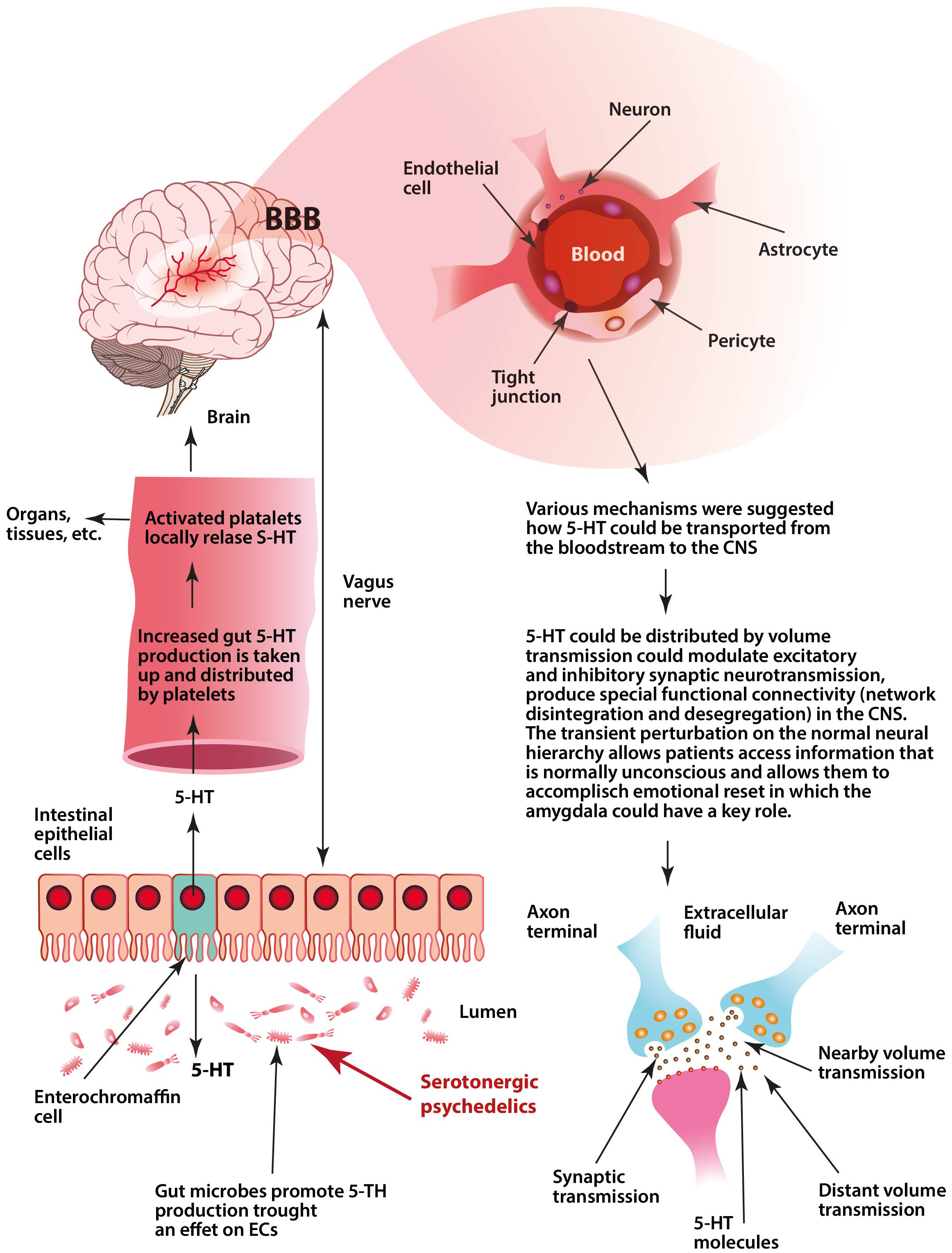
Fig. 1.

Visualization of the key aspects of our hypothesis. Serotonergic psychedelics could affect gut microbes that produce a transient increase of serotonin (5-HT) by host enterochromaffin cells (ECs). This increased gut 5-HT production—which is taken up and distributed by platelets—may work as a hormone-like regulatory signal that could influence membrane structure (that modifies membrane signaling processes) and membrane permeability in the host organs and tissues and in the brain. Increased plasma 5-HT levels could enhance permeability of microvessel endothelial cells and the blood-brain barrier (BBB). Transiently increased permeability of the BBB allows for plasma 5-HT to enter the central nervous system (CNS) and be distributed by the volume transmission (VT). Then, this gut-derived 5-HT could transiently modulate excitatory and inhibitory neurotransmission, produce special network disintegration in the CNS mainly within the default mode network (DMN), and promote structural and functional neural plasticity. This transient perturbation of the normal neural hierarchy (reduced connectivity between the frontal cortex and the lower brain areas, i.e., inhibition of top-down control) allows patients access to suppressed fear information and perform an emotional reset, in which the amygdala may have a key function.

Diverse medications and environmental factors can perturb 5-HT biosynthesis in the gut that could cause dysbiosis, which impairs the serotonergic gut-brain axis and produces alterations in platelet-dependent signal processes, including changes in vascular permeability throughout the whole body as well as in the BBB [240, 241, 242].

We should consider that in the case under discussion, i.e., serotonergic psychedelics, we are not talking about dysbiosis, but a transient modulation of 5-HT biosynthesis by serotonergic psychedelics in the gut.

Serotonergic psychedelics could modulate the gut microbiota and excitatory and inhibitory neurons in the ENS. This may influence the microbiota that promotes 5-HT biosynthesis from ECs that supply 5-HT to the mucosa, lumen, and circulating platelets [18]. Recently, we raised the idea that a large part of 5-HT-produced by ECs that is taken up and distributed by platelets-may operate as a hormone-like regulatory signal for the whole body [7]. This platelet-mediated distribution of intestinal 5-HT is dependent on the intestine’s actual health condition and influences membrane permeability throughout our organization.

5-HT has a strong lipid membrane affinity, influences membrane structure properties that modify membrane signaling processes, and affects vascular permeability [28, 29, 30, 31, 32]. Consequently, the hormone-like regulatory signal by platelet 5-HT can modulate countless signal processes in the whole body. Nevertheless, here we focus on membrane permeability, particularly on BBB permeability.

Lipid rafts may also play an important function in serotonergic psychedelic mechanisms. Increased plasma 5-HT levels induced by serotonergic psychedelics may modulate the permeability and fluidity of lipid rafts in plasma membrane subregions that participate dynamically in cell signaling and molecular trafficking operations in various cell membranes like platelets and the BBB [243, 244, 245, 246].

Several studies have found that increased plasma 5-HT levels can increase the permeability of microvessel endothelial cells as well as the BBB [118, 119, 128, 129, 247]. 5-HT injected intravenously in rats produces an increase in 5-HT and 5-HIAA (5-Hydroxyindoleacetic acid) in the brain [120].

Platelets are the key regulators of plasma 5-HT concentration [45, 248]. The regulation of the SERT activity on platelets has a key role to stabilize the concentration of plasma 5HT. It has been proposed that 5-HT regulates its own plasma concentration by modulating the uptake properties of platelet SERT, and that SERT does not regulate plasma 5-HT levels [249]. Since SERT is present in the BBB, it was proposed that 5-HT may enter into the extracellular fluid of the CNS [123].

Serotonergic psychedelics could increase the circulating concentration of PAF. Increases in 5-HT in the gut and, as a result, the increased level of 5-HT carried by platelets in the blood can act as an inflammatory signal [250, 251]. This could increase the circulating concentration of PAF, so PAF can also contribute to inflammation-mediated tissue injury, including BBB breakdown [116].

Volume transmission is a general mode of intercellular communication that takes place in the extracellular fluid and in the cerebrospinal fluid of the brain [201, 203, 210]. In the CNS, serotonergic transmission is mainly achieved by a non-synaptic VT mechanism [252]. Neuropsychoactive drugs act rather as VT signals [253].

We hypothesize (see Fig. 1) that the increased level of 5-HT—induced by serotonergic psychedelics in the gut and carried by platelets—produces a transient increase in permeability in microvessel endothelial cells within the BBB. This lets 5-HT enter into the extracellular fluid of the CNS and may act via volume transmission (diffusion mechanism). As a result, serotonin has a wide range of effects in the CNS that can modulate both excitatory and inhibitory neurotransmission, in which the DMN and amygdala may have key roles.

Our hypothesis may provide a possible mechanism for how psychedelics could create a transient disintegration in the normal neural network by reducing connectivity between the frontal cortex and the lower brain areas and promoting structural and functional neural plasticity, which allows patients access to suppressed fear information and perform an emotional reset [169, 170, 171]. Finally, it seems that there are important differences between the therapeutic mechanisms by which SSRIs mitigate negative emotions and those by psilocybin, since the latter allows patients to process traumatic memories and stressful experiences [184].

15. The Limitations of this Hypothesis

The limitations of this new complex hypothesis are that many parts are difficult to substantiate with scientific literature and that findings from cited animal and cell research might not translate to the complex physiology of humans. For example, the role of volumetric transport in brain processes is not yet known, and research has been neglected to this day, although it is one of the major and widespread forms of intercellular communication in the CNS. Monoamine neurotransmission, such as dopamine, norepinephrine, and serotonin, basically performs VT that could modulate glutamate and GABA neurons in the mammalian CNS. Furthermore, 5-HT is essentially produced in the gut, which is distributed by platelets and affects the regulation of the whole body and the brain, and the existing literature suggests that 5-HT may enter the brain via the BBB and could affect brain processes. Is it possible that a portion of brain serotonin comes from the gut? Studies have also suggested that BBB permeability can be regulated continuously and extensively, allowing types of molecules to enter the CNS that we would not believe.

One may argue that such enhancement of BBB permeability by 5-HT that is derived from ECs and conveyed by platelets may cause neuronal toxicity through the passage of peripheral cytokines and toxic substances into the brain. However, it is possible that 5-HT and other various molecules can selectively enter the brain through as yet unknown mechanisms.

Author Contributions

IB developed the concept. NCsN, PB and IB contributed to data collection, analysis and interpretation of results, and manuscript preparation.

Ethics Approval and Consent to Participate

Not applicable.

Acknowledgment

Not applicable.

Funding

This research was supported by Charles University project Cooperatio SVV.

Conflict of Interest

The authors declare no conflict of interest.

Publisher’s Note: IMR Press stays neutral with regard to jurisdictional claims in published maps and institutional affiliations.

References
[1]
John Jayakumar JAK, Panicker MM, Basu B. Serotonin 2A (5-HT2A) receptor affects cell-matrix adhesion and the formation and maintenance of stress fibers in HEK293 cells. Scientific Reports. 2020; 10: 21675.
[2]
Donovan LL, Johansen JV, Ros NF, Jaberi E, Linnet K, Johansen SS, et al. Effects of a single dose of psilocybin on behaviour, brain 5-HT2a receptor occupancy and gene expression in the pig. European Neuropsychopharmacology. 2021; 42: 1–11.
[3]
Vollenweider FX, Preller KH. Psychedelic drugs: neurobiology and potential for treatment of psychiatric disorders. Nature Reviews Neuroscience. 2020; 21: 611–624.
[4]
Preller KH, Razi A, Zeidman P, Stämpfli P, Friston KJ, Vollenweider FX. Effective connectivity changes in LSD-induced altered states of consciousness in humans. Proceedings of the National Academy of Sciences. 2019; 116: 2743–2748.
[5]
Schindler EAD, Wallace RM, Sloshower JA, D’Souza DC. Neuroendocrine associations underlying the persistent therapeutic effects of classic serotonergic psychedelics. Frontiers in Pharmacology. 2018; 9: 177.
[6]
Tylš F, Páleníček T, Horáček J. Psilocybin – Summary of knowledge and new perspectives. European Neuropsychopharmacology. 2014; 24: 342–356.
[7]
Szőke H, Kovács Z, Bókkon I, Vagedes J, Szabó AE, Hegyi G, et al. Gut dysbiosis and serotonin: intestinal 5-HT as a ubiquitous membrane permeability regulator in host tissues, organs, and the brain. Reviews in the Neurosciences. 2020; 31: 415–425.
[8]
Császár N, Bókkon I. Gut Serotonin as a General Membrane Permeability Regulator. Current Neuropharmacology. 2022; 20: 269–271.
[9]
Kuypers KPC. Psychedelic medicine: the biology underlying the persisting psychedelic effects. Medical Hypotheses. 2019; 125: 21–24.
[10]
Sanz C, Zamberlan F, Erowid E, Erowid F, Tagliazucchi E. The experience elicited by hallucinogens presents the highest similarity to dreaming within a large database of psychoactive substance reports. Frontiers in Neuroscience. 2018; 12: 7.
[11]
Fidalgo S, Ivanov DK, Wood SH. Serotonin: from top to bottom. Biogerontology. 2013; 14: 21–45.
[12]
Maffei ME. 5-Hydroxytryptophan (5-HTP): Natural occurrence, analysis, biosynthesis, biotechnology, physiology and toxicology. International Journal of Molecular Sciences. 2020; 22: 181.
[13]
Berger M, Gray JA, Roth BL. The Expanded Biology of Serotonin. Annual Review of Medicine. 2009; 60: 355–366.
[14]
Gershon MD. 5-Hydroxytryptamine (serotonin) in the gastrointestinal tract. Current Opinion in Endocrinology, Diabetes & Obesity. 2013; 20: 14–21.
[15]
Mawe GM, Hoffman JM. Serotonin signalling in the gut—functions, dysfunctions and therapeutic targets. Nature Reviews Gastroenterology & Hepatology. 2013; 10: 473–486.
[16]
Lund ML, Egerod KL, Engelstoft MS, Dmytriyeva O, Theodorsson E, Patel BA, et al. Enterochromaffin 5-HT cells – a major target for GLP-1 and gut microbial metabolites. Molecular Metabolism. 2018; 11: 70–83.
[17]
Martin AM, Young RL, Leong L, Rogers GB, Spencer NJ, Jessup CF, et al. The diverse metabolic roles of peripheral serotonin. Endocrinology. 2017; 158: 1049–1063.
[18]
Yano JM, Yu K, Donaldson GP, Shastri GG, Ann P, Ma L, et al. Indigenous bacteria from the gut microbiota regulates host serotonin biosynthesis. Cell. 2015; 161: 264–276.
[19]
Kanova M, Kohout P. Serotonin-Its synthesis and roles in the healthy and the critically Ill. International Journal of Molecular Sciences. 2021; 22: 4837.
[20]
Garcia-Garcia AL, Soiza-Reilly M. Serotonin subsystems modulate diverse and opposite behavioral functions. ACS Chemical Neuroscience. 2019; 10: 3061–3063.
[21]
Lv J, Liu F. The role of serotonin beyond the central nervous system during embryogenesis. Frontiers in Cellular Neuroscience. 2017; 11: 74.
[22]
Herr N, Bode C, Duerschmied D. The effects of serotonin in immune cells. Frontiers in Cardiovascular Medicine. 2017; 4: 48.
[23]
Wong DW, Soga T, Parhar IS. Aging and chronic administration of serotonin-selective reuptake inhibitor citalopram upregulate Sirt4 gene expression in the preoptic area of male mice. Frontiers in Genetics. 2015; 6: 281.
[24]
Trakhtenberg EF, Goldberg JL. The role of serotonin in axon and dendrite growth. International Review of Neurobiology. 2012; 38: 105–126.
[25]
Sikander A, Rana SV, Prasad KK. Role of serotonin in gastrointestinal motility and irritable bowel syndrome. Clinica Chimica Acta. 2009; 403: 47–55.
[26]
Azouzi S, Santuz H, Morandat S, Pereira C, Côté F, Hermine O, et al. Antioxidant and membrane binding properties of serotonin protect lipids from oxidation. Biophysical Journal. 2017; 112: 1863–1873.
[27]
Huether G, Schuff-Werner P. Platelet serotonin acts as a locally releasable antioxidant. Advances in Experimental Medicine and Biology. 1996; 86: 299–306.
[28]
Peters GH, Wang C, Cruys-Bagger N, Velardez GF, Madsen JJ, Westh P. Binding of serotonin to lipid membranes. Journal of the American Chemical Society. 2013; 135: 2164–2171.
[29]
Dey S, Surendran D, Engberg O, Gupta A, Fanibunda SE, Das A, et al. Altered membrane mechanics provides a receptor‐independent pathway for serotonin action. Chemistry. 2021; 27: 7533–7541.
[30]
Retamal JS, Grace MS, Dill LK, Ramirez-Garcia P, Peng S, Gondin AB, et al. Serotonin-induced vascular permeability is mediated by transient receptor potential vanilloid 4 in the airways and upper gastrointestinal tract of mice. Laboratory Investigation. 2021; 101: 851–864.
[31]
Majno G, Palade GE. Studies on Inflammation. 1. The effect of histamine and serotonin on vascular permeability: an electron microscopic study. The Journal of Biophysical and Biochemical Cytology. 1961; 11: 571–605.
[32]
Engberg O, Bochicchio A, Brandner A-F, Gupta A, Dey S, Böckmann RA, et al. Serotonin alters the phase equilibrium of a ternary mixture of phospholipids and cholesterol. Frontiers in Physiology. 2020; 11: 578868.
[33]
Koupenova M, Clancy L, Corkrey HA, Freedman JE. Circulating platelets as mediators of immunity, inflammation, and thrombosis. Circulation Research. 2018; 122: 337–351.
[34]
Ali RA, Wuescher LM, Worth RG. Platelets: essential components of the immune system. Current Trends in Immunology. 2015; 16: 65–78.
[35]
Mezger M, Nording H, Sauter R, Graf T, Heim C, von Bubnoff N, et al. Platelets and immune responses during thromboinflammation. Frontiers in Immunology. 2019; 10: 1731.
[36]
Franco AT, Corken A, Ware J. Platelets at the interface of thrombosis, inflammation, and cancer. Blood. 2015; 126: 582–588.
[37]
Heijnen H, van der Sluijs P. Platelet secretory behaviour: as diverse as the granules … or not? Journal of Thrombosis and Haemostasis. 2015; 13: 2141–2151.
[38]
Morrell CN, Aggrey AA, Chapman LM, Modjeski KL. Emerging roles for platelets as immune and inflammatory cells. Blood. 2014; 123: 2759–2767.
[39]
Jedlitschky G, Greinacher A, Kroemer HK. Transporters in human platelets: physiologic function and impact for pharmacotherapy. Blood. 2012; 119: 3394–3402.
[40]
Shah BH, Rasheed H, Rahman IH, Shariff AH, Khan FL, Rahman HB, et al. Molecular mechanisms involved in human platelet aggregation by synergistic interaction of platelet-activating factor and 5-hydroxytryptamine. Experimental & Molecular Medicine. 2001; 33: 226–233.
[41]
Khait VD, Huang Y, Zalsman G, Oquendo MA, Brent DA, Harkavy-Friedman JM, et al. Association of serotonin 5-HT2a receptor binding and the T102C polymorphism in depressed and healthy caucasian subjects. Neuropsychopharmacology. 2005; 30: 166–172.
[42]
Spigset O. Serotonin 5-HT2a receptor binding in platelets from healthy subjects as studied by [3H]-Lysergic Acid Diethylamide ([3H]-LSD): Intra- and interindividual variability. Neuropsychopharmacology. 1997; 16: 285–293.
[43]
Coccaro E. Impulsive aggression in personality disorder correlates with platelet 5-HT2a receptor binding. Neuropsychopharmacology. 1997; 16: 211–216.
[44]
Li N, Wallén NH, Ladjevardi M, Hjemdahl P. Effects of serotonin on platelet activation in whole blood. Blood Coagulation & Fibrinolysis. 1997; 8: 517–524.
[45]
Brenner B, Harney JT, Ahmed BA, Jeffus BC, Unal R, Mehta JL, et al. Plasma serotonin levels and the platelet serotonin transporter. Journal of Neurochemistry. 2007; 102: 206–215.
[46]
Lopez-Vilchez I, Diaz-Ricart M, White JG, Escolar G, Galan AM. Serotonin enhances platelet procoagulant properties and their activation induced during platelet tissue factor uptake. Cardiovascular Research. 2009; 84: 309–316.
[47]
Maouia A, Rebetz J, Kapur R, Semple JW. The immune nature of platelets revisited. Transfusion Medicine Reviews. 2020; 34: 209–220.
[48]
Witte A, Chatterjee M, Lang F, Gawaz M. Platelets as a novel source of pro-inflammatory chemokine CXCL14. Cellular Physiology and Biochemistry. 2017; 41: 1684–1696.
[49]
Clemetson KJ, Clemetson JM, Proudfoot AEI, Power CA, Baggiolini M, Wells TNC. Functional expression of CCR1, CCR3, CCR4, and CXCR4 chemokine receptors on human platelets. Blood. 2000; 96: 4046–4054.
[50]
Randriamboavonjy V, Fleming I. Platelet communication with the vascular wall: role of platelet-derived microparticles and non-coding RNAs. Clinical Science. 2018; 132: 1875–1888.
[51]
Ponomareva AA, Nevzorova TA, Mordakhanova ER, Andrianova IA, Rauova L, Litvinov RI, et al. Intracellular origin and ultrastructure of platelet-derived microparticles. Journal of Thrombosis and Haemostasis. 2017; 15: 1655–1667.
[52]
Miliotis S, Nicolalde B, Ortega M, Yepez J, Caicedo A. Forms of extracellular mitochondria and their impact in health. Mitochondrion. 2019; 48: 16–30.
[53]
Boudreau LH, Duchez AC, Cloutier N, Soulet D, Martin N, Bollinger J, et al. Platelets release mitochondria serving as substrate for bactericidal group IIA-secreted phospholipase A2 to promote inflammation. Blood. 2014; 124: 2173–2183.
[54]
Cognasse F, Hamzeh-Cognasse H, Lafarge S, Chavarin P, Cogné M, Richard Y, et al. Human platelets can activate peripheral blood B cells and increase production of immunoglobulins. Experimental Hematology. 2007; 35: 1376–1387.
[55]
León-Ponte, M, Ahern GP, O’Connell PJ. Serotonin provides an accessory signal to enhance T-cell activation by signaling through the 5-HT7 receptor. Blood. 2007; 109: 3139–3146.
[56]
Canobbio I. Blood platelets: Circulating mirrors of neurons? Research and Practice in Thrombosis and Haemostasis. 2019; 3: 564–565.
[57]
Ponomarev ED. Fresh evidence for platelets as neuronal and innate immune cells: Their role in the activation, differentiation, and deactivation of Th1, Th17, and Tregs during tissue inflammation. Frontiers in Immunology. 2018; 9: 406.
[58]
Pavlovic V, Ciric M, Jovanovic V, Trandafilovic M, Stojanovic P. Platelet-rich fibrin: Basics of biological actions and protocol modifications. Open Medicine. 2021; 16: 446–454.
[59]
Izzi B, Tirozzi A, Cerletti C, Donati MB, de Gaetano G, Hoylaerts MF, et al. Beyond haemostasis and thrombosis: platelets in depression and its co-morbidities. International Journal of Molecular Sciences. 2020; 21: 8817.
[60]
Dukhinova M, Kuznetsova I, Kopeikina E, Veniaminova E, Yung AWY, Veremeyko T, et al. Platelets mediate protective neuroinflammation and promote neuronal plasticity at the site of neuronal injury. Brain, Behavior, and Immunity. 2018; 74: 7–27.
[61]
Owens MJ, Nemeroff CB. The serotonin transporter and depression. Depression and Anxiety. 1998; 8: 5–12.
[62]
Cook EHJr, Fletcher KE, Wainwright M, Marks N, Yan SY, Leventhal BL. Primary structure of the human platelet serotonin 5-HT2A receptor: identify with frontal cortex serotonin 5-HT2A receptor. Journal of Neurochemistry. 1994; 63: 465–469.
[63]
Andres AH, Rao ML, Ostrowitzki S, Entzian W. Human brain cortex and platelet serotonin2 receptor binding properties and their regulation by endogenous serotonin. Life Sciences. 1993; 52: 313–321.
[64]
Zhuang X, Xu H, Fang Z, Xu C, Xue C, Hong X. Platelet serotonin and serotonin transporter as peripheral surrogates in depression and anxiety patients. European Journal of Pharmacology. 2018; 834: 213–220.
[65]
Peitl V, Getaldić-Švarc B, Karlović D. Platelet Serotonin Concentration is Associated with Illness Duration in Schizophrenia and Chronological Age in Depression. Psychiatry Investigation. 2020; 17: 579–586.
[66]
Maurer-Spurej E, Pittendreigh C, Misri S. Platelet serotonin levels support depression scores for women with postpartum depression. Journal of Psychiatry and Neuroscience. 2007; 32: 23–29.
[67]
Gomez-Eguilaz M, Ramon-Trapero JL, Perez-Martinez L, Blanco JR. The microbiota-gut-brain axis and its great projections. Revista de Neurología. 2019; 68: 111–117.
[68]
Cryan JF, O’Riordan KJ, Cowan CSM, Sandhu KV, Bastiaanssen TFS, Boehme M, et al. The microbiota-gut-brain axis. Physiological Reviews. 2019; 99: 1877–2013.
[69]
Spencer NJ, Hibberd TJ, Travis L, Wiklendt L, Costa M, Hu H, et al. Identification of a rhythmic firing pattern in the enteric nervous system that generates rhythmic electrical activity in smooth muscle. The Journal of Neuroscience. 2018; 38: 5507–5522.
[70]
Morarach K, Mikhailova A, Knoflach V, Memic F, Kumar R, Li W, et al. Diversification of molecularly defined myenteric neuron classes revealed by single-cell RNA sequencing. Nature Neuroscience. 2021; 24: 34–46.
[71]
Wei GZ, Martin KA, Xing PY, Agrawal R, Whiley L, Wood TK, et al. Tryptophan-metabolizing gut microbes regulate adult neurogenesis via the aryl hydrocarbon receptor. Proceedings of the National Academy of Sciences. 2021; 118: e2021091118.
[72]
Singh R, Zogg H, Wei L, Bartlett A, Ghoshal UC, Rajender S, et al. Gut microbial dysbiosis in the pathogenesis of gastrointestinal dysmotility and metabolic disorders. Journal of Neurogastroenterology and Motility. 2021; 27: 19–34.
[73]
Wilkins LJ, Monga M, Miller AW. Defining dysbiosis for a cluster of chronic diseases. Scientific Reports. 2019; 9: 12918.
[74]
Peng J, Xiao X, Hu M, Zhang X. Interaction between gut microbiome and cardiovascular disease. Life Sciences. 2018; 214: 153–157.
[75]
Carding S, Verbeke K, Vipond DT, Corfe BM, Owen LJ. Dysbiosis of the gut microbiota in disease. Microbial Ecology in Health and Disease. 2015; 26: 26191.
[76]
Scher JU, Ubeda C, Artacho A, Attur M, Isaac S, Reddy SM, et al. Decreased bacterial diversity characterizes the altered gut microbiota in patients with psoriatic arthritis, resembling dysbiosis in inflammatory bowel disease. Arthritis & Rheumatology. 2015; 67: 128–139.
[77]
Abrahamsson TR, Jakobsson HE, Andersson AF, Björkstén B, Engstrand L, Jenmalm MC. Low gut microbiota diversity in early infancy precedes asthma at school age. Clinical & Experimental Allergy. 2014; 44: 842–850.
[78]
Carroll IM, Ringel-Kulka T, Siddle JP, Ringel Y. Alterations in composition and diversity of the intestinal microbiota in patients with diarrhea-predominant irritable bowel syndrome. Neurogastroenterology & Motility. 2012; 24: 521–e248.
[79]
Sjöstedt P, Enander J, Isung J. Serotonin reuptake inhibitors and the gut microbiome: significance of the gut microbiome in relation to mechanism of action, treatment response, side effects, and tachyphylaxis. Frontiers in Psychiatry. 2021; 12: 682868.
[80]
Ait Chait Y, Mottawea W, Tompkins TA, Hammami R. Unravelling the antimicrobial action of antidepressants on gut commensal microbes. Scientific Reports. 2020; 10: 17878.
[81]
McGovern AS, Hamlin AS, Winter G. A review of the antimicrobial side of antidepressants and its putative implications on the gut microbiome. Australian & New Zealand Journal of Psychiatry. 2019; 53: 1151–1166.
[82]
Lukić I, Getselter D, Ziv O, Oron O, Reuveni E, Koren O, et al. Antidepressants affect gut microbiota and Ruminococcus flavefaciens is able to abolish their effects on depressive-like behavior. Translational Psychiatry. 2019; 9: 133.
[83]
Karine de Sousa A, Rocha JE, Gonçalves de Souza T, Sampaio de Freitas T, Ribeiro-Filho J, Melo Coutinho HD. New roles of fluoxetine in pharmacology: Antibacterial effect and modulation of antibiotic activity. Microbial Pathogenesis. 2018; 123: 368–371.
[84]
Lee SM, Dong TS, Krause-Sorio B, Siddarth P, Milillo MM, Lagishetty V, et al. The intestinal microbiota as a predictor for antidepressant treatment outcome in geriatric depression: a prospective pilot study. International Psychogeriatrics. 2022; 34: 33–45.
[85]
Sjöstedt P, Enander J, Isung J. Serotonin reuptake inhibitors and the gut microbiome: Significance of the gut microbiome in relation to mechanism of action, treatment response, side effects, and tachyphylaxis. Frontiers in Psychiatry. 2021;12: 682868.
[86]
Munoz-Bellido JL, Munoz-Criado S, Garcı̀a-Rodrı̀guez JA. Antimicrobial activity of psychotropic drugs: selective serotonin reuptake inhibitors. International Journal of Antimicrobial Agents. 2000; 14: 177–180.
[87]
Ameen S, Praharaj SK. Functional auditory hallucinations in a case of serotonin syndrome. The Journal of Neuropsychiatry and Clinical Neurosciences. 2013; 25: E60–E61.
[88]
Monji A, Kato T, Mizoguchi Y, Horikawa H, Seki Y, Kanba S. Visual and auditory hallucinations during normal use of paroxetine for treatment of major depressive disorder. The Journal of Neuropsychiatry and Clinical Neurosciences. 2011; 23: E14–E15.
[89]
Tripp AC, Golden SA. Transient complex visual hallucinations with venlafaxine treatment: a case report. The Primary Care Companion to The Journal of Clinical Psychiatry. 2009; 11: 83.
[90]
Schuld A, Archelos JJ, Friess E. Visual hallucinations and psychotic symptoms during treatment with selective serotonin reuptake inhibitors: is the sigma receptor involved? Journal of Clinical Psychopharmacology. 2000; 20: 579–580.
[91]
Generoso JS, Giridharan VV, Lee J, Macedo D, Barichello T. The role of the microbiota-gut-brain axis in neuropsychiatric disorders. Brazilian Journal of Psychiatry. 2021; 43: 293–305.
[92]
Mathee K, Cickovski T, Deoraj A, Stollstorff M, Narasimhan G. The gut microbiome and neuropsychiatric disorders: implications for attention deficit hyperactivity disorder (ADHD). Journal of Medical Microbiology. 2020; 69: 14–24.
[93]
Yuan X, Kang Y, Zhuo C, Huang X, Song X. The gut microbiota promotes the pathogenesis of schizophrenia via multiple pathways. Biochemical and Biophysical Research Communications. 2019; 512: 373–380.
[94]
Fowlie G, Cohen N, Ming X. The perturbance of microbiome and gut-brain axis in autism spectrum disorders. International Journal of Molecular Sciences. 2018; 19: E2251
[95]
Spielman LJ, Gibson DL, Klegeris A. Unhealthy gut, unhealthy brain: the role of the intestinal microbiota in neurodegenerative diseases. Neurochemistry International. 2018; 120: 149–163.
[96]
Sudo N, Chida Y, Aiba Y, Sonoda J, Oyama N, Yu X, et al. Postnatal microbial colonization programs the hypothalamic-pituitary-adrenal system for stress response in mice. The Journal of Physiology. 2004; 558: 263–275.
[97]
Huang F, Wu X. Brain neurotransmitter modulation by gut microbiota in anxiety and depression. Frontiers in Cell and Developmental Biology. 2021; 9: 649103
[98]
Carlson AL, Xia K, Azcarate-Peril MA, Rosin SP, Fine JP, Mu W, et al. Infant gut microbiome composition is associated with non-social fear behavior in a pilot study. Nature Communications. 2021; 12: 3294.
[99]
Hoban AE, Moloney RD, Golubeva AV, McVey Neufeld KA, O’Sullivan O, Patterson E, et al. Behavioural and neurochemical consequences of chronic gut microbiota depletion during adulthood in the rat. Neuroscience. 2016; 339: 463–477.
[100]
Jenkins T, Nguyen JC, Polglaze KE, Bertrand PP. Influence of tryptophan and serotonin on mood and cognition with a possible role of the gut-brain axis. Nutrients. 2016; 8: 56.
[101]
Schemann M, Frieling T, Enck P. To learn, to remember, to forget—how smart is the gut? Acta Physiologica. 2020; 228: e13296.
[102]
Dinan TG, Stilling RM, Stanton C, Cryan JF. Collective unconscious: how gut microbes shape human behavior. Journal of Psychiatric Research. 2015; 63: 1–9.
[103]
Legan TB, Lavoie B, Mawe GM. Direct and indirect mechanisms by which the gut microbiota influence host serotonin systems. Neurogastroenterology and Motility. 2022. (in press)
[104]
De Vadder F, Grasset E, Mannerås Holm L, Karsenty G, Macpherson AJ, Olofsson LE, et al. Gut microbiota regulates maturation of the adult enteric nervous system via enteric serotonin networks. Proceedings of the National Academy of Sciences. 2018; 115: 6458–6463.
[105]
Foong JPP, Hung LY, Poon S, Savidge TC, Bornstein JC. Early life interaction between the microbiota and the enteric nervous system. American Journal of Physiology-Gastrointestinal and Liver Physiology. 2020; 319: G541–G548.
[106]
Mishima Y, Ishihara S. Enteric Microbiota-mediated serotonergic signaling in pathogenesis of irritable bowel syndrome. International Journal of Molecular Sciences. 2021; 22: 10235.
[107]
Mandić AD, Woting A, Jaenicke T, Sander A, Sabrowski W, Rolle-Kampcyk U, et al. Clostridium ramosum regulates enterochromaffin cell development and serotonin release. Scientific Reports. 2019; 9: 1177.
[108]
Hata T, Asano Y, Yoshihara K, Kimura-Todani T, Miyata N, Zhang XT, et al. Regulation of gut luminal serotonin by commensal microbiota in mice. PLoS ONE. 2017; 12: e0180745.
[109]
Wikoff WR, Anfora AT, Liu J, Schultz PG, Lesley SA, Peters EC, et al. Metabolomics analysis reveals large effects of gut microflora on mammalian blood metabolites. Proceedings of the National Academy of Sciences. 2009; 106: 3698–3703.
[110]
Reigstad CS, Salmonson CE, III JFR, Szurszewski JH, Linden DR, Sonnenburg JL, et al. Gut microbes promote colonic serotonin production through an effect of short‐chain fatty acids on enterochromaffin cells. The FASEB Journal. 2015; 29: 1395–1403.
[111]
Clarke G, Grenham S, Scully P, Fitzgerald P, Moloney RD, Shanahan F, et al. The microbiome-gut-brain axis during early life regulates the hippocampal serotonergic system in a sex-dependent manner. Molecular Psychiatry. 2013; 18: 666–673.
[112]
Kanwar JR, Sriramoju B, Kanwar RK. Neurological disorders and therapeutics targeted to surmount the blood-brain barrier. International Journal of Nanomedicine. 2012; 7: 3259–3278.
[113]
Galea I. The blood–brain barrier in systemic infection and inflammation. Cellular & Molecular Immunology. 2021; 18: 2489–2501.
[114]
Kelesidis T, Papakonstantinou V, Detopoulou P, Fragopoulou E, Chini M, Lazanas MC, et al. The role of platelet-activating factor in chronic inflammation, immune activation, and comorbidities associated with HIV infection. Aids Reviews. 2015; 17: 191–201.
[115]
Brailoiu E, Barlow CL, Ramirez SH, Abood ME, Brailoiu GC. Effects of platelet-activating factor on brain microvascular endothelial cells. Neuroscience. 2018; 377: 105–113.
[116]
Sharma J, Young DM, Marentette JO, Rastogi P, Turk J, McHowat J. Lung endothelial cell platelet-activating factor production and inflammatory cell adherence are increased in response to cigarette smoke component exposure. American Journal of Physiology-Lung Cellular and Molecular Physiology. 2012; 302: L47–L55.
[117]
Deng Y, Fang W, Li Y, Cen J, Fang F, Lv P, Gong S, Mao L. Blood-brain barrier breakdown by PAF and protection by XQ-1H due to antagonism of PAF effects. European Journal of Pharmacology. 2009; 616: 43-47.
[118]
Sharma HS, Westman J, Cervós Navarro J, Dey PK, Nyberg F. Probable involvement of serotonin in the increased permeability of the blood—brain barrier by forced swimming. an experimental study using Evans blue and 131i-sodium tracers in the rat. Behavioural Brain Research. 1995; 72: 189–196.
[119]
Sharma HS, Olsson Y, Kumar Dey P. Changes in blood-brain barrier and cerebral blood flow following elevation of circulating serotonin level in anesthetized rats. Brain Research. 1990; 517: 215–223.
[120]
Bulat M, Supek Z. Passage of 5-hydroxytryptamine through the blood-brain barrier, its metabolism in the brain and elimination of 5-hydroxyindoleacetic acid from the brain tissue. Journal of Neurochemistry. 1968; 15: 383–389.
[121]
Bulat M, Supek Z. The penetration of 5-hydroxytryptamine through the blood-brain barrier. Journal of Neurochemistry. 1967; 14: 265–271.
[122]
Nakatani Y, Sato-Suzuki I, Tsujino N, Nakasato A, Seki Y, Fumoto M, et al. Augmented brain 5-HT crosses the blood-brain barrier through the 5-HT transporter in rat. European Journal of Neuroscience. 2008; 27: 2466–2472.
[123]
Young LW, Darios ES, Watts SW. An immunohistochemical analysis of SERT in the blood–brain barrier of the male rat brain. Histochemistry and Cell Biology. 2015; 144: 321–329.
[124]
Puhm F, Boilard E, Machlus KR. Platelet extracellular vesicles: Beyond the blood. Arteriosclerosis, Thrombosis, and Vascular Biology. 2021; 41: 87–96.
[125]
Cullivan S, Murphy CA, Weiss L, Comer SP, Kevane B, McCullagh B, et al. Platelets, extracellular vesicles and coagulation in pulmonary arterial hypertension. Pulmonary Circulation. 2021; 11: 20458940211021036.
[126]
Żmigrodzka M, Witkowska-Piłaszewicz O, Winnicka A. Platelets extracellular vesicles as regulators of cancer progression-an updated perspective. International Journal of Molecular Sciences. 2020; 21: 5195.
[127]
Kopeikina E, Ponomarev ED. The role of platelets in the stimulation of neuronal synaptic plasticity, electric activity, and oxidative phosphorylation: Possibilities for new therapy of neurodegenerative diseases. Frontiers in Cellular Neuroscience. 2021; 15: 680126.
[128]
Fang W, Zhang R, Sha L, Lv P, Shang E, Han D, et al. Platelet activating factor induces transient blood-brain barrier opening to facilitate edaravone penetration into the brain. Journal of Neurochemistry. 2014; 128: 662–671.
[129]
Fang W, Geng X, Deng Y, Li Y, Shang E, Cen J, et al. Platelet activating factor induces blood brain barrier permeability alteration in vitro. Journal of Neuroimmunology. 2011; 230: 42–47.
[130]
Brust P, Friedrich A, Krizbai IA, Bergmann R, Roux F, Ganapathy V, et al. Functional expression of the serotonin transporter in immortalized rat brain microvessel endothelial cells. Journal of Neurochemistry. 2000; 74: 1241–1248.
[131]
Galvão-Coelho NL, Marx W, Gonzalez M, Sinclair J, de Manincor M, Perkins D, et al. Classic serotonergic psychedelics for mood and depressive symptoms: a meta-analysis of mood disorder patients and healthy participants. Psychopharmacology. 2021; 238: 341–354.
[132]
Stenbæk DS, Madsen MK, Ozenne B, Kristiansen S, Burmester D, Erritzoe D, et al. Brain serotonin 2a receptor binding predicts subjective temporal and mystical effects of psilocybin in healthy humans. Journal of Psychopharmacology. 2021; 35: 459–468.
[133]
Ross S. Therapeutic use of classic psychedelics to treat cancer-related psychiatric distress. International Review of Psychiatry. 2018; 30: 317–330.
[134]
Garcia-Romeu A, Griffiths R, Johnson M. Psilocybin-occasioned mystical experiences in the treatment of tobacco addiction. Current Drug Abuse Reviews. 2014; 7: 157–164.
[135]
Nichols D, Johnson M, Nichols C. Psychedelics as medicines: an emerging new paradigm. Clinical Pharmacology & Therapeutics. 2017; 101: 209–219.
[136]
Halberstadt AL. Recent advances in the neuropsychopharmacology of serotonergic hallucinogens. Behavioural Brain Research. 2015; 277: 99–120.
[137]
López-Giménez JF, González-Maeso J. Hallucinogens and serotonin 5-HT2A receptor-mediated signaling pathways. Current Topics in Behavioral Neurosciences. 2018; 36: 45–73.
[138]
González-Maeso J, Weisstaub NV, Zhou M, Chan P, Ivic L, Ang R, et al. Hallucinogens recruit specific cortical 5-HT2a receptor-mediated signaling pathways to affect behavior. Neuron. 2007; 53: 439–452.
[139]
Smith R. Agonist properties of N,N-Dimethyltryptamine at serotonin 5-HT2a and 5-HT2C receptors. Pharmacology Biochemistry and Behavior. 1998; 61: 323–330.
[140]
Preller KH, Herdener M, Pokorny T, Planzer A, Kraehenmann R, Stämpfli P, et al. The fabric of meaning and subjective effects in LSD-induced states depend on serotonin 2a receptor activation. Current Biology. 2017; 27: 451–457.
[141]
Vollenweider FX, Vollenweider-Scherpenhuyzen MFI, Bäbler A, Vogel H, Hell D. Psilocybin induces schizophrenia-like psychosis in humans via a serotonin-2 agonist action. NeuroReport. 1998; 9: 3897–3902.
[142]
Gewirtz JC, Marek GJ. Behavioral evidence for interactions between a hallucinogenic drug and group II metabotropic glutamate receptors. Neuropsychopharmacology. 2000; 23: 569-576.
[143]
Halberstadt AL, Geyer MA. Effect of hallucinogens on unconditioned behavior. Current Topics in Behavioral Neurosciences. 2018; 36: 159–199.
[144]
Halberstadt AL, Geyer MA. Multiple receptors contribute to the behavioral effects of indoleamine hallucinogens. Neuropharmacology. 2011; 61: 364–381.
[145]
Inserra A, De Gregorio D, Gobbi G. Psychedelics in psychiatry: Neuroplastic, immunomodulatory, and neurotransmitter mechanisms. Pharmacological Reviews. 2021; 73: 202–277.
[146]
Martin DA, Nichols CD. Psychedelics recruit multiple cellular types and produce complex transcriptional responses within the brain. EBioMedicine. 2016; 11: 262–277.
[147]
Canal CE. Serotonergic psychedelics: Experimental approaches for assessing mechanisms of action. New Psychoactive Substances. 2018; 10: 227–260.
[148]
dos Santos RG, Osório FL, Crippa JAS, Hallak JEC. Classical hallucinogens and neuroimaging: a systematic review of human studies: hallucinogens and neuroimaging. Neuroscience & Biobehavioral Reviews. 2016; 71: 715–728.
[149]
Alves PN, Foulon C, Karolis V, Bzdok D, Margulies DS, Volle E, et al. An improved neuroanatomical model of the default-mode network reconciles previous neuroimaging and neuropathological findings. Communications Biology. 2019; 2: 370.
[150]
Roy AK, Shehzad Z, Margulies DS, Kelly AMC, Uddin LQ, Gotimer K, et al. Functional connectivity of the human amygdala using resting state fMRI. NeuroImage. 2009; 45: 614–626.
[151]
Bzdok D, Laird AR, Zilles K, Fox PT, Eickhoff SB. An investigation of the structural, connectional, and functional subspecialization in the human amygdala. Human Brain Mapping. 2013; 34: 3247–3266.
[152]
Qi C, Wang Z, Bai W, Liu T, Zheng X. Reduced information transmission of medial prefrontal cortex to basolateral amygdala inhibits exploratory behavior in depressed rats. Frontiers in Neuroscience. 2020; 14: 608587.
[153]
Santos A, Granell E, Gómez-Ansón B, Crespo I, Pires P, Vives-Gilabert Y, et al. Depression and anxiety scores are associated with amygdala volume in Cushing’s syndrome: Preliminary study. BioMed Research International. 2017; 2017: 2061935.
[154]
Holmes AJ, Lee PH, Hollinshead MO, Bakst L, Roffman JL, Smoller JW, et al. Individual differences in amygdala-medial prefrontal anatomy link negative affect, impaired social functioning, and polygenic depression risk. Journal of Neuroscience. 2012; 32: 18087–18100.
[155]
Schumann CM, Bauman MD, Amaral DG. Abnormal structure or function of the amygdala is a common component of neurodevelopmental disorders. Neuropsychologia. 2011; 49: 745–759.
[156]
Beesdo K, Lau JYF, Guyer AE, McClure-Tone EB, Monk CS, Nelson EE, et al. Common and distinct amygdala-function perturbations in depressed vs anxious adolescents. Archives of General Psychiatry. 2009; 66: 275–285.
[157]
Drevets WC. Neuroimaging abnormalities in the amygdala in mood disorders. Annals of the New York Academy of Sciences. 2003; 985: 420–444.
[158]
Murray RJ, Brosch T, Sander D. The functional profile of the human amygdala in affective processing: Insights from intracranial recordings. Cortex. 2014; 60: 10–33.
[159]
Pessoa L, Adolphs R. Emotion processing and the amygdala: from a ‘low road’ to ‘many roads’ of evaluating biological significance. Nature Reviews Neuroscience. 2010; 11: 773–782.
[160]
Averbeck BB, Seo M. The statistical neuroanatomy of frontal networks in the macaque. PLoS Computational Biology. 2008; 4: e1000050.
[161]
Diano M, Celeghin A, Bagnis A, Tamietto M. Amygdala response to emotional stimuli without awareness: Facts and interpretations. Frontiers in Psychology. 2017; 7: 2029.
[162]
De Gregorio D, Aguilar-Valles A, Preller KH, Heifets BD, Hibicke M, Mitchell J, et al. Hallucinogens in mental health: Preclinical and clinical studies on LSD, psilocybin, MDMA, and ketamine. The Journal of Neuroscience. 2021; 41: 891–900.
[163]
Mueller F, Lenz C, Dolder PC, Harder S, Schmid Y, Lang UE, et al. Acute effects of LSD on amygdala activity during processing of fearful stimuli in healthy subjects. Translational Psychiatry. 2017; 7: e1084.
[164]
Bershad AK, Preller KH, Lee R, Keedy S, Wren-Jarvis J, Bremmer MP, et al. Preliminary report on the effects of a low dose of LSD on resting-state amygdala functional connectivity. Biological Psychiatry: Cognitive Neuroscience and Neuroimaging. 2020; 5: 461–467.
[165]
Grimm O, Kraehenmann R, Preller KH, Seifritz E, Vollenweider FX. Psilocybin modulates functional connectivity of the amygdala during emotional face discrimination. European Neuropsychopharmacology. 2018; 28: 691–700.
[166]
Roseman L, Nutt DJ, Carhart-Harris RL. Quality of acute psychedelic experience predicts therapeutic efficacy of psilocybin for treatment-resistant depression. Frontiers in Pharmacology. 2018; 8: 974.
[167]
Roseman L, Haijen E, Idialu-Ikato K, Kaelen M, Watts R, Carhart-Harris R. Emotional breakthrough and psychedelics: Validation of the Emotional Breakthrough Inventory. Journal of Psychopharmacology. 2019; 33: 1076–1087.
[168]
Kraehenmann R, Preller KH, Scheidegger M, Pokorny T, Bosch OG, Seifritz E, et al. Psilocybin-induced decrease in amygdala reactivity correlates with enhanced positive mood in healthy volunteers. Biological Psychiatry. 2015; 78: 572–581.
[169]
Carhart-Harris RL, Roseman L, Bolstridge M, Demetriou L, Pannekoek JN, Wall MB, et al. Psilocybin for treatment-resistant depression: fMRI-measured brain mechanisms. Scientific Reports. 2017; 7: 13187.
[170]
Alonso JF, Romero S, Mañanas M, Riba J. Serotonergic psychedelics temporarily modify information transfer in humans. International Journal of Neuropsychopharmacology. 2015; 18: 1–9.
[171]
Császár-Nagy N, Kapócs G, Bókkon I. Classic psychedelics: the special role of the visual system. Reviews in the Neurosciences. 2019; 30: 651–669.
[172]
Albert PR, Le François B, Vahid-Ansari F. Genetic, epigenetic and posttranscriptional mechanisms for treatment of major depression: the 5-HT1a receptor gene as a paradigm. Journal of Psychiatry and Neuroscience. 2019; 44: 164–176.
[173]
Zheng H, Onoda K, Wada Y, Mitaki S, Nabika T, Yamaguchi S. Serotonin-1a receptor C-1019G polymorphism affects brain functional networks. Scientific Reports. 2017; 7: 12536.
[174]
Miller MW, Sperbeck E, Robinson ME, Sadeh N, Wolf EJ, Hayes JP, et al. 5-HT2A gene variants moderate the association between PTSD and reduced default mode network connectivity. Frontiers in Neuroscience. 2016; 10: 299.
[175]
Yan C-G, Chen X, Li L, Castellanos FX, Bai TJ, Bo QJ, et al. Reduced default mode network functional connectivity in patients with recurrent major depressive disorder. Proceedings of the National Academy of Sciences. 2019; 116: 9078–9083.
[176]
Li SY, Zhu Y, Wang YL, Lü PP, Zuo WB, Li FY. Dysfunctional resting-state connectivity of default mode network in adolescent patients with first-episode drug-naive major depressive disorder. Zhonghua Yi Xue Za Zhi. 2017; 97: 3538–3542.
[177]
Hamilton JP, Furman DJ, Chang C, Thomason ME, Dennis E, Gotlib IH. Default-mode and task-positive network activity in major depressive disorder: Implications for adaptive and maladaptive rumination. Biological Psychiatry. 2011; 70: 327–333.
[178]
Sheline YI, Price JL, Yan Z, Mintun MA. Resting-state functional MRI in depression unmasks increased connectivity between networks via the dorsal nexus. Proceedings of the National Academy of Sciences. 2010; 107: 11020–11025.
[179]
Shao L, Liao C, Gregg I, Davoudian PA, Savalia NK, Delagarza K, et al. Psilocybin induces rapid and persistent growth of dendritic spines in frontal cortex in vivo. Neuron. 2021; 109: 2535–2544.e4.
[180]
Carhart-Harris RL, Muthukumaraswamy S, Roseman L, Kaelen M, Droog W, Murphy K, et al. Neural correlates of the LSD experience revealed by multimodal neuroimaging. Proceedings of the National Academy of Sciences. 2016; 113: 4853–4858.
[181]
Winkelman MJ. The mechanisms of psychedelic visionary experiences: hypotheses from evolutionary psychology. Frontiers in Neuroscience. 2017; 11: 539.
[182]
Gallagher J. Magic mushrooms can ‘reset’ depressed brain. 2017. Available at: https://www.bbc.com/news/health-41608984 (Accessed: 14 October 2017).
[183]
Madsen MK, Stenbæk DS, Arvidsson A, Armand S, Marstrand-Joergensen MR, Johansen SS, et al. Psilocybin-induced changes in brain network integrity and segregation correlate with plasma psilocin level and psychedelic experience. European Neuropsychopharmacology. 2021; 50: 121–132.
[184]
Roseman L, Demetriou L, Wall MB, Nutt DJ, Carhart-Harris RL. Increased amygdala responses to emotional faces after psilocybin for treatment-resistant depression. Neuropharmacology. 2018; 142: 263–269.
[185]
Healy CJ. The acute effects of classic psychedelics on memory in humans. Psychopharmacology. 2021; 238: 639–653.
[186]
Young KD, Friedman ES, Collier A, Berman SR, Feldmiller J, Haggerty AE, et al. Response to SSRI intervention and amygdala activity during self-referential processing in major depressive disorder. NeuroImage: Clinical. 2020; 28: 102388.
[187]
Ma Y. Neuropsychological mechanism underlying antidepressant effect: a systematic meta-analysis. Molecular Psychiatry. 2015; 20: 311–319.
[188]
Godlewska BR, Norbury R, Selvaraj S, Cowen PJ, Harmer CJ. Short-term SSRI treatment normalises amygdala hyperactivity in depressed patients. Psychological Medicine. 2012; 42: 2609–2617.
[189]
Murphy SE, Norbury R, O’Sullivan U, Cowen PJ, Harmer CJ. Effect of a single dose of citalopram on amygdala response to emotional faces. British Journal of Psychiatry. 2009; 194: 535–540.
[190]
de Vos CMH, Mason NL, Kuypers KPC. Psychedelics and neuroplasticity: A systematic review unraveling the biological underpinnings of psychedelics. Frontiers in Psychiatry. 2021; 12: 724606.
[191]
Martin DA, Nichols CD. The effects of hallucinogens on gene expression. Current Topics in Behavioral Neurosciences. 2018; 36: 137–158.
[192]
Francescangeli J, Karamchandani K, Powell M, Bonavia A. The serotonin syndrome: From molecular mechanisms to clinical practice. International Journal of Molecular Sciences. 2019; 20: 2288.
[193]
Machado-Vieira R, Baumann J, Wheeler-Castillo C, Latov D, Henter ID, Salvadore G, et al. The timing of antidepressant effects: A comparison of diverse pharmacological and somatic treatments. Pharmaceuticals. 2010; 3: 19–41.
[194]
Artin H, Zisook S, Ramanathan D. How do serotonergic psychedelics treat depression: the potential role of neuroplasticity. World Journal of Psychiatry. 2021; 11: 201–214.
[195]
Malcolm B, Thomas K. Serotonin toxicity of serotonergic psychedelics. Psychopharmacology. 2022; 239: 1881–1891.
[196]
Silins E, Copeland J, Dillon P. Qualitative review of serotonin syndrome, ecstasy (MDMA) and the use of other serotonergic substances: Hierarchy of risk. Australian & New Zealand Journal of Psychiatry. 2007; 41: 649–655.
[197]
Makunts T, Jerome L, Abagyan R, de Boer A. Reported cases of serotonin syndrome in MDMA users in FAERS Database. Frontiers in Psychiatry. 2022; 12: 824288.
[198]
Alpár A, Zahola P, Hanics J, Hevesi Z, Korchynska S, Benevento M, et al. Hypothalamic CNTF volume transmission shapes cortical noradrenergic excitability upon acute stress. EMBO Journal. 2018; 37: e100087.
[199]
Taber KH, Hurley RA. Volume transmission in the brain: beyond the synapse. The Journal of Neuropsychiatry and Clinical Neurosciences. 2014; 26: iv–i4.
[200]
Agnati LF, Guidolin D, Maura G, Marcoli M, Leo G, Carone C, et al. Information handling by the brain: proposal of a new “paradigm” involving the roamer type of volume transmission and the tunneling nanotube type of wiring transmission. Journal of Neural Transmission. 2014; 121: 1431–1449.
[201]
Fuxe K, Borroto-Escuela DO, Romero-Fernandez W, Zhang W, Agnati LF. Volume transmission and its different forms in the central nervous system. Chinese Journal of Integrative Medicine. 2013; 19: 323–329.
[202]
De-Miguel FF, Fuxe K. Extrasynaptic neurotransmission as a way of modulating neuronal functions. Frontiers in Physiology. 2012; 3: 16.
[203]
Bókkon I, Antal I. Schizophrenia: redox regulation and volume transmission. Current Neuropharmacology. 2011; 9: 289–300.
[204]
Agnati LF, Fuxe K. Volume transmission as a key feature of information handling in the central nervous system possible new interpretative value of the Turing’s B-type machine. Progress in Brain Research. 2000; 128: 3–19.
[205]
Fuxe K, Agnati LF. Volume transmission in the brain, novel mechanisms for neural transmission. Raven Press: New York. 1991.
[206]
Vizi ES. Modulation of cortical release of acetylcholine by noradrenaline released from nerves arising from the rat locus coeruleus. Neuroscience. 1980; 5: 2139–2144.
[207]
Vizi E. Presynaptic modulation of neurochemical transmission. Progress in Neurobiology. 1979; 12: 181–290.
[208]
Paton WDM, Vizi ES. The inhibitory action of noradrenaline and adrenaline on acetylcholine output by guinea-pig ileum longitudinal muscle strip. British Journal of Pharmacology. 1969; 35: 10–28.
[209]
Borroto-Escuela DO, Agnati LF, Bechter K, Jansson A, Tarakanov AO, Fuxe K. The role of transmitter diffusion and flow versus extracellular vesicles in volume transmission in the brain neural–glial networks. Philosophical Transactions of the Royal Society B: Biological Sciences. 2015; 370: 20140183.
[210]
Borroto-Escuela DO, Perez De La Mora M, Manger P, Narváez M, Beggiato S, Crespo-Ramírez M, et al. Brain dopamine transmission in health and Parkinson’s disease: Modulation of synaptic transmission and plasticity through volume transmission and dopamine heteroreceptors. Frontiers in Synaptic Neuroscience. 2018; 10: 20.
[211]
Fuxe K, Dahlström AB, Jonsson G, Marcellino D, Guescini M, Dam M, et al. The discovery of central monoamine neurons gave volume transmission to the wired brain. Progress in Neurobiology. 2010; 90: 82–100.
[212]
Fuxe K, Borroto-Escuela D. Volume transmission and receptor-receptor interactions in heteroreceptor complexes: understanding the role of new concepts for brain communication. Neural Regeneration Research. 2016; 11: 1220–1223.
[213]
Fuxe K, Borroto-Escuela DO, Tarakanov A, Fernandez WR, Manger P, Rivera A, et al. Understanding the balance and integration of volume and synaptic transmission. Relevance for psychiatry. Neurology, Psychiatry and Brain Research. 2013; 19: 141–158.
[214]
Hornung J-P. The neuronatomy of the serotonergic system. In Müller CP, Jacobs BL (eds) Handbook of the Behavioral Neurobiology of Serotonin (pp. 51–64). Elsevier: Amsterdam. 2010.
[215]
Séguéla P, Watkins KC, Descarries L. Ultrastructural relationships of serotonin axon terminals in the cerebral cortex of the adult rat. Journal of Comparative Neurology. 1989; 289: 129–142.
[216]
Smiley JF, Goldman-Rakic PS. Serotonergic axons in monkey prefrontal cerebral cortex synapse predominantly on interneurons as demonstrated by serial section electron microscopy. The Journal of Comparative Neurology. 1996; 367: 431–443.
[217]
Quentin E, Belmer A, Maroteaux L. Somato-dendritic regulation of raphe serotonin neurons; A key to antidepressant action. Frontiers in Neuroscience. 2018; 12: 982.
[218]
Fuxe K, Dahlström A, Höistad M, Marcellino D, Jansson A, Rivera A, et al. From the Golgi–Cajal mapping to the transmitter-based characterization of the neuronal networks leading to two modes of brain communication: Wiring and volume transmission. Brain Research Reviews. 2007; 55: 17–54.
[219]
De-Miguel FF, Leon-Pinzon C, Torres-Platas SG, Del-Pozo V, Hernández-Mendoza GA, Aguirre-Olivas D, et al. Extrasynaptic communication. Frontiers in Molecular Neuroscience. 2021; 14: 638858.
[220]
Del-Bel E, De-Miguel FF. Extrasynaptic neurotransmission mediated by exocytosis and diffusive release of transmitter substances. Frontiers in Synaptic Neuroscience. 2018; 10: 13.
[221]
Trueta C, De-Miguel FF. Extrasynaptic exocytosis and its mechanisms: a source of molecules mediating volume transmission in the nervous system. Frontiers in Physiology. 2012; 3: 319.
[222]
Pérez de la Mora M, Jacobsen KX, Crespo-Ramírez M, Flores-Gracia C, Fuxe K. Wiring and volume transmission in rat amygdala. Implications for fear and anxiety. Neurochemical Research. 2008; 33: 1618–1633.
[223]
Lyall LM, Wyse CA, Graham N, Ferguson A, Lyall DM, Cullen B, et al. Association of disrupted circadian rhythmicity with mood disorders, subjective wellbeing, and cognitive function: a cross-sectional study of 91 105 participants from the UK Biobank. The Lancet Psychiatry. 2018; 5: 507–514.
[224]
Anderson KN, Bradley AJ. Sleep disturbance in mental health problems and neurodegenerative disease. Nature and Science of Sleep. 2013; 5: 61–75.
[225]
Franzen PL, Buysse DJ. Sleep disturbances and depression: risk relationships for subsequent depression and therapeutic implications. Dialogues in Clinical Neuroscience. 2008; 10: 473–481.
[226]
Olbrich S, Arns M. EEG biomarkers in major depressive disorder: Discriminative power and prediction of treatment response. International Review of Psychiatry. 2013; 25: 604–618.
[227]
Viot-Blanc V. Modeles biologiques de la depression: l’effet des antidepresseurs sur le sommeil [Biological models of depression: effect of antidepressants on sleep]. Encephale. 1995; 7: 35–40.
[228]
Hutka P, Krivosova M, Muchova Z, Tonhajzerova I, Hamrakova A, Mlyncekova Z, et al. Association of sleep architecture and physiology with depressive disorder and antidepressants treatment. International Journal of Molecular Sciences. 2021; 22: 1333.
[229]
Steiger A, Pawlowski M. Depression and sleep. International Journal of Molecular Sciences. 2019; 20: 607.
[230]
Steiger A, Kimura M. Wake and sleep EEG provide biomarkers in depression. Journal of Psychiatric Research. 2010; 44: 242–252.
[231]
Palagini L, Baglioni C, Ciapparelli A, Gemignani A, Riemann D. REM sleep dysregulation in depression: State of the art. Sleep Medicine Reviews. 2013; 17: 377–390.
[232]
Corsi-Cabrera M, Velasco F, del Río-Portilla Y, Armony JL, Trejo-Martínez D, Guevara MA, et al. Human amygdala activation during rapid eye movements of rapid eye movement sleep: an intracranial study. Journal of Sleep Research. 2016; 25: 576–582.
[233]
Maquet P, Péters J, Aerts J, Delfiore G, Degueldre C, Luxen A, et al. Functional neuroanatomy of human rapid-eye-movement sleep and dreaming. Nature. 1996; 383: 163–166.
[234]
Kraehenmann R. Dreams and psychedelics: Neurophenomenological comparison and therapeutic implications. Current Neuropharmacology. 2017; 15: 1032–1042.
[235]
Kraehenmann R, Pokorny D, Vollenweider L, Preller KH, Pokorny T, Seifritz E, et al. Dreamlike effects of LSD on waking imagery in humans depend on serotonin 2a receptor activation. Psychopharmacology. 2017; 234: 2031–2046.
[236]
Kometer M, Vollenweider FX. Serotonergic hallucinogen-induced visual perceptual alterations. Current Topics in Behavioral Neurosciences. 2018; 36: 257–282.
[237]
Kometer M, Pokorny T, Seifritz E, Volleinweider FX. Psilocybin-induced spiritual experiences and insightfulness are associated with synchronization of neuronal oscillations. Psychopharmacology. 2015; 232: 3663–3676.
[238]
Jalal B. The neuropharmacology of sleep paralysis hallucinations: serotonin 2a activation and a novel therapeutic drug. Psychopharmacology. 2018; 235: 3083–3091.
[239]
McCulloch DE, Grzywacz MZ, Madsen MK, Jensen PS, Ozenne B, Armand S, et al. Psilocybin-induced mystical-type experiences are related to persisting positive effects: A quantitative and qualitative report. Frontiers in Pharmacology. 2022; 13: 841648.
[240]
Cussotto S, Clarke G, Dinan TG, Cryan JF. Psychotropics and the Microbiome: a Chamber of Secrets… Psychopharmacology. 2019; 236: 1411–1432.
[241]
Karl JP, Hatch AM, Arcidiacono SM, Pearce SC, Pantoja-Feliciano IG, Doherty LA, et al. Effects of psychological, environmental and physical stressors on the gut microbiota. Frontiers in Microbiology. 2018; 9: 2013.
[242]
Ge X, Ding C, Zhao W, Xu L, Tian H, Gong J, et al. Antibiotics-induced depletion of mice microbiota induces changes in host serotonin biosynthesis and intestinal motility. Journal of Translational Medicine. 2017; 15: 13.
[243]
Liu JJ, Hezghia A, Shaikh SR, Cenido JF, Stark RE, Mann JJ, et al. Regulation of monoamine transporters and receptors by lipid microdomains: implications for depression. Neuropsychopharmacology. 2018; 43: 2165–2179.
[244]
Ceñido JF, Itin B, Stark RE, Huang Y, Oquendo MA, John Mann J, et al. Characterization of lipid rafts in human platelets using nuclear magnetic resonance: a pilot study. Biochemistry and Biophysics Reports. 2017; 10: 132–136.
[245]
Allen JA, Yadav PN, Roth BL. Insights into the regulation of 5-HT2a serotonin receptors by scaffolding proteins and kinases. Neuropharmacology. 2008; 55: 961–968.
[246]
Bodin S, Tronchère H, Payrastre B. Lipid rafts are critical membrane domains in blood platelet activation processes. Biochimica et Biophysica Acta (BBA) - Biomembranes. 2003; 1610: 247–257.
[247]
Sharma HS, Dey PK. Influence of long-term immobilization stress on regional blood-brain barrier permeability, cerebral blood flow and 5-HT level in conscious normotensive young rats. Journal of the Neurological Sciences. 1986; 72: 61–76.
[248]
Mercado CP, Kilic F. Molecular mechanisms of SERT in platelets: Regulation of plasma serotonin levels. Molecular Interventions. 2010; 10: 231–241.
[249]
Lowery CL 3rd, Woulfe D, Kilic F. Responses of plasma catecholamine, serotonin, and the platelet serotonin transporter to cigarette smoking. Frontiers in Neuroscience. 2019; 13: 32.
[250]
Wan M, Ding L, Wang D, Han J, Gao P. Serotonin: A potent immune cell modulator in autoimmune diseases. Frontiers in Immunology. 2020; 11: 186
[251]
Shajib MS, Khan WI. The role of serotonin and its receptors in activation of immune responses and inflammation. Acta Physiologica. 2015; 213: 561–574.
[252]
Vizi ES, Fekete A, Karoly R, Mike A. Non-synaptic receptors and transporters involved in brain functions and targets of drug treatment. British Journal of Pharmacology. 2010; 160: 785–809.
[253]
Zoli M, Jansson A, Syková E, Agnati LF, Fuxe K. Volume transmission in the CNS and its relevance for neuropsychopharmacology. Trends in Pharmacological Sciences. 1999; 20: 142–150.
Share
Back to top