


1 Department of Neurology, Second Hospital of Tianjin Medical University, 300211 Tianjin, China
2 Department of Rehabilitation, Second Hospital of Tianjin Medical University, 300211 Tianjin, China
†These authors contributed equally.
Academic Editor: Rafael Franco
Abstract
Background: This study aims to explore the features of
gait disorders with cerebral small vessel disease (CSVD), and results from
magnetic resonance imaging (MRI) with diffusion tensor imaging (DTI) were
analyzed. Methods: The 139 patients with CSVD were divided into
two groups by the Tinetti scale scores: the gait disorder (GD) group with a score
Keywords
- cerebral small vessel disease
- gait disorder
- WMH
- lacunes
- CMB
- EPVS
- total MRI burden
- DTI
Cerebral small vessel disease (CSVD) is a common type of cerebrovascular disease which occurs among older adults. It is a syndrome of clinical, neuroimaging, and pathological changes caused by intracranial arteriole, capillary, venule, and arteriovenous anastomotic branch [1]. The clinical manifestations lack specificity; some patients may have no symptoms in the early stage of the disease, often missed diagnoses, some can be manifested as acute vascular occlusion caused by stroke, and some can be manifested as a progressive impairment of neurological function, which may be insidious and chronic. Gait disorders are the second most common problem after cognitive impairment in CSVD [2] and are often associated with cognitive decline. One study [3] found a direct correlation between CSVD and mild Parkinsonian signs (MPS), such as limb stiffness, tremors, and postural imbalance. Abnormal gaits can seriously decrease patients’ quality of life and increase the financial burden of families and caregivers, which can lead to an increased risk of falls, frequent accidental injuries, and increased mortality. The lesions may cause such symptoms as abnormal gait, pseudobulbar paralysis, cognitive impairment, affective disorder, urinary and stool disorders, etc., causing a decrease in the ability to function in everyday life [4].
With the development of neuroimaging, especially magnetic resonance imaging (MRI), more CSVDs are found in clinical work. The main imaging features of CSVD are recent small subcortical infarcts, lacune of presumed vascular origin (lacune), white matter hyperintensities of presumed vascular origin (WMH), cerebral microbleeds (CMB), enlarged perivascular spaces (EPVS), and brain atrophy. Among them, lacunes, WMH, CMB, and EPVS are representative. These CSVD features are sometimes mixed and are related to vascular risk factors, so CSVD should be treated as a whole-brain disease [5]. Klarenbeek and Staals [6, 7] proposed the CSVD image total burden and scoring method. That is, the degree of these four features was divided into sub-indicators to evaluate the coexistence of the severity on MRI. It can reflect the brain damage of CSVD more completely.
Compared with the normal sequences of MRI, diffusion tensor imaging (DTI) is a non-invasive assessment of the structural integrity of tissue, and it can evaluate the microstructural changes in the brain and provide information for the severity and prognosis of disease. The main clinical parameters of DTI are fractional anisotrophy (FA), mean diffusion (MD), axial diffusion (AD), and radial diffusion (RD) [8].
FA is the most commonly used index of anisotropy, with a range of values 0~1. Lower FA and higher MD values suggest that the connectivity of the brain microstructure is poor. This provides the possibility for the early diagnosis and recognition of disease, disease prediction, and treatment evaluation [9].
The clinical data of patients with CSVD were analyzed retrospectively to study the risk factors of CSVD-related gait disorders and their correlation with imaging findings to improve the overall understanding of the clinical symptoms of CSVD for early interventions, improving the life quality of patients.
According to the CSVD diagnostic criteria [10], details of 139
patients with CSVD who were admitted to our outpatient department and ward from
May 2019 to December 2020 were collected and assessed by the Tinetti scale [11].
The Tinetti balance and gait analysis can reflect individual static and dynamic
balance ability and gait function, with a total score of 28 points. A total
Tinetti scale score
The patients in this study were aged 50–80 years and excluded those who had symptomatic stroke, Parkinson’s disease, antipsychotic medication, traumatic brain injury, encephalitis, brain tumor, traffic hydrocephalus, and other causes of dementia in the previous six months to rule out cardiogenic cerebral and arterial–arterial embolisms caused by stenosis of the great arteries. Other systemic diseases that affect movement and gait were also excluded, such as cervical and lumbar spine disease, arthritis, joint injury, and severe osteoporosis. General physical and nervous system examinations were carried out, including the evaluation and examination of motor and cognitive functions. All subjects were examined with 3.0T head MRI and magnetic resonance angiography (MRA) except for those with intracranial vascular stenosis. A professional neurologist made the diagnosis. Patients had to cooperate with the scoring and MRI examinations for inclusion in the study.
The clinical data of patients diagnosed with CSVD were retrospectively analyzed. The ethics committee approved this research protocol.
A series of scales were used to score the motor and non-motor functions of these patients, and the MRI was analyzed.
The gait disorder was mainly assessed by the Tinetti balance and gait analysis scale to reflect the patients’ static and dynamic balance abilities and gait function. The Tinetti balance and gait analysis comprised two major parts, with a total score of 28 points: a balance function test (nine items, 0–16 points) and a gait test (eight items, 0–12 points).
The severe gait disorder of CSVD was mainly manifested as Parkinson’s disease-like symptoms of the lower limbs; therefore, the Unified Parkinson’s Disease Rating Scale (UPDRS) part III was used to evaluate the motor function [12].
The Barthel Index (BI) score was adopted to assess the patients’ capacity to live daily [13].
The cognitive dysfunction of patients with CSVD is often less than the degree of
dementia, so the Montreal Cognitive Assessment (MoCA) score [14] can better
reflect the degree of cognitive impairment. The MoCA score ranges from 0 to 30. A
score
All subjects underwent a head MRI with a 3.0T magnetic resonance scanner (GE
HealthCare, Chicago, IL, USA), including T1- and T2-fluid attenuated inversion recovery
(FLAIR), susceptibility weighted imaging (SWI), diffusion weighted imaging (DWI),
DTI, and MRA. The T1 image parameters were: TR = 8.2 ms, TE = 3.2 ms, TI
= 450 ms, slice thickness = 1.0 mm, gap = 0, flip angle = 12°, field of
vision (FOV) = 256
Imaging evaluations included the characteristic image markers of CSVD: lacunes,
WMH, CMB, and EPVS. The WMH were evaluated on axial T2-weighted and FLAIR
sequences by the Fazekas rating scale [15]: 0 = absence of lesions, 1 =
nonconfluent lesions, 2 = confluent lesions, and 3 = diffuse lesions. Fazekas
grade
We carried out DTI analysis. Using FSL software (www.fmrib.ox.ac.uk/fsl), we performed the following steps. First of all, the brain extraction tool (http://fsl.fmrib.ox.ac.uk/fsl/fslwiki/BET) is used to extract the B0 image, remove the scalp and skull and extract the brain tissue. Second, the FSL “Head motion and Eddy current Correction” tool (http://fsl.fmrib.ox.ac.uk/fsl/fslwiki/eddy) was used as a precaution to reduce inconsistent image distortion. All images were aligned to the FA standard template by nonlinear registration after using the Functional Magnetic Resonance Imaging of the Brain (FMRIB) toolbox [16]. Third, we fitted the tensor model with the DTIFit command in the FSL FDT toolbox (http://fsl.fmrib.ox.ac.uk/fsl/fslwiki/FDT). Finally, the diffusion tensor images and parameters are obtained.
Tract-based spatial statistics (TBSS) analysis using the Oxford Center for
Functional Magnetic Resonance Imaging of the Brain (FMRIB) Software Library (FSL)
toolbox (http://www.fmrib.ox.ac.uk/) was performed to generate skeletonized
fractional anisotropy (FA), mean diffusion (MD), axial diffusion
(AD), and radial diffusion (RD). First, all participants’ FA data were registered
to a common space (the FMRIB58_FA MNI space template) using a combination of
affine and non-linear registration to align all FA images by the FLIRT and FNIRT
tools. After registration, the images of all subjects were aligned to the
standard MNI152 space (1
We performed various operations to control the quality of the MRI data. First, the basic parameters (resolution, dimension information, etc.), the number of gradient directions, b value, data signal-to-noise ratio, artifacts, and head motion were examined. Second, corrections of eddy and motion were performed during the data preprocessing phase after eliminating all problematic subjects. Finally, all image layers were checked after performing the first step quality check of TBSS analysis, and all missing or distorted images were excluded. These steps were screened by two experienced neuro physicians who were unaware of the clinical data.
The SPSS version 22.0 (IBM Corp., Armonk, NY, USA) statistical software was used
to carry out the descriptive analysis of each variable and the differences
between GN group (normal gait group) and GD group (gait disorder group) were
compared. The normally distributed continuous variables were expressed as mean
(standard deviations, SD) and were compared using independent Student’s
t-test. The non-normally distributed continuous variables were expressed
as median (interquartile ranges, IQR) and were compared using the Wilcoxon
rank-sum test (such as comparison of UPDRS-III). In addition, the categorical
data were expressed as numbers with percentages and the chi-squared
(
There were 40 males and 36 females among the 76 GN patients, and the mean age
was 62.83
| Characteristics | GN group (n = 76) | GD group (n = 63) | p value | |
| Gender-male, n (%) | 40 (52.63) | 35 (55.56) | 0.119 | 0.731 |
| Age-year, mean (SD) | 62.83 (10.82) | 63.78 (6.29) | 0.616 | 0.539 |
| Year of education, mean (SD) | 10.13 (3.69) | 9.97 (4.35) | 0.235 | 0.815 |
| Hypertension, n (%) | 36 (47.37) | 47 (74.60) | 10.621 | 0.001 |
| Diabetes, n (%) | 10 (13.16) | 18 (28.57) | 5.087 | 0.024 |
| Coronary heart disease, n (%) | 6 (7.89) | 12 (19.05) | 2.559 | 0.110 |
| Cerebral infarction, n (%) | 9 (11.84) | 22 (34.92) | 10.588 | 0.001 |
| Cerebral hemorrhage, n (%) | 2 (2.63) | 4 (6.35) | 1.153 | 0.283 |
| Hypercholesterolemia, n (%) | 11 (14.47) | 15 (23.81) | 1.974 | 0.160 |
| MoCA, mean (SD) | 24.33 (3.63) | 22.27 (5.08) | 2.782 | 0.006 |
| BI, mean (SD) | 88.95 (8.05) | 74.35 (8.56) | 10.343 | |
| UPDRS-III, median (IQR) | 2.5 (0, 6) | 13 (11, 18) | 9.639 | |
| Tinetti-Total scale, mean (SD) | 26.38 (1.74) | 18.62 (3.41) | 17.314 | |
| Tinetti-Gait scale, mean (SD) | 11.16 (0.90) | 8.00 (1.56) | 14.922 | |
| Tinetti-Balance, mean (SD) | 15.21 (1.07) | 10.62 (2.39) | 15.032 | |
| Note: GN group, normal gait group; GD group, gait disorder group; MoCA, Cognitive Assessment score; BI, Barthel Index; UPDRS-III, Unified Parkinson’s Disease Rating Scale (Part III). Values are presented as the number (percentage, %), mean (standard deviations, SD) and median (interquartile ranges, IQR). | ||||
The mean BI of the GD group was 74.35
The MRI findings of the GD group showed that WMH of Fazekas grade 2–3 existed
in 52 cases (82.54%), lacunes in 28 cases (44.44%), CMB in 19 cases (30.16%),
and EPVS in 11 cases (17.46%). Most of these MRI features are mixed in GD group.
In the GN group, WMH of Fazekas grade 2–3 existed in 32 cases (42.11%), lacunes
in 18 cases (23.68%), CMB in 18 cases (23.68%), and EPVS in 8 cases (10.53%).
There were significant differences between the two groups (p
| MRI | GN group (n = 76) | GD group (n = 63) | p value | ||
| MRI feature | |||||
| WMH Fazekas 2–3, n (%) | 32 (42.11) | 52 (82.54) | 23.552 | ||
| Lacunes, n (%) | 18 (23.68) | 28 (44.44) | 6.705 | 0.010 | |
| CMB, n (%) | 18 (23.68) | 19 (30.16) | 0.739 | 0.390 | |
| EPVS, n (%) | 8 (10.53) | 11 (17.46) | 1.403 | 0.236 | |
| Total CSVD Burden | 17.856 | 0.001 | |||
| Grade 0, n (%) | 31 (40.79) | 7 (11.11) | |||
| Grade 1, n (%) | 24 (31.58) | 22 (34.92) | |||
| Grade 2, n (%) | 11 (14.47) | 17 (26.98) | |||
| Grade 3, n (%) | 10 (13.16) | 16 (25.40) | |||
| Grade 4, n (%) | 0 (0) | 1 (1.59) | |||
| Diffusion Tensor Imaging | |||||
| FA, mean (SD) | 0.54 (0.039) | 0.51 (0.035) | 4.728 | ||
| MD, mean (SD)* | 0.74 (0.055) | 0.79 (0.055) | 5.336 | ||
| AD, mean (SD)* | 1.19 (0.095) | 1.13 (0.119) | 3.305 | 0.0012 | |
| RD, mean (SD)* | 0.50 (0.049) | 0.54 (0.054) | 4.574 | ||
| Note: GN group, normal gait group; GD group, gait disorder group; WMH, white
matter hyperintensities; CMB, cerebral microbleeds; EPVS, enlarged perivascular
spaces; FA, fractional anisotrophy; MD, mean diffusion; AD, axial diffusion; RD,
radial diffusion. Values are presented as the number (percentage, %) and mean
(standard deviations, SD). * is represented as *10 | |||||
The DTI parameters between the GD and GN groups were analyzed using TBSS to
assess the differences in voxel WM integrity. The values of FA, MD, AD and RD are
shown in Table 2. The GD group had lower FA values in bilateral anterior thalamic
radiation, bilateral corticospinal tract, bilateral cingulate gyrus, bilateral
hippocampus, forceps major, forceps minor, bilateral inferior fronto-occipital
fasciculus, bilateral inferior longitudinal fasciculus, bilateral superior
longitudinal fasciculus, bilateral uncinate fasciculus, and bilateral superior
longitudinal fasciculus (temporal part) (corrected for family-wise error (FEW),
p
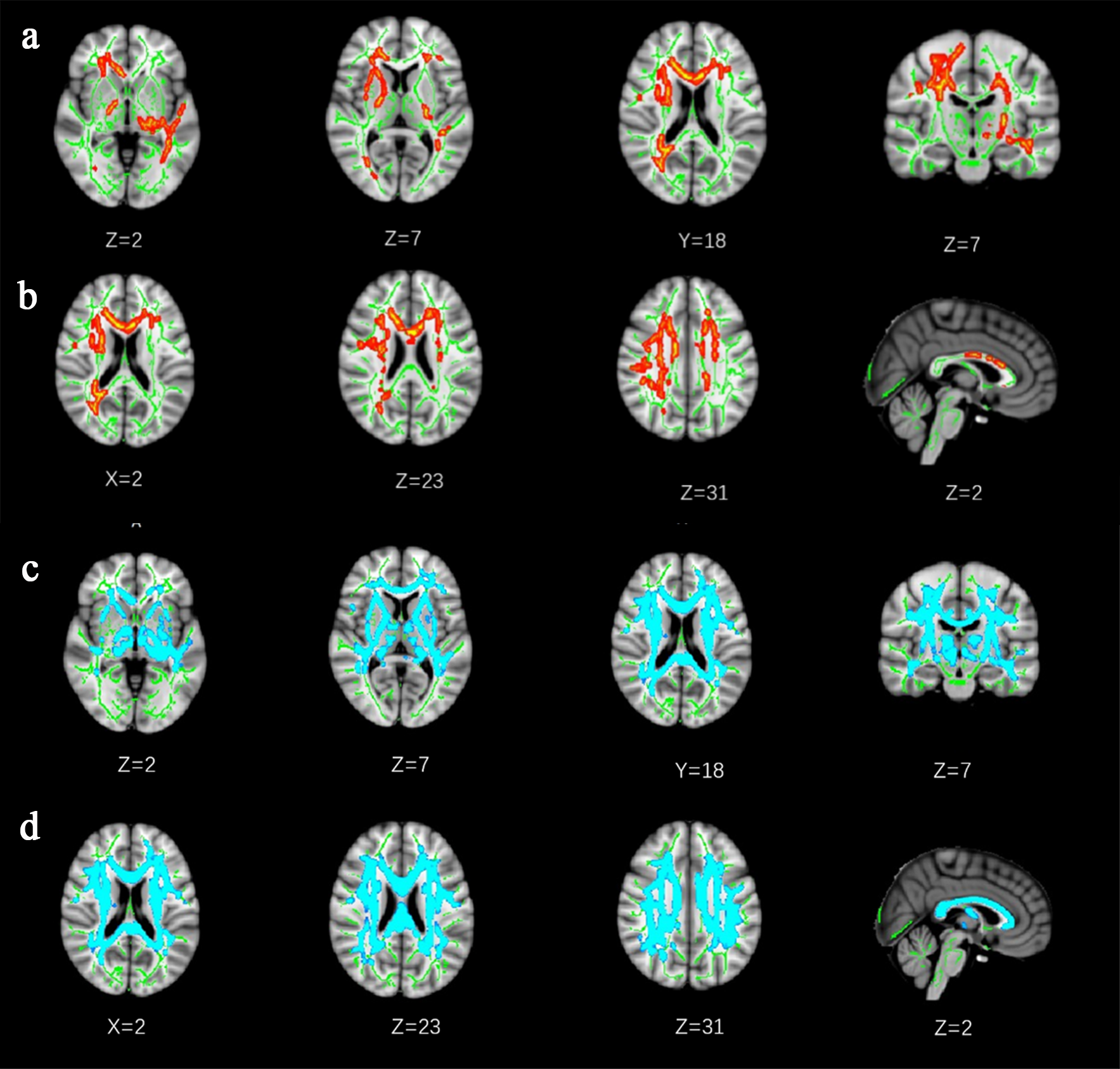 Fig. 1.
The Voxel-wise Tract-Based Spatial Statistics results in
fractional anisotrophy (FA) and mean diffusion (MD) between normal gait group and
gait disorder group. Lines (a) and (b) showed the differences in FA between groups.
Green represented the mean white matter skeleton of all subjects. Red-yellow
voxels (thickened for better visibility) represented the white matter regions
with decreased FA in the gait disorder group compared with normal gait group (FWE
corrected, p
There were significant correlations between MoCA and the gait scale, including
UPDRS (r = –0.210, p = 0.014), and total gait (r =
0.201, p = 0.002) scores when corrected by age, sex, education, and
vascular risk factors. The correlation between DTI and the gait scale scores
corrected by age, sex, education, and vascular risk factors was analyzed. The
values of FA and MD were chosen to represent the DTI measurements because FA, MD,
AD, and RD were based on the same principle. Significant correlations were
observed between FA and the gait scale, including the BI (r = 0.294,
p
In this study, the most relevant items of the Tinetti gait score in the GD patients were decreases in step height and length and increases in gait width bases with a short gait. The RUN DMC study of Helena et al. [18] also found similar patterns. Studies have found that some patients may develop vascular Parkinson’s syndrome. In this study, patients with gait disorders were accompanied by an increase in the UPDRS III score, mainly lower limb motor disorders, so patients should be further followed up to see whether this would develop into vascular Parkinsonism [19].
The risk factors for CSVD are similar to those for cerebrovascular disease, including hypertension, diabetes, dyslipidemia, vascular history, arteriosclerosis, and aging. This research shows that hypertension, diabetes, and cerebral infarction in the GD group were significantly higher than in the GN group. The Health ABC study [20] and the leukoaraiosis and disability study [21] also found these risk factors. Diabetes mellitus and hypertension were associated with ischemic microvascular disease and, therefore, patients with those conditions were more likely to develop vascular Parkinson’s syndrome. These studies found that hypertension, diabetes, and other risk factors could cause gait disorders in CSVD, so the occurrence and development of gait disorders can be avoided by preventing and treating these risk factors, reducing older adults’ falls and a series of other complications. Pinter et al. [22] analyzed 678 community health subjects aged 71–74 and found that age and sex significantly affected gait speed in patients with CSVD. In the present study, the gait disorder had no correlation with age or sex, which might be related to the criteria for admission and the selection of the control group.
Cognitive dysfunction may exist in patients with CSVD. In this study, the MoCA was used to evaluate patients’ cognitive function, and the MoCA score in the GD group was lower than in the GN group, and the correlation between MoCA and the gait scale scores were significant. Grau-Olivares et al. [23] assessed neuropsychological abnormalities in 40 patients with lacunar infarctions and found that mild neuropsychological disturbances are not infrequent (57.5%) in acute lacunar infarcts, especially in patients with atypical lacunar syndrome and pure motor hemiparesis. Motor function decline in patients of lacunar infarction is often accompanied by cognitive decline. Mielke et al. [24] collected data from 1478 older people aged 70–89 and found that a faster pace was associated with better memory, executive function, and overall cognitive ability, concluding gait speed is a reliable, achievable, and non-invasive risk factor for cognitive decline. As gait disorders and cognitive function decline, so does the ability to function in everyday life [25]. CSVD is frequently accompanied by anxiety, depression, and other emotional abnormalities [10], but whether these anomalies influence gait disorders remains to be confirmed by further studies.
Because the early clinical symptoms of CSVD are mild or asymptomatic, the onset is often ignored. Therefore, neuroimaging has become an essential means of early diagnosis of CSVD and asymptomatic angioneuropathy. The neuroimaging markers of CSVD are usually presented by MRI. The MRI markers for patients with cerebrovascular disease were published in 2013 [26] and included lacunes, WMH, CMB, and EPVS. A study of 678 community health subjects aged 71–74 found that WMH in these older adults appears to be the primary force of gait impairment [22]. Lacunes are common in asymptomatic older patients and are associated with an increased risk of stroke, gait disorders, and dementia [27]. In this study, WMH, and lacunes in the GD group were higher than in the GN group. However, in this univariate analysis, the CMB and EPVS were not found to be associated with gait disorders, but they may be involved in the overall MRI burden, along with other imaging features, in the destruction of gait-related basal ganglia and cortical circuits, causing neural network damage. The total MRI burden was different in the GD and GN groups, indicating the increase of the total MRI burden was related to the severity of gait disorders.
The most important predictor of gait dysfunction is WMH. Serious WMH related to
gait disorders are found in the internal capsule, the radiographic coronal
region, the periventricular area, the frontal lobe, and the genu of the corpus
callosum. Even if the WM fibers look intact on the normal sequences of MRI, the
damage of the WM fibers’ integrity could be seen on the DTI sequence [28], which
is increasingly being used to give quantitative information about the state of
the WM [29]. We performed TBSS analyses in patients with CSVD to differentiate
the extent of WM fiber damage [30, 31]. The results showed the WM fiber bundles
were affected when gait abnormality occurred. Comparing the FA and MD values of
the two groups, the GD group had more severe damage of the fiber bundles in
almost all parts of the brain than the GN group. The damaged areas included
bilateral anterior thalamic radiation, bilateral corticospinal tract, bilateral
cingulate gyrus, bilateral hippocampus, forceps major, forceps minor, bilateral
inferior fronto-occipital fasciculus, bilateral inferior longitudinal fasciculus,
bilateral superior longitudinal fasciculus, bilateral uncinate fasciculus, and
bilateral superior longitudinal fasciculus (temporal part) (corrected for FWE,
p
In this DTI analysis, it was found that FA was positively correlated with the BI
score (r = 0.294, p
There are some limitations in the present research. The sample size of the
present study was small, which may have caused a deviation in the results. Due to
the lack of tracking analysis on the symptom development of these patients in the
present study, further studies with larger sample sizes and continuous follow-up
are needed to further investigate the development and prognosis of gait
disorders, cognitive impairment, and other emotional abnormalities. Because of
the limitations of the original image data, only 32 gradient directions were
collected and the b value was 1000 s/mm
In conclusion, the present study revealed that patients with CSVD and gait disorders had a higher incidence of previous hypertension, diabetes, and cerebral infarction. Patients with gait disorders had lower daily living ability and higher UPDRS III scores than those with CSVD alone, accompanied by cognitive dysfunction. The MRI findings of patients with CSVD and gait disorders were more obviously accompanied by WMH of Fazekas grade 2–3 and lacunes. Patients with CSVD and gait disorders had a more significant total MRI burden than GN patients. These patients had more severe damage to the fiber bundles in almost all parts of the brain using DTI analysis. It has also been suggested that WMH, especially damage to the fibers’ microstructures, may play an important role in gait disorders.
Conception and design of the research—PZ, YG, XL. Acquisition of data—WF, PZ, YG. Analysis and interpretation of the data—PZ, YG. Statistical analysis—XX, XT, YD. Obtaining financing—XL. Writing of the manuscript—PZ, YG, XL. Critical revision of the manuscript for intellectual content—XL. All authors read and approved the final draft.
We confirm that we have read the Editorial Policy pages. This study was conducted with approval from the Ethics Committee of Second Hospital of Tianjin Medical University (No. 2016YFC1300604). This study was conducted in accordance with the declaration of Helsinki. Written informed consent was obtained from all participants.
We would like to acknowledge the hard and dedicated work of all the staff that implemented the intervention and evaluation components of the study.
Funding of the study was provided by the National Key Research and development Plan “New Strategy on Pathogenesis and Clinical Diagnosis and Treatment of Chronic Cerebral Small Vessels” (No. 2016YFC1300604).
The authors declare no conflict of interest.
References
Publisher’s Note: IMR Press stays neutral with regard to jurisdictional claims in published maps and institutional affiliations.