


 , Alexander Ghannam 2, Mathew Wooster 3, Nicolas H. Pope 2,*
1 Department of Surgery, Division of General Surgery, University of Florida Health Jacksonville, Jacksonville, FL 32209, USA
2 Department of Surgery, Division of Cardiothoracic Surgery, Medical University of South Carolina, Charleston, SC 29425, USA
3 Department of Surgery, Division of Vascular Surgery, Medical University of South Carolina, Charleston, SC 29425, USA
Abstract
Congenital anomalies in the thoracic aorta, although rare, can present challenging clinical scenarios. Current literature suggests that an aberrant aortic anatomy may be associated with higher rates of aneurysm formation; however, specific screening and management guidelines have yet to be established.
This report presents a case of a 61-year-old male who experienced progressive dysphagia and was diagnosed with an aberrant right-sided aortic arch accompanied by a 5.8 cm descending thoracic aortic aneurysm. Successful endovascular repair was performed with no postoperative complications.
Endovascular repair may be a successful treatment option for these patients, although further studies with long-term follow-up are needed.
Keywords
- thoracic aortic aneurysm
- aortic aneurysm
- congenital
- endovascular
- case report
Congenital anomalies in the aortic arch are exceedingly rare, affecting only 0.01% to 0.1% of the general population [1]. Among these, right-sided aortic arches are particularly uncommon, with an incidence of 0.05% to 0.1% [2]. Therefore, owing to the rarity of these arch anomalies, limited data exist on the surveillance, management, and long-term outcomes of these patients. This report presents a case of a symptomatic right-sided aortic arch aneurysm and describes our approach to intervention.
Our patient is a 61-year-old African American male with a history of chronic obstructive pulmonary disease, hypertension, diabetes, and a 20-pack-per-year smoking history, who initially presented to his primary care provider with complaints of progressive dysphagia over the previous two years. An esophagogastroscopy was performed, with unremarkable results. Computed tomography angiography (CTA) revealed a right-sided aortic arch with an associated 5.8 cm descending thoracic aortic aneurysm, tapering to 3.0 cm at the right mainstem bronchus. The aorta demonstrated a mirror image of normal vascular anatomy, including a left brachiocephalic artery, right common carotid artery, and right subclavian artery (Fig. 1).
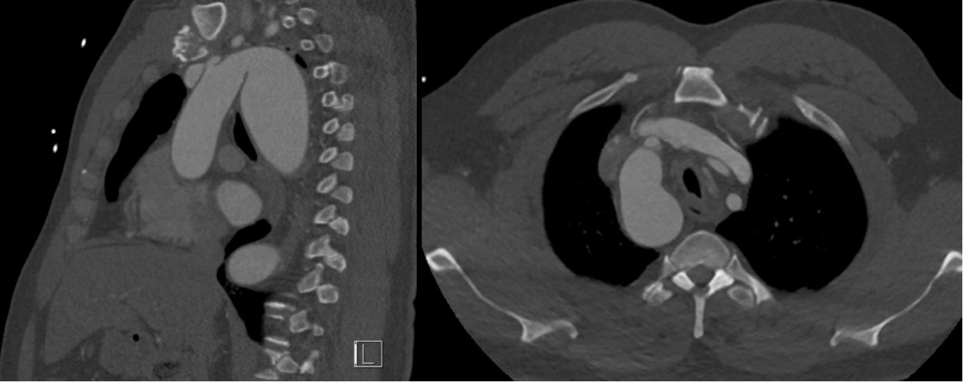 Fig. 1.
Computed tomography angiography (sagittal: left; axial: right) image of the 5.8 cm descending thoracic right-sided aortic aneurysm.
The echocardiography and carotid ultrasound results were unremarkable; however, pulmonary function testing demonstrated severe obstructive lung disease. A multidisciplinary cardiac and vascular surgery team deemed the patient a candidate for thoracic endovascular aortic repair (TEVAR) rather than open surgery, given the poor functional and respiratory status of the patient.
Three weeks following initial presentation, the patient was admitted to a hybrid operating suite and induced with general anesthesia. The right brachial artery was surgically exposed and accessed using an 8 Fr catheter. The right femoral artery was accessed percutaneously and utilized for large-bore intra-arterial access. Additionally, the right femoral vein was cannulated to advance a transvenous pacing wire into the right ventricle. Intravascular ultrasound (IVUS) and angiography were both employed to define the anatomy and confirm measurements of the aortic diameter. After performing the measurements, a 32 mm by 109 mm Cook graft (Cook Inc., Bloomington, IN, USA) was chosen and deployed to ensure the proximal extent landed between the right common carotid and right subclavian artery, covering the right subclavian artery (Fig. 2).
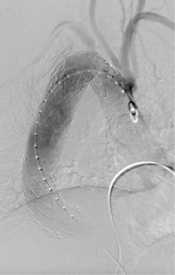 Fig. 2.
Intraoperative angiography image of successful graft deployment.
Notably, rapid ventricular pacing was used during deployment of the proximal stent graft due to the unusual anatomy, a short landing zone, and significant angulation of the arch. An additional 34 mm by 161 mm graft was used for distal extension, ensuring the preservation of large intercostal arteries. In situ laser fenestration of the right subclavian artery was attempted but was unsuccessful due to distal right subclavian artery tortuosity. The postoperative course was unremarkable, and the patient was discharged home four days later on dual antiplatelet therapy, with the reported symptoms resolved. Surveillance CTA at one month demonstrated excellent aneurysm exclusion (Fig. 3), and the patient remained asymptomatic at the follow-up.
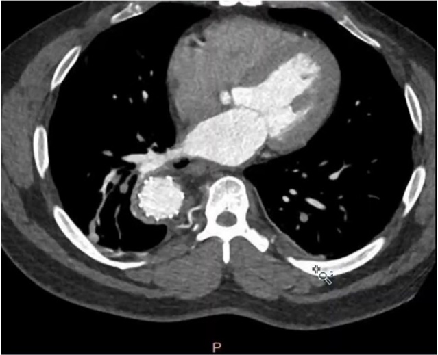 Fig. 3.
Postoperative computed tomography angiography (CTA) (1-month) image after endovascular repair.
The pharyngeal arteries of the mammalian embryo act as the foundation for the formation of the major vasculature in the human head, neck, and thorax. Given the complexity of maturation and function, aberrations are possible and are usually associated with genetic syndromes, such as DiGeorge syndrome, or congenital cardiac anomalies, including tetralogy of Fallot and truncus arteriosus [1]. The development of a right-sided aortic arch results from the continued patency of the right dorsal aorta, with regression of the left fourth pharyngeal arch [3]. Several subtypes of right-sided aortic arches exist, with the most common variant being the right aortic arch with mirror image vascular branching [3], as seen in our patient.
The clinical presentation of aortic arch anomalies varies widely. Anomalies that pose physiological implications, especially those involving vascular rings or cardiac malformations, typically manifest early in infancy. Meanwhile, symptoms such as dysphagia and stridor often arise due to tracheoesophageal compression [4]. In our case, a right-sided aortic arch with mirror-image normal vascular anatomy did not initially cause vascular issues. Instead, the formation of an aneurysm led to dysphagia, prompting presentation. The absence of prior cross-sectional imaging contributed to the delay in diagnosis, highlighting the rarity and significance of late presentations. This case underscores the importance of considering anatomically abnormal yet physiologically functioning aortic anomalies, which can evade detection and manifest later in life, necessitating inclusion in clinical differential diagnoses.
Recent studies have suggested a correlation between aortic arch anomalies and
aneurysmal disease. Indeed, a 2021 retrospective analysis of 21,336 CTA scans
from patients aged 50 to 85 at a single institution found that the prevalence of
thoracic aortic aneurysms was more than double in those with arch anomalies
compared to those without (10.8% vs. 4.1%; p
CTA is the primary non-invasive imaging modality for evaluating thoracic aortic
abnormalities [5]. Notably, the threshold for surgical intervention in sporadic
aortic root and ascending aortic aneurysms has been lowered from 5.5 cm to 5.0 cm
in select patients, according to the 2022 ACC/AHA guidelines, with even lower
thresholds recommended in certain cases involving heritable thoracic aortic
diseases [6]. Rapid aneurysm growth, which is defined as
TEVAR is a well-established treatment for descending thoracic aortic aneurysms, regardless of anatomical variation or the presence of heritable syndromes. Retrospective studies have demonstrated that TEVAR is a viable intervention for high-risk patients with genetic aortopathies, such as Marfan syndrome, Loeys–Dietz syndrome, and Ehlers–Danlos syndrome [7]. However, the use of TEVAR in patients with congenital anatomical anomalies is less frequently reported. We suggest this may be due to the technical expertise required to navigate atypical or unfamiliar vascular anatomy. Our case demonstrates that successful TEVAR is feasible in such settings, provided the operator has experience managing aberrant and tortuous anatomy. In this case, we believe that rapid ventricular pacing contributed to procedural success by stabilizing mediastinal structures and minimizing the risk of stent graft migration. This technique may be a valuable adjunct during stent deployment in patients with complex aortic arch anatomy.
Right-sided aortic arch pathology is extremely rare and poorly documented, leaving providers without recommendations for surveillance and management. The increased risk of aneurysmal disease associated with right-sided aortic arches highlights a significant gap in care due to the absence of tailored guidelines. Despite this, right-sided aortic arch aneurysms can successfully be treated using endovascular techniques following appropriate planning and with relevant experience.
Data sharing is not applicable to this article, as no datasets were generated or analyzed during the current study.
MK (conception, data acquisition, manuscript drafting, and critical revision), AG (conception, data acquisition, manuscript drafting, and critical revision), MW (critical revision and clinical design), NP (critical revision and clinical design). All authors read and approved the final manuscript. All authors have participated sufficiently in the work and agreed to be accountable for all aspects of the work.
The study was conducted in accordance with the principles of the Declaration of Helsinki. According to the University of Florida Health Jacksonville institution, case reports do not require IRB approval and do not fall under the federal definition of research. Regarding written consent, written procedural consent was obtained from this patient, granting permission for the procedure as well as the use of material for educational purposes. Verbal consent was also obtained regarding the potential use of this case for research and education.
Not applicable.
This research received no external funding.
The authors declare no conflict of interest.
Supplementary material associated with this article can be found, in the online version, at https://doi.org/10.31083/HSF49909.
References
Publisher’s Note: IMR Press stays neutral with regard to jurisdictional claims in published maps and institutional affiliations.