


1 Department of Gynecology, Second Affiliated Hospital of Xinjiang Medical University, State Key Laboratory of Pathogenesis, Prevention and Treatment of High Incidence Diseases in Central Asia, 830000 Urumqi, Xinjiang, China
Abstract
Background: This study aimed to investigate the utility of transvaginal
ultrasound and hysteroscopy in diagnosing abnormal uterine bleeding in
peri-menopausal women. Methods: Between September 2021 and March 2023,
176 women presenting with abnormal uterine bleeding at the Second Affiliated
Hospital of Xinjiang Medical University were subjected to both hysteroscopy and
transvaginal ultrasound examinations. Results: There was a significant
discrepancy between the pathological diagnoses and the findings from transvaginal
ultrasound (p
Keywords
- uterus
- AUB
- hysteroscopy
- transvaginal ultrasound
- peri-menopausal abnormal uterine bleeding
Perimenopause represents a pivotal transition in a woman’s life, marked by a natural shift from regular menstrual cycles to anovulatory cycles, characterised by irregular and often excessive menstruation, culminating in permanent infertility [1]. This phase typically commences 2 to 8 years prior to menopause and concludes one year thereafter [2]. Abnormal uterine bleeding (AUB) is a prevalent gynaecological condition among peri-menopausal women. During this period, ovarian function declines gradually, leading to a decrease in estradiol levels, which may result in abnormal uterine bleeding. The prevalence of AUB ranges from 10% to 30% among women of childbearing age, escalating to up to 90% among peri-menopausal women. Although AUB is predominantly attributed to benign lesions, it can also emanate from malignant lesions, such as atypical endometrial hyperplasia or endometrial carcinoma [3]. In 2011, the International Federation of Gynecology and Obstetrics (FIGO) introduced a novel classification system for AUB, subsequently endorsed by the American Congress of Obstetrics and Gynecology (ACOG) in 2012. This system, referred to by the acronym PALM-COEIN (endometrial polyp, adenomyosis, leiomyoma, malignancy and hyperplasia, coagulopathy, ovulatory dysfunction, endometrial, iatrogenic, and not yet classified), categorises AUB based on its suspected aetiology: polyp, adenomyosis, leiomyoma, malignancy and hyperplasia, coagulopathy, ovulatory dysfunction, endometrial, iatrogenic, and not yet classified [4]. Consequently, peri-menopausal AUB can significantly impair the physical and mental health of affected women. Therefore, early detection and diagnosis are paramount for the management of endometrial malignancies. Currently, the primary clinical diagnostic techniques include transvaginal ultrasound, hysteroscopy, and diagnostic uterine curettage, with hysteroscopy and transvaginal ultrasound being the most frequently utilised ancillary examination methods [5]. Hysteroscopy is regarded as the gold standard in the diagnosis and treatment of female endometrial lesions [6]. Transvaginal ultrasound plays a crucial role in the diagnosis of AUB, offering a comprehensive assessment of the uterus and its surrounding structures. Nevertheless, the accuracy of transvaginal ultrasound in diagnosing uterine lesions is suboptimal, with inconsistent diagnostic criteria and repeatability. In contrast, hysteroscopy proves more efficacious than transvaginal ultrasound in diagnosing endometrial diseases, albeit with limitations such as high costs, reduced convenience, and lower patient tolerance [7]. The aim of this study was to evaluate the efficacy of transvaginal ultrasound and hysteroscopy in the etiological diagnosis of peri-menopausal AUB, thereby providing a scientific foundation for the early diagnosis and treatment of perimenopausal abnormal uterine haemorrhage.
Between September 2021 and March 2023, a retrospective study was conducted at the Second Affiliated Hospital of Xinjiang Medical University, involving 176 perimenopausal women experiencing abnormal uterine bleeding. Inclusion criteria included: ① women presenting with abnormal uterine bleeding; ② those with definitive pathological diagnosis results; ③ perimenopausal women aged between 40–59 years; ④ those who underwent transvaginal ultrasound and hysteroscopy. Exclusion criteria encompassed: ① unknown pathological diagnosis; ② women not in the peri-menopausal period; ③ non-uterine sources of bleeding; ④ absence of transvaginal ultrasonography or hysteroscopy.
Performed by an ultrasound specialist with over five years of experience in the ultrasound diagnosis department of the Second Affiliated Hospital of Xinjiang Medical University, Doppler colour ultrasound was utilised individually. The probe frequency ranged from 5.0–9.0 MHz. Comprehensive examination techniques included oblique, cross-sectional, and longitudinal scans, conducted after gently inserting the transvaginal ultrasound probe into the vaginal fornix or onto the cervical surface. The assessment focused on the uterus’s shape, intrauterine cavity echoes, lesion location, endometrial thickness measurement, and the presence of abnormal lesions like polyps, hyperplasia, cancer, etc. Additionally, the examination sought to identify abnormalities in the bilateral adnexa and pelvic cavity. TVU is pivotal in diagnosing structural causes of AUB (PALM category) and evaluating nonstructural causes such as ovulatory dysfunction [8]. The classification of intracavitary fluid, structure, echogenicity, border, and the endometrial–myometrial boundary was based on the International Endometrial Tumor Analysis (IETA) System, utilising B-mode ultrasound [9]. During evaluation, clinicians ensured the uterus occupied two-thirds of the screen for optimal clarity [10, 11]. Vascular patterns identified included: scattered vessel pattern: indicative of endometrial hyperplasia, characterised by widespread vasculature within the endometrium. Multiple Vascular Pattern: associated with endometrial cancer, featuring multiple vessels at the endometrium and the myometrial–endometrial interface due to angiogenic proliferation within and around tumour tissue. Pedicle Vascular Pattern: a single vessel penetrating the endometrium. Circular Vascular Pattern: increased peripheral vascularity around the tumour, typical of uterine fibroids [9]. On TVU, polyps may manifest as non-specific endometrial thickening or focal masses within the endometrial cavity, potentially matching the surrounding endometrium in echogenicity with smooth borders, though they may also appear heterogeneous. Endometrial polyps are often characterised by a dominant central feeder vessel, visible via color flow Doppler techniques. While there are no definitive imaging criteria for adenomyosis diagnosis, the most specific indication is subendometrial echogenic linear striations. An ill-defined endometrial/myometrial border also suggests adenomyosis. Leiomyomas are distinguished by a pseudocapsule providing a well-defined border on ultrasound. They often absorb ultrasound beams, resulting in a shadowing effect behind the myoma, sometimes referred to as a “Venetian-blind effect” [12].
Gynaecologists from the Second Affiliated Hospital of Xinjiang Medical University, boasting over a decade of extensive clinical experience, performed hysteroscopy (Olympus Corp., Shinjuku, Tokyo, Japan) under anaesthesia. The flow rate and pressure of the liquid distension medium were regulated to 200–400 mL/min and 100–150 mmHg, respectively, utilising 0.9% sodium chloride solution for cervical canal dilation. Following cervical canal dilation, the hysteroscope was connected to observe the endometrium’s thickness and colour, the openings of the bilateral fallopian tubes, the angles of the uterus, the shape of the uterine cavity, the cervical canal, and to examine the size of intrauterine lesions and the distribution of blood vessels. Electrosurgical resection was performed on the abnormal areas, and diagnostic curettage of the uterine cavity wall was conducted to obtain tissue samples from the endometrium and suspicious lesions within the uterine cavity for pathological examination. Under hysteroscopic examination, most submucous fibroids manifest as spherical masses protruding into the uterine cavity. Diagnostic hysteroscopy cannot conclusively diagnose adenomyosis due to its limitation to the superficial endometrial layer [13]. However, typical hysteroscopic findings may include: (i) an irregular endometrium with tiny surface openings; (ii) pronounced hypervascularisation; (iii) an endometrial “strawberry” pattern; (iv) a fibrous cystic appearance of intrauterine lesions following 3–5 episodes of intramyometrial haemorrhage; (v) haemorrhagic cystic lesions presenting a dark blue or chocolate brown appearance [14]. Uterine polyps are characterised as individualised endometrial outgrowths that can emerge throughout the uterine cavity, consisting variably of stroma, glands, and blood vessels, with their proportional composition influencing their hysteroscopic appearance [15]. In cases of endometrial hyperplasia, the hysteroscopic view may reveal local or diffuse thickening of the endometrium, occasionally with single or multiple protrusions, which could be honeycombed or mesh-like. If the appearance is greyish white or yellow, or if there is a heterogeneous distribution of large blood vessels, atypical endometrial hyperplasia or endometrial cancer should be strongly suspected [16].
Tissue samples were fixed in 10% formaldehyde, embedded in paraffin, and subjected to routine sectioning and staining. An Olympus optical microscope (Olympus Corp., Shinjuku, Tokyo, Japan) was used for examination. The final pathological diagnosis served as the gold standard.
Statistical analysis was conducted using SPSS 26.0 (IBM Corp., Armonk, NY, USA).
The rates of count data were expressed in percentages (%). The Chi-square test
was employed to analyse the diagnostic efficiency differences between methods.
Kappa coefficients were used to assess the consistency of analysis, with
According to the study flowchart (Fig. 1), collected the patient information and screened the patients eligible for the study. Based on clinical data of 176 patients, we were collected for their age, body mass index (BMI), age at menarche, pregnancy history, and accompanying symptoms. The average age of the 176 patients in the study was 47.26, average body mass index (BMI) 25.21. All the information met the inclusion criteria of this study. The average age of menarche 13.74, more than two pregnancies on average (Table 1).
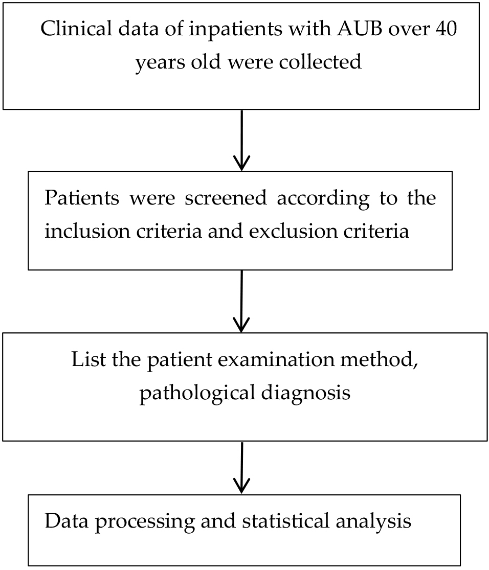 Fig. 1.
Study flowchart. AUB, abnormal uterine bleeding.
| Patient (n) | Age (years) | BMI (kg/m |
Age at menarche (years) | Number of pregnancies (n) | Number of deliveries (n) | A history of abortion (n) | Hypertension (n) | Glycuresis (n) | |||
| With | Without | With | Without | With | Without | ||||||
| 176 | 47.26 |
25.21 |
13.74 |
2.58 |
1.42 |
36 | 140 | 20 | 156 | 6 | 170 |
BMI, body mass index.
The findings of this study revealed a significant discrepancy between the
pathological diagnosis and the outcomes obtained from transvaginal ultrasound
(p
| Grouping | Pathological diagnosis | p | OR (95% CI) | Kappa | |||
| Pathological | Physiological | ||||||
| Transvaginal ultrasound | |||||||
| Abnormal | 139 (95.2) | 7 (4.8) | 41.523 | 0.000 | 17.375 (6.114–49.377) | 0.475 | |
| Normal | 16 (53.3) | 14 (46.7) | |||||
| Hysteroscope | |||||||
| Abnormal | 147 (96.7) | 5 (3.3) | 79.229 | 0.000 | 58.800 (17.174–201.315) | 0.669 | |
| Normal | 8 (33.3) | 16 (66.7) | |||||
| Two kinds of combination |
|||||||
| Abnormal | 151 (97.4) | 4 (2.6) | 108.101 | 0.000 | 160.438 (36.748–700.449) | 0.784 | |
| Normal | 4 (19.0) | 17 (81.0) | |||||
Note: * indicates transvaginal ultrasound combined with hysteroscopy. OR, odds ratio; 95% CI, 95% confidence interval.
In the context of diagnosing peri-menopausal AUB, transvaginal ultrasound demonstrated a sensitivity of 89.7%, a specificity of 66.7%, and a negative predictive value of 46.7%. The diagnostic sensitivity and specificity of hysteroscopy were superior, at 94.8% and 76.2% respectively, although the negative predictive value remained relatively low at 66.7%. The combined diagnostic methodology exhibited enhanced sensitivity, specificity, and negative predictive value at 97.4%, 81.0%, and 81.0% respectively, surpassing the performance of either transvaginal ultrasound or hysteroscopy when used independently, as illustrated in Table 3.
| Grouping | Pathological diagnosis | Sensitivity (%) | Specificity (%) | Positive predictive value (%) | Negative predictive value (%) | ||
| Pathological | Physiological | ||||||
| Transvaginal ultrasound | |||||||
| Abnormal | 139 (95.2) | 7 (4.8) | 89.7 | 66.7 | 95.2 | 46.7 | |
| Normal | 16 (53.3) | 14 (46.7) | |||||
| Hysteroscope | |||||||
| Abnormal | 147 (96.7) | 5 (3.3) | 94.8 | 76.2 | 96.7 | 66.7 | |
| Normal | 8 (33.3) | 16 (66.7) | |||||
| Two kinds of combination |
|||||||
| Abnormal | 151 (97.4) | 4 (2.6) | 97.4 | 81.0 | 97.4 | 81.0 | |
| Normal | 4 (19.0) | 17 (81.0) | |||||
Note: * indicates transvaginal ultrasound combined with hysteroscopy. AUB, abnormal uterine bleeding.
Concerning the etiological diagnosis of AUB attributable to endometrial polyps,
hysteroscopy achieved a concordance rate of 93.3%, significantly exceeding that
of transvaginal ultrasound, which stood at 77.3%. This difference was
statistically significant (
| Pathological diagnosis | Number of cases | Transvaginal ultrasound | Hysteroscope | p | OR (95% CI) | |||
| In accordance with | Not in accordance with | In accordance with | Not in accordance with | |||||
| Uterine leiomyoma | 19 | 17 (89.5) | 2 (10.5) | 19 (100) | 0 (0.0) | 2.111 | 0.146 | 0.895 (0.767–1.044) |
| Endometrial polyps | 75 | 58 (77.3) | 17 (22.7) | 70 (93.3) | 5 (6.7) | 7.670 | 0.006 | 0.244 (0.085–0.701) |
| Adenomyosis | 4 | 3 (75.0) | 1 (25.0) | 4 (100) | 4 (0.0) | 1.143 | 0.285 | 0.750 (0.426–1.321) |
| Endometrial malignant transformation | 7 | 4 (57.1) | 3 (42.9) | 6 (85.7) | 1 (14.3) | 1.400 | 0.237 | 0.222 (0.017–2.970) |
| Atypical hyperplasia of endometrium | 6 | 4 (66.7) | 2 (33.3) | 5 (83.3) | 1 (16.7) | 0.444 | 0.505 | 0.400 (0.0267–6.176) |
| Benign hyperplasia | 42 | 30 (71.4) | 12 (28.6) | 35 (83.3) | 7 (16.7) | 1.700 | 0.192 | 0.500 (0.175–1.432) |
| Normal endometrium | 21 | 14 (66.7) | 7 (33.3) | 16 (76.2) | 5 (23.8) | 0.467 | 0.495 | 0.625 (0.161–2.419) |
| Else | 2 | 1 (50.0) | 1 (50.0) | 2 (100) | 0 (0.0) | 1.333 | 0.248 | 0.500 (0.125–1.999) |
During the peri-menopausal period, ovarian function diminishes progressively, leading to imbalances in estrogen and progesterone levels. This hormonal disruption can cause endometrial polyps, simple and complex endometrial hyperplasia, and in some instances, atypical hyperplasia and endometrial carcinoma. Consequently, a subset of peri-menopausal women may develop AUB, which significantly impacts their physical and psychological well-being and overall quality of life. Therefore, identifying the underlying cause promptly is imperative. The primary objective of evaluating AUB accurately is to diagnose the bleeding’s aetiology, facilitate the early detection of serious conditions such as endometrial atypical hyperplasia and cancer, and enable the implementation of suitable treatment strategies [17].
The diagnosis of peri-menopausal AUB relies on the patient’s history, gynaecological examination, and appropriate laboratory and imaging investigations, with the final pathological diagnosis serving as the gold standard. Transvaginal ultrasound has emerged as the preferred initial imaging modality for patients with AUB, owing to its excellent feasibility, cost-effectiveness, safety, non-invasiveness, and repeatability. It is particularly advantageous in the primary screening for the aetiology of AUB, capable of diagnosing structural causes of abnormal bleeding such as polyps, adenomyosis, leiomyomas, hyperplasia, and malignancy, as well as aiding in the diagnosis of ovulatory dysfunction [8]. Despite its strengths, the specificity and sensitivity of transvaginal ultrasound are limited, and it alone cannot definitively exclude organic causes of AUB [18]. Hysteroscopy, a minimally invasive technique, allows direct observation of the endometrium’s thickness and colour, the opening of the bilateral fallopian tubes, uterine angles, cavity shape, and cervical canal. It excels in detecting small lesions that transvaginal ultrasound may miss, and electroresection or curettage of lesions can enhance the sensitivity and accuracy of the aetiological diagnosis of AUB. Despite its growing popularity, hysteroscopy faces limitations in clinical practice, such as cost, technical complexity, invasiveness, and patient tolerance.
Wang et al. [19] demonstrated that combining transvaginal ultrasound,
enhanced by the K-means clustering colour image segmentation algorithm, with
hysteroscopy significantly increased diagnostic sensitivity. This study’s
results, encompassing 176 peri-menopausal AUB patients, revealed 21 cases of
normal endometrium, 42 of benign endometrial hyperplasia, 75 of endometrial
polyp, 7 of endometrial carcinoma, 4 of adenomyosis, 6 of atypical endometrial
hyperplasia, 19 of uterine leiomyoma, and 2 of other uterine lesions. In
diagnosing AUB caused by endometrial polyps, the concordance rates for
hysteroscopy and transvaginal ultrasound were 93.3% and 77.3%, respectively,
with the difference being statistically significant (
This study was conducted at a tertiary hospital and involved transvaginal ultrasound performed by a technician with over five years of experience, alongside hysteroscopy carried out by a seasoned gynaecologist with more than a decade of expertise. Consequently, the findings are both accurate and reliable. Additionally, the research offers invaluable insights by juxtaposing the outcomes of both examinations against pathological diagnoses. Nonetheless, the study is marred by the generally low quality of the included studies and the application of inclusion and exclusion criteria that overlook potential confounding factors, such as menstrual status. These oversights may introduce bias into the study’s conclusions. Thus, given the limitations in the quality and scope of this research, further investigations are warranted to corroborate and refine these findings.
In conclusion, according to the study of 176 women with AUB during perimenopause, we know thantransvaginal ultrasound and hysteroscopy possess significant diagnostic utility in identifying the aetiology of peri-menopausal AUB. Transvaginal ultrasound, being both safe and relatively cost-effective, offers considerable benefits in the initial screening for the causes of AUB. However, the potential for missed or incorrect diagnoses remains. Although hysteroscopy is more expensive and less convenient, its integration with pathological examination significantly enhances the detection rates of cancerous and precancerous lesions. The combined application of transvaginal ultrasound and hysteroscopy improves the diagnostic accuracy for endometrial lesions. Therefore, it is recommended that this combined approach be employed in the evaluation of peri-menopausal AUB patients to promptly identify the cause of AUB and initiate appropriate treatment.
The data that support the findings of this study are available from Second Affiliated Hospital of Xinjiang Medical University but restrictions apply to the availability of these data, which were used under license for the current study and so are not publicly available. Data are however available from the authors upon reasonable request and with permission of Second Affiliated Hospital of Xinjiang Medical University.
BT: conceptualization, data curation, formal analysis, investigation, methodology, writing - original draft, writing - review & editing. QHZ: conception, funding acquisition, resources. Both authors contributed to editorial changes in the manuscript. Both authors have participated sufficiently in the work and agreed to be accountable for all aspects of the work. Both authors read and approved the final manuscript.
Informed consent was obtained from all individual participants included in the study. The Ethics Committee of the Second Affiliated Hospital of Xinjiang Medical University granted approval for this study (Ethics Approval Number: KY2024032030).
We extend our sincere thanks to Changsheng Xu for his contribution the fomal analysis in this manuscript. We extend our sincere thanks to everyone who supported us in the preparation of this manuscript.
The study was funded by Natural Science Foundation of Xinjiang Uygur Autonomous Region (2023D01C122) and Department of Gynecology, Second Affiliated Hospital of Xinjiang Medical University, State Key Laboratory of Pathogenesis, Prevention and Treatment of High Incidence Diseases in Central Asia (SKL-HIDCA-2022-GJ4) and Open topic of the Key Laboratory of Neurological Diseases in Xinjiang (XJDX1711-2260). And the “Tianshan Talents” medical and health high-level personnel training plan (TSYC202301B128).
The authors declare no conflict of interest.
References
Publisher’s Note: IMR Press stays neutral with regard to jurisdictional claims in published maps and institutional affiliations.