


 , Mo Zhu 2,*
1 Department of Radiology, Suzhou Xiangcheng District People's Hospital, 215131 Suzhou, Jiangsu, China
2 Department of Radiology, The First Affiliated Hospital of Soochow University, 215006 Suzhou, Jiangsu, China
†These authors contributed equally.
Abstract
Background: The present exploration is aimed to determine whether
diffusion kurtosis imaging (DKI)-derived mean diffusivity (MD) and mean kurtosis
(MK) are possible parameters for the invasive breast cancer grading and whether
MD/MK is related to breast cancer clinical-pathologic factors including estrogen
receptor (ER), progesterone receptor (PR), human epidermal growth factor receptor
2 (HER-2) and Ki-67. Methods: Tumors from 108 invasive breast carcinoma
patients (45.6
Keywords
- breast cancer
- diffusion kurtosis imaging
- magnetic resonance imaging
- prognostic factors
It has been shown that evaluation of the degree of malignancy of breast cancer contributes to the diagnosis and treatment of breast cancer patients. Conventional methods are not effective enough for evaluation of breast lesions [1, 2, 3]. Therefore, it is essential for radiologist to improve the accuracy of diagnosis and classification of breast cancer, especially in invasive breast cancer.
Magnetic resonance imaging (MRI) is extensively used to diagnose suspicious breast lesions [4, 5, 6, 7, 8]. Diffusion weighted magnetic resonance imaging (DWI) is also used to diagnose suspicious breast lesions. The conventional DWI model always assumes a Gaussian diffusion of water protons. Thus, the DWI signal mono exponentially decreases with increasing b value. However, water diffusion in the living tissues is restricted by tissue microstructure and a non-Gaussian distribution of water displacement profile was discovered in the presence of various barriers. In 2005, Jensen et al. [9] reported a non-Gaussian diffusion-weighted model called diffusion kurtosis imaging (DKI). Compared to DWI in assessing extracellular water diffusion, DKI may improve the understanding of water diffusion characteristics of the local microenvironment by calculating and analyzing the coefficients of kurtosis and diffusion. Furthermore, it has been demonstrated that DKI may provide the non-Gaussian diffusion behavior in various tissues including liver [10, 11], brain [12, 13, 14], and prostate [15, 16, 17]. Moreover, it has been shown that DKI may offer better characterization of tumors than traditional DWI [18, 19].
The traditional DWI model is based on the Gaussian behavior of water diffusion, which makes the diffusion of water molecules without any restriction. However, in living tissues, diffusion is usually limited by tissue microstructure and shows non-Gaussian behavior. The DKI model can quantify the amount of water molecules deviating from the normal distribution, which can reflect the subtle changes of human environment more truly and accurately. DKI imaging has two important parameters, mean diffusivity (MD) and mean kurtosis (MK). MD reflects the overall dispersion level and resistance of the molecule, and only represents the size of the dispersion. The larger the value, the stronger the diffusion ability of water molecules. MK is the most critical parameter of DKI technology, which is defined as the average value of diffusion kurtosis in all directions and is considered as an indicator of the complexity of tissue microstructure. The size of MK depends on the complexity of tissue microstructure in the region of interest. The more complex the structure (such as the lower differentiation degree of cancer cells and the higher cell density), the more significant the diffusion restriction of non-Gaussian water molecules and the larger the MK. If DKI parameters can precisely predict proliferative activity as well as histologic malignancy grade of breast cancers, it will be a great aid in making clinical decision. However, few studies have evaluated association between different clinical-pathologic risk factors of breast cancer and the DKI parameters [18, 19, 20]. In the present study, we aimed to evaluate the role of DKI in the diagnosis and characteristics of breast carcinoma. We also evaluated the relationship between breast cancer-associated pathologic factors and DKI parameters.
This is a prospective study. Institutional review board has approved the
investigation and written informed consent from patients were obtained. A total
of 178 patients with suspected breast cancer who received treatment in our
hospital from August 2015 to August 2018 were collected. According to the
exclusion criteria, 60 cases of fibroma were removed, 7 cases of inflammatory
pseudotumor were removed, and 3 cases of image artifacts were assessed for large
impact. 108 female patients (mean of age = 45.6
A 3.0-T MR imager (Magnetom Skyra; Berlin, Germany) with a dedicated four-channel bilateral breast coil in the axial orientation was used to perform MR imaging examinations. T2-weighted images acquirement and dynamic contrast-enhanced T1-weighted MRI was conducted as described previously [21].
Lesion samples were obtained after breast MRI. The lesion was removed surgically. Or after local anesthesia, the lesion was obtained by ultrasound-guided needle biopsy. The tumors (n = 108) used in this study were defined as grade II and grade III, according to the formation of glandular ducts, nuclear shape and size and chromatin, chromatin increase and nuclear division, etc. Estrogen receptor (ER), progesterone receptor (PR), human epidermal growth factor receptor 2 (HER-2) and Ki-67 antigens results were obtained from the cancer registry having been processed in pathology department.
Positive judgment of ER and PR: when calculating the proportion of positive
cells in total cells, 0% is recorded as negative (–) if no positive cells are
found; if the rate of positive cells
Statistical analyses were implemented with SPSS software (version 20.0, Chicago,
IL, USA). Continuous variables were reported as the mean
| Parameter | MD (10 |
MK (10 | |||
|---|---|---|---|---|---|
| Statistic | p value | Statistic | p value | ||
| ER | Negative (n = 25) | 0.095 | 0.200 | 0.096 | 0.200 |
| Positive (n = 83) | 0.086 | 0.196 | 0.088 | 0.168 | |
| PR | Negative (n = 29) | 0.072 | 0.200 | 0.184 | 0.013 |
| Positive (n = 79) | 0.063 | 0.200 | 0.086 | 0.200 | |
| HER-2 | Negative (n = 70) | 0.115 | 0.022 | 0.067 | 0.200 |
| Positive (n = 38) | 0.097 | 0.200 | 0.080 | 0.200 | |
| Ki-67 | Low (n = 40) | 0.105 | 0.200 | 0.054 | 0.200 |
| High (n = 68) | 0.089 | 0.200 | 0.064 | 0.200 | |
| Grade II (n = 44) | 0.122 | 0.098 | 0.094 | 0.200 | |
| Grade III (n = 64) | 0.091 | 0.200 | 0.128 | 0.011 | |
Totally, 108 patients with breast tumors were included: 90 infiltrating ductal carcinomas, 2 in situ ductal carcinomas, 15 infiltrating lobular carcinomas, and 1 in situ lobular carcinoma. As shown in Table 2, there was a significant difference between grade II and Grade III tumors, depending on the patient’s menstrual status. However, as shown in Table 2, there was no significant difference between Grade II and Grade III tumors, based on patient age and tumor size. TRA images, Dynamic contrast enhancement (DCE) maps, MD and MK maps, and the pathology images of a 34 years old woman with infiltrating ductal carcinomas in right breast were shown in Figs. 1,2.
| Characteristic | Grade II | Grade III | p value | |
|---|---|---|---|---|
| Patient age | 46.5 |
47.1 |
p | |
| Menstrual status | p | |||
| Premenopausal (n = 63) | 32 | 31 | ||
| Postmenopausal (n = 45) | 12 | 33 | ||
| Lesion characteristics | ||||
| Size (cm) | 3.1 |
3.5 |
p | |
| Shape | p | |||
| Round | 4/44 | 6/64 | ||
| Oval | 9/44 | 11/64 | ||
| Irregular | 31/44 | 47/64 | ||
| MD (10 |
1.17 |
0.82 |
p | |
| MK | 0.85 |
1.01 (0.85–1.16) | p | |
Note: MD, mean diffusivity; MK, mean kurtosis.
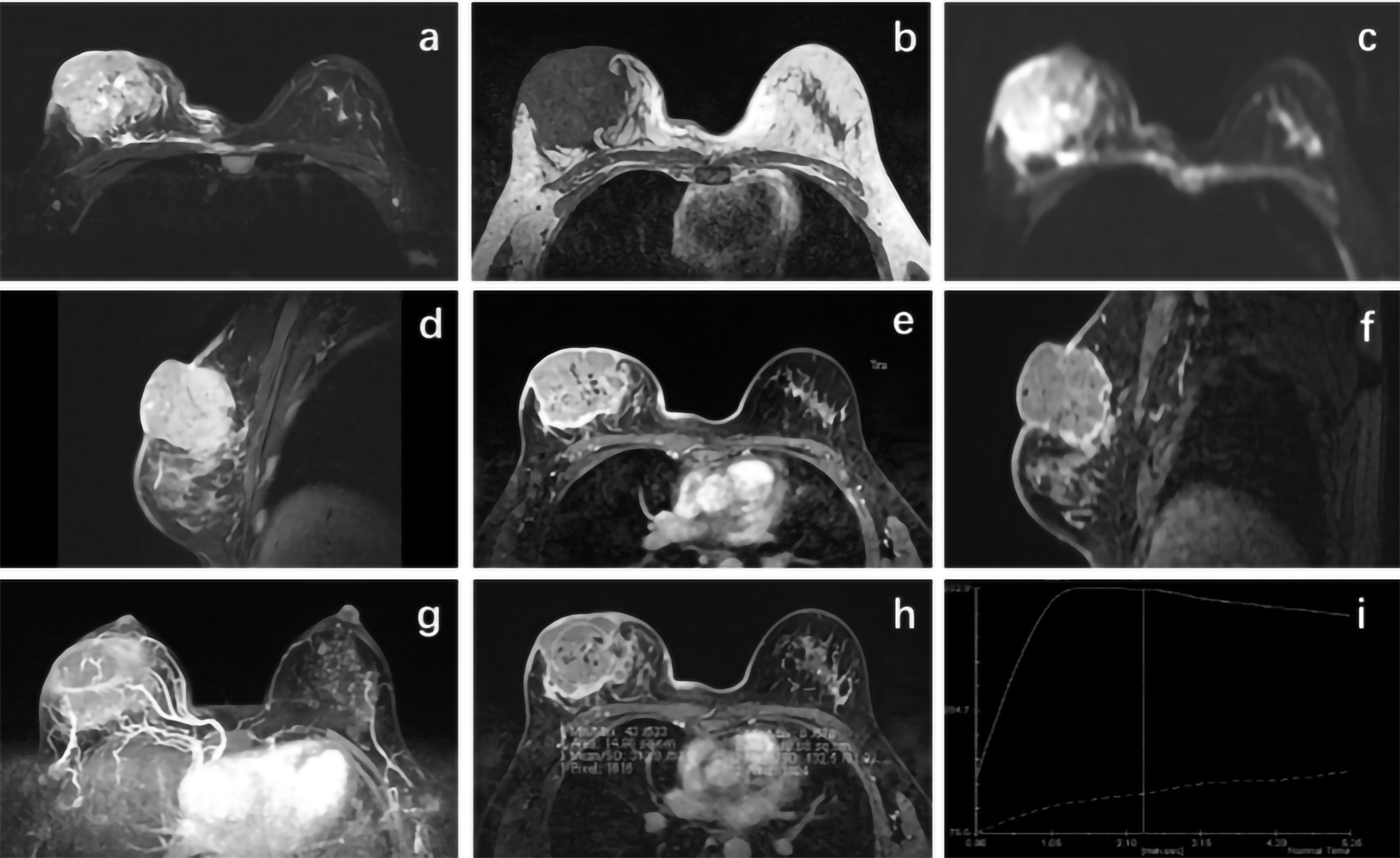 Fig. 1.
Transverse (TRA) images showing the pathology and dynamic contrast enhancement (DCE) of a 34 years old woman with right breast infiltrating ductal carcinomas. T2-weighted TRA short TI inversion recovery (STIR) image (a) T1-weighted TRA image (b), T1-weighted TRA image (c), DWI TRA image (d), T1-weighted TRA contrast image (e), and T1-weighted SAG contrast image (f) of a 34 years old woman with infiltrating ductal carcinomas in right breast. In the upper outer quadrant of the right breast, there was an irregular mass surrounded by disorder structures, and an irregular nodular shadow below the mass. The lesion showed strong enhancement. Dynamic contrast enhancement (DCE) of a 34-year old woman with infiltrating ductal carcinomas in right breast. The DCE maps (g), enhancement maps (h) and contrast enhancement curves (i) of maximum intensity projection (MIP) of a 34-year old woman with infiltrating ductal carcinomas in right breast.
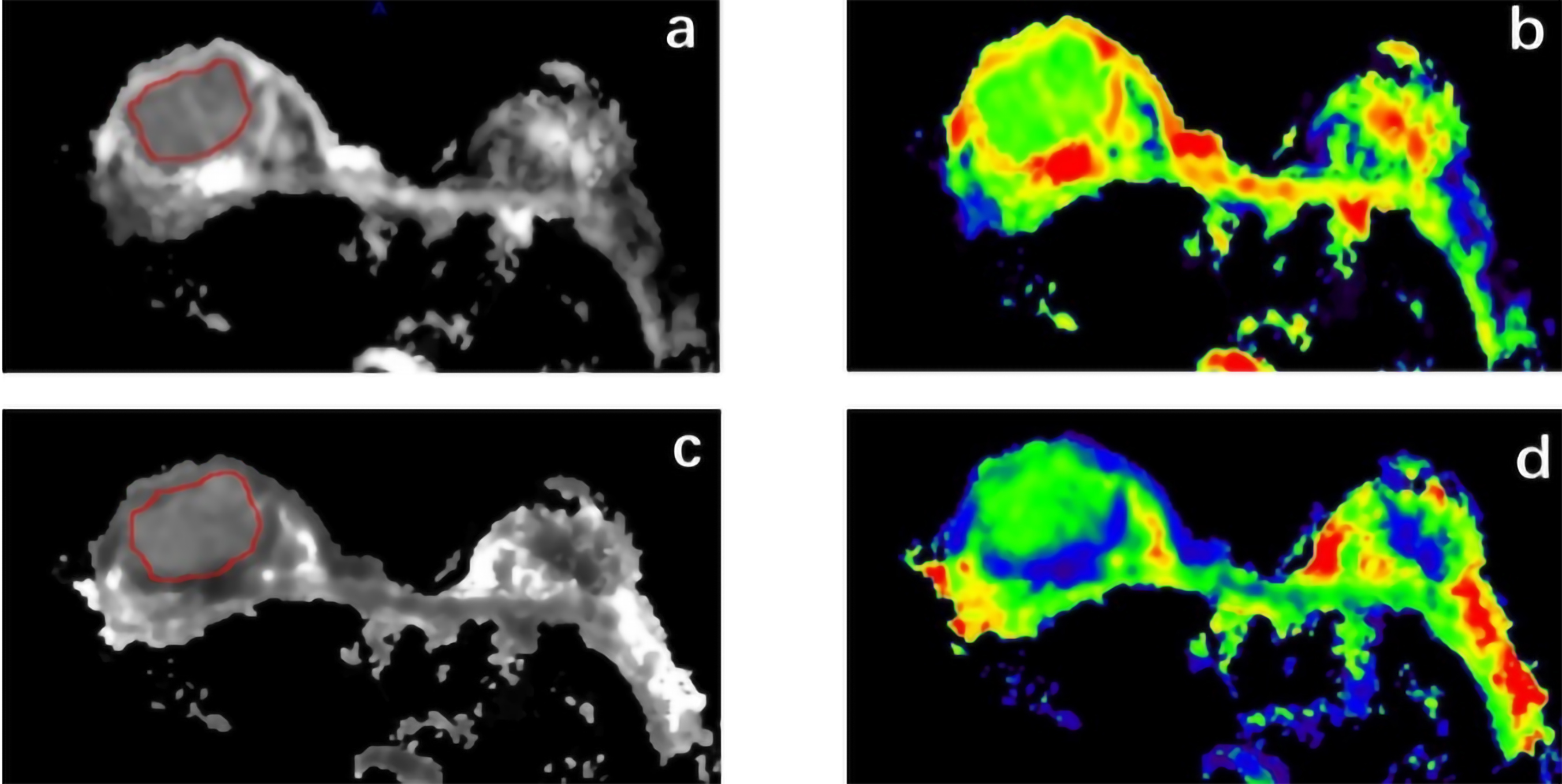 Fig. 2.
MD and MK maps of a 34-years old woman with infiltrating ductal carcinomas in right breast. MD images showed that the lesion was slightly hypointense. MK image shows slightly hyperintense lesion. MD map (a), MK maps (b), MD pseudo color map (c), MK pseudo color map (d) of a 34-years old woman with infiltrating ductal carcinomas in right breast.
As shown in Table 1, MD values were significantly decreased in grade III tumors
(0.82
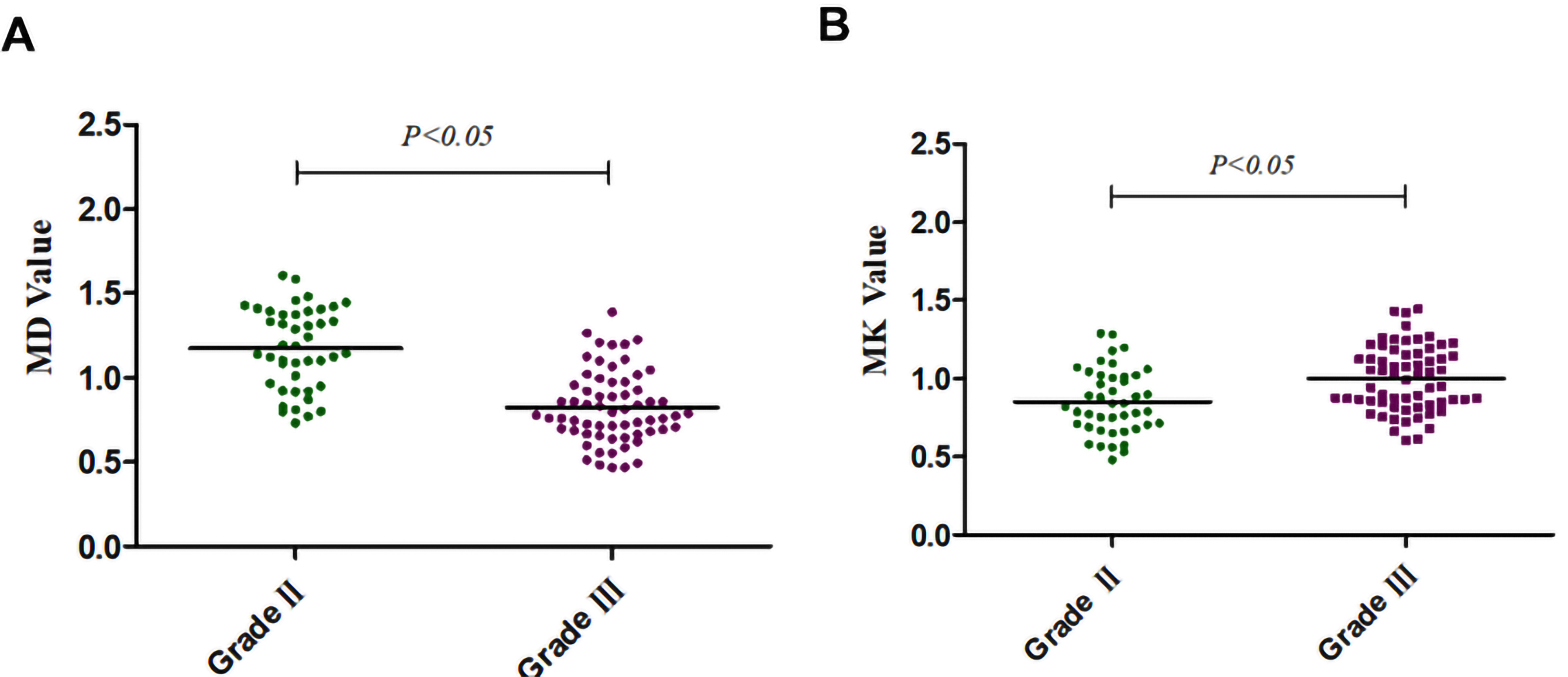 Fig. 3.
Different MD and MK in grade II and grade III breast cancer patients. Summarized data showing MD values (A) and MK values (B) in grade II and grade III breast cancer patients.
Then, the relationship of MD and MK with the expressions (positive and negative)
of ER, PR, HER-2 and Ki-67 were analyzed. The routine HE staining and IHC
staining of grade II tumors with ER (1+), PR (–), Ki-67 (3+, about 70%), and
HER-2 (3+) was demonstrated in Fig. 4A. As shown in Fig. 4B,C, MD was negatively
associated with Ki-67 level (r = –0.39, p
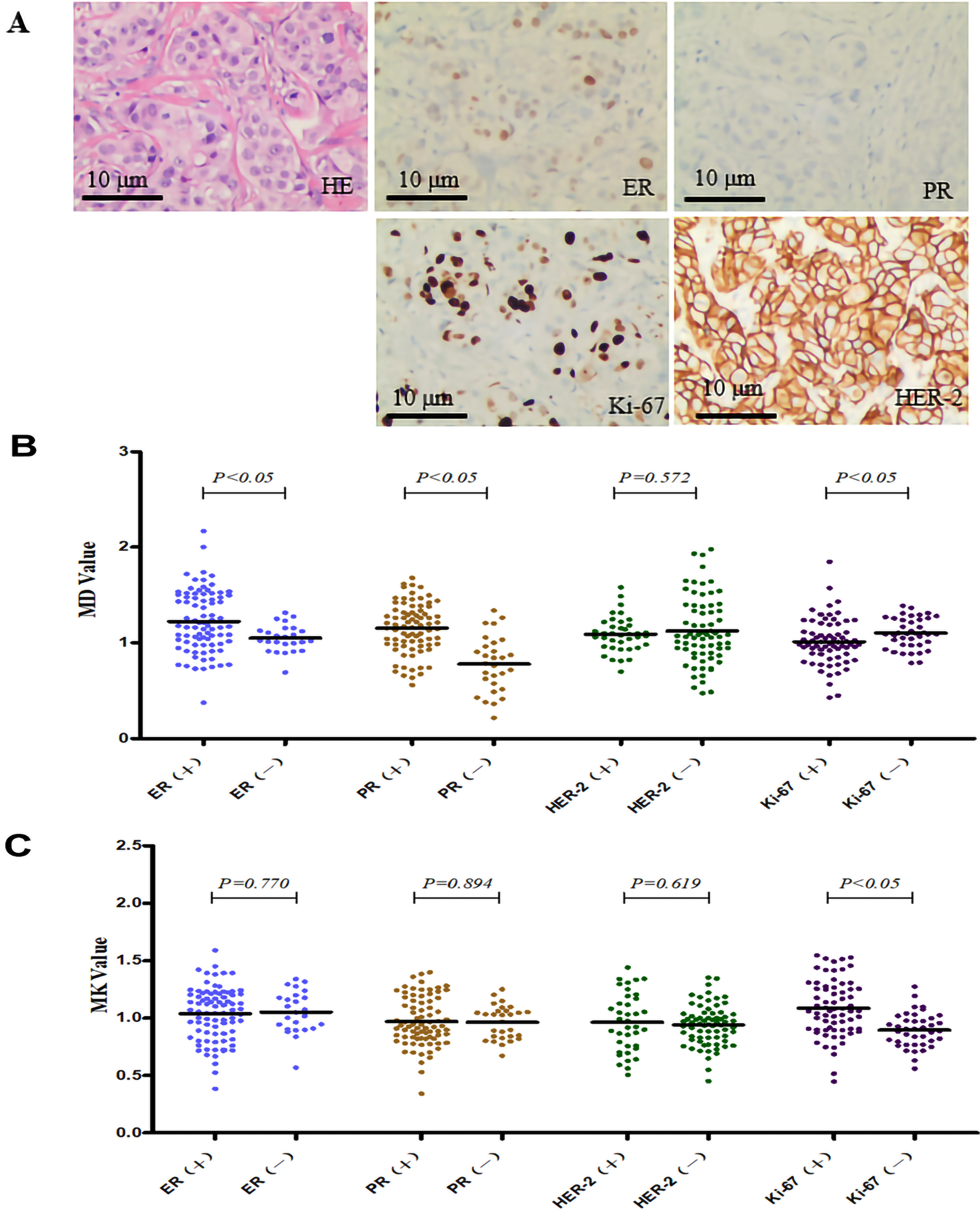 Fig. 4.
Correlation of MD and MK with the expression of ER, PR, and
Ki-67. (A) The pathological images 200
| Parameter | MD (10 |
MK | |
|---|---|---|---|
| ER | |||
| Negative (n = 25) | 1.05 |
1.05 | |
| Positive (n = 83) | 1.22 |
1.04 | |
| p value | 0.010 | 0.770 | |
| power value | - | 0.961 | |
| PR | |||
| Negative (n = 29) | 0.78 |
1.03 (0.83–1.06) | |
| Positive (n = 79) | 1.15 |
0.97 | |
| p value | 0.000 | 0.917 | |
| power value | - | 0.957 | |
| HER-2 | |||
| Negative (n = 70) | 1.07 |
0.94 | |
| Positive (n = 38) | 1.09 |
0.96 | |
| p value | 0.997 | 0.619 | |
| power value | 0.915 | 0.936 | |
| Ki-67 | |||
| Low (n = 40) | 1.10 |
0.90 | |
| High (n = 68) | 1.01 |
1.09 | |
| p value | 0.040 | 0.000 | |
Note: ER, estrogen receptor; PR, progesterone receptor; HER-2, human epidermal growth factor receptor 2; MD, mean diffusivity; MK, mean kurtosis.
Traditional breast cancer screening, classification and diagnosis have always relied on X-ray imaging and ultrasound. However, with the progress of the times, the accuracy and sensitivity of traditional diagnostic techniques are increasingly high, and the traditional diagnosis is faced with great challenges [18]. For radiologists, it is necessary to improve the accuracy of breast cancer diagnosis and pathological grading. Accurate description of breast tumor types and degree of malignancy is conducive to the formulation of treatment plans and improvement of treatment outcomes. DKI, with its non-invasive imaging function, can provide valuable information about water diffusion characteristics in tumor microenvironment for diagnosis. In the present project, we investigated the association between DKI-derived parameters MD and MK with tumor grade and the expression of breast cancer-related proteins including Ki-67, ER, PR, and HER-2. Our results show that MD was obviously lower in grade III breast cancer than that in grade II breast cancer, while MK was prominently higher in grade III breast cancer than that in grade II breast cancer. MD was negatively correlated with Ki-67 level, while MK was positively associated with Ki-67 level. In addition, MD was positively associated with the level of ER and PR, but not HER-2. A study has found that the MD value of benign breast lesions is higher than that of malignant breast lesions, while the MK value of malignant breast lesions is significantly higher than that of benign breast lesions. The MD and MK values of benign breast lesions are significantly different from those of malignant breast lesions, and only the MK value is statistically different between fibroadenoma and fibrocystic breast lesions [18]. Therefore, the results of this study and previous studies show that DKI may serve as a useful tool in the diagnosis for breast cancer.
Firstly, our study focused on two main parameters: MD and MK, which was calculated by DKI model. Diffusivity is the diffusion coefficient with correction of non-Gaussian bias, and Kurtosis quantifies the deviation of tissue diffusion from a Gaussian pattern [15]. The present study demonstrated that MD was significantly decreased, while MK was significantly increased in grade III patients in contrast to grade II patients. It has been well accepted that DKI, a non-invasive functional imaging technique, could help us to figure out the properties of water diffusion in microenvironment of tumors. This finding is consistent with a previous study demonstrating that high-grade tumors are identified with evident variation of nuclear pleomorphic and high mitotic counts and without tubule and gland formation [24]. Indeed, DKI could provide us with the characteristics of tissue water diffusion [9, 25]. Specifically, the characterization of the water diffusion of the microenvironment can help us to understand the pathological status of breast lesions. Because of a reduction of the extracellular space, cell proliferation in high-grade tumor is promoted, and motion of water molecules is restricted. The cellular density is much higher in high-grade tumors than that in low-grade tumors. These changes suggest that tissue complexity was increased at the microstructural level in grade III tumors and exhibited higher kurtosis and lower diffusivity. Therefore, this study suggests that the change in tissue complexity at the microstructure level results in higher MK values and lower MD values in the tissues of grade 3 breast cancer cases compared with grade 2 breast cancer cases. In addition, Spearman correlation analysis results in this study showed that MD value was negatively correlated with the pathological grade of breast cancer, while MK value was positively correlated with the pathological grade, which proved that DKI had a certain guiding value in evaluating the degree of breast cancer lesions.
It has been reported that high Ki-67 level represents high recurrence ratio and low survival rate [26, 27, 28, 29], and detection of Ki-67 positivity is helpful in evaluating prognosis of breast cancer patients. The results of this study showed that MD value was negatively correlated with Ki-67 expression, while MK value was positively correlated with Ki-67 expression. Our result is consistent with previous studies demonstrating that MK was positively correlated with histological grade and Ki-67, while MD was negatively correlated with histological grade and Ki-67 [19]. This may be caused by that tumor tissue becomes more complex due to tumor cell proliferation and vascular hyperplasia. The positive association between MK value and Ki-67 and the negative association between MD value and Ki-67 in advanced tumors may have certain value to evaluate the tumor grade in response to tumor proliferative activity, and provide the basis for later treatment.
In addition to Ki-67 factor, ER, PR and HER-2 are also widely used prognostic markers of breast cancer, and there are many studies related to imaging. And ER and PR are widely used markers in determining therapeutic response to hormone therapies [20, 30, 31, 32]. The results of this study showed that there were significant statistical differences in MD values between negative and positive expression groups of ER, PR and Ki-67 factors, but no statistical differences between negative and positive expression groups of HER-2 factors. MK showed statistically significant difference only between the negative and positive expression groups of Ki-67 factor, but no statistically significant difference between the negative and positive expression groups of ER, PR and HER-2 factor. The results with no significant difference were tested by the T-test of efficacy analysis, and the effect values were all greater than 0.9. Therefore, p value greater than or equal to 0.05 was the true negative result. In addition, in the current study, MD was positively correlated with ER, PR positive expression, indicating that diffusion kurtosis MRI could be of interest to discriminate hormone receptor-positive breast cancers. However, the present study has a few limitations. When we do magnetic resonance imaging of breast, we first judge the condition of breast cancer by using the T2WI image, and then decide whether to use the longer DKI scanning sequence or not. The lack of experienced doctor’s guidance caused the omission of some cases. In order to improve the systematicness and scientificity of this study, attention should be paid to the collection and study of early and small cases in the future.
In summary, this exploration shows the quantitative analysis of DKI parameters has great value in the evaluation on the classification of invasive breast carcinoma. DKI can provide help in characterization and diagnosis of breast lesions. However, further studies are needed to further explore the value of DKI in breast carcinoma with larger sample size.
The datasets used and/or analyzed during the current study are available from the corresponding author on reasonable request.
YG did data collection, data collation and manuscript writing. NW did the sorting of patient data, statistical analysis and manuscript revision. GYH reviewed the draft and checked the results of the data analysis. XMW did had a hand in revising the first draft and designing of the work. CHH examined the overall framework of the manuscript and the interpretation of data for the work. JS verified the study design and involved in drafting the manuscript. WGT analyzed the data and revised it critically for important intellectual content. LY did was involved in the collection of patient image information and involved in drafting the manuscript. XXM did was involved in delineating the extent of the lesion and involved in drafting the manuscript. QQW was involved in image acquisition and image processing and revised in drafting the manuscript. MZ determined the method and purpose of the research, constructed the overall framework of the research, and provided the research funding. All authors read and approved the final manuscript. All authors contributed to editorial changes in the manuscript.
This study was approved by the Ethics Committee. The ethics approval number is 073. Written informed consent from patients were obtained.
Not applicable.
This work was supported by the Jiangsu Provincial Health Commission’s Elderly Health Project (LK2021017).
The authors declare no conflict of interest.
References
Publisher’s Note: IMR Press stays neutral with regard to jurisdictional claims in published maps and institutional affiliations.