


 , Jakub Toczek 1, Zenon Czuba 2, Rafał Stojko 1
1 Department of Gynecology, Obstetrics and Oncological Gynecology, Medical University of Silesia in Katowice, 40-211 Katowice, Poland
2 Department of Microbiology and Immunology, Medical University of Silesia in Katowice, 41-808 Zabrze, Poland
Academic Editor: Ugo Indraccolo
Abstract
Objective: The aim of this study was to understand the pathophysiologic mechanisms of macrophage inflammatory protein 1 alpha (MIP1a) and beta (MIP1b) chemokines in endometriosis, and to understand the immuno-pathophysiology underlying the progression of this disease. Methods: Analyses and conclusions outlined in this study were based on in vitro experiments conducted using supernatants collected from cultured lymphocytes taken from women with endometriosis and from healthy donors. The study group included 30 patients meeting the outlined inclusion criteria who were diagnosed at various clinical stages of endometriosis following a laparoscopic procedure and subsequent histopathological examination. The control group were 50 patients with infertility but without endometriosis. Blood samples were taken and the resulting buffy coat was used to establish lymphocyte cultures. After plating the cells, phytohaemagglutinin (PHA) was added and after 24 hours of incubation, supernatants were collected by centrifugation and subjected to analysis. Results: MIP1a and MIP1b levels in PHA-stimulated lymphocyte cultures from women with endometriosis were elevated when compared to controls, and this difference was highly statistically significant for both cytokines (p = 0.00001 and p = 0.000026, respectively). Additionally, we analyzed the correlation between the occurrence of endometriosis and PHA-stimulated chemokine concentration in lymphocyte culture supernatants. Both MIP1a and MIP1b exhibited a statistical significance with the presence of endometriosis (p = 0.00001). Conclusions: In this study, we observed a significant increase in secretion of selected chemokine factors in in vitro cultures of lymphocytes from women diagnosed with endometriosis. This feature may indicate an essential role for these chemotactic factors in the pathogenesis of endometriosis. To fully understand the influence of MIP1a and MIP1b on disease progression, it will be necessary in future studies to determine chemokine concentrations at each stage of endometriosis.
Keywords
- macrophages
- chemokines
- endometriosis
- immune pathway
- laparoscopy
Endometriosis is a chronic estrogen-dependent disease characterized by the occurrence of endometrial tissue outside the uterus [1]. It is estimated that approximately 10 million women in the United States suffer from endometriosis, which is principally diagnosed in the third and fourth decade of their lives [2].
Currently, the vast majority of treatment options rely on contraceptive drugs, or therapeutics that suppress release of the estrogen in the ovaries, because endometriosis is widely viewed as a progressive, chronic problem caused by high estrogen concentrations. It has been shown that ectopically growing endometrial cells are resistant to immune responses elicited by inflammatory cells. Moreover, these cells also display an increased proliferative potential as well as increased expression of aromatase receptors. This latter feature leads to an increase in estrogen expression promoted by prostaglandin E2. Current findings indicate that endometriosis risk is six times higher in patients with a first-degree relative diagnosed with severe endometriosis suggesting that conditions that promote endometriosis can result from inherited, or acquired, genetic factors. Gene polymorphisms linked to detoxification, expression of estrogen receptors, cytokines, immune-modulating proteins (e.g., Toll-like receptors), as well as factors related to attraction and invasion have been linked to endometriosis. Cytokine release dysfunction is believed to be an important aspect of many disorders, including endometriosis and inflammatory cell response is likely an important aspect in the progression of this disorder [3, 4].
The principal participants and communicators in immune signaling cascades are cytokines. Although most of these polypeptides are produced by immune cells, some cytokines are also released by mononuclear cells. Cytokines are important in coordinating immune cell functions to obtain appropriate host immune response to infection, or trauma, through autocrine and/or paracrine signaling. Based on their role in regulating immunity, cytokines can be classified as either pro or anti-inflammatory. Pro-inflammatory cytokines include interleukin-1, tumor necrosis factor 1 alpha, interferon-gamma, and granulocyte monocyte colony stimulating factor. These cytokines signal to other immune cells involved in the recruitment of pro-inflammatory mediators at the site of injury. This results in an initiation and escalation of inflammation in response to infection or trauma. Cytokines regulating the intensity and length of the inflammatory response through blocking the activity of pro-inflammatory cytokines are termed anti-inflammatory cytokines, although some of them also play pro-inflammatory roles and these are IL-4, IL-6, and IL-10 [5]. Other chemokines, such as monocyte chemoattractant protein 1 (MCP1), IL-8, stromal cell-derived factor 1 (SDF-1) can recruit immune cells to an injured area and stimulate production of additional cytokines [6].
Immune factors can also play an important role in the development of endometriosis. Normal immune response to either pathogens or injury may disrupt the delicate balance between pro and anti-inflammatory cytokines and immune response regulators. Previous studies have indicated an increased concentration of leukocytes, as well as dysregulation of immunological activity, in women with endometriosis [7]. Peritoneal fluid taken from these patients contains higher concentrations of pro-inflammatory and angiogenic cytokines which contribute to the pathogenesis of endometriosis. These cytokines are most likely produced by immune cells, such as macrophages, and/or by the lesion itself. Moreover, peritoneal fluid taken from women with endometriosis contains components that cause monocytes to differentiate into macrophages rather than dendritic cells, even in the presence of cytokines that differentiate dendritic cells in vitro [8]. In this study, we have examined selected chemokines that appear to play important roles in the creation and survival of endometriosis lesions. The principal mediators of monocyte and T-cell migration to the areas of inflammation are members of the chemokine sub-families CCL2 (monocyte chemoattractant protein 1 (MCP-1)), CCL5 (regulated on activation, normal T cell expressed and secreted (RANTES)) and CCL3 (macrophage inflammatory protein 1-alpha (MIP-1a)) [9].
CCL2 and its receptors (CCR2, CCR4, and CCR5) have been reported to attract myeloid-derived monocytic suppressor cells (Mo-MDSC) in various disease models. CCR5, one of the most important receptors, binds three chemokines, specifically, CCL3, CCL4 and CCL5 [10]. These cytokines have a strong ability to suppress anti-tumor immunity in various types of cancer, and the use of CCR5 inhibitors could bring clinical benefits for cancer patients. Also, during inflammatory or infectious conditions, CCR5 and its chemokines play an important role in immunosuppression. One recent study of melanoma showed that CCR5 and its ligands can recruit MDSC to neoplastic lesions both in vivo and in vitro, and enhance the immunosuppressive function of MDSC [11].
As endometriosis has been reported to be a chronic inflammatory disease with malignant activities, we have chosen to focus this study on examination of CCL3 (MIP1a and MIP1b) in endometriosis. The principal goal of this research study was to systematize knowledge concerning pathophysiologic mechanisms of selected chemokine activity in women with endometriosis. An improved understanding of the origin of endometriosis may result in the implementation of new, non-invasive diagnostic methods, and more effective, dedicated therapeutics.
Our study group consisted of randomly selected patients diagnosed with endometriosis during laparoscopic procedure and histopathological examination in different clinical stages of illness, who were patients of hospital of the Order of Brothers Hospitallers in Katowice.
All participants provided informed consent prior to participation in the study. This study was conducted in accordance with the principles contained in the Helsinki Declaration, and the study protocol was approved by the Ethics Committee of the Medical University of Silesia in Katowice, approval no. KNW/0022/KB1/100/17 of October 3, 2017.
Control group consisted of patients during infertility diagnostics, where endometriosis was excluded. In our study this group of women were suitable to compare with women suffering from endometriosis.
Criteria for inclusion in the endometriosis group were no active autoimmune disease, no chronic inflammatory disease, no immune system modelling medication such as vaccines, immunosuppressants, and steroids for at least 3 months before participation in the study, age from 18 to 50 years old, and a diagnosis of endometriosis following laparoscopy. Before beginning the study patients were informed concerning the nature of the research and possible risks. Patients were asked to sign a written consent to participate in the study.
Peripheral blood (10 mL) was collected using anticoagulant (heparin). After collection, this material was transported at a temperature of 24 °C to the laboratory of the Department of Microbiology and Immunology of Medical Faculty in Zabrze, where density gradient centrifugation was performed. Lymphocytes cultures were established from the buffy coat at one 4-well plate per patient. After plating lymphocytes, phytohaemagglutinin (PHA) was added to two of the wells and after 24 hr cultures were centrifuged and the supernatant was removed. Both experimental and control samples were cultured with and without PHA stimulation for comparative determination.
MIP-1a and MIP-1b were measured using the Bio-Plex multiplex assay system (Bio-Rad Laboratories, Hercules, California, USA) with the use of xMAP bead-based technology. This approach includes the use of sets of beads created by two fluorescent dyes in differing proportions. Beads can be further coupled with reagents specific for each bioassay. In the sandwich methodology used in this study, one antibody was attached to sets of beads in the same color and another antibody is attached a fluorescent tag. The use of multicolor beads enables multiplexed detection of many substances simultaneously in one sample (see Fig. 1).
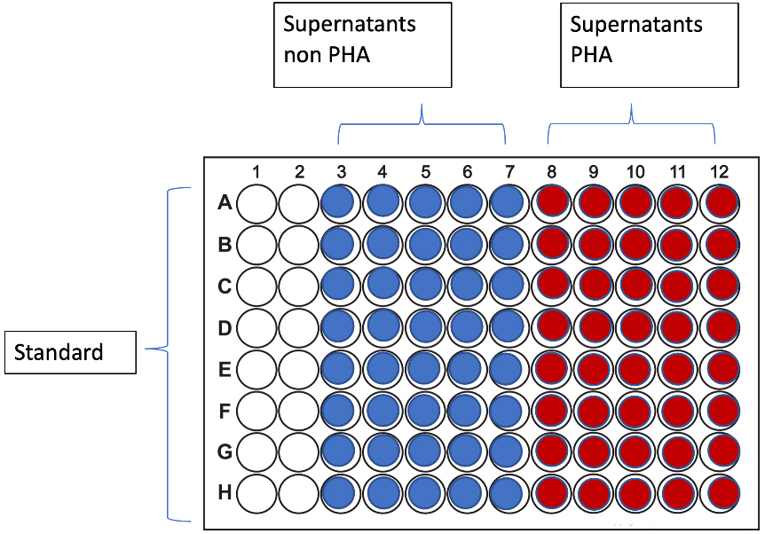 Fig. 1.
Layout of multi-well plate used in testing.
The results were compiled and analyzed using ANOVA statistical tests to test relationships between groups.
Based on the outlined inclusion criteria, the study included 80 patients. 30 of the enrolled patients has diagnosed cases of endometriosis, and 50 healthy women were included in the control group. The average age of endometriosis patients was 33.41 years (SD = 3.21) and the average age of control group was slightly lower at 31.39 years (SD = 5.1).
We analyzed the concentration of MIP1a and MIP1b in supernatants obtained from
lymphocyte cultures of women with endometriosis and healthy controls. The mean
cytokine concentration in each control (C) and test (T) groups is presented in
Fig. 2. All results were tested for normal data distribution using the
Shapiro-Wilk test. Results of p
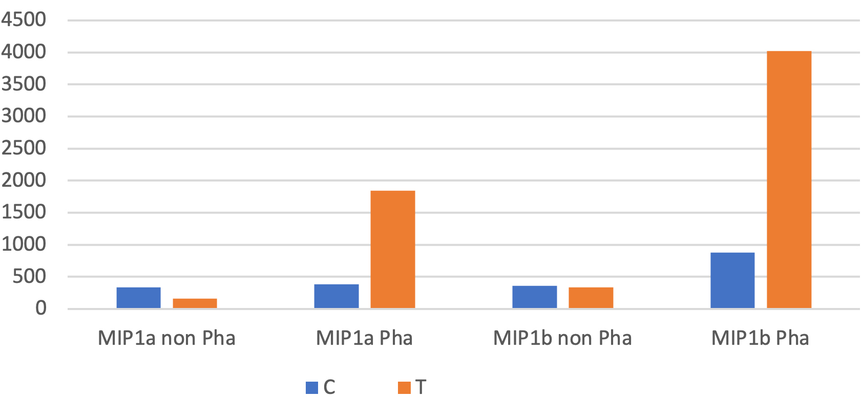 Fig. 2.
The average concentration of MIP1a and MIP1b in supernatants.
By comparing both with and without PHA stimulation supernatants, statistically significant changes in the concentration of both chemokines were observed in the endometriosis patient group. Variance was analyzed for PHA stimulation and the occurrence of endometriosis. This analysis is presented in Table 1.
| aNOVA for p |
Supernatants | Chemokines | aNOVA for all groups for PHA and occurrence of endometriosis |
| 0.266196 | Healthy/endometriosis | MIP-1a | 0.00001 |
| 000001 | healthy with PHA/endometriosis with PHA | ||
| 0.868461 | Healthy/endometriosis | MIP-1b | 0.00001 |
| 0.000026 | healthy with PHA/endometriosis with PHA |
By analyzing the variance, we noted statistically significant higher concentrations of MIP1a and MIP1b in PHA stimulated cultures in the endometriosis group in comparison to control cultures with PHA stimulation at the level of p = 0.00001 and p = 0.000026, respectively.
Additionally, we analyzed the statistical correlation in relationship between the occurrence of endometriosis and PHA stimulation on the concentration of chemokines in lymphocyte cultures. In both tested cytokines statistically significant results were observed (p = 0.00001).
Understanding mechanisms that regulate recruitment of macrophages to endometrial lesions may provide an increased understanding of disease progression. Many abnormally expressed molecules responsible for macrophage recruitment have been identified in endometrial lesions. For example, increased secretion of monocyte chemoattractant protein 1 (MCP-1, also known as chemokine ligand 2 (CCL-2)) from peritoneal macrophages of women with endometriosis may influence paracrine and autocrine signaling resulting in macrophage accumulation within the peritoneal cavity.
Endometrial cells from taken from endometriosis lesions are more resistant to the cytotoxic action of peritoneal macrophages. Cytotoxicity of natural killer (NK) cells and macrophages were inversely correlated to the stage of endometrial disease. Protection against the cytotoxic effects of NK cells and macrophages appears to be provided by a dysregulation of the MHC I complex within ectopic endometrial cells [12, 13].
Akoum et al. [14] analyzed 57 infertility or pelvic pain patients with laparoscopically-confirmed endometriosis and compared them with 44 fertile women with no evidence of endometriosis. This study showed a statistically significant increase in MCP-1 and MIP secretion from lymphocytes taken from endometriosis patients. Resultant MIP concentrations in plasma were also higher in patients with endometriosis (163.0 to 788 pg/mL) than in control group (0.0 to 355 pg/mL); however, such increases were statistically significant only at the earliest stages of endometriosis. However, increased chemotactic activity, defined as the average count of migrating cells, was noticed at all stages of endometriosis compared to controls. The authors concluded that endometriosis is associated with increased levels and activity of MCP-1 in peripheral blood, and increased expression and activation of this cytokine may play an important role in the inflammatory process related to this condition [15].
In a different retrospective control case study, samples of peritoneal fluid
were collected during laparoscopy and subsequently evaluated [16]. Data obtained
showed clear patterns of increased cytokine concentrations in the peritoneal
fluid of women with endometriosis. Most notably, a significant increase in EGF,
FGF-2, Il-1
The Malutan et al. [18] study, conducted on a group
of 160 women, focused on the evaluation of concentrations of various chemokines
and interferons in the serum of patients with diagnosed endometriosis. These
researchers showed that women with endometriosis have higher levels of
IFN-
A limitation of this study is that it was conducted on a relatively small number of subjects due to the large costs of outlined laboratory procedures. Because of this small study group size, future studies will need to be conducted on a greater patient population. Additionally, we believe that future efforts will need to be extended to endometriosis patients from different social and geographical backgrounds as this study is focused only on patients from one clinic in Silesia. Future studies should also investigate other immunological pathways including additional chemokines.
In this study we measured significant increases in in vitro secretion of selected chemotactic factors MIP1a and MIP1b by lymphocytes harvested from women with endometriosis. This finding suggests an important role for these factors in the pathogenesis of the disease, and could open a new direction for alternative treatment strategies. To better understand their influence on disease progression will require determining chemokine concentrations at each stage of endometriosis. Further research in this direction may help to develop an advanced, non-invasive diagnostic tool to diagnose early stage endometriosis.
MS—extraction and drafting of the manuscript, statistical analysis; JT—analysis of data, manuscript revision; ZC—methodology, determination of samples; RS—design and revision. All authors read and approved the final manuscript.
All respondents gave their informed consent to participate in the study. The study was conducted in accordance with the principles contained in the Helsinki Declaration, and the study protocol was approved by the Ethics Committee of the Medical University of Silesia in Katowice, approval no. KNW / 0022 / KB1 / 100/17.
Many thanks to the team of the Department of Microbiology and Immunology in Zabrze under the leadership of Zenon Czuba for the preparation of the laboratory procedures, in-vitro lymphocyte culture, and sample measurements.
This research received no external funding.
The authors declare no conflict of interest.
References
Publisher’s Note: IMR Press stays neutral with regard to jurisdictional claims in published maps and institutional affiliations.