IMR Press / CEOG / Volume 48 / Issue 4 / DOI: 10.31083/j.ceog4804133
Open Access Original Research
Age and anti-Műllerian hormone: prediction of cumulative pregnancy outcome in in vitro fertilization with diminished ovarian reserve
Show Less
1 Center of Reproductive Medicine, Guangzhou Women and Children’s Medical Center, Guangzhou Medical University, 510623 Guangzhou, Guangdong, China
*Correspondence: sunling6299@163.com; sunling6299@gwcmc.org (Ling Sun)
Clin. Exp. Obstet. Gynecol. 2021, 48(4), 835–841; https://doi.org/10.31083/j.ceog4804133
Submitted: 12 November 2020 | Revised: 10 December 2020 | Accepted: 23 December 2020 | Published: 15 August 2021
Copyright: © 2021 The Author(s). Published by IMR Press.
This is an open access article under the CC BY 4.0 license (https://creativecommons.org/licenses/by/4.0/).
Abstract

Background: To determine the effects of AMH and age on IVF outcomes in women with DOR and whether the cumulative pregnancy potential after consecutive IVF cycles is age or AMH level depended. Methods: Between January 2014 and December 2018, 466 infertility patients (1004 IVF/ICSI cycles) with AMH 1.1 ng/mL were recruited and firstly divided into two groups (Group 1: AMH <0.6 ng/mL and Group 2: AMH 0.6–1.1 ng/mL). Furthermore, they were stratified into another four groups (A1, A2; B1, B2) based on age (Group A: <38 y, Group B: 38 y). Laboratory parameters and cumulative pregnancy chances were retrospectively evaluated. Results: Regardless of age, number of retrieved oocytes, 2 pronuclear zygotes (2PN), embryos, good-quality embryos per cycle, implantation rate (IR), clinical pregnancy rate (CPR) and ongoing pregnancy rate (OPR) per embryo transfer (ET) and per patient were significantly lower in Group 1 than those in Group 2 (P < 0.001). However, in women <38 years, there were no significant differences in IR, CPR and OPR per ET and per patient between Group A1 and A2 (P > 0.05). The cumulative OPR per patient increased to 60% in Group A2 and 51.7% in Group A1 after the third transfer but reached a plateau (24.8% in Group B2 and 11.8% in Group B1) after the second ET. Conclusions: For advanced-age women with DOR, AMH level showed as a strong predictor of both ovarian response and pregnancy rate. However, even with extremely lower AMH value, younger women still had higher potential to get pregnancy chance through more attempts at subsequent IVF treatments.

Keywords
Age
Anti-Műllerian hormone
Diminished ovarian reserve
IVF
Ongoing pregnancy rate
1. Introduction

A reliable predictor of in vitro fertilization (IVF) outcomes would be useful in order to optimize individualized treatment protocols and assist in counseling patients about their chances of having successful IVF treatment. Therefore, several “ovarian reserve markers” have been investigated for the ability to predict ongoing pregnancy or live birth rates after assisted reproduction treatment (ART). Examples include follicular phase serum follicle-stimulating hormone (FSH), ultrasound assessment of the antral follicle count (AFC) and others [1, 2]. However, maternal age at embryo transfer is still the most accurate established predictor [3, 4].

Recently, anti-Műllerian hormone (AMH) has been proposed as a good predictor of both ovarian reserve and response [5, 6]. AMH has been shown to be a better indicator of a patient’s follicular response to controlled ovarian stimulation (COS) with gonadotropins than the basal levels of FSH, estradiol, inhibin B, or age [7, 8]. However, conflicting results exist regarding the association of AMH with the outcome of pregnancy after IVF treatment. Although some studies have shown that AMH is a good predictor of ovarian response and outcome of pregnancy [9, 10], others found that AMH had poor accuracy for the prediction of embryo quality and ongoing pregnancy [10, 11], suggesting that AMH is a marker of oocyte quantity rather than quality [12]. Pregnancy outcome in IVF cycles is highly associated with oocyte and embryo quality, and many studies described age but not AMH as a good predictor of quality of both oocyte and embryo [13].

Diminished ovarian reserve (DOR) defined by American Society for Reproductive Medicine (ASRM) describes women of reproductive age with regular menses have reduced fecundability and/or poor ovarian response (POR) to gonadotropin stimulation compared with women of similar age [14]. It refers to a decline in reproductive potential largely ascribed to the natural ovarian aging process [15], which is distinct from either menopause or premature ovarian failure [16].

POR is usually identified by a low follicular response to ovarian stimulation, resulting in reduction of retrieved oocytes during IVF treatment [17]. Consensus on its definition by the European Society of Human Reproduction and Embryology (ESHRE) recommended the minimum criteria (Bologna criteria). At least two of the following three conditions should be met to predict POR: (1) advanced age (40 years) or any other risk factor; (2) low antral follicle count (AFC <5–7) or AMH <0.5–1.1 ng/mL; (3) prior poor ovarian response (3 oocytes after a conventional stimulation) [18].

DOR is used to describe a decreased state of ovarian reserve on the basis of ovarian reserve testing before cycle start, while POR is the poor response according to the result to ovarian stimulation during IVF. They are not interchangeable since there might exists a mismatch between patients with DOR and their ovarian response in IVF cycles.

The most common causes of DOR are different in patients of different ages. For patients of advanced age, DOR seems mostly to be correlated with the natural decline of ovarian function caused by increasing chronological age, and the decrease of oocyte number was accompanied by a significant decline in quality [19, 20]. However, in younger patients, it is most often related to heredity, environment and iatrogenic factors, such as previous pelvic or ovarian surgery [21]. Although the number of follicles was found to be decreased in these situations, the quality of oocytes was not affected. Therefore, young women with low AMH value would be predicted to have favorable IVF outcome due to a well-preserved oocyte competence and comparable good-quality embryos. A few studies have explored various indicators that predict the pregnancy rate in patients with DOR in IVF cycles. Among them, AMH as one of the most commonly ordered tests of ovarian reserve was debated on its prediction power. Ulrich et al. [22], highlighted the limitations of AMH especially for poor responders. However, more data are needed to provide sufficient evidence to suggest the use of AMH for DOR patients.

The aim of this study was to evaluate the role of serum AMH concentration and age in predicting pregnancy outcome in women with diminished ovarian reserve and whether the pregnancy rate after consecutive IVF cycles is age and/or AMH dependent.

2. Materials and methods
2.1 Subject recruitment

This study was declared exempt by the ethics committee of the Guangzhou Women and Children’s Medical Center, China, and written informed consent was obtained from all patients. 466 women undergoing IVF/ICSI treatment during the period from January 2014 to December 2018 were enrolled in the study. All women had serum AMH taken on day 2–5 of their menstrual cycle.

The inclusion criteria were as follows: patient aged 20–45 years, AMH 1.1 ng/mL, body mass index of 18–30 kg/m2 and normal gynecological ultrasound. Patients with polycystic ovary syndrome (PCOS), premature ovarian failure (POF), hydrosalpinx, intrauterine adhesions, systemic illnesses, chromosomal abnormalities and/or cycles canceled before oocyte retrieval were excluded.

Patients were first divided into two groups (Group 1: AMH <0.6 ng/mL and Group 2: AMH 0.6–1.1 ng/mL) and then stratified into four groups (A1, A2; B1, B2) based on age (Group A: <38 y, Group B: 38 y).

2.2 AMH measurement

Measure of AMH levels on the day 2–5 of menstrual cycle was carried out in the endocrinology lab of our hospital using the AMH/MIS commercially enzyme-linked immunosorbent assay kit (Kangrun Biotech, Guangzhou, China) following the manufacturer’s instructions. The lowest and highest detection level of this assays was 0.06 ng/mL and 16.0 ng/mL, respectively. The intra- and inter-assay coefficients of variation were 10% and 15%, respectively.

2.3 Stimulated cycles

The stimulation protocol was a GnRH-antagonist protocol, the starting gonadotropin dosage was dependent on the patients’ age, weight and BMI, and adjusted according to ovarian response. A single injection of 250 mg of human chorionic gonadotropin (hCG, choriogonadotropin alpha, Merck Serono) was administered when two follicles of 18 mm were observed. Oocyte retrieval took place about 36 hours after the hCG administration. Fertilization was assessed 19 ± 1 hours after insemination, and embryos with two pronuclei were cultured individually. On day 3 after insemination, the embryo quality was assessed using the grading system (Istanbul consensus) [23], and one or two cleavage embryos of the best quality were transferred. Remaining embryos were cryopreserved by vitrification.

2.4 Outcome measure

A serum beta-hCG test was performed 14 days after transfer. Clinical and ongoing pregnancy was confirmed by transvaginal ultrasound at 5 to 6 weeks after transfer. A patient with no ongoing pregnancy at the end of the stimulated cycle period and with surplus cryopreserved embryos could undergo cryopreserved replacement cycles within 1 year of the patient’s start of treatment. The main outcome was ongoing pregnancy rate arising from all fresh and subsequent frozen embryo transfers within 1 year.

2.5 Data analysis

Statistical analysis was performed with the assistance of SPSS software (Chicago, IL, USA) version 19 for Windows, applying parametric and nonparametric tests when appropriate. Continuous variables were expressed as absolute numbers, mean ± standard deviation (SD), and analyzed by Student’s t test. Categorical variables were expressed as percentages and analyzed with the chi-square test or Fisher exact test depending on the sample size. Statistical significance was defined as a two-sided P value less than 0.05.

3. Results
3.1 Patients

Table 1 summarizes patient demographic characteristics for the four different AMH groups. 209 patients in Group 1 undergoing 520 cycles had extremely low AMH levels (<0.6 ng/mL), whereas 257 patients in Group 2 undergoing 484 cycles had low AMH levels (0.6–1.1 ng/mL). Women in Group 1 with extremely low AMH levels had lower antral follicular count (AFC) (3.2 ± 1.6 vs 4.7 ± 2.2, P < 0.001) and higher baseline FSH (mIU/mL) (10.2 ± 6.2 vs 7.9 ± 4.4, P < 0.001) than those in Group 2. The mean age was 38.7 ± 5.0 years [median: 40] in Group 1 and 36.7 ± 5.2 years [median: 37] in Group 2 (P > 0.05). The Percentage of women who were between 41–45 years of age was higher in Group 1 than that in Group 2 (36.4% vs 23.7%, P < 0.01). More patients with menstrual factors in Group 1 than Group 2 (38.3% vs 25.3%, P < 0.01). Other similar background characteristics (type and etiology of infertility disorder) were listed in these two groups.

Table 1.Patients characteristics in 466 women with AMH level (<0.6 ng/mL) and (0.6–1.1 ng/mL).
Group 1 Group 2 P
(AMH <0.6 ng/mL) (AMH 0.6–1.1 ng/mL)
No. of patients 209 257
OPU cycles 520 484
AFC 3.2 ± 1.6 4.7 ± 2.2 <0.001
Baseline FSH (mIU/mL) 10.2 ± 6.2 7.9 ± 4.4 <0.001
Age group (years; n, %)
21–25 1 (0.5) 6 (2.3) 0.209
26–30 14 (7.2) 30 (11.7) 0.068
31–35 37 (17.7) 67 (26.1) 0.031
36–40 71 (34.0) 87 (33.9) 0.978
41–45 76 (36.4) 61 (23.7) 0.003
46–50 10 (4.8) 6 (2.3) 0.149
Mean ± SD 38.7 ± 5.0 36.7 ± 5.2 0.452
Median 40 37
Type of fertility disorder (n, %)
Primary 56 (26.8) 84 (32.7) 0.168
Secondary 153 (73.2) 173 (67.3) 0.168
Etiology (n, %)
Male factors 12 (5.7) 25 (9.7) 0.113
Tubal factors 98 (46.9) 144 (56.0) 0.049
Menstrual factors 80 (38.3) 65 (25.3) 0.003
Endometriosis 16 (7.7) 22 (8.6) 0.723
Unexplained 3 (1.4) 1 (0.4) 0.476
3.2 Cycle parameters in two AMH groups

Comparisons of cycle parameters between Group 1 and Group 2 are shown in Table 2. Number of retrieved oocytes, 2 pronuclear zygotes (2PN), embryos and good-quality embryos per cycle were significantly lower in Group 1 than those in Group 2 (all P < 0.001). Implantation rate (16.7% vs 25.6%,), clinical pregnancy rate (CPR) (27% vs 37.1%), ongoing pregnancy rate (OPR) (18.2% vs 31.3%) per embryo transfer (ET) cycle and CPR (33.9% vs 55.5%), OPR (26.1% vs 46.7%) per patient were significantly higher in Group 2 than Group 1 (all P < 0.001). The cancelled transfer cycle rate was significantly higher in Group 1 than Group 2 (90.0% vs 78.3%, P < 0.001).

Table 2.Comparisons of cycles parameters between two AMH groups.
Group 1 Group 2 P
(AMH <0.6 ng/mL) (AMH 0.6–1.1 ng/mL)
OPU cycles 520 484
Fresh embryo transfer cycles (n, %) 52 (22) 105 (30) 0.021
Cancelled transfer cycles rate 468/520 (90.0%) 379/484 (78.3%) <0.001
Frozen embryo transfer cycles (n, %) 184 (78) 237 (69) 0.021
Oocytes retrieved per cycle 2.2 ± 1.7 4.0 ± 2.9 <0.001
2PN per cycle 1.4 ± 1.2 2.7 ± 2.1 <0.001
Embryos per cycle 1.2 ± 1.2 2.3 ± 1.8 <0.001
Good-quality embryos per cycle 0.5 ± 0.7 0.9 ± 1.0 <0.001
Implantation rate 68/408 (16.7%) 160/624 (25.6%) <0.001
Clinical pregnancy rate per ET 56/236 (27.0%) 127/342 (37.1%) <0.001
Ongoing pregnancy rate per ET 43/236 (18.2%) 107/342 (31.3%) <0.001
Miscarriage rate 13/56 (23.2%) 20/342 (5.8%) 0.226
Clinical pregnancy rate per patient 56/165 (33.9%) 127/229 (55.5%) <0.001
Ongoing pregnancy rate per patient 43/165 (26.1%) 107/229 (46.7%) <0.001
3.3 Cycle parameters in subgroups according to age and AMH level

Patients were then stratified into four subgroups depending on age (<38 y or 38 y). Table 3 shows mean age, AFC, basal hormonal profiles and cycle parameters in each subgroup. As expected, patients with extremely low AMH level had lower AFC and higher basal FSH levels than those with low AMH levels (0.6–1.1 ng/mL) regardless of age. There were no significant differences in cycle cancellation due to zero oocyte retrieval, fertilization failure or embryonic arrest between the AMH <0.6 ng/mL and AMH 0.6–1.1 ng/mL subgroups regardless of age. The percentage of patients with embryos transferred was significantly lower in Group A1 than that in Group A2 (76.6% vs 91.9%, P < 0.01).

Table 3.Cycles parameters in 466 women according age and AMH level.
Group A (Age <38 y) Group B (Age 38 y)
A1 A2 P B1 B2 P
(AMH <0.6 ng/mL) (AMH 0.6–1.1 ng/mL) (AMH <0.6 ng/mL) (AMH 0.6–1.1 ng/mL)
Patients 77 136 132 121
OPU cycles 169 230 351 254
Age (years) 33.2 ± 3.0 32.6 ± 3.4 0.146 42.0 ± 2.4 41.3 ± 0.4 0.93
AFC 3.7 ± 1.8 5.4 ± 2.2 0.04 2.9 ± 1.4 4.0 ± 1.9 0.016
bFSH (mIU/mL) 10.5 ± 5.9 7.9 ± 5.1 0.08 10.0 ± 6.4 7.9 ± 3.4 0.002
bLH (mIU/mL) 3.9 ± 2.1 3.1 ± 1.6 0.03 3.7 ± 2.5 3.5 ± 2.3 0.07
bE2 (pmol/L) 155.7 ± 124.2 164.0 ± 146.6 0.526 171.6 ± 160.9 167.6 ± 137.8 0.205
No oocyte (n, %) 5 (6.5) 1 (0.7) 0.05 10 (7.6) 6 (5.0) 0.393
No 2PN (n, %) 4 (5.2) 5 (3.7) 0.597 6 (4.6) 7 (5.8) 0.655
No embryo (n, %) 9 (11.7) 5 (3.7) 0.05 10 (7.6) 4 (3.3) 0.227
Fresh embryo transfer cycles (n, %) 21 (23.9) 67 (35.3) 0.057 31 (21.0) 38 (25) 0.404
Frozen embryo transfer cycles (n, %) 67 (76.1) 123 (64.7) 0.057 117 (79.1) 114 (75) 0.404
3.4 IVF outcome comparisons between age-stratified groups

Analysis of pregnancy rates in patients within age-stratified groups is shown in Table 4. Among women under or above 38 years of age, number of retrieved oocytes, 2PN, embryos per cycle were significantly lower in the extremely low AMH (<0.6 ng/mL) groups than in the low AMH (0.6–1.1 ng/mL) groups (A1 vs A2, p < 0.05; B1 vs B2, P < 0.001). Number of good-quality embryos per cycle in B2 was significantly higher in Group B2 than that in Group B1(0.7 ± 0.9 vs 0.4 ± 0.6, P < 0.001), whereas there was no significant difference between Group A1 and A2 (0.7 ± 0.9 vs 1.0 ± 1.1, P > 0.05). In women older than 38 years of age, implantation rate (17.8% vs 9.4%), CPR (25.7% vs 15.3%), OPR (19.7% vs 8.1%) per ET cycle and CPR (37.5% vs 18.9%), OPR (28.8% vs 11.3%) per patient were significantly higher in Group B2 than Group B1 (P < 0.01). However, in patients younger than 38 years, implantation rate (27.6% vs 31.4%), CPR (40.9% vs 46.3%), OPR (35.2% vs 40.5%) per cycle and CPR (61% vs 70.4%), OPR (52.5% vs 61.6%) per patient were not statically different between Group A1 and A2 (P > 0.05). The cancelled transfer cycle rate was significantly higher in Group A1 than Group A2 (87.6% vs 70.9%, P < 0.001) and higher in Group B1 than Group B2 (91.2% vs 85%, P = 0.019), respectively. The percentage of patients without embryos transferred was significantly higher in Group A1 than that in Group A2 (23.4% vs 8.1%, P < 0.01).

Table 4.Comparisons of IVF outcomes in age-stratified groups.
Group A (Age <38 y) Group B (Age 38 y)
A1 A2 P B1 B2 P
(AMH <0.6 ng/mL) (AMH 0.6–1.1 ng/mL) (AMH <0.6 ng/mL) (AMH 0.6–1.1 ng/mL)
Oocytes retrieved per cycle 2.8 ± 2.1 4.7 ± 3.1 0.009 1.9 ± 1.3 3.2 ± 2.4 <0.001
2PN per cycle 1.9 ± 1.5 3.2 ± 2.3 0.008 1.2 ± 1.0 2.1 ± 1.8 <0.001
Embryos per cycle 1.5 ± 1.4 2.7 ± 1.9 0.032 1.0 ± 1.0 1.9 ± 1.6 <0.001
Good-quality embryos per cycle 0.7 ± 0.9 1.0 ± 1.1 0.178 0.4 ± 0.6 0.7 ± 0.9 <0.001
Cancelled transfer cycles rate 148/169 (87.6%) 163/230 (70.9%) <0.001 320/351 (91.2%) 216/254 (85%) 0.019
Implantation rate 45/163 (27.6%) 113/360 (31.4%) 0.383 23/245 (9.4%) 47/264 (17.8%) 0.006
Clinical pregnancy rate per ET 36/88 (40.9%) 88/190 (46.3%) 0.399 20/148 (13.5%) 39/152 (25.7%) 0.008
Ongoing pregnancy rate per ET 31/88 (35.2%) 77/190 (40.5%) 0.399 12/148 (8.1%) 30/152 (19.7%) 0.004
Miscarriage rate 5/36 (13.9%) 11/88 (12.5%) 0.834 8/20 (40%) 9/39 (23.1%) 0.174
Without embryos transferred rate per patient 18/77 (23.4%) 11/136 (8.1%) 0.002 26/132 (19.7%) 17/121 (14.0%) 0.232
Clinical pregnancy rate per patient 36/59 (61%) 88/125 (70.4%) 0.205 20/106 (18.9%) 39/104 (37.5%) 0.003
Ongoing pregnancy rate per patient 31/59 (52.5%) 77/125 (61.6%) 0.244 12/106 (11.3%) 30/104 (28.8%) 0.002
3.5 Trend of cumulative ongoing pregnancy rates per patient

Fig. 1A presents the trend of COPR observed following more than five oocyte pick-up (OPU) and Fig. 1B shows the COPR in different transfer cycles.

Fig. 1 shows the trend of COPR observed following more than five oocyte pick-up (OPU) and four ET cycles. Analysis revealed that COPR per patient increased along with consecutive treatments respectively in the four subgroups, especially in Group A2 (young women
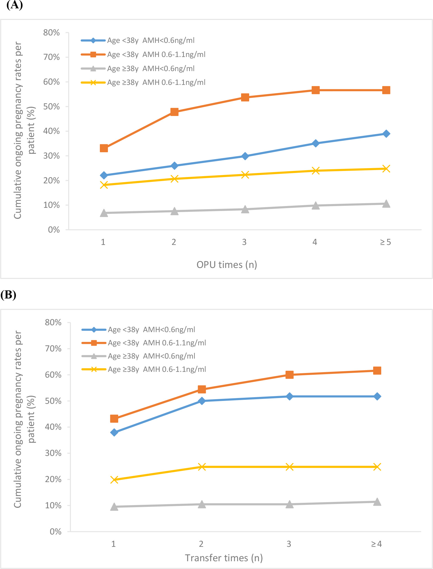
 <38 with AMH 0.6–1.1 ng/mL). Regardless of AMH level, the COPR in women above 38 years slightly increased with following ET cycles as the trend was close to a horizon line, while the COPR in women younger than 38 years obviously improved with increasing number of cycles. The COPR per patient increased to 60% in Group A2 and 51.7% in Group A1 after the third transfer in women <38 years but reached a plateau (24.8% in Group B2 and 11.8% in Group B1) after the second ET in women above 38 years.

Fig. 1.

Cumulative ongoing pregnancy rates per patient following consecutive OPU (A) and ET cycles (B) for women with AMH <1.1 ng/mL.

4. Discussion

Our goal was to investigate the ability of AMH and age to predict IVF pregnancy rates in women with poor ovarian reserve. However, the cut-off values of AMH for predicting poor ovarian response (POR) varied differently from 0.1 to 2.97 ng/mL [24]. According to the Bologna criteria of POR (AMH <0.5–1.1 ng/mL), we recruited patients with AMH 1.1 ng/mL in this study [18]. Another previous study suggested an AMH cut-off level for POR was 0.61 ng/mL and there was significant difference in pregnancy rate below and above this cut-off level [25]. Hence, our study divided patients into two groups based on AMH level <0.6 ng/mL or 0.6–1.1 ng/mL.

Regardless of age, number of retrieved oocytes, 2PN, embryos, good-quality embryos per cycle, IR, CPR and OPR per ET and per patient were significantly lower in patients with AMH <0.6 ng/mL than those with AMH 0.6–1.1 ng/mL. There is a consensus that AMH is a good biomarker of ovarian response but there is still disagreement on its use as a predictor of pregnancy rates in patients undergoing IVF cycles [10, 26]. Our results demonstrate that for women with diminished ovarian reserve, serum AMH value seemed to be closely related with response to ovarian stimulation and IVF pregnancy outcomes. However, when comparing the two groups at baseline, we found that the difference between the two groups was not only due to AMH levels, but also because patients with lower AMH levels were slightly older on average and higher percentage of women between 41–45 y. A meta-analysis supported that age is the most important predictor of IVF outcomes [27]. As an age-specific value, the AMH level was proved to be negatively correlated with female age. Therefore, the positive association between ovarian reserve and pregnancy rate with serum AMH could be also variable depending on age. According to the study of Franasiak, the prevalence of aneuploidy relative to female age demonstrated that the no euploid embryo rate was lowest (2% to 6%) in women aged 26 to 37 and the percent of aneuploid embryos was significantly higher after 38 years [28]. Pairwise comparison in our study showed that in the same AMH range, the pregnancy chance of women above 38 y was significantly lower than that of women under 38 years. Considering the interaction between age and AMH, it may be useful to consider both AMH level and age (38 y or <38 y) instead of using AMH alone.

Analysis of the 4 subgroups revealed several interesting trends. First, in women older than 38, not only the ovarian response but also IVF outcomes were significantly lower in patients with AMH <0.6 ng/mL than those with AMH 0.6–1.1 ng/mL. Sezai’s study also suggested that AMH could accurately predict pregnancy rates for advanced age women [29]. In women younger than 38, although the ovarian response (e.g., number of retrieved oocytes, 2PN and embryos per cycle) were also significantly lower in patients with AMH <0.6 ng/mL than those with AMH 0.6–1.1 ng/mL, there were no significant differences in number of good-quality embryos per cycle and no difference in pregnancy outcomes (IR, CPR and OPR per ET and per patient) within the two AMH level groups.

It was worth noting that the cancelled transfer cycle rate were high in all 4 subgroups, especially in patients with lower AMH. Daney de Marcillac found that women with a decreased AMH level had a higher cancellation rate [30]. One reason for that was no embryo or no good quality embryo formed during IVF. Lower AMH levels in various studies have been found to be associated with poor embryo quality [14]. Although the percentage of patients without embryos transferred was significantly higher in patients <38 y with AMH <0.6 ng/mL, the pregnancy rates of this subgroup were significantly higher than those of patients 38 y with AMH 0.6–1.1 ng/mL. These results suggested that even low AMH in young patients does not affect the number of good embryos per cycle or the likelihood of pregnancy. For young patients, the effect of serum AMH in predicting pregnancy outcomes appears to remain limited.

The main causes of AMH decrease were heterogeneous in patients of different ages. For patients in advanced age, the decrease of AMH level was mostly correlated with the natural decline of ovarian function caused by increasing chronological age, and the decrease of oocytes number was accompanied by a significant decline in quality [19, 20]. Female fecundity largely decreases in women of advanced reproductive age primarily as a result of high rate of oocyte abnormalities in chromosome alignment and microtubular matrix composition [31] as well as embryo aneuploidy [32]. However, in young patients, it was often related to heredity, environment and iatrogenic factors, such as previous pelvic ovarian surgery [21]. Although the number of follicles decreased, the quality of oocytes was not affected. Therefore, young women with low AMH value had favorable IVF outcome due to a well-preserved oocyte competence and comparable good-quality embryos.

Recent study found that poor ovarian reserve in young patients does not mean poor IVF outcomes [33]. Study on 202 infertile women younger than 35 found the time to pregnancy was significantly longer in the very low AMH group than in the normal AMH group [34]. Our study also suggested that young patients, even with extremely low AMH, were still likely to benefit from more IVF cycles. COPR in patients under 38 increased by more than 50% after multiple oocyte retrieval cycles reaching 57%. Similarly, Steiner et al. [35] concluded that women with AMH <0.7 ng/mL have no significant difference in probability of pregnancy by 12 cycles compared to those with normal AMH value. Therefore, AMH does not appear to be an appropriate marker to counsel young patient for their IVF outcomes. Regardless of AMH level, the COPR of patients over 38 years did not change significantly with the increase of oocyte retrieval cycles as the trend was close to a horizon line. Multi-cycle treatment had slightly effect on improving the pregnancy outcome in women above 38 years due to the high rate of embryo aneuploidy [28]. That suggested the significance of AMH value alone in guiding elderly women for further IVF treatment was also very limited.

The main limitation of this historical cohort study is the relatively lower sample size in Group A1 which may limit the statistical power. Furthermore, we did not concern canceled cycles per cycle start and previous cycle attempts in other clinics, which could affect the evaluation of time to get pregnancy.

5. Conclusions

Combination of AMH and age was more reliable in the prediction of IVF outcomes in women with diminished ovarian reserve than AMH alone. For women over 38 years old, AMH and age are both useful in predicting the success of IVF treatment, whereas AMH does not appear to have much predictive utility in women under 38 years old. This should be considered when counseling women and their partners about clinical pregnancy outcomes.

Author contributions

LS: conception and design of the study. YD: data collection, statistical analysis, construction of Figure and Tables. PLL: follow-up of enrolled subjects. MNY, ZHC: interpretation of data. YD, ZHO drafted the article, and AM, LS revised it critically. All authors reviewed the manuscript and approved the version to be published.

Ethics approval and consent to participate

This study was declared exempt by the ethics committee of the Guangzhou Women and Children’s Medical Center, China, and written informed consent was obtained from all patients.

Acknowledgment

Thanks to all the peer reviewers for their opinions and suggestions.

Funding

This study was supported by institutional funding of Youth medicine research and development program for Reproductive Medicine from Chinese Medical Association (17020150684), Health Science and technology project of Guangzhou (20211A011026 and 20201A011029).

Conflict of interest

The authors declare no conflict of interest.

References
[1]
Sharif K, Elgendy M, Lashen H, Afnan M. Age and basal follicle stimulating hormone as predictors of in vitro fertilisation outcome. British Journal of Obstetrics and Gynaecology. 1998; 105: 107–112.
[2]
Nahum R, Shifren JL, Chang Y, Leykin L, Isaacson K, Toth TL. Antral follicle assessment as a tool for predicting outcome in IVF-is it a better predictor than age and FSH? Journal of Assisted Reproduction and Genetics. 2001; 18: 151–155.
[3]
Lintsen AM, Eijkemans MJ, Hunault CC, Bouwmans CA, Hakkaart L, Habbema JD, et al. Predicting ongoing pregnancy chances after IVF and ICSI: a national prospective study. Human Reproduction. 2007; 22: 2455–2462.
[4]
Nelson SM, Anderson RA, Broekmans FJ, Raine-Fenning N, Fleming R, La Marca A. Anti-Mullerian hormone: clairvoyance or crystal clear? Human Reproduction. 2012; 27: 631–636.
[5]
Lekamge DN, Barry M, Kolo M, Lane M, Gilchrist RB, Tremellen KP. Anti-Mullerian hormone as a predictor of IVF outcome. Reproductive Biomedicine Online. 2007; 14: 602–610.
[6]
Kwee J, Schats R, McDonnell J, Themmen A, de Jong F, Lambalk C. Evaluation of anti-Mullerian hormone as a test for the prediction of ovarian reserve. Fertility and Sterility. 2008; 90: 737–743.
[7]
Anckaert E, Smitz J, Schiettecatte J, Klein BM, Arce J. The value of anti-Mullerian hormone measurement in the long GnRH agonist protocol: association with ovarian response and gonadotrophin-dose adjustments. Human Reproduction. 2012; 27: 1829–1839.
[8]
Hazout A, Bouchard P, Seifer DB, Aussage P, Junca AM, Cohen-Bacrie P. Serum antimullerian hormone/mullerian-inhibiting substance appears to be a more discriminatory marker of assisted reproductive technology outcome than follicle-stimulating hormone, inhibin B, or estradiol. Fertility and Sterility. 2004; 82, 1323–1329.
[9]
Kini S, Li HWR, Morrell D, Pickering S, Thong KJ. Anti-mullerian hormone and cumulative pregnancy outcome in in-vitro fertilization. Journal of Assisted Reproduction and Genetics. 2010; 27: 449–456.
[10]
Arce JC, La Marca A, Mirner Klein B, Nyboe Andersen A, Fleming R. Antimullerian hormone in gonadotropin releasing-hormone antagonist cycles: prediction of ovarian response and cumulative treatment outcome in good-prognosis patients. Fertility and Sterility. 2013; 99: 1644–1653.
[11]
Hamdine O, Eijkemans MJC, Lentjes EGW, Torrance HL, Macklon NS, Fauser B, et al. Antimullerian hormone: prediction of cumulative live birth in gonadotropin-releasing hormone antagonist treatment for in vitro fertilization. Fertility and Sterility. 2015; 104: 891–898.e2.
[12]
Smeenk JM, Sweep FC, Zielhuis GA, Kremer JA, Thomas CM, Braat DD. Antimullerian hormone predicts ovarian responsiveness, but not embryo quality or pregnancy, after in vitro fertilization or intracyoplasmic sperm injection. Fertility and Sterility. 2007; 87: 223–226.
[13]
Scheffer JB, Scheffer BB, de Carvalho RF, Rodrigues J, Grynberg M, Mendez Lozano DH. Age as a predictor of embryo quality regardless of the quantitative ovarian response. International Journal of Fertility & Sterility. 2017; 11: 40–46.
[14]
Practice Committee of the American Society for Reproductive Medicine. Testing and interpreting measures of ovarian reserve: a committee opinion. Fertility and Sterility. 2015; 103: e9–e17.
[15]
te Velde ER, Scheffer GJ, Dorland M, Broekmans FJ, Fauser BC. Developmental and endocrine aspects of normal ovarian aging. Molecular and Cellular Endocrinology. 1998; 145: 67–73.
[16]
Tal R, Seifer DB. Ovarian reserve testing: a user’s guide. American Journal of Obstetrics and Gynecology. 2017; 217: 129–140.
[17]
Devine K, Mumford SL, Wu M, DeCherney AH, Hill MJ, Propst A. Diminished ovarian reserve in the United States assisted reproductive technology population: diagnostic trends among 181,536 cycles from the Society for Assisted Reproductive Technology Clinic Outcomes Reporting System. Fertility and Sterility. 2015; 104: 612–619.e3.
[18]
Ferraretti AP, La Marca A, Fauser BCJM, Tarlatzis B, Nargund G, Gianaroli L. ESHRE consensus on the definition of ‘poor response’ to ovarian stimulation for in vitro fertilization: the Bologna criteria. Human Reproduction. 2011; 26: 1616–1624.
[19]
Keefe D, Kumar M, Kalmbach K. Oocyte competency is the key to embryo potential. Fertility and Sterility. 2015; 103: 317–322.
[20]
Duncan FE, Jasti S, Paulson A, Kelsh JM, Fegley B, Gerton JL. Age-associated dysregulation of protein metabolism in the mammalian oocyte. Aging Cell. 2017; 16: 1381–1393.
[21]
El-Toukhy T, Khalaf Y, Hart R, Taylor A, Braude P. Young age does not protect against the adverse effects of reduced ovarian reserve—an eight year study. Human Reproduction. 2002; 17: 1519–1524.
[22]
Ulrich ND, Marsh EE. Ovarian reserve testing: a review of the options, their applications, and their limitations. Clinical Obstetrics & Gynecology. 2019; 62: 228–237.
[23]
Alpha Scientists in Reproductive Medicine, ESHRE Special Interest Group Embryology. Istanbul consensus workshop on embryo assessment: proceedings of an expert meeting. Reproductive BioMedicine Online. 2011; 22: 632–646.
[24]
La Marca A, Sunkara SK. Individualization of controlled ovarian stimulation in IVF using ovarian reserve markers: from theory to practice. Human Reproduction Update. 2014; 20: 124–140.
[25]
Salmassi A, Mettler L, Hedderich J, Jonat W, Deenadayal A, von Otte S, et al. Cut-off levels of anti-Mullerian hormone for the prediction of ovarian response, in vitro fertilization outcome and ovarian hyperstimulation syndrome. International Journal of Fertility & Sterility. 2015; 9: 157–167.
[26]
Reichman DE, Goldschlag D, Rosenwaks Z. Value of antimullerian hormone as a prognostic indicator of in vitro fertilization outcome. Fertility and Sterility. 2014; 101: 1012–1018. e1.
[27]
van Loendersloot LL, van Wely M, Limpens J, Bossuyt PM, Repping S, van der Veen F. Predictive factors in in vitro fertilization (IVF): a systematic review and meta-analysis. Human Reproduction Update. 2010; 16: 577–589.
[28]
Franasiak JM, Forman EJ, Hong KH, Werner MD, Upham KM, Treff NR, et al. The nature of aneuploidy with increasing age of the female partner: a review of 15,169 consecutive trophectoderm biopsies evaluated with comprehensive chromosomal screening. Fertility and Sterility. 2014; 101: 656–663.e1.
[29]
Sahmay S, Oncul M, Tuten A, Tok A, Acikgoz AS, Cepni I. Anti-mullerian hormone levels as a predictor of the pregnancy rate in women of advanced reproductive age. Journal of Assisted Reproduction and Genetics. 2014; 31: 1469–1474.
[30]
Daney de Marcillac F, Pinton A, Guillaume A, Sagot P, Pirrello O, Rongieres C. What are the likely IVF/ICSI outcomes if there is a discrepancy between serum AMH and FSH levels? A multicenter retrospective study. Journal of Gynecology Obstetrics and Human Reproduction. 2017; 46: 629–635.
[31]
Battaglia DE, Goodwin P, Klein NA, Soules MR. Influence of maternal age on meiotic spindle assembly in oocytes from naturally cycling women. Human Reproduction. 1996; 11: 2217–2222.
[32]
Benadiva CA, Kligman I, Munné S. Aneuploidy 16 in human embryos increases significantly with maternal age. Fertility and Sterility. 1996; 66: 248–255.
[33]
Pereira N, Setton R, Petrini AC, Lekovich JP, Elias RT, Spandorfer SD. Is anti-Mullerian hormone associated with IVF outcomes in young patients with diminished ovarian reserve? Women’s Health. 2016; 12: 185–192.
[34]
Koo HS, Song IO, Cha SH, Park CW, Kim HO. The likelihood of achieving pregnancy through timed coitus in young infertile women with decreased ovarian reserve. Clinical and Experimental Reproductive Medicine. 2018; 45: 31–37.
[35]
Steiner AZ, Pritchard D, Stanczyk FZ, Kesner JS, Meadows JW, Herring AH, et al. Association between Biomarkers of Ovarian Reserve and Infertility among Older Women of Reproductive Age. Journal of the American Medical Association. 2017; 318: 1367–1376.
Share
Back to top