


Clinical and Experimental Obstetrics & Gynecology (CEOG) is published by IMR Press from Volume 47 Issue 1 (2020). Previous articles were published by another publisher on a subscription basis, and they are hosted by IMR Press on imrpress.com as a courtesy and upon agreement with S.O.G.
 , Ju Tae Seo 4
1 Laboratory of Reproductive Medicine, Cheil General Hospital & Women’s Healthcare Center, College of Medicine, Dankook University, Seoul, Korea
2 Department of Obstetrics and Gynecology, Cheil General Hospital & Women’s Healthcare Center, College of Medicine, Dankook University, Seoul, Korea
3 Department of Urology, Cheil General Hospital & Women’s Healthcare Center, College of Medicine, Dankook University, Seoul, Korea
4 Department of Biological Science, College of Natural Science, Ajou University, Suwon, Korea
Abstract
Purpose: Vaginal lubricants are widely used by women to resolve intercourse difficulties or as performance enhancers. However, little information is available regarding the safety of over-the-counter (OTC) lubricants. Therefore, in this study, a set of commercial lubricant products available on the Korean online market were selected and evaluated in vitro for their safety relevant to their osmolality. Materials and Methods: Five commercially available gel-based OTC lubricant products were included in this study. Osmolality was determined using a vapor pressure 5520 osmometer. Cell viability was measured by MTT assay and Western blotting. Human vaginal tissue integrity was measured by immunohistochemistry. Results: Four OTC lubricant showed hyper-osmolality, while one OTC lubricant was within the minimum WHO-recommended standards. The findings showed that four hyperosmolal OTC vaginal lubricants showed cellular damage to the cultured vaginal cell line and human vaginal explant. One iso-osmolal OTC vaginal lubricant showed less cellular damage to both cultured vaginal cell lines and human vaginal explants. Conclusion: Present study showed that the osmolality of OTC vaginal lubricant is a major factor affecting epithelial cell survival and tissue integrity. The effect of each ingredient contained in OTC lubricants was not considered. Thus, additional research is needed to evaluate formulae to enable production of safer OTC vaginal lubricants.
Keywords
- Vagina
- Lubricant
- Osmolality
- Sexual problems
- Tissue integrity
Vaginal lubricants are widely used by women to resolve intercourse difficulties or as performance enhancers [1]. Up to 46% of women between the ages of 18 to 45 years experience dyspareunia [2]. Decreased vaginal lubrication has numerous causes, including advancing age, hormonal changes, menopause, breastfeeding, stress, conditions such as inflammatory bowel disease, diabetes, chronic heart failure, and multiple sclerosis, and iatrogenic causes such as the use of radiation, chemotherapy, and antidepressants [3]. Clinicians often prescribe the use of over-the-counter (OTC) lubricants to alleviate symptoms. In the United States, it is estimated that 62% of women have used a lubricant during sexual activities and 25.3% have used a lubricant during the previous month [4].
Sexual problems are more common among infertile couples, with an incidence ranging from 5% to 55%. Sexual dysfunction can be the cause of infertility or the result of it [5, 6]. Thus, the use of vaginal lubricant can improve sexual activity and alleviate distress in infertile women. However, little information is available regarding the safety of OTC lubricants. In the United States, OTC lubricants are categorized as medical devices by the U.S. Food and Drugs Administration. In Korea, OTC lubricants are categorized as medical devices or cosmetics. Therefore, the safety testing performed on these products is often limited. The high osmotic property of lubricants has been known as a major cause of vaginal damage and subsequent increased susceptibility for infection. The lubricants’ ability to cause irritation (assessed based on the mucus production) has been reported on by Adriaens and Remon. Their report showed a significant, quadratic relationship between product osmolality and its ability to cause irritation. A hypo-osmolar lubricant caused negative mucus production, while a highly hyperosmotic lubricant resulted in severe irritation and tissue damage. An iso-osmolar lubricant caused no changes [7]. Dezzutti et al. showed that hyper-osmolar lubricants were associated with cellular toxicity and vaginal epithelial damage while showing no anti-viral activity [8].
In this study, a set of commercial lubricant products available on the Korean online market were selected and evaluated in vitro for their safety relevant to their osmolality.
Five commercially available gel-based OTC lubricant products were included in this study. All products were purchased online and the product name and manufacturer name were blinded to avoid bias on the part of the researcher regarding the products and to ensure fairness of evaluation.
Osmolality was determined using a vapor pressure 5520 osmometer calibrated with opti-mole 290 and 1,000 mmol/kg osmolality standards. The products were diluted ten-fold (vol/vol) with deionized water before testing since the osmometer has an upper limit of 3,200 mmol/kg.
A human vaginal epithelial cell line (VK2/E6E7 cells) was purchased from and cultured in keratinocyte-serum free medium with 0.1 ng/mL human recombinant epidermal growth factor, 0.05 mg/mL bovine pituitary extract, and 100 units/mL each of penicillin and streptomycin at 37°C with 5% CO2 in a high humidity environment. All experiments were performed with VK2/E6E7 cells during the exponential growth phase (48 hours after plating). Lubricants were diluted with the same culture medium and applied on cultured cells with a 5% (vol/vol) final concentration over 48 hours.
Cell viability was measured using the MTT assay. Briefly, about 1×10⁴ cells/well were seeded in 96-well plates and incubated in a humidified atmosphere containing 5% CO2 overnight. After the culture medium was removed, 200 μL of 0.5 mg/mL MTT [3-(4,5-dimethylthiazol-2-yl)-2,5-diphenyltetrazolium bromide] solution was added to each well, and the mixtures were further incubated at 37°C for four hours. The reaction was stopped by replacing the MTT solution with dimethyl sulfoxide (DMSO). Formazan released from the cells was measured using a microplate reader at 570 nm.
Cells grown in plastic dishes were harvested and washed with ice-cold phosphate-buffered saline (PBS). Whole-cell lysates were obtained through lysis in the lysis buffer containing 50 mM Tris, pH 6.8, 150 mM NaCl, 5% sodium deoxycholate, 1% NP-40, and 1 mM ethylene glycol tetra-acetic acid supplemented with a mixture of protease and phosphatase inhibitors containing 100 mM 4-(2-aminoethyl) benzenesulfonyl fluoride hydrochloride, 80 uM aprotinin, 5 mM bestatin, 1.5 mM E-64, 2 mM leupeptin, and 1 mM pepstatin A. After one hour incubation on ice, the lysates were centrifuged at 13,000 rpm for 20 min at 4°C, and the protein content in the supernatant was determined using the Bio-Rad protein assay. The sample buffer containing 0.25 M Tris-HCl, pH 6.8, 4% SDS, 40% glycerol, 10% mercapto-ethanol, and 0.005% bromophenol blue was added to each whole-cell lysate, boiled for three minute, and resolved using SDS-polyacrylamide gel electrophoresis (40 μg protein/lane). The proteins resolved on the gel were electro-transferred onto nitrocellulose blotting membranes and immunoblotted with anti-bcl-2 (1:1000 dilution), anti-cleaved poly ADP ribose polymerase (PARP) (1:1000 dilution), anti-caspase 7 (1:1000 dilution), or anti-occludin (1:500 dilution) antibodies. The antibody-antigen complexes were visualized with appropriate HRP-conjugated secondary antibodies (1:5000 dilution) and using enhanced chemiluminescence according to the manufacturer’s instructions.
Human vaginal tissue explants obtained from three independent patients who underwent routine hysterectomies were dissected and cultured as described by Introini et al. [9] Briefly, vaginal tissues were cut in blocks of 4-5 mm in width. Six vaginal tissues were obtained from each patient. Vaginal tissues were cultured in 37°C, 5% CO2 incubator with RPM I1,640 medium (5% FBS, 10% MEM-nonessential amino acids, 10% fungizone, 10% MEM-sodium pyruvate, and 1% gentamicin). Vaginal tissue blocks were cultured in the cell culture insert and the culture insert was added into the 24-well dish. The mucosal side of the vaginal tissues were placed face upwards. The medium was used to cover the intact vaginal tissue as a control, while the mucosal sides of the other experimental tissues were covered with one lubricant each. After 48 hours of culture, tissues were fixed in 4% paraformaldehyde for 24 hours and embedded with paraffin according to the standard paraffin block method [10]. Paraffin blocks were cut in 5-μm thickness. The paraffin sections were treated with 4% H2O2 for five minutes to remove the residual peroxidase. After washing with distilled water three times, the paraffin sections were incubated with 1⁄500-diluted primary antibody in a humidified chamber at room temperature for 12 hours. For immunohistochemistry, primary mouse monoclonal antibody against bcl-2 and cleaved caspase-7 were utilized. The sections were then reacted with secondary antibodies that are included in the LSAB-HRP detection kit for 15 minutes according to the manufacturer’s instruction. After washing with PBS three times, chromogen development with diaminobenzidine was performed. The sections were then counter-stained with hematoxylin for ten seconds, sealed with Permount solution, and observed under a light microscope. All primary antibodies were diluted with a solution containing carrier proteins, such as 3% bovine serum albumin to reduce the non-specific binding. Positive staining was analyzed using Image pro plus 4.5 software. Briefly, a percentage of positive stained cells were calculated using the formula (number of positive stained cells/total number of detected cells) under the microscopic images (×200). Weak brown stains were excluded from the counting. Data were analyzed from three independent vaginal tissues.
T-test was used to assess the differences between the study and control groups. Statistical analysis was performed using SPSS PC Version 9.0. P < 0.05 was considered to be statistically significant.
The osmolality of OTC vaginal lubricants was measured using a vapor pressure 5520 osmometer (Table 1). All OTC lubricants showed high viscosity; they were therefore diluted 1/10-fold. Surprisingly, product no. 2 showed an osmolality of 5,275 ± 25 mmol/kg, while the osmolality for product no. 5 was not determined, as its osmolality exceeded the optimal range of the osmometer. Products no. 1, 3, and 4 showed an osmolality of 275 ± 25 mmol/kg, 1,800 ± 32 mmol/kg, and 1,233 ± 11 mmol/kg, respectively. Among all products tested, only one product was within the minimum WHO-recommended standards (˂ 380 mmol/kg) [3].
| Product no. | Osmolality (mmol/kg) |
|---|---|
| 1 | 275 ± 21 |
| 2 | 5,272 ± 76 |
| 3 | 1,800 ± 32 |
| 4 | 1,233 ± 11 |
| 5 | ND* |
VK2/E6E7 cells were cultured with 5% dilutions of five OTC vaginal lubricants over 48 hours. As shown in Figure 1A, no treated control cells showed typical epithelial cell features (oval shape and plenty of cytoplasm). After treatment with the OTC vaginal lubricant products, all the treated cells underwent morphological changes, except for the cells treated with product no. 1. Over 50% of cells were detached from the culture dish plate and the remaining cells shrank into star-shaped structures. Among the cells treated with product no. 4, only 10-20% of the cells were detached and the cells showed less morphological changes than the cells treated with the other three products (no. 2, 3, and 5).
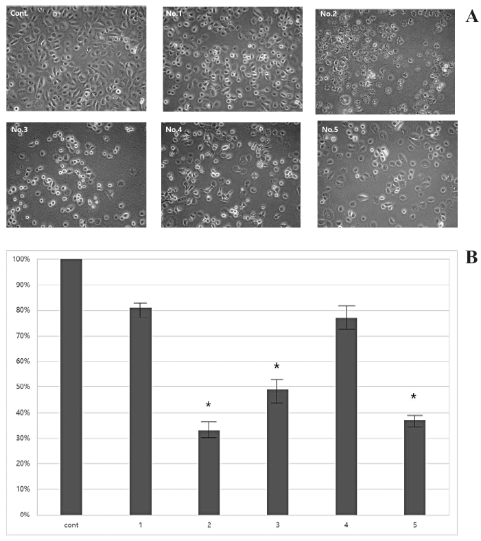 Figure 1.
— Effect of OTC vaginal lubricants on the cultured human vaginal cell lines. (A) VK2/E6E7 cells are cultured with 5% dilutions of five OTC vaginal lubricants over 48 hours (×200). (B) Effect of OTC vaginal lubricants on viability of the cultured human vaginal cell lines. The data of bars are presented as mean ± SD. * represents p < 0.05, compared with control.
To measure cell viability quantitatively, the authors performed the MTT assay and immunoblotting analysis. After treatment with OTC vaginal lubricants over 48 hours, they compared cell viability with the viability in the untreated controls (revised 100% viability). As shown in Figure 1B, the cells treated with products no. 2, 3, and 5 showed significantly decreased cell viability (33.1 ± 7.6%, 49.3 ± 11.5 %, and 37.8 ± 5%, p < 0.005). The cells treated with products no. 1 and 4 showed decreased viability, but the difference was not statistically significant (80.9 ± 5.3%, 77.7% ± 9.8%, p > 0.005) compared to the controls. These data were confirmed through Western blotting; as seen in Figure 2, the apoptosis or cell death-related molecules increased in the cells that were treated with the OTC vaginal lubricants having hyper-osmolality. The cells treated with product no. 3 did not express cleaved PARP. Occludin is an important tight junction-associated protein [11]. Expression of occludin protein was increased in all the cells treated with the lubricants, while the untreated controls showed no expression of the occludin molecule.
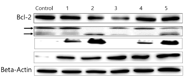 Figure 2.
— Western blotting analysis for expression of Bcl-2, caspase 7, cleaved PARP, and occludin in VK2/E6E7 cells after 48-hour treatment of five OTC vaginal lubricants. β-actin serves as an internal control. A single band for each antibody is developed.
Five OTC vaginal lubricants were placed on top of the mucosal side of human vaginal tissues and cultured for 48 hours. As shown in Figure 3A, the untreated control showed intact and thick epithelial areas on the upper side of the stromal region, with high expression of bcl-2. The tissues treated with product no. 1 showed that the outer mucosal layer had partially vanished, while the bcl-2 level was high, the same as in the untreated controls. The remaining four tissue samples showed vanishing and damage of the epithelial and mucosal layers. The expression levels of bcl-2 were determined using bcl-2 positive cells in three different microscopic fields. The untreated controls and the tissues treated with product no. 1 showed higher expression of bcl-2 (100% and 93.7 ± 11.2)(Figure 3B). The tissues treated with products no. 2, 3, 4, and 5 showed lower expression of bcl-2 than the untreated controls (12.4 ± 4.2%, 10.7 ± 2.4%, 61.4 ± 11.3%, and 59.2 ± 5.4% respectively, p < 0.05). Positive expression of cleaved caspase 3 was detected in all tissues, including the untreated controls (Figure 4A and B; 13.7 ± 8.2%). Except for the tissues treated with product no. 1 (18.9 ± 3.5%), the tissues treated with the remaining four OTC lubricants showed significantly increased cleaved caspase 3 expression compared to the untreated controls (no. 2, 57.1 ± 13.2%; no. 3, 31.2 ± 2.1%; no. 4, 47.3 ± 7.1%; no. 5, 78.1 ± 14.2%, p < 0.05).
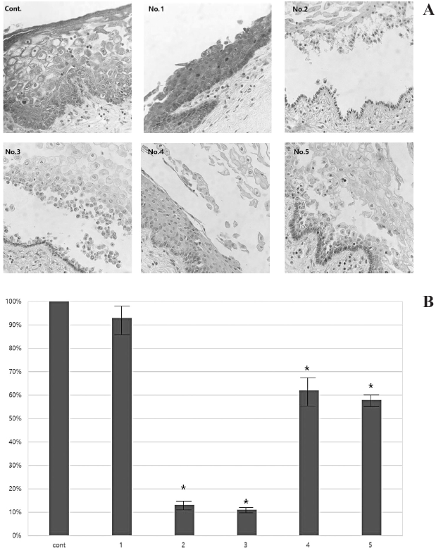 Figure 3.
— Histological sections of vagina after 48 hours of treatment with different OTC vaginal lubricants. (A) Ultrastructural histological sections for expression of bcl-2 on human vaginal tissues (×200). The brown colored cells represent positive expression of bcl-2. (B) Expression of bcl-2 is analyzed by number of cells with brown-stained/total number of cells under the microscopic images (×200). Weak brown stains are excluded from the count. The data of bars are presented as mean ± SD. * represents p < 0.05, compared with control.
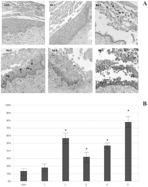 Figure 4.
— Histological sections of vagina after 48 hours of treatment with different OTC vaginal lubricants. (A) Ultrastructural histological sections for expression of cleaved caspase 3 on human vaginal tissues (×200). The brown colored cells represent positive expression of cleaved caspase 3. (B) Expression of cleaved caspase 3 is analyzed by number of cells with brown-stained/total number of cells under the microscopic images (×200). Weak brown stains are excluded from the count. The data of bars are presented as mean ± SD. * represents p < 0.05, compared with control.
Vaginal lubricants intended for use during sexual activity are readily available for purchase in drug stores, through large retail chains, and over the internet. Women use vaginal lubricants to reduce vaginal dryness. Available pre-clinical and clinical data support that hyperosmolar vaginal products may be related to safety issues, and may also have detrimental effects on sperm motility, viability, and chromatin quality [1].
The present findings showed that four of the five OTC vaginal lubricants tested were hyperosmolar and resulted in cellular damage to the cultured vaginal cell line and human vaginal explant. One iso-osmolar OTC vaginal lubricant had less effect on both cultured vaginal cell lines and human vaginal explants.
Addition of OTC vaginal lubricants immediately induced morphological changes in the cultured vaginal cell line. The cell cytoplasm shrank and some cells showed rough surfaces. After 48 hours of treatment, cell death was seen in approximately 50% of the cells in the hyper-osmolar OTC lubricant-treated group. The cells treated with the iso-osmolar OTC vaginal lubricant showed fewer morphological changes and almost 80% of the cells survived with the same duration of treatment. Cellular viability was also measured 48 hours after OTC vaginal lubricant treatment. The cells treated with hyper-osmolar OTC vaginal lubricant showed decreased viability. To clarify the effect of OTC vaginal lubricants on cell death, the authors measured the expression of apoptosis-related proteins by the cells. The anti-apoptotic protein bcl-2 was detected in all the experimental conditions. The cells treated with products no. 2, 3, and 4 showed decreased expression of bcl-2, while the bcl-2 expression in the cells treated with products no. 1 and 5 was unchanged. The authors also measured the expression of other apoptosis-related proteins such as caspase-7 and cleaved PARP. In the case of caspase-7, the cleaved form was detected in the controls and in the cells treated with products no. 1, 2, 3, and 5. The cleaved form of PARP was detected in the cells treated with products no. 1, 2, 4, and 5. No. 1 and 4, which had relatively lower osmolality than the others, showed decreased level of expression of the cleaved form of PARP. The cleaved form of PARP was not detected in the cells treated with product no. 3 even when cleaved caspase 7 was detected in the same cells, making the data seemingly ambiguous. PARP is known to have the function of DNA repair and mediates apoptosis or necrosis of cells. Twelve DNA damage can induce an apoptosis initiation event that is mediated by caspase-3 activation and PARP cleavage. However, significant damage to the DNA repairing capacity resulted in cell necrosis without PARP cleavage.13 Therefore, the authors suggested that the OTC vaginal lubricants, regardless of their osmolality, could induce cellular change and hyper-osmolarones could even bring about cellular damage such as apoptosis or necrosis.
Occludin is a protein that is encoded by the OCLN gene in humans [14]. It is a 65-kDa (522-amino acid polypeptide) integral plasma-membrane protein located at the tight junctions [15]. In the present experimental condition, all OTC vaginal lubricant treated cells showed expression of occludin. It has already been mentioned in several studies that expression of occludin increases to protect cellular integrity and epithelial restitution under stressful conditions [16, 17].
As shown in Figures 3 and 4, treatment with OTC vaginal lubricants affected mucosal epithelial layers of the human vaginal tissue explants regardless of their osmolality. Product no. 1 that had iso-osmolality showed less epithelial dysfunction. Bcl-2 was highly expressed in the untreated control tissues and the tissue treated with product no. 1, but the mucosal epithelial layer was partially separated from its epithelium in the tissue treated with product no. 1 (Figure 3A). Minimum expression of cleaved caspase-3 was seen in the untreated controls and the tissues treated with product no. 1. These data showed that the osmolality of OTC vaginal lubricant is a major factor affecting epithelial cell survival and tissue integrity.
Dezzutti et al. reported that hyper-osmolality of OTC vaginal lubricants was associated with cellular toxicity and could potentially lead to increased susceptibility to HIV-1 and other sexually transmitted diseases [8]. Indeed, persons who consistently use hyper-osmolal lubricants showed a higher incidence of sexually transmitted diseases. In the present study, the extent of cellular or tissue damage did not correspond to the osmolality of the lubricants. There are different ingredients in every OTC vaginal lubricant, the major ingredients being water, glycerin, and other moisturizers. Some lubricants have aloe barbadensis leaf juice, alcohols, and natural flavors as additional ingredients. Especially, aloe vera is known to have a healing effect on skin wounds [18, 19]. However, these ingredients may bring about discordance between the osmolality and cellular toxicity. One of the OTC vaginal lubricants showed iso-osmolality. However, despite the physiological osmolality, it showed minimum cytotoxicity on the mucosal epithelium of vaginal tissue.
In summary, OTC vaginal lubricants are very frequently used by individuals who find intercourse inconvenient due to dryness and vaginal atrophy. In the present study, the authors were concerned with the high osmolality of OTC vaginal lubricants and evaluated their effects on the vaginal mucosal layer. The hyper-osmolality of OTC vaginal lubricants is a major cause of dysfunction of the vaginal mucosal layer. Therefore, the authors suggest that frequent use hyper-osmolar OTC vaginal lubricants may actually have an aggravating effect on vaginal dryness and infection. One of the limitations of this study is that it did not include all the OTC vaginal lubricants available in Korea; additionally, the effect of each ingredient contained in these products was not considered. Thus, additional research is needed to evaluate formulae to enable production of safer OTC vaginal lubricants.