

Clinical and Experimental Obstetrics & Gynecology (CEOG) is published by IMR Press from Volume 47 Issue 1 (2020). Previous articles were published by another publisher on a subscription basis, and they are hosted by IMR Press on imrpress.com as a courtesy and upon agreement with S.O.G.

1 Department of Radiology,Adnan Menderes University, Aydın, Turkey
2 Department of Gynecology and Obstetrics, Adnan Menderes University, Aydın, Turkey
Abstract
Introduction: This study was aimed to preliminarily examine the effect of laser-assisted three area zona thinning (LAZT) during the cleavage stage of embryo development and the hatching process in mice based on female age. Materials and Methods: C57BL female mice of two different age groups (6-11 and 28-31 weeks) were superovulated and then mated. The 8-cell stage embryos were treated with LAZT in three areas of zona pellucida (ZP) at 120º intervals. The control group was embryos without LAZT. Results: In old mice, LAZT significantly increased blastocyst formation and hatching compared to the control group (82% vs. 38% in number of expanding blastocyst and 66% vs. 20% in number of hatching blastocyst). Conclusion: These results show that multi-area LAZT results in a significant improvement of blastocyst formation and hatching in mice compared to controls.
Keywords
- Hatching
- IVF
- Laser
Over the last 40 years, much research has been aimed towards the improvement of implantation potential regarding preimplantation stage embryos in in vitro fertilization-embryo transfer (IVF-ET). One breakthrough in this field came with the development of a technique known as assisted hatching (AH). Defects in the hatching stage have been considered a possible cause of implantation failure in assisted reproductive technology (ART). AH helps this hatching process and induces embryo implantation as an artificial rupture technique of the ZP.
Since the first report was published by Cohen et al. in 1988 [1], a variety of AH techniques have been introduced. However, while some studies have proposed AH as a method for improving the implantation capacity and pregnancy outcome in human IVF-ET [2], others have found no effects [3] or insufficient evidence to determine any impact of AH on implantation or pregnancy outcome, including live birth rates [4]. Thus, the routine use of AH is still debatable. Also, the time and the method of AH treatment is controversial. Initially, AH is performed by thinning or opening ZP by mechanical [5] or chemical methods [6]. However, these methods are not only technically inconvenient, but also pose the risk of embryo damage. Recently, AH using lasers was the most popular method because laser AH (LAH) is simple and rapid [7].
In this context, another important focus in LAH treatment is the area of ZP that is thinned or opened. Generally, it is currently understood that larger holes are more effective for AH [5]. Nevertheless, the effect of LAH still remains controversial due to heterogeneous study design in different AH indications [8] and methods [2].
This study aimed to examine the effect of laser-assisted three areas zona thinning (LAZT) during the cleavage stage on embryo development and the hatching process in mice based on age.
All experiments with mice were conducted in accordance with the Guide for the Care and Use of Laboratory Animals of National Institute Health, approved by the PNUH Institutional Animal Care and Use Committee.
In all experiments, C57BL inbred mice were purchased from Korea Experimental Animal Center (Daegu, Korea). The mice were maintained on a light-dark cycle, with light on at 5:00 AM and off at 7:00 PM, and with food and water available ad libitum.
Female mice were divided into two groups of young (6-11 weeks old) and old (28-31 weeks old). The mice were superovulated by intraperitoneal injection with 5 IU pregnant mare’s serum gonadotropin (PMSG), followed by injection of 5 IU hCG 48 hours later, and then they were immediately paired with an individual male. Eighteen hours after hCG injection, female mice were checked the presence of vaginal plug. The mice with a confirmed vaginal plug were considered to have mated and they were sacrificed by cervical dislocation. Thereafter, cumulus-enclosed one-cell embryos (zygotes) were retrieved from the oviductal ampullae and denuded by incubation for one minute with 0.1% hyaluronidase in Dulbecco’s phosphate buffer saline. Zygotes were pooled and washed three times in P1 medium with 10% serum substitute supplement.
The only healthy zygotes were cultured in 30 μL of P1 medium with 10% SSS for the first two days, and then blastocyst medium with 10% SSS for the later two days under paraffin-oil at 37°C in a 5% CO2 incubator, and the media were changed daily. Blastocyst formation and hatching rates were determined 96 hours and 120 hours after zygote collection, respectively.
LAZT was treated at good quality 8-cell embryos of grade A and B using OCTAX NaviLase in three areas of ZP at a 120º interval. The original culture dish containing the 8-cell embryos was placed on an inverted microscope equipped with a laser system. The point of treatment on the ZP was carefully focused on and treated with the laser. At this time, the embryos were not stabilized with the holding pipette. One area on the ZP for LAZT was randomly chosen. The ZP was thinned to more than 70% of its initial thickness using a 1.48 μm wavelength diode laser by making 7-8 consequent holes of 15-20 μm on one area of the ZP. All zona thinning was performed at the empty region without contact to the blastomere to minimize the damage. The power of the laser was 100% and the pulse duration was 500 μs (Figure 1).
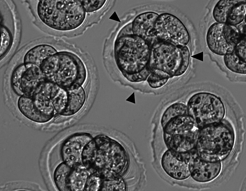 Figure 1.
— Laser-assisted three area zona thinning (LAZT) at the 8-cell embryos stage in the mouse. LAZT was performed on zona pellucida by making 7-8 consequent holes of 15-20 μm per one area at three areas (arrow head) (×200 magnification).
Statistical analysis used SPSS program (version 19.0) and all data were presented as a mean ± SD. Comparisons of the blastocyst formation, hatching rates, and pregnancy outcome were analyzed by λ2-test and t-test. P < 0.05 was considered to be statistically significant.
This study was investigated to determine whether three area LAZT at the 8-cell embryo stage affects development and hatching rate in mice at a certain age.
In young mice (6-11 weeks old), LAZT had no effect on blastocyst formation rate (92.0%), but promoted a significant increase in hatching rate (76.0%) compared to the control group (84.0% and 36.0%, respectively) (p < 0.05)
(Table 1).
| Control | LAZT | p-value | |
|---|---|---|---|
| No. of 8-cell embryos at 60 h (%) | 50 | 50 | |
| No. of expanding blastocyst at 96 h (%) | 42 (84.0) | 46 (92.0) | NS |
| No. of hatching blastocyst at 120h (%) | 18 (36.0) | 38 (76.0) | < 0.05 |
Time after zygote collection. P-value was calculated using λ2-test. LAZT: laser-assisted zona thinning.
In old mice, however, both blastocyst formation (82.0%) and hatching rate (66.0%) were significantly increased in the LAZT group compared to those (38.0% and 20.0%, respectively) of control group (p < 0.05 and p < 0.01, respectively) (Table 2).
| Control | LAZT | p-value | |
|---|---|---|---|
| No. of 8-cell embryos at 60 h (%) | 50 | 50 | |
| No. of expanding blastocyst at 96 h (%) | 19 (38.0) | 41 (82.0) | < 0.05 |
| No. of hatching blastocyst at 120h (%) | 10 (20.0) | 33 (66.0) | < 0.01 |
Time after zygote collection. P-value was calculated using λ2-test. LAZT: laser-assisted zona thinning.
This study shows that LAZT on three areas of the ZP at the 8-cell embryo stage significantly improves blastocyst formation and hatching rate in the mouse. The effect was more significant in aged mice compared to young ones.
Zona hardening is a process that prevents the natural hatching of blastocysts, and has been known to occur during in vitro culture [9], after cryopreservation [10], or in advanced maternal age [5].
The age of potentially pregnant females is one of determinants for the use of AH [11], so this study preliminarily investigated the effect of the proposed LAZT method on aged female mice (28-31 weeks old) and confirmed an improvement in blastocyst formation and hatching rate. In some studies, harmful [12] and minimal changes in the clinical pregnancy rate were observed in the AH group compared to the control group for patients less than 37 years old [13]. These results strongly suggest that advanced female age could be a key determinant for the use of AH. Kanyo et al. reported positive effects in regards to pregnancy rates in frozen embryo transfer cycles for old women, and considers LAH a method suitable to overcome the negative effect of zona hardening [14].
In the present study, LAZT increased the hatching rate in the mouse. This effect of LAZT may have an effect on the pregnancy rate. Miyata et al. showed that the initiation of hatching occurred earlier in the AH group compared to the blastocysts without AH [15]. It has been hypothesized that electrical stimulation may induce egg activation and stimulate embryo development rate. Thus, further study is needed to elucidate clearly a correlation between increased hatching rate and pregnancy outcome after LAZT treatment.
Another interesting finding in the present study is the importance of the site and size of AH. A natural hatching site has been known to be in proximity to the inner cell mass (ICM) of blastocysts in humans and at the side of opposite to the ICM in mice. AH performed near ICM significantly improved the hatching rate compared to AH opposite to the ICM [15]. Considering that embryo transfer into the uterus in case of 4-cell or 8-cell embryo, the exact site of hatching is unknown. It is very difficult to determine the correct site of AH that hatching will occur. In this respect, zona thinning at three areas in the present study seems to be one of the solutions to increase the hatching possibility of AH-treated 8-cell embryos. Indeed, the hatching rate in LAZT at three areas was remarkably higher than that in the control group in the present study.
In determining hole size for AH, a general consensus may be that larger holes are preferable [5, 16]. In general, holes greater than one-quarter [8], one-third [17], two-thirds or one-half of the zona circumference [16, 18] resulted in successful hatching or a higher pregnancy rate in humans IVFET. However, zona opening or thinning of a large size during or after AH may cause damage to the embryos, including trapping of more of the larger ICM [18, 19]. This result suggests that the choice of an appropriate size for AH should be considered to minimize the risk of direct damage. In this regard, the present study performed zona thinning to 70-90% of initial ZP thickness by making two consequent holes of 15-20 μm at the empty region without contacting the blastomere.
One of main concerns in the use of LAH, however, is whether thermal damage can result in an adverse effect on embryos [6, 20, 21], and in turn perinatal and neonatal outcomes [22, 23]. The fact that embryos at the 8-cell stage respond to thermal shock by the induction of heat shock protein is thought to reduce the risk of adverse effects by multiple lasers during AH [24]. Indeed, this concern for the use of lasers has been lowered by several studies. Kanyó and Konc showed that there was no evidence of increased chromosomal abnormalities or congenital malformations for 134 children born after laser AH [23]. Zhou et al. reported that no adverse effects were identified on the perinatal outcomes after LAH treatment using a retrospective cohort study for a total of 699 women with 392 infants delivered [25]. Jwa et al. showed in a retrospective cohort analysis of 35, 488 LAH cycles that AH alone does not increase the risk of major congenital anomalies [26]. These results suggest that LAH may be a safe treatment, however these results do not absolutely preclude the possibility for other underlying abnormalities and further long-term studies are required.
In conclusion, this study shows that LAZT at three areas on 8-cell embryos results in a significant improvement of blastocyst formation and hatching compared to controls in the mouse. This effect of LAZT was more significant in aged mice than young mice. This study suggests that zona thinning at multiple areas rather than a single point on ZP at the cleavage stage may improve the potential of successful embryo hatching.