

 , Hong Mo Ku 2, Ji Sun Kwon 3
1 Department of Neurology, International St. Mary’s Hospital, Catholic Kwandong University College of Medicine, 22711 Incheon, Republic of Korea
2 Institute for Cognitive Intervention, International St. Mary’s Hospital, 22711 Incheon, Republic of Korea
3 Department of Neurology, Misodle Geriatric Hospital, 08250 Seoul, Republic of Korea
Abstract
Increased adenosine deaminase (ADA) activity in cerebrospinal fluid (CSF) has been used as a biomarker of tuberculous meningitis and other etiologies, including viral, fungal, and neoplastic meningitis. Gout can influence inflammatory pathways, altering biochemical markers such as ADA. This report describes a unique case of varicella-zoster virus (VZV) meningitis with gout associated with elevated ADA levels.
A 37-year-old man presented with a one-week history of headache, fever, and chills. His medical history included gout, diagnosed three years earlier. CSF analysis from the first lumbar puncture revealed lymphocytic pleocytosis, elevated protein, mildly decreased glucose, and ADA activity of 7.4 IU/L. The patient developed worsening headache and vomiting, prompting a second lumbar puncture. Repeat CSF analysis showed persistent lymphocytosis, mildly decreased glucose, and increased ADA activity (9.1 IU/L). The patient also experienced an acute gout flare during hospitalization, which required antigout therapy. The subsequent CSF polymerase chain reaction (PCR) confirmed VZV meningitis.
The patient responded well to symptomatic treatment. Serum ADA levels were also elevated (14.7 IU/L), likely reflecting systemic inflammation due to gout.
Systemic inflammation, which includes the meninges and joints, had been postulated to increase protein diffusion through the vascular walls and had been associated with increased ADA activity in CSF and serum. To the best of our knowledge, this is one of the few reports on VZV meningitis and gout associated with elevated ADA levels.
Keywords
- adenosine deaminase
- gout
- varicella-zoster virus
- meningitis
- case report
Adenosine deaminase (ADA) is an enzyme that catalyzes deamination of adenosine
during the formation of inosine and plays an important role in the proliferation,
differentiation, and activation of T lymphocytes [1, 2]. Increased cerebrospinal fluid (CSF) ADA level of
The varicella-zoster virus (VZV) is the causative agent of varicella during primary infection and herpes zoster upon reactivation. Following primary infection, VZV establishes lifelong latency in the dorsal root ganglia, with reactivation typically triggered by immunosuppression, advanced age, or stress. While reactivation most commonly presents as shingles, it can lead to neurological complications, including VZV meningitis. VZV meningitis is characterized by inflammation of the meninges and may present with headache, fever, neck stiffness, and, occasionally, neurological deficits. The pathogenesis of VZV meningitis involves the spread of reactivated virus from neural ganglia to the central nervous system (CNS), triggering an inflammatory immune response in the meninges. Diagnosis relies on a combination of clinical assessment, CSF analysis, and advanced molecular techniques. Polymerase chain reaction (PCR) for VZV DNA in CSF is the diagnostic gold standard [3].
Gout is linked to systemic inflammation and VZV meningitis with gout can influence inflammatory pathways, potentially altering biochemical markers such as ADA. This report describes a unique case of VZV meningitis associated with gout and elevated ADA levels in serum and CSF. Clinicians are encouraged to integrate ADA findings with a comprehensive diagnostic workup and to remain cautious to interpret ADA levels in the CSF.
A 37-year-old man presented with headache, fever, and chills for one week. His
medical history was unremarkable except for gout, diagnosed three years ago. His
physical and neurological examination showed unremarkable findings including the
absence of skin lesions, except for mild neck stiffness. On arrival, his vital
signs were stable except for a fever of 37.8 °C. Laboratory examination only
showed an elevated uric acid level at 9.1 mg/dL (reference value 3.5–8 mg/dL)
(Table 1). Brain imaging was also unremarkable (Fig. 1). Lumbar puncture yielded
an opening pressure of 200 mmHg (reference value 50–180 mmHg), a
clear appearance, and a composition of 500 white blood cells (WBCs)/mm3
(88% lymphocytes, reference value 0–5 cells/mm3), 50 red blood cells
(RBCs)/mm3 (reference value 0 cells/mm3), glucose of 48 mg/dL
(reference value 40–70 mg/dL, serum glucose 105 mg/dL), protein of 117 mg/dL
(reference value 15–45 mg/dL) and ADA of 7.4 IU/L (reference value
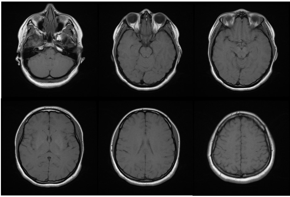 Fig. 1.
Brain magnetic resonance imaging of the patient showed no specific abnormalities.
| ADA (IU/L) | VZV IgG | VZV IgM | VZV PCR | Tb PCR | Uric acid (mg/dL) | |
| Serum at admission | None | None | None | None | None | 9.1 |
| CSF at admission | 7.4 | + | - | + | - | None |
| CSF 3 days after admission | 9.1 | + | - | + | - | None |
| Serum 13 days after admission | 14.7 | + | - | - | - | 11.3 |
ADA, adenosine deaminase; CSF, cerebrospinal fluid; PCR, polymerase chain reaction; Tb, tuberculosis; VZV, varicella-zoster virus; IgM, immunoglobulin M; IgG, immunoglobulin G. Reference value; ADA at serum: 0–4 IU/L, ADA at CSF: 0–2.5 IU/L, Uric acid at serum: 3.5–8 mg/dL.
The CARE Checklist has been attached as Supplementary Material associated with this article.
Initially, he was diagnosed as viral meningitis and treated with antipyretics and analgesics such as acetaminophen. However, on the third day of admission, he complained severe diffuse headache, nausea, and vomiting. He had a blood pressure of 130/80 mmHg, heart rate of 90 beats/minute, respiratory rate of 21 breaths/minute and body temperature of 38.3 °C. A second lumbar puncture yielded an opening pressure of 210 mmHg, a clear appearance, and a composition of 370 WBCs/mm3 (89% lymphocytes), 120 RBCs/mm3, glucose of 45 mg/dL (serum glucose 111 mg/dL), protein of 64.6 mg/dL, and ADA of 9.1 IU/L (Table 1). Compared to the first lumbar puncture, the WBC count and protein were reduced, and the RBCs were increased, however the CSF finding patterns (slightly low glucose level and lymphocyte predominant leukocytosis) were similar to the first lumbar puncture except for the higher activity of ADA.
He was treated with antipyretics, analgesics, and antituberculosis medication because of the possibility of tuberculous meningitis (i.e., isoniazid 300 mg, rifampicin 600 mg, and pyrazinamide 1500 mg) with pyridoxine 50 mg. On the seventh day of admission, his headache gradually improved, and we continued antituberculosis medication. His body temperature showed 37.3 °C. On the 13th day of admission, he complained of tenderness and swelling on the first digit of the left foot. His body temperature showed 37.0 °C. The uric acid level at this time increased to 11.3 mg/dL (Table 1). After adding antigout medication, his toe pain resolved. In both the first and second lumbar punctures, the CSF PCR for tuberculosis was negative, but VZV PCR was positive. In addition, the serum VZV immunoglobulin G was positive. The serum ADA level was 14.7 IU/L (Table 1). He was diagnosed as VZV meningitis with gout. His clinical symptoms gradually improved and we discontinued the antituberculosis medication after checking VZV PCR results. Thereafter, we gave conservative management with antipyretics and his headache gradually resolved.
The worsening headache, persistent lymphocytosis, mildly decreased glucose level, and increased ADA activity made us decide to add antituberculosis medication. In gout, high uric acid levels can damage cells and tissues, leading to systemic and local inflammation; this inflammation can increase the activity of ADA, leading to increased serum and CSF levels of ADA [4]. The exact mechanism of increased CSF ADA activity in patients with gout and VZV meningitis is not fully understood. The underlying mechanism between gout-induced systemic inflammation and increased ADA activity in the CSF involves the interplay of gout-induced systemic inflammation, cytokine production and blood-brain barrier alterations [5]. The systemic inflammation, which includes the meninges and joints, has been postulated to increase protein diffusion through the vascular walls and had been associated with increased ADA activity in CSF and serum.
Previous reports have described cases of increased ADA activity in aseptic meningitis caused by enterovirus and VZV [6, 7], but the mechanisms were not explained. To the best of our knowledge, this is one of the few reports on VZV meningitis associated with increased ADA levels in a patient with gout.
In some cases, the difficulty in differentiating between tuberculous and VZV meningitis is a concern, because both conditions can present with similar CSF characteristics, such as lymphocytic predominance, increased ADA activity, and glucose consumption. Moreover, skin lesions can be absent in up to half of patients with VZV meningitis, as observed in our case.
In areas where tuberculosis is endemic, determining ADA levels in the CSF is often used for initial screening for tuberculous meningitis [8]. Research indicates that elevated ADA levels in CSF may also be associated with other infectious etiologies, such as fungal meningitis (e.g., cryptococcosis) and viral infections, particularly in cases of intense immune activation. Additionally, non-infectious conditions, including certain malignancies (e.g., lymphomas or leukemias with CNS involvement) and autoimmune diseases (e.g., systemic lupus erythematosus with neuropsychiatric manifestations), have been reported to cause increased ADA activity.
The usefulness of ADA level in the CSF remains a topic of debate, because several reported cases of nontuberculous meningitis with increased ADA, including the present case. These findings highlight the need to interpret ADA levels in the context of the clinical presentation and other diagnostic findings. Clinicians are encouraged to integrate ADA findings with a comprehensive diagnostic workup and to remain cautious to interpret ADA levels. However, we believed that determination of ADA level can be helpful in guiding timely antituberculosis treatment.
This case highlights several unique features and challenges in the diagnosis and management of VZV meningitis in a patient with coexisting gout. The coexistence of these two conditions presented diagnostic dilemmas, particularly in the interpretation of ADA levels in the CSF. While elevated ADA activity is a well-established marker for tuberculous meningitis, it has also been associated with other infectious and inflammatory conditions, as seen in this patient. Additionally, the absence of vesicular skin lesions—a hallmark of VZV infection—in up to half of VZV meningitis cases further obscured the clinical picture. The mildly increased ADA levels (7.4–9.1 IU/L) in the absence of CSF tuberculosis PCR results initially complicated the diagnosis, prompting empirical antituberculosis treatment.
Future research should focus on elucidating the mechanisms behind ADA elevation in non-tuberculous meningitis, particularly in cases with concurrent systemic inflammatory conditions like gout. Additionally, investigating the interplay between hyperuricemia, inflammation, and ADA activity may shed light on the broader implications of gout in infectious diseases.
Systemic inflammation, which includes the meninges and joints, had been postulated to increase protein diffusion through the vascular walls and had been associated with increased ADA activity in CSF and serum. Clinicians should be cautious to interpret ADA levels in the CSF and consider other etiologies elevating ADA levels. To the best of our knowledge, this is one of the few reports on VZV meningitis associated with increased ADA levels in a patient with gout.
The data used to support the findings of this study are available from the corresponding author upon request.
BDK designed the study. HMK and JSK acquired the data. BDK, HMK and JSK analyzed the data. BDK drafted the article. HMK and JSK critically revised the article. BDK reviewed the submitted version of manuscript. All authors contributed to important editorial changes in the manuscript. All authors read and approved the final manuscript. All authors have participated sufficiently in the work and agreed to be accountable for all aspects of the work.
All procedures performed in this study involving human participants were in accordance with the ethical standards of the institutional or national research committee and with the Declaration of Helsinki. The study was reviewed and approved by the human subjects’ institutional review boards of International St. Mary’s Hospital, Catholic Kwandong University College of Medicine (Ethical approval number: IS22MSMV0080). Written informed consent was obtained from the patient.
Not applicable.
This research received no external funding.
The authors declare no conflict of interest.
Supplementary material associated with this article can be found, in the online version, at https://doi.org/10.31083/BJHM50384.
References
Publisher’s Note: IMR Press stays neutral with regard to jurisdictional claims in published maps and institutional affiliations.