IMR Press / FBL / Volume 30 / Issue 10 / DOI: 10.31083/FBL39948
Open Access Review
Temporal Orchestration of Immunity: How Circadian Clocks Coordinate Immune Responses
Show Less
Affiliation
1 School of Basic Medical Sciences, Suzhou Medical College of Soochow University, 215123 Suzhou, Jiangsu, China
2 School of Life Sciences, Suzhou Medical College of Soochow University, 215123 Suzhou, Jiangsu, China
3 MOE Key Laboratory of Geriatric Diseases and Immunology, School of Basic Medical Sciences, Suzhou Medical College of Soochow University, 215123 Suzhou, Jiangsu, China
*Correspondence: liuchao@suda.edu.cn (Chao Liu); ybzhongsd@suda.edu.cn (Yingbin Zhong)
Front. Biosci. (Landmark Ed) 2025, 30(10), 39948; https://doi.org/10.31083/FBL39948
Submitted: 24 April 2025 | Revised: 10 July 2025 | Accepted: 18 July 2025 | Published: 31 October 2025
Copyright: © 2025 The Author(s). Published by IMR Press.
This is an open access article under the CC BY 4.0 license.
Abstract

As an evolutionarily conserved timekeeping system, the circadian clock orchestrates physiological adaptations to diurnal environmental cues through transcriptional-translational feedback loops (TTFLs). Accumulating evidence reveals that circadian regulation governs immunological processes, with the immune system—a critical host defense mechanism—exhibiting robust circadian rhythmicity in functional organization. This review synthesizes recent advances in circadian modulation of pathogen-host interactions, immune cell trafficking, effector functions, circadian light hygiene—gut immune crosstalk, and tumor immunobiology. We examine the bidirectional crosstalk between circadian oscillators and immune pathways while addressing the clinical implications for immune-related pathologies. Significantly, we advocate chrono-immunotherapy as a transformative paradigm that leverages circadian principles to optimize therapeutic timing, enhancing efficacy while minimizing adverse effects. Future research directions aimed at elucidating mechanistic foundations and accelerating clinical translation are outlined. A comprehensive understanding of circadian-immune system dynamics not only provides fundamental insights into biological regulation but also establishes a chronobiological framework for precision medicine in immune-mediated disorders.

Keywords
circadian clock
immune regulation
host-pathogen interactions
chronotherapy
immunotherapy
immune system diseases
1. Introduction

The circadian rhythm constitutes an endogenous ~24-hour biological oscillator that enables organisms to entrain physiological processes to periodic environmental cues (e.g., photic and thermal fluctuations), thereby optimizing adaptive behaviors and maintaining metabolic homeostasis—a fundamental biological mechanism critical for health maintenance and disease prevention [1]. This phylogenetically conserved timekeeping mechanism is ubiquitously observed across taxonomic kingdoms, from prokaryotic cyanobacteria to complex mammalian systems [2].

At the molecular level, mammalian circadian regulation operates through a hierarchically organized network of cellular oscillators. The suprachiasmatic nucleus (SCN) of the hypothalamus functions as the central pacemaker, coordinating peripheral clocks through integrated neural and humoral signalling pathways [3, 4]. The core molecular mechanism involves transcription-translation feedback loops (TTFLs), wherein clock gene products autoregulate their expression through sequential post-translational modifications, including phosphorylation and regulated nuclear translocation [4]. Notably, while circadian systems arose through convergent evolution in diverse taxa, they exhibit conserved architectural principles characterized by autoregulatory genetic networks mediated by Per-Arnt-Sim (PAS) domain-containing transcriptional regulators [5]. The groundbreaking work of Nobel laureates Hall, Rosbash, and Young, utilizing Drosophila melanogaster models, identifying period gene-encoded components as essential for establish the autonomous circadian oscillator [6].

Accumulating evidence demonstrates that circadian rhythms represent a fundamental regulatory layer governing virtually all biological processes [7]. Circadian disruption has been etiologically linked to numerous metabolic and endocrine disorders, including obesity, metabolic syndrome, obstructive sleep apnea (OSA), and type 2 diabetes mellitus [8, 9, 10, 11, 12]. This pervasive regulatory influence highlights the circadian system’s integral role in maintaining cellular homeostasis, coordinating physiological functions, and integrating metabolic-endocrine signaling, thereby revealing novel therapeutic targets for associated pathologies [7].

Of particular relevance, circadian oscillations exert precise temporal control over immune system function, modulating host defense mechanisms against inflammatory processes and pathogenic challenges from bacterial, parasitic, and viral agents [13, 14, 15]. Notably, viral replication cycles exhibit distinct temporal regulation patterns, with certain pathogens employing chronobiological strategies to establish either acute or persistent infections [14]. These findings underscore the critical importance of elucidating circadian-immune system interactions in contemporary biomedical research.

This review synthesizes current knowledge regarding circadian regulation of immunological processes. We systematically evaluate: (1) circadian-mediated modulation of host-pathogen interactions; (2) temporal control of leukocyte functional dynamics; (3) circadian light hygiene and gut-immnue crosstalk; and (4) chronobiological regulation of anti-tumor immunity. Furthermore, we examine the clinical implications of circadian biology in immunotherapeutic strategies and chronotherapeutic interventions through a translational medicine perspective. Finally, we identify key research directions to advance understanding of circadian-immune crosstalk, with particular emphasis on mechanistic studies of temporal regulation and its therapeutic applications in precision chrono-immunology.

2. The Circadian Clock System and Its Molecular Mechanisms

The mammalian circadian timing system comprises three functionally integrated components: (1) input pathway that transduce environmental signals (e.g., light-dark cycles) to the central pacemaker; (2) a central pacemaker generating self-sustained oscillations; and (3) output pathway that regulate rhythmic physiological, metabolic, and behavioral processes [16]. While the suprachiasmatic nucleus (SCN) of the hypothalamus functions as the dominant circadian pacemaker in mammals, autonomous circadian oscillators exist in peripheral tissues [4]. The SCN synchronizes these peripheral clocks through coordinated neuroendocrine and autonomic nervous signaling [6]. Environmental zeitgebers (“time-givers”), particularly photic stimuli and feeding cycles, entrain the central oscillator, which in turn phase-locks peripheral oscillators via coupled transcriptional-translational feedback loops (TTFLs) [17] (Fig. 1).
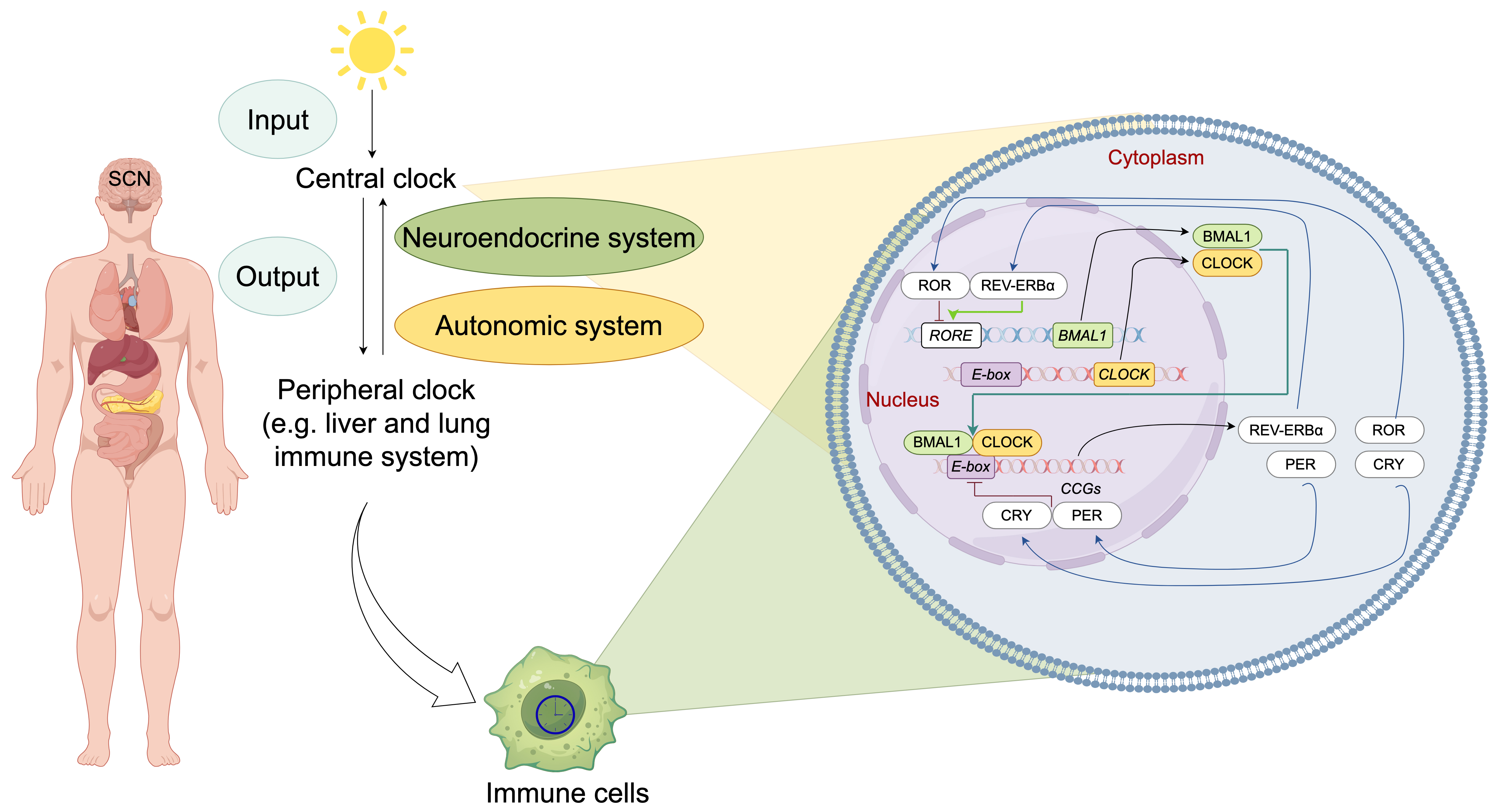
Fig. 1.

Molecular architecture and systemic organization of the mammalian circadian clock system. The mammalian circadian timing system operates through a hierarchically organized network, with the central oscillator in the suprachiasmatic nucleus (SCN) serving as the master pacemaker. The SCN integrates photic input and synchronizes peripheral oscillators through dual efferent pathways: (1) autonomic neural signaling and (2) neuroendocrine regulation. At the molecular level, cell-autonomous circadian rhythms are generated by evolutionarily conserved transcriptional-translational feedback loops (TTFLs) comprising core clock components: CLOCK-BMAL1 heterodimers function as primary transcriptional activators by binding to E-box enhancer elements (CACGTG) in promoter regions, thereby initiating the expression of core clock repressors Period [PER1-3] and Cryptochrome [CRY1-2] and numerous clock-controlled genes (CCGs). Accumulated PER-CRY protein complexes form repressive complexes that inhibit CLOCK-BMAL1 transcriptional activity, thereby inhibit their own transcription. Concurrently, CLOCK-BMAL1 activates transcription of nuclear receptors (REV-ERBα/β, RORα/β/γ). These regulators competitively bind ROR-responsive elements (ROREs) in the BMAL1 promoter. Illustration was created using Microsoft PowerPoint. CRY, Cryptochrome; PER, Period; ROR, Retinoic acid-related orphan receptor; CLOCK, Circadian locomotor output cycles kaput; BMAL1, Brain and muscle ARNT-like 1.

At the molecular level, cell-autonomous circadian oscillators in both SCN neurons and peripheral cells share a conserved core mechanism centered on interlocking TTFLs [4] (Fig. 1). In the primary transcriptional loop, the core transcriptional activators circadian locomotor output cycles kaput (CLOCK) and brain and muscle ARNT-like 1 (BMAL1) form heterodimers that bind E-box regulatory elements, driving transcriptional activation of numerous clock-controlled genes (CCGs) [11, 18]. Period (PER1-3) and Cryptochrome (CRY1-2) genes, transcripted by CLOCK and BMAL1 heterodimers, accumulate progressively to repress their own transcription, establishing a negative feedback mechanism [19]. Additionally, the CLOCK-BMAL1 complex regulates nuclear receptor superfamily members, including reverse orientation c-erbA gene (REV-ERBα/β, as know as Nr1d1/2) and retinoic acid-related orphan receptor (RORα/β/γ) isoforms [18]. These receptors compete for ROR response elements (ROREs) in the BMAL1 promoter: REV-ERBs repress, whereas RORs activate transcription, forming an auxiliary feedback circuit that stabilizes circadian periodicity [18]. Complementing transcriptional regulation, core clock proteins undergo extensive post-translational modifications (e.g., phosphorylation, acetylation, ubiquitination, and SUMOylation), introducing regulatory complexity via modulation of protein stability and subcellular localization [18, 20].

3. Circadian Regulation of Pathogen-Host Interactions

The circadian-immune axis exhibits bidirectional crosstalk that profoundly modulates host defense mechanisms against bacterial, parasitic, and viral infections, with significant implications for inflammatory pathogenesis [21].

3.1 Bacterial Infections

Experimental studies using Salmonella enterica serovar Typhimurium infection mice models demonstrate circadian gating of immune responses, with enhanced antibacterial activity during the active phase (zeitgeber time [ZT] 0–12) and attenuated responses during rest (ZT12–24) [13]. Notably, infection-responsive circadian oscillations in antimicrobial peptide genes (e.g., Reg3γ) expression directly regulate pathogen colonization patterns in colonic tissues [13]. Genetic ablation of core clock components Clock in mice leads to attenuated production of proinflammatory cytokines (e.g., IL-6, TNF, and IL-1) post-infection, accompanied by disrupted circadian rhythmicity in immune-metabolic genes expression and local infection-site transcriptomes [13].

BMAL1 deficiency in macrophages exacerbates LPS-induced sepsis in mice [22]. Mechanistically, BMAL-deficient macrophages exhibit upregulatiton of C-X-C motif chemokine ligand 2 (CXCL2), which enhances polymorphonuclear neutrophils recruitment and neutrophil extracellular traps (NETs) formation via binding to the chemokine receptor C-X-C motif chemokine receptor 2 (CXCR2), thereby exacerbating lung injury in sepsis model [22]. Additionally, BMAL1 deficiency has been linked to aggravated Porphyromonas Gingivalis-induced atherosclerosis via promotion of oxidative stress [23], and BMAL1 dysfunction in circadian-disrupted animal model intensifies inflammatory response to Helicobacter pylori infection [24]. Furthermore, BMAL1 regulates coagulation factor biosynthesis in the mouse liver during Streptococcus oralis infection [25].

Melatonin, an indoleamine with circadian rhythmicity in pineal gland synthesis and secretion, orchestrates immune modulation via pleiotropic pathways and exhibits broad-spectrum antibacterial activity [26, 27]. For instance, melatonin treatment has been shown to protect to hippocampal neurons in rats infected with Klebsiella pneumoniae [28]. Its immunomodulatory adjuvant effects have been investigated in patients with Helicobacter pylori [29, 30], Mycobacterium tuberculosis [31], and Clostridium perfringens infections [32]. Melatonin administration also attenuates sepsis-induced spleen apoptosis and reduces inflammatory infiltration in multiple tissues [26].

3.2 Parasitic Infection

Host circadian regulation of immune functions plays a decisive role in modulating the progression and outcome of parasitic infections [33]. For example, circadian rhythmicity of immune system has been shown to influence developmental cycles of Plasmodium species within erythrocytes by temporally controlling parasite maturation [34]. Similarly, infection kinetics of the helminth Trichuris muris exhibit time-of-day-dependent variations in worm expulsion rates, a phenomenon mediated by circadian regulation of dendritic cell [35]. In Leishmania major infections, macrophage- and neutrophil-expressed BMAL1 orchestrates infection susceptibility through dual mechanisms: modulating cellular recruitment via chemokine expression rhythms and establishing circadian control over parasitic invasion efficiency [15].

Experimental evidence indicates that pinealectomy or the administration of the melatonin receptor inhibitor luzindole desynchronizes the cell cycle of Plasmodium falciparum and reduce parasitemia [36]. Erythrocytes exposed to melatonin or related indolic compounds exhibit an increase in P. falciparum multinucleated forms [37], whereas high doses melatonin treatment prevents hepatocyte apoptosis induced by malaria infection in mice [38]. Melatonin administration has also been shown to reduce hepatic necrosis areas in Entamoeba histolytica infected hamsters [39], decrease blood trypomastigotes counts in Trypanosoma cruzi infected rats [40], suppress cholangiocarcinoma and liver injury in Opisthorchis viverrine-infected hamsters [41], and enhance cellular immunity by stimulating CD4+ and CD8+ cell production in Toxoplasma gondii-infected rats [42].

3.3 Viral Infections

The replication, transcription, and translation of viral particles are inherently contingent upon host cellular machinery [14]. Experimental studies have established that CLOCK histone acetyltransferase complex is integral to transcriptional regulation of herpes simplex virus (HSV) throughout its replicative cycle, whereby CLOCK knockdown significantly attenuates viral gene expression [43]. Circadian regulatory systems exert dual mechanistic effects on viral pathogenesis: they not only govern the expression of host factors essential for viral replication [44, 45], but also modulate host inflammatory responses that dictate influenza infection outcomes [46].

In bone marrow-derived macrophages (BMDMs), BMAL1 has been identified as a critical regulator of cell-specific innate immune defenses against respiratory syncytial virus (RSV) and parainfluenza virus type 3 (PIV3) [47]. Bmal1-deficient embryonic fibroblasts exhibit altered viral susceptibility, with enhanced replication efficiency of RSV and PIV3 [47]. Accumulating evidence shows that NLRP3 inflammasome activation during SARS-CoV-2 infection is significantly associated with COVID-19 disease severity [48, 49]. Noably, Rev-erbα downregulates NLRP3 inflammasome activity and subsequent cytokine release [50], while circadian-regulated angiotensin converting enzyme 2 (ACE2) expression patterns directly influence SARS-CoV-2 replication dynamics and clinical progression [51].

Melatonin modulates immunometabolic processes through multi-target mechanisms: enhancing BMAL1-mediated regulation of mitochondrial bioenergetics [52], activating Sirtuin-3/SOD2 axis to optimize tricarboxylic acid cycle flux and oxidative phosphorylation efficiency [53]. Thereby supporting antiviral responses against Venezuelan equine encephalitis (VEE), respiratory syncytial virus (RSV), and Ebola virus infections [54, 55, 56]. This indoleamine may also reduce the risk of and aid in the treatment of COVID-19 and other RNA viral infections [57].

The circadian modulation of pathogen-host interactions highlights its systemic impact on immune regulation. Beyond infections diseases, the circadian clock further orchestrates immune cell dynamics, as discussed below.

4. Circadian Modulation of Immune Cell Activity

The circadian clock system not only orchestrates the body’s defense response to pathogenic invasions but also exerts profound regulatory effects on both innate and adaptive immune responses by temporally controlling immune cell populations and their functional dynamics (Table 1, Ref. [13, 15, 19, 22, 58, 59, 60, 61, 62, 63, 64, 65, 66, 67, 68, 69, 70, 71, 72, 73, 74, 75, 76, 77, 78, 79, 80, 81, 82, 83, 84, 85, 86, 87, 88, 89, 90, 91, 92, 93, 94, 95, 96, 97, 98, 99, 100, 101]). Emerging evidence indicates that circadian rhythmicity governs immune cell trafficking, spatial organization, clonal expansion, lineage commitment, and functional competence through integrated mechanisms involving intrinsic cellular oscillators and extrinsic microenvironmental zeitgebers [21, 58, 59, 102]. At the molecular level, core clock components (e.g., BMAL1 and CLOCK) directly regulate the transcriptional activity of immunologically relevant genes, including those involved in NF-κB signaling pathways and pattern recognition receptor expression [103].

Table 1. Effects of Clock genes on cell immunity.
Gene Model Tissue/cells Immune function References
Bmal1 Bmal1-⁣/- Peripheral blood, spleen Reduced B cell numbers, impaired B cell development [60]
Neutrophils, macrophages Loss of time-dependent neutrophil and macrophage infiltration after Leishmania infection [15]
Bmal1f⁢l/f⁢l; Lyz2-Cre Neutrophils, macrophages Upregulated CXCL2 expression, increased recruitment of polymorphonuclear neutrophils and the formation of neutrophil extracellular traps (NETs) after Sepsis-induced acute lung injury [22]
Macrophages Increased IL-6, TNFα after LPS stimulation [61, 62, 63]
Increased ROS, HIF1a, Il1β, and Il6 and after LPS stimulation [64, 65]
Increased proinflammatory miR-155 cluster [63]
Increased atypical inflammasome-mediated pyrodeath and lethality [66]
Increased the expression of PD-L1 [67]
Increased mitochondrial dysfunction [68]
Monocytes Increased proinflammatory cytokines after L. monocytogenes infection [59]
Neutrophils Enhanced April expression; immature of neutrophils when treated with TLR7/8 agonist [69]
Bmal1f⁢l/f⁢l; hMRP8-cre Neutrophils Impaired clearance of aged neutrophils [70]
Bmal1f⁢l/f⁢l; CCSP-iCre Club-cell Loss of rhythmic expression of CXCL5 and enhanced neutrophil responses to LPS [58]
Bmal1f⁢l/f⁢l; ERCre+ Lung fibroblasts Increased CXCL5 expression, exacerbated neutrophil recruitment and elevated NF-κB signaling activity [71]
Bmal1f⁢l/f⁢l; Cd4-Cre Lymphocytes Ablated the overall rhythmicity of T cell and B cell numbers in lymph nodes (LNs); [72]
Bmal1f⁢l/f⁢l; xCd19-Cre Loss of oscillation in CCR7 and impeded lymphocyte homing;
Loss of oscillations in S1pr1 and lymphocyte egress
Rorc-Cre ILC3s Reduced ILC3 subsets and IL-17- and IL-22-producing ILC3; [73, 74]
Altered diurnal patterns of Proteobacteria and Bacteroidetes;
Reduced expression of Reg3b, Reg3g, Muc3 and Muc13
Clock ClockΔ⁢19/Δ⁢19 T cells Loss of the circadian rhythm of T-cell proliferation following T-cell receptor stimulation [75]
Macrophages Reduced secretion of IL-6 and TNF-α and induction of Il-6, Il-1β, Tnfα, Cxcl-1, Ifn-β, and Che-mokine (C-C motif) ligand 2 (Ccl2) in response to LPS stimulation or Salmonella infection [13]
Expressed higher levels of scavenger receptors and impaired cholesterol metabolism and enhanced atherosclerosis [76]
Hepatocytes, MEFs Reduced NFκB activity [77]
MEFs Reduced inflammatory cytokines after LPS [78]
BMMCs Loss of the circadian rhythm of IL-33-mediated IL-6, IL-13, and TNF-α production [79]
Loss of time-dependent variation in IgE-mediated allergic reactions [80]
Skin γ/δ T-cell Reduced expression of IL-23R [81]
Per1 Per1-⁣/- Splenic NK cells Loss of the circadian rhythm of IFN-γ, perforin, and granzyme B [19]
Monocytes/macrophages Increased Ccr2 expression levels; Increased TNFα, IL-1β, IL-6, MCP-1 after LPS treatment [82]
siRNA T cells Increased the protein secretion of Th1 cytokines, such as IFN-γ and IL-2 and partially rescued the hydrocortison-induced effects on Th1 differentiation [83]
Per2 Per2B⁢r⁢d⁢m⁢1 Macrophages Decreased levels of Tlr9 mRNA and reduced TNFα, IL-12 expression after CpG ODNs (TLR9 ligand) induction [84]
Increased IL-6 after lipoteichoic acid treatment and loss of IL-6 rhythms [85]
BMMCs Loss of diurnal anaphylactic cutaneous reaction [86]
Per2-⁣/- Splenocytes Reduced expression of IFN-γ, IL-1β after LPS treatment [87]
Cry Cry1-⁣/-; Cry2 -⁣/- Fibroblasts Increased expression of IL-6, TNF-α, and iNOS and activated NF-κB and PKA signaling [88]
Macrophages Increased expression of IL-6, Cxcl1, and iNOS
MEFs, Splenocytes Increased T cell cellularity; [89]
Increased TNFα levels;
Increased LPS hypersensitivity
Rorc Rorc-⁣/- T cells Reduced severity of experimental autoimmune encephalomyelitis (EAE) and absence of tissue-infiltrating Th17 Cells [90]
Lymphoid tissue Thymocyte development and lymphoid organogenesis [91, 92, 93]
Increased apoptosis of thymocytes and lack lymphnodes and Peyer’s patches; [94, 95]
Impaired TCRα repertoire
Rorα Rorα-⁣/- Lymphocytes Defective T and B cell development; [96]
Increased IFN-γ production after TCR stimulation
BMMCs Increased amount of TNF-α and IL-6 upon activation [96]
Macrophages Increased amount of TNF-α and IL-6 upon activation [96]
Rorα-⁣/-; Rag2-⁣/- Serum Elevated serum IgG levels were [96]
Rev-erbα Rev-erbα-⁣/- T cells Increased expression of Nfiil3, reduced intestinal Th17 cell frequencies and increased susceptibility to inflammatory disease [97]
Macrophages Activation of the NF-κB/NLRP3 axis and increased the severity of colitis [98]
Increased the expression of Ccl2 [99]
Diminished LPS-evoked IL-6 response rhythm [62]
Rorc-Cre ILC3s Perturbed enteric ILC3 subsets [73, 74]
Agonists Macrophages Inhibited IL-6 protein secretion; Reduced expression of Il-6, Cxcl11, Ccl2, Cxcl6, Il19 [62]
Suppressed IL-1β production [100]
Attenuates DSS-induced colitis [98]
Suppressed the expression of Ccl2 [99]
BMMCs Inhibited IgE and IL-33 mediated activation [101]

Notes: ILC3, Group 3 innate lymphoid cells; MEF, mouse embryonic fibroblast; BMMCs, one marrow–derived cultured mast cell.

4.1 Neutrophils

Neutrophils represent 40–70% of human leukocytes and function as key effectors in innate immune defenses against bacterial and fungal pathogens [104]. During Leishmania infection, the host cellular circadian clock modulates expression of CD206 and CD11b—surface markers mediating pathogen adherence and phagocytosis—thereby orchestrating the circadian-dependent recruitment of neutrophils and proinflammatory CD206+ peritoneal macrophages to infection sites [15]. In pulmonary inflammation, neutrophil infiltration exhibits circadian periodicity driven by rhythmic CXCL5 secretion from lung epithelial cells [58, 71, 105].

Sepsis models reveal that macrophage-specific Bmal1 deficiency upregulates CXCL2 expression, promoting CXCR2-mediated polymorphonuclear neutrophil recruitment and neutrophil extracellular trap (NET) formation, which exacerbates pulmonary damage [22]. Bmal1-deficient neutrophils exhibit impaired senescence and dysregulated proinflammatory responses, potentially contributing to the pathogenesis of systemic lupus erythematosus [69]. Additionally, hypothalamic regulation of neutrophil phagocytic activity depends on both endogenous melatonin rhythms and exogenous photoperiod cues [106, 107]. Melatonin protects neutrophils from oxidative stress-induced apoptosis and restores functions such as phagocytosis, degranulation, and NETosis in glutathione (GSH)/glutathione reductase (GR)-deficient neutrophils by regulating reactive oxygen species (ROS) levels [108].

4.2 Monocytes/Macrophages

Monocytes, the largest leukocytes in peripheral circulation, originate from hematopoietic stem cell-derived progenitors in bone marrow and serve as precursors for tissue macrophages and dendritic cells. These phagocytic cells play critical roles in eliminating cellular debris and senescent cells via efferocytosis. The inflammatory Ly6C+hi monocyte subset exhibits circadian-regulated trafficking patterns mediated by chemotactic signaling pathways [59]. Specifically, CCR2 receptor interactions with its ligands CCL2/CCL8 drive monocyte trafficking to inflammatory loci. Following Listeria monocytogenes peritoneal infection, the BMAL1-CLOCK heterodimer orchestrates circadian recruitment of polycomb repressive complex 2 (PRC2)—a transcriptional repressor of Ccl2—thereby modulating Ly6C+hi monocyte peritoneal infiltration into peritoneum [59].

Experimental models demonstrate that LPS-induced endotoxic shock exhibits marked temporal variation in severity [109], with macrophages serving as primary effectors in innate immune activation [62]. Mechanistic studies show that TLR4-expressing splenocytes (predominatly monocytes/macrophages) exhibit circadian-regulated cytokine secretion of TNF-α and IL-6 following LPS stimulation, indicating circadian modulation of TLR4 downstream signaling [61]. Notably, IL-6 hyperresponsiveness occurs under disrupted light-dark cycles or SCN ablation, highlighting the role of central circadian regulation [110, 111]. Current evidence indicates that macrophage metabolic reprogramming through HIF-1α-mediated glycolytic upregulation coordinates pro-inflammatory responses, with circadian-regulated metabolic enzyme expression priming inflammatory potential [112, 113].

Bmal1 deficiency induces a pro-inflammatory metabolic shift in macrophage, characterized by enhanced glycolysis and mitochondrial respiration. The BMAL1-HIF-1α regulatory axis has emerged as a therapeutic target for inflammatory disorders [65, 68]. Studies have shown that BMAL1 regulates macrophage polarization via the glycolytic pathway in alcoholic liver disease [114]. Furthermore, Bmal1 Deficiency exacerbates noncanonical inflammasome-mediated macrophage pyroptosis and mouse lethality via Rev-erbα-C/EBPβ-SAA1 axis [66].

4.3 Lymphocytes

Lymphocytes display circadian-regulated trafficking patterns between lymph nodes through coordinated entry and egress mechanisms. The circadian-regulated egress of lymphocytes reaches its peak during species-specific behavioral rest phases (diurnal period in mice and nocturnal phase in humans) [72, 115], resulting in maximal lymph node cellularity during each organism’s active phase [72, 115, 116]. Current evidence identifies two principal regulatory pathways governing this rhythmic trafficking: (1) Lymphocyte homing mediated by circadian oscillations in chemokine receptor-ligand interactions (CCR7/CCL21 and CXCR4/CXCL12), and (2) Lymphocyte egress controlled by rhythmic sphingosine-1-phosphate receptor 1 (S1PR1) expression [72]. Circadian coordination occurs through synchronized peak expression of CCR7 on T/B lymphocytes and its ligand CCL21 in high endothelial venules (HEV) [72]. Genetic ablation of Bmal1 in CD4+ T cells abolishes both mRNA expression and surface protein oscillations of CCR7 [72]. Notably, β2-adrenergic receptor (β2-AR)-deficient lymphocytes exhibit accelerated, arrhythmic transit through lymph nodes [117]. The S1P gradient between circulatory compartments and tissues directs lymphocyte migration via S1PR1 signaling [118], with receptor expression rhythms transcriptionally regulated by core clock components BMAL1 and CLOCK in lymphocytes [72]. These mechanistic insights collectively demonstrate that circadian control of lymphocyte recirculation integrates cell-autonomous clock mechanisms with rhythmic microenvironmental signal.

Experimental evidence shows that Mop3-⁣/- (Bmal1 knockout) mice exhibit significant reductions in B-cell populations within peripheral blood, splenic, and bone marrow compartments, whereas T-cell numbers remain unaffected [60]. Notably, lethally irradiated BALB/c Rag2-⁣/- recipients reconstituted with Mop3-⁣/- bone marrow cells (BMCs), as well as B-cell-specific Bmal1-deficient murine models, display no significant defects in B-cell differentiation or functional capacity [60, 119]. Conversely, transplantation of wild-type BALB/c BMCs into irradiated Mop3-⁣/- recipients impairs B-cell developmental progression [60]. Notably, BMAL1 expression peaks during the early light phase in murine bone marrow, temporally coinciding with enhanced proliferation and differentiation signals in B-cell progenitors. This rhythmic BMAL1 expression may serve as a physiological gatekeeper, aligning bone marrow niche activity with systemic cues to optimize B cell output. Thus, the reduction in B cell numbers observed in Bmal-knockout models not only reflects a loss of gene function but also signifies the disruption of this tightly regulated temporal framework [59, 60, 72].

Naive CD4+ T lymphocytes differentiate into Th17 effector cells, specialized producers of interleukin-17 (IL-17) that mediate proinflammatory immune responses [120]. The nuclear receptor RORγt serves as a master transcriptional regulator for both Th17 cells and type 3 innate lymphoid cells (ILC3s), with ILC3-derived IL-22 inhibiting Th17 polarization [121, 122]. Cellular differentiation is further modulated by NFIL3/E4BP4-mediated transcriptional repression of Rorc (encoding RORγt) in CD4+ T cells [97].

Circadian regulation is manifested through reciprocal expression patterns: Nfil3 transcription oscillates with nocturnal predominance, whereas Rorc expression peaks during photophase, establishing an anti-phase relationship [97]. This temporal control is mechanistically linked to circadian clock components: REV-ERBα directly suppresses Nfil3 transcription, while Rev-erbα expression requires BMAL1/CLOCK heterodimers [11, 60]. Consistently, genetic disruption of Rev-erbα or ClockΔ19 mutation elevates NFIL3 levels in T cells [97]. Furthermore, melatonin regulates Th17 differentiation through REV-ERBα-mediated pathways [123].

5. Circadian Light Hygiene and Gut-Immnue Crosstalk

Host circadian rhythms are intricately intertwined with the gut microbiome, which exhibits diurnal oscillations in community composition, gene expression, and metabolite production [124, 125]. These microbial rhythms-particularly in the biosynthesis of short—chain fatty acids (SCFAs) like butyrate and acetate—modulate host immune regulation by altering histone acetylation, promoting Treg differentiation, and influencing dendritic cell activation.

Environmental light cues, especially photoperiod length and nocturnal light pollution, are critical for synchronizing both host and microbial circadian clocks. Disruption in light hygiene (e.g., shift work, screen light exposure, irregular sleep) has been shown to reduce microbiota diversity, decrease SCFA levels, and compromise intestinal epithelial integrity [126, 127]. Such disturbances may exacerbate mucosal inflammation and increase susceptigility to infections or autoimmune disorders.

During the COVID-19 pandemic, reduced outdoor light exposure, social isolation, and disrupted sleep cycles led to widespread circadian misalignment. Concomitantly, melatonin suppression, gut dysbiosis, and increased intestinal permeability were observed in numerous patients and convalescents [128]. Emerging evidence further indicates that post-COVID individuals exhibit heightened sensitivity to light cues, highlighting long-term vulnerability of the light–microbiome–immune axis [129].

Notably, melatonin serves as a key mediator in this tripartite relationship. Beyond its role in regulating sleep–wake cycles, melatonin modulates gut immune responses by preserving tight junction protein expression, mitigating ROS-induced damage, and restoring microbial eubiosis [130]. Chronobiological interventions designed to maintain microbial rhythmicity—such as timed light exposure, scheduled meal timing, and melatonin supplementation—hold promise for alleviating post-infectious or stress-induced immune dysregulation.

6. Circadian Clocks and Tumor Immunity

The circadian clock acts as a pivotal regulator of systemic homeostasis, and its dysregulation promotes tumorigenesis and malignant progression through chronoimmunological perturbations, thereby increasing cancer risk [131, 132]. Murine models exemplify the empirical validation of circadian oncology principles, where the timing of tumor implantation dictates neoplastic growth dynamics, underscoring the temporal dependency of oncological processes [133].

Circadian rhythm disruption (CRD) induces multilevel dysregulation of tumor-associated immune cells while fostering a protumoral and immunosuppressive tumor immune microenvironment (TIME) (Fig. 2). This pathological niche, enriched with dysregulated cytokine/chemokine networks, establishes a self-reinforcing loop that accelerates malignant transformation [134, 135]. CRD-mediated erosion of circadian macrophage polarization rhythms significantly reduces the M1/M2 index (proinflammatory/anti-inflammatory) in splenic and tumoral compartments, creating an immunosuppressive landscape conducive to tumor expansion [136]. Furthermore, CRD disrupts transcriptional-translational coordination of perforin in natural killer (NK) cells, impairing circadian cytotoxic rhythms and accelerating tumor immune evasion [137].
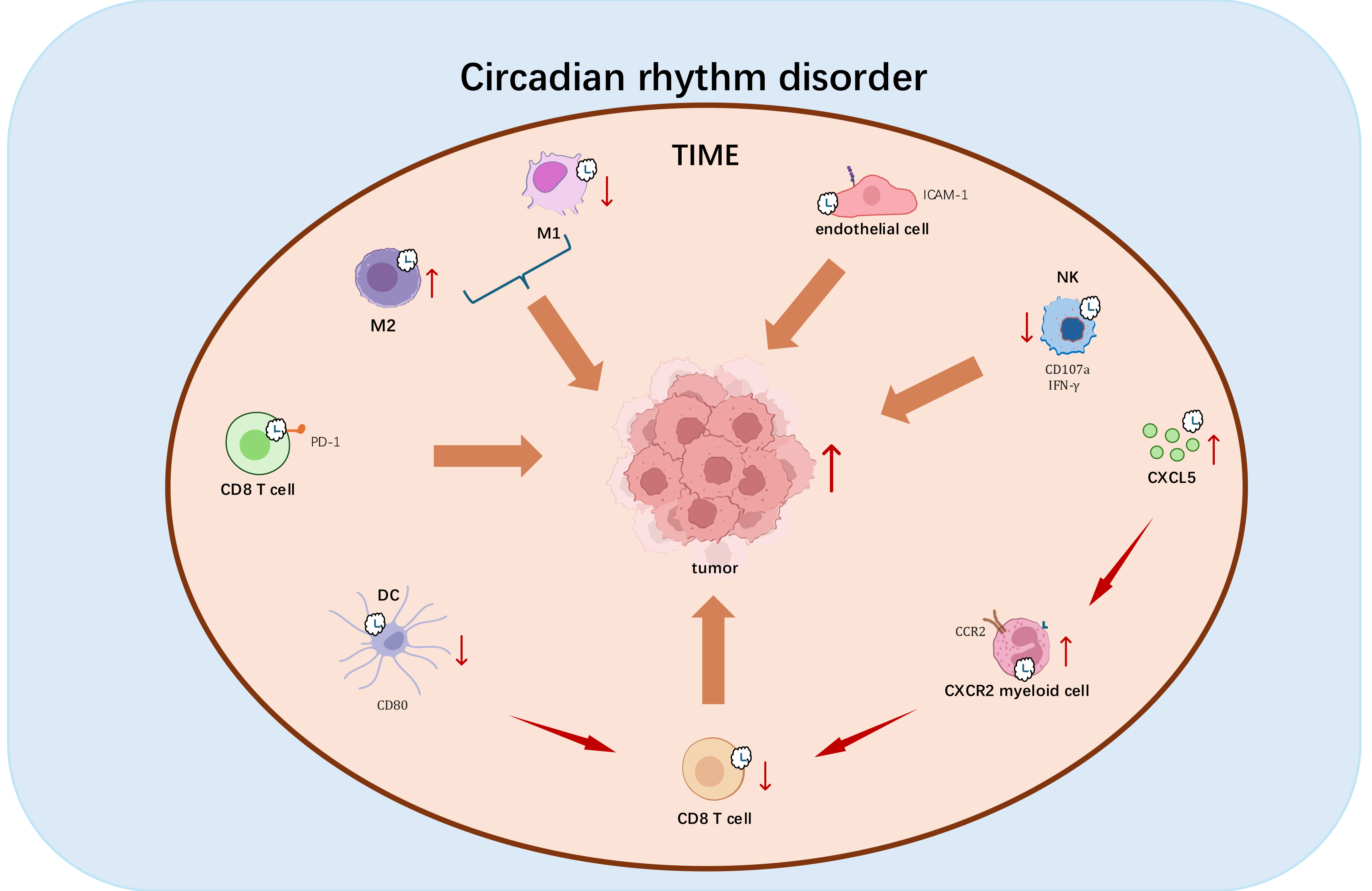
Fig. 2.

Mechanistic impact of Circadian rhythm disruption (CRD) on tumorgenesis. CRD exerts multifaceted immunosuppressive effects that promote tumor progression through the following mechanisms: (1) Disrupting M1/M2 macrophage polarization toward tumor-promoting anti-inflammatory phenotypes; (2) Disrupting circadian-regulated perforin expression in natural killer cells, compromising their tumoricidal capacity; (3) Impairing rhythmic ICAM-1 expression, thereby disrupting leukocyte trafficking; (4) Dysregulating circadian surface expression of CD80 and PD-1, attenuating CD8+ T cell activation and potentiating immune checkpoint pathways; (5) Inducing tumor-specific CXCL5 overexpression that facilitates CXCR2+ myeloid-derived suppressor cell recruitment, establishing an immunosuppressive niche; (6) Directly inhibiting cytotoxic T lymphocyte infiltration. These coordinated perturbations in chronoimmunological regulation establish a permissive microenvironment for multifactorial immunosuppression and malignant progression. Red vertical upward arrows indicate cell activation or gene upregulation; red vertical downward arrows indicate cell activity inhibition or gene downregulation; brown arrows indicate promotion. Diagram was assembled in Microsoft PowerPoint.

Emerging evidence highlights TIME modulation and immune infiltration dynamics as circadian-regulated hallmarks of cancer. Endothelial cells exhibit circadian ICAM-1 expression patterns that govern leukocyte trafficking rhythms into tumors [138]. CD8+ T cells demonstrate time-of-day-dependent functional oscillations, transitioning from immunosuppressed morning states to enhanced anti-tumor activity in the evening [138]. BMAL1-mediated circadian control of CD80 in dendritic cells coordinates rhythmic CD8+ T cell priming in tumor-draining lymph nodes, establishing temporal windows for optimal anti-tumor immunity [133]. CRD-induced cytokine network perturbations disrupt leukocyte circadian trafficking, reducing circulatory immune cell numbers and tumor infiltration capacity [139]. Mechanistically, CRD-driven TIME reprogramming involves activation of CXCL5-CXCR2 axis, which recruits immunosuppressive myeloid cells that suppress T cell responses and block CD8+ T cell infiltration [139]. These findings establish the clinical relevance of clock genes and their transcriptional targets (e.g., CXCL5, CD80) as prognostic chronotherapeutic biomarkers in oncology [140, 141, 142, 143].

Collectively, circadian regulatory networks represent a fundamental axis in cancer biology. Deciphering circadian-cancer crosstalk may unveil chronotherapeutic strategies for precision oncology, leveraging temporal biology to optimize immune activation and tumor suppression.

7. Conclusions

The circadian clock acts as a central regulator of immune homeostasis, exerting both direct cellular and systemic modulatory effects. Beyond the established crosstalk between core clock components and immune cells, emerging evidence highlights circadian-mediated immunomodulation through neuroendocrine, metabolic, and hypoxic signaling axes. For example, glucocorticoids (GCs) rhythmically control T-cell trafficking via GR-mediated binding to glucocorticoid response elements (GREs) in the IL-7Rα locus enhancer, thereby regulating CXCR4 expression cycles [116, 144]. A bidirectional regulatory loop exists between hypoxia-inducible factor-1α (HIF-1α) and core circadian machinery [145], with experimental models showing that HIF-1α forms a transcriptional complex with ARNTL1/BMAL1 and CLOCK. This complex binds E-box elements in the CXCR4 promoter to orchestrate leukocyte migration, a process modulated by circadian-controlled ROS levels and p38MAPK/MK2 phosphorylation cascades that stabilize HIF-1α [146, 147, 148]. Additionally, circadian-driven sympathetic activity releases phase-dependent norepinephrine, which activates β2-adrenergic receptors (β2-ARs) on lymph node lymphocytes [105, 149]. This neuroimmune interaction upregulates CCR7/CXCR4 chemokine receptors through cAMP-PKA signaling, retaining lymphocytes within lymphoid compartments [105, 115, 117].

Chronoimmunological research has uncovered significant temporal variations in immune regulation with direct clinical relevance. The CXCL12-CXCR4 axis has emerged as a therapeutic target in strategies combining CXCR4 inhibitors with immunotherapy to overcome treatment resistance [139]. Circadian synchronization studies reveal robust oscillatory expression patterns of the Pdcd1 gene (encoding PD-1) in activated CD8+ T cells, with corresponding diurnal variations in the efficacy of anti-PD-1 and CAR T-cell therapies [138]. Vaccination timing has been shown to influence tumor growth modulation in preclinical models [133, 138], while circadian dysregulation is implicated in severe COVID-19 pathogenesis [150]. Melatonin administration holds therapeutic potential for mitigating cytokine storms in critical cases [151], and nocturnal bright light therapy—by suppressing plasma melatonin—has been proposed as adjuvant for malaria eradication [152]. These findings underscore the transformative potential of temporally optimized therapies for immune-related disorders.

Substantial mysteries remain regarding the bidirectional circadian-immune system regulation. While core clock components like BMAL1 are established modulators of immune cell rhythms, the role of auxiliary clock proteins in functional attenuation require systematic investigation. Methodologically rigorous approaches—such as cell-type-specific circadian disruption models—are essential to map clock protein domains to immunoregulatory phenotypes. Single-cell transcriptomics should be prioritized to dissect cell-type-specific circadian-immune crosstalk, including how tissue-resident macrophages maintain local rhythms independent of SCN signals control. Development of small-molecule chronotherapeutics (e.g., BMAL1 stabilizers or CRY degraders) may offer precision strategies for autoimmune diseases. Elucidating these mechanisms could advance our understanding of disease pathogenesis and identify novel therapeutic targets.

Recent studies highlight the importance of nocturnal pineal melatonin and cortisol profiles in resetting immune cells for diurnal function, with implications for cancer and neurodegenerative conditions [153]. Melatonin peaks during early sleep, followed by a cortisol rise during the cortisol awakening response. Like gut-derived butyrate, melatonin inhibits nuclear translocation of glucocorticoid receptor-alpha (GR-α) [153, 154], indicating that factors suppressing pineal melatonin (e.g., aging, type 2 diabetes) [128] may disinhibit cortisol-mediated immune regulation. Clarifying how circadian alterations impact immune cell gene expression and mitochondrial function represents a key future direction.

Understanding how environmental zeitgebers (e.g., light exposure, gut microbial rhythms) integrate into circadian immune networks—as discussed in Section 5—will be essential for developing holistic chrono-immunological interventions. Such insights may bridge basic chronobiology with translational medicine, fostering temporally optimized strategies to enhance immune health.

Author Contributions

YJ, CL, and YZ contributed to the conceptualization and writing of the manuscript. YJ and YZ assembled the figures and tables. All authors contributed to editorial changes in the manuscript. All authors read and approved the final manuscript. All authors have participated sufficiently in the work and agreed to be accountable for all aspects of the work.

Ethics Approval and Consent to Participate

Not applicable.

Acknowledgment

We would like to thank the peer reviewers for their constructive feedback, which greatly improved the quality and clarity of this manuscript.

Funding

This work was supported by grants from the National Key R&D Program of China (2019YFA0800400), the National Natural Science Foundation of China (NSFC) (31871186), the Hui-Chun Chin and Tsung-Dao Lee Chinese Undergraduate Research Endowment (CURE), the Priority Academic Program Development (PAPD) of Jiangsu Higher Education Institutions (YX13400214).

Conflict of Interest

The authors declare no conflict of interest.

Declaration of AI and AI-Assisted Technologies in the Writing Process

During the preparation of this work the authors used DeepSeek in order to check spell and grammar. After using this tool, the authors reviewed and edited the content as needed and takes full responsibility for the content of the publication.

References
[1]
Roenneberg T, Merrow M. The Circadian Clock and Human Health. Current Biology: CB. 2016; 26: R432–43. https://doi.org/10.1016/j.cub.2016.04.011.
[2]
Golden SS, Canales SR. Cyanobacterial circadian clocks–timing is everything. Nature Reviews. Microbiology. 2003; 1: 191–199. https://doi.org/10.1038/nrmicro774.
[3]
Honma S, Ono D, Suzuki Y, Inagaki N, Yoshikawa T, Nakamura W, et al. Suprachiasmatic nucleus: cellular clocks and networks. Progress in Brain Research. 2012; 199: 129–141. https://doi.org/10.1016/B978-0-444-59427-3.00029-0.
[4]
Hastings MH, Maywood ES, Brancaccio M. Generation of circadian rhythms in the suprachiasmatic nucleus. Nature Reviews. Neuroscience. 2018; 19: 453–469. https://doi.org/10.1038/s41583-018-0026-z.
[5]
Young MW, Kay SA. Time zones: a comparative genetics of circadian clocks. Nature Reviews. Genetics. 2001; 2: 702–715. https://doi.org/10.1038/35088576.
[6]
Arjona A, Sarkar DK. Are circadian rhythms the code of hypothalamic-immune communication? Insights from natural killer cells. Neurochemical Research. 2008; 33: 708–718. https://doi.org/10.1007/s11064-007-9501-z.
[7]
Panda S. Circadian physiology of metabolism. Science (New York, N.Y.). 2016; 354: 1008–1015. https://doi.org/10.1126/science.aah4967.
[8]
Moeller JS, Bever SR, Finn SL, Phumsatitpong C, Browne MF, Kriegsfeld LJ. Circadian Regulation of Hormonal Timing and the Pathophysiology of Circadian Dysregulation. Comprehensive Physiology. 2022; 12: 4185–4214. https://doi.org/10.1002/cphy.c220018.
[9]
Li X, Liu X, Meng Q, Wu X, Bing X, Guo N, et al. Circadian clock disruptions link oxidative stress and systemic inflammation to metabolic syndrome in obstructive sleep apnea patients. Frontiers in Physiology. 2022; 13: 932596. https://doi.org/10.3389/fphys.2022.932596.
[10]
Reinke H, Asher G. Crosstalk between metabolism and circadian clocks. Nature Reviews. Molecular Cell Biology. 2019; 20: 227–241. https://doi.org/10.1038/s41580-018-0096-9.
[11]
Patke A, Young MW, Axelrod S. Molecular mechanisms and physiological importance of circadian rhythms. Nature Reviews. Molecular Cell Biology. 2020; 21: 67–84. https://doi.org/10.1038/s41580-019-0179-2.
[12]
Chaix A, Lin T, Le HD, Chang MW, Panda S. Time-Restricted Feeding Prevents Obesity and Metabolic Syndrome in Mice Lacking a Circadian Clock. Cell Metabolism. 2019; 29: 303–319.e4. https://doi.org/10.1016/j.cmet.2018.08.004.
[13]
Bellet MM, Deriu E, Liu JZ, Grimaldi B, Blaschitz C, Zeller M, et al. Circadian clock regulates the host response to Salmonella. Proceedings of the National Academy of Sciences of the United States of America. 2013; 110: 9897–9902. https://doi.org/10.1073/pnas.1120636110.
[14]
Zhuang X, Edgar RS, McKeating JA. The role of circadian clock pathways in viral replication. Seminars in Immunopathology. 2022; 44: 175–182. https://doi.org/10.1007/s00281-021-00908-2.
[15]
Kiessling S, Dubeau-Laramée G, Ohm H, Labrecque N, Olivier M, Cermakian N. The circadian clock in immune cells controls the magnitude of Leishmania parasite infection. Scientific Reports. 2017; 7: 10892. https://doi.org/10.1038/s41598-017-11297-8.
[16]
Asher G, Sassone-Corsi P. Time for food: the intimate interplay between nutrition, metabolism, and the circadian clock. Cell. 2015; 161: 84–92. https://doi.org/10.1016/j.cell.2015.03.015.
[17]
Partch CL, Green CB, Takahashi JS. Molecular architecture of the mammalian circadian clock. Trends in Cell Biology. 2014; 24: 90–99. https://doi.org/10.1016/j.tcb.2013.07.002.
[18]
Crane BR, Young MW. Interactive features of proteins composing eukaryotic circadian clocks. Annual Review of Biochemistry. 2014; 83: 191–219. https://doi.org/10.1146/annurev-biochem-060713-035644.
[19]
Logan RW, Wynne O, Levitt D, Price D, Sarkar DK. Altered circadian expression of cytokines and cytolytic factors in splenic natural killer cells of Per1(-/-) mutant mice. Journal of Interferon & Cytokine Research: the Official Journal of the International Society for Interferon and Cytokine Research. 2013; 33: 108–114. https://doi.org/10.1089/jir.2012.0092.
[20]
Kubra S, Zhang H, Si Y, Gao X, Wang T, Pan L, et al. REGγ regulates circadian clock by modulating BMAL1 protein stability. Cell Death Discovery. 2021; 7: 335. https://doi.org/10.1038/s41420-021-00704-9.
[21]
Scheiermann C, Gibbs J, Ince L, Loudon A. Clocking in to immunity. Nature Reviews. Immunology. 2018; 18: 423–437. https://doi.org/10.1038/s41577-018-0008-4.
[22]
Zeng T, Liang L, Deng W, Xie M, Zhao M, Wang S, et al. BMAL1 plays a crucial role in immune homeostasis during sepsis-induced acute lung injury. Biochemical Pharmacology. 2024; 226: 116379. https://doi.org/10.1016/j.bcp.2024.116379.
[23]
Xie M, Tang Q, Nie J, Zhang C, Zhou X, Yu S, et al. BMAL1-Downregulation Aggravates Porphyromonas Gingivalis-Induced Atherosclerosis by Encouraging Oxidative Stress. Circulation Research. 2020; 126: e15–e29. https://doi.org/10.1161/CIRCRESAHA.119.315502.
[24]
Li T, Shao W, Li S, Ma L, Zheng L, Shang W, et al. H. pylori infection induced BMAL1 expression and rhythm disorder aggravate gastric inflammation. EBioMedicine. 2019; 39: 301–314. https://doi.org/10.1016/j.ebiom.2018.11.043.
[25]
Chen L, Li S, Nie J, Zhao J, Yu S, Li Y, et al. Bmal1 Regulates Coagulation Factor Biosynthesis in Mouse Liver in Streptococcus oralis Infection. Frontiers in Cellular and Infection Microbiology. 2020; 10: 530190. https://doi.org/10.3389/fcimb.2020.530190.
[26]
Carrillo-Vico A, Lardone PJ, Alvarez-Sánchez N, Rodríguez-Rodríguez A, Guerrero JM. Melatonin: buffering the immune system. International Journal of Molecular Sciences. 2013; 14: 8638–8683. https://doi.org/10.3390/ijms14048638.
[27]
Vielma JR, Bonilla E, Chacín-Bonilla L, Mora M, Medina-Leendertz S, Bravo Y. Effects of melatonin on oxidative stress, and resistance to bacterial, parasitic, and viral infections: a review. Acta Tropica. 2014; 137: 31–38. https://doi.org/10.1016/j.actatropica.2014.04.021.
[28]
Wu UI, Mai FD, Sheu JN, Chen LY, Liu YT, Huang HC, et al. Melatonin inhibits microglial activation, reduces pro-inflammatory cytokine levels, and rescues hippocampal neurons of adult rats with acute Klebsiella pneumoniae meningitis. Journal of Pineal Research. 2011; 50: 159–170. https://doi.org/10.1111/j.1600-079X.2010.00825.x.
[29]
Osadchuk MA, Sibriaev AA, Kireeva NV, Kvetnoĭ IM. The influence of melatonin included in the combined treatment of antichelicobaterial therapy on immunohistochemical characteristics of gastric epitheliocytes from patients with duodenal ulcer. Klinicheskaia Meditsina. 2012; 90: 48–52.
[30]
Chojnacki C, Popławski T, Blasiak J, Chojnacki J, Reiter RJ, Klupinska G. Expression of melatonin synthesizing enzymes in Helicobacter pylori infected gastric mucosa. BioMed Research International. 2013; 2013: 845032. https://doi.org/10.1155/2013/845032.
[31]
Ozkan E, Yaman H, Cakir E, Deniz O, Oztosun M, Gumus S, et al. Plasma melatonin and urinary 6-hydroxymelatonin levels in patients with pulmonary tuberculosis. Inflammation. 2012; 35: 1429–1434. https://doi.org/10.1007/s10753-012-9456-3.
[32]
Regodón S, Ramos A, Míguez MP, Carrillo-Vico A, Rosado JA, Jardín I. Vaccination prepartum enhances the beneficial effects of melatonin on the immune response and reduces platelet responsiveness in sheep. BMC Veterinary Research. 2012; 8: 84. https://doi.org/10.1186/1746-6148-8-84.
[33]
Carvalho Cabral P, Tekade K, Stegeman SK, Olivier M, Cermakian N. The involvement of host circadian clocks in the regulation of the immune response to parasitic infections in mammals. Parasite Immunology. 2022; 44: e12903. https://doi.org/10.1111/pim.12903.
[34]
Smith LM, Motta FC, Chopra G, Moch JK, Nerem RR, Cummins B, et al. An intrinsic oscillator drives the blood stage cycle of the malaria parasite Plasmodium falciparum. Science (New York, N.Y.). 2020; 368: 754–759. https://doi.org/10.1126/science.aba4357.
[35]
Hopwood TW, Hall S, Begley N, Forman R, Brown S, Vonslow R, et al. The circadian regulator BMAL1 programmes responses to parasitic worm infection via a dendritic cell clock. Scientific Reports. 2018; 8: 3782. https://doi.org/10.1038/s41598-018-22021-5.
[36]
Srinivasan V, Spence DW, Moscovitch A, Pandi-Perumal SR, Trakht I, Brown GM, et al. Malaria: therapeutic implications of melatonin. Journal of Pineal Research. 2010; 48: 1–8. https://doi.org/10.1111/j.1600-079X.2009.00728.x.
[37]
Schuck DC, Jordão AK, Nakabashi M, Cunha AC, Ferreira VF, Garcia CRS. Synthetic indole and melatonin derivatives exhibit antimalarial activity on the cell cycle of the human malaria parasite Plasmodium falciparum. European Journal of Medicinal Chemistry. 2014; 78: 375–382. https://doi.org/10.1016/j.ejmech.2014.03.055.
[38]
Guha M, Maity P, Choubey V, Mitra K, Reiter RJ, Bandyopadhyay U. Melatonin inhibits free radical-mediated mitochondrial-dependent hepatocyte apoptosis and liver damage induced during malarial infection. Journal of Pineal Research. 2007; 43: 372–381. https://doi.org/10.1111/j.1600-079X.2007.00488.x.
[39]
França-Botelho AC, França JL, Oliveira FMS, Franca EL, Honório-França AC, Caliari MV, et al. Melatonin reduces the severity of experimental amoebiasis. Parasites & Vectors. 2011; 4: 62. https://doi.org/10.1186/1756-3305-4-62.
[40]
Santello FH, Frare EO, dos Santos CD, Toldo MPA, Kawasse LM, Zucoloto S, et al. Melatonin treatment reduces the severity of experimental Trypanosoma cruzi infection. Journal of Pineal Research. 2007; 42: 359–363. https://doi.org/10.1111/j.1600-079X.2007.00427.x.
[41]
Laothong U, Pinlaor P, Boonsiri P, Pairojkul C, Priprem A, Johns NP, et al. Melatonin inhibits cholangiocarcinoma and reduces liver injury in Opisthorchis viverrini-infected and N-nitrosodimethylamine-treated hamsters. Journal of Pineal Research. 2013; 55: 257–266. https://doi.org/10.1111/jpi.12068.
[42]
Baltaci AK, Bediz CS, Mogulkoc R, Kurtoglu E, Pekel A. Effect of zinc and melatonin supplementation on cellular immunity in rats with toxoplasmosis. Biological Trace Element Research. 2003; 96: 237–245. https://doi.org/10.1385/BTER:96:1-3:237.
[43]
Kalamvoki M, Roizman B. The histone acetyltransferase CLOCK is an essential component of the herpes simplex virus 1 transcriptome that includes TFIID, ICP4, ICP27, and ICP22. Journal of Virology. 2011; 85: 9472–9477. https://doi.org/10.1128/JVI.00876-11.
[44]
Zhuang X, Rambhatla SB, Lai AG, McKeating JA. Interplay between circadian clock and viral infection. Journal of Molecular Medicine (Berlin, Germany). 2017; 95: 1283–1289. https://doi.org/10.1007/s00109-017-1592-7.
[45]
Borrmann H, McKeating JA, Zhuang X. The Circadian Clock and Viral Infections. Journal of Biological Rhythms. 2021; 36: 9–22. https://doi.org/10.1177/0748730420967768.
[46]
Sengupta S, Tang SY, Devine JC, Anderson ST, Nayak S, Zhang SL, et al. Circadian control of lung inflammation in influenza infection. Nature Communications. 2019; 10: 4107. https://doi.org/10.1038/s41467-019-11400-9.
[47]
Majumdar T, Dhar J, Patel S, Kondratov R, Barik S. Circadian transcription factor BMAL1 regulates innate immunity against select RNA viruses. Innate Immunity. 2017; 23: 147–154. https://doi.org/10.1177/1753425916681075.
[48]
Pan P, Shen M, Yu Z, Ge W, Chen K, Tian M, et al. SARS-CoV-2 N protein promotes NLRP3 inflammasome activation to induce hyperinflammation. Nature Communications. 2021; 12: 4664. https://doi.org/10.1038/s41467-021-25015-6.
[49]
van den Berg DF, Te Velde AA. Severe COVID-19: NLRP3 Inflammasome Dysregulated. Frontiers in Immunology. 2020; 11: 1580. https://doi.org/10.3389/fimmu.2020.01580.
[50]
Pourcet B, Zecchin M, Ferri L, Beauchamp J, Sitaula S, Billon C, et al. Nuclear Receptor Subfamily 1 Group D Member 1 Regulates Circadian Activity of NLRP3 Inflammasome to Reduce the Severity of Fulminant Hepatitis in Mice. Gastroenterology. 2018; 154: 1449–1464.e20. https://doi.org/10.1053/j.gastro.2017.12.019.
[51]
Zhou P, Yang XL, Wang XG, Hu B, Zhang L, Zhang W, et al. Addendum: A pneumonia outbreak associated with a new coronavirus of probable bat origin. Nature. 2020; 588: E6. https://doi.org/10.1038/s41586-020-2951-z.
[52]
Reiter RJ, Sharma R, Ma Q, Rosales-Corral S, Acuna-Castroviejo D, Escames G. Inhibition of mitochondrial pyruvate dehydrogenase kinase: a proposed mechanism by which melatonin causes cancer cells to overcome cytosolic glycolysis, reduce tumor biomass and reverse insensitivity to chemotherapy. Melatonin Research. 2019; 2: 105–119. https://doi.org/10.32794/mr11250033.
[53]
Anderson G, Reiter RJ. Melatonin: Roles in influenza, Covid-19, and other viral infections. Reviews in Medical Virology. 2020; 30: e2109. https://doi.org/10.1002/rmv.2109.
[54]
Bonilla E, Valero-Fuenmayor N, Pons H, Chacín-Bonilla L. Melatonin protects mice infected with Venezuelan equine encephalomyelitis virus. Cellular and Molecular Life Sciences: CMLS. 1997; 53: 430–434. https://doi.org/10.1007/s000180050051.
[55]
Huang SH, Cao XJ, Liu W, Shi XY, Wei W. Inhibitory effect of melatonin on lung oxidative stress induced by respiratory syncytial virus infection in mice. Journal of Pineal Research. 2010; 48: 109–116. https://doi.org/10.1111/j.1600-079X.2009.00733.x.
[56]
Tan DX, Korkmaz A, Reiter RJ, Manchester LC. Ebola virus disease: potential use of melatonin as a treatment. Journal of Pineal Research. 2014; 57: 381–384. https://doi.org/10.1111/jpi.12186.
[57]
DiNicolantonio JJ, McCarty M, Barroso-Aranda J. Melatonin may decrease risk for and aid treatment of COVID-19 and other RNA viral infections. Open Heart. 2021; 8: e001568. https://doi.org/10.1136/openhrt-2020-001568.
[58]
Gibbs J, Ince L, Matthews L, Mei J, Bell T, Yang N, et al. An epithelial circadian clock controls pulmonary inflammation and glucocorticoid action. Nature Medicine. 2014; 20: 919–926. https://doi.org/10.1038/nm.3599.
[59]
Nguyen KD, Fentress SJ, Qiu Y, Yun K, Cox JS, Chawla A. Circadian gene Bmal1 regulates diurnal oscillations of Ly6C(hi) inflammatory monocytes. Science (New York, N.Y.). 2013; 341: 1483–1488. https://doi.org/10.1126/science.1240636.
[60]
Sun Y, Yang Z, Niu Z, Peng J, Li Q, Xiong W, et al. MOP3, a component of the molecular clock, regulates the development of B cells. Immunology. 2006; 119: 451–460. https://doi.org/10.1111/j.1365-2567.2006.02456.x.
[61]
Keller M, Mazuch J, Abraham U, Eom GD, Herzog ED, Volk HD, et al. A circadian clock in macrophages controls inflammatory immune responses. Proceedings of the National Academy of Sciences of the United States of America. 2009; 106: 21407–21412. https://doi.org/10.1073/pnas.0906361106.
[62]
Gibbs JE, Blaikley J, Beesley S, Matthews L, Simpson KD, Boyce SH, et al. The nuclear receptor REV-ERBα mediates circadian regulation of innate immunity through selective regulation of inflammatory cytokines. Proceedings of the National Academy of Sciences of the United States of America. 2012; 109: 582–587. https://doi.org/10.1073/pnas.1106750109.
[63]
Curtis AM, Fagundes CT, Yang G, Palsson-McDermott EM, Wochal P, McGettrick AF, et al. Circadian control of innate immunity in macrophages by miR-155 targeting Bmal1. Proceedings of the National Academy of Sciences of the United States of America. 2015; 112: 7231–7236. https://doi.org/10.1073/pnas.1501327112.
[64]
Early JO, Menon D, Wyse CA, Cervantes-Silva MP, Zaslona Z, Carroll RG, et al. Circadian clock protein BMAL1 regulates IL-1β in macrophages via NRF2. Proceedings of the National Academy of Sciences of the United States of America. 2018; 115: E8460–E8468. https://doi.org/10.1073/pnas.1800431115.
[65]
Timmons GA, Carroll RG, O’Siorain JR, Cervantes-Silva MP, Fagan LE, Cox SL, et al. The Circadian Clock Protein BMAL1 Acts as a Metabolic Sensor In Macrophages to Control the Production of Pro IL-1β. Frontiers in Immunology. 2021; 12: 700431. https://doi.org/10.3389/fimmu.2021.700431.
[66]
Shim DW, Eo JC, Kim S, Hwang I, Nam B, Shin JE, et al. Deficiency of circadian clock gene Bmal1 exacerbates noncanonical inflammasome-mediated pyroptosis and lethality via Rev-erbα-C/EBPβ-SAA1 axis. Experimental & Molecular Medicine. 2024; 56: 370–382. https://doi.org/10.1038/s12276-024-01162-w.
[67]
Deng W, Zhu S, Zeng L, Liu J, Kang R, Yang M, et al. The Circadian Clock Controls Immune Checkpoint Pathway in Sepsis. Cell Reports. 2018; 24: 366–378. https://doi.org/10.1016/j.celrep.2018.06.026.
[68]
Alexander RK, Liou YH, Knudsen NH, Starost KA, Xu C, Hyde AL, et al. Bmal1 integrates mitochondrial metabolism and macrophage activation. eLife. 2020; 9: e54090. https://doi.org/10.7554/eLife.54090.
[69]
Nakabo S, Sandoval-Heglund D, Hanata N, Brooks S, Hoffmann V, Zhang M, et al. The circadian clock gene BMAL1 modulates autoimmunity features in lupus. Frontiers in Immunology. 2024; 15: 1465185. https://doi.org/10.3389/fimmu.2024.1465185.
[70]
Adrover JM, Del Fresno C, Crainiciuc G, Cuartero MI, Casanova-Acebes M, Weiss LA, et al. A Neutrophil Timer Coordinates Immune Defense and Vascular Protection. Immunity. 2019; 51: 966–967. https://doi.org/10.1016/j.immuni.2019.11.001.
[71]
Cox SL, O’Siorain JR, He Y, Lordan R, Naik A, Tang SY, et al. Circadian disruption in lung fibroblasts enhances NF-κB activity to exacerbate neutrophil recruitment. FASEB Journal: Official Publication of the Federation of American Societies for Experimental Biology. 2023; 37: e22753. https://doi.org/10.1096/fj.202201456R.
[72]
Druzd D, Matveeva O, Ince L, Harrison U, He W, Schmal C, et al. Lymphocyte Circadian Clocks Control Lymph Node Trafficking and Adaptive Immune Responses. Immunity. 2017; 46: 120–132. https://doi.org/10.1016/j.immuni.2016.12.011.
[73]
Godinho-Silva C, Domingues RG, Rendas M, Raposo B, Ribeiro H, da Silva JA, et al. Light-entrained and brain-tuned circadian circuits regulate ILC3s and gut homeostasis. Nature. 2019; 574: 254–258. https://doi.org/10.1038/s41586-019-1579-3.
[74]
Teng F, Goc J, Zhou L, Chu C, Shah MA, Eberl G, et al. A circadian clock is essential for homeostasis of group 3 innate lymphoid cells in the gut. Science Immunology. 2019; 4: eaax1215. https://doi.org/10.1126/sciimmunol.aax1215.
[75]
Fortier EE, Rooney J, Dardente H, Hardy MP, Labrecque N, Cermakian N. Circadian variation of the response of T cells to antigen. Journal of Immunology (Baltimore, Md.: 1950). 2011; 187: 6291–6300. https://doi.org/10.4049/jimmunol.1004030.
[76]
Pan X, Jiang XC, Hussain MM. Impaired cholesterol metabolism and enhanced atherosclerosis in clock mutant mice. Circulation. 2013; 128: 1758–1769. https://doi.org/10.1161/CIRCULATIONAHA.113.002885.
[77]
Spengler ML, Kuropatwinski KK, Comas M, Gasparian AV, Fedtsova N, Gleiberman AS, et al. Core circadian protein CLOCK is a positive regulator of NF-κB-mediated transcription. Proceedings of the National Academy of Sciences of the United States of America. 2012; 109: E2457–65. https://doi.org/10.1073/pnas.1206274109.
[78]
Bellet MM, Zocchi L, Sassone-Corsi P. The RelB subunit of NFκB acts as a negative regulator of circadian gene expression. Cell Cycle (Georgetown, Tex.). 2012; 11: 3304–3311. https://doi.org/10.4161/cc.21669.
[79]
Kawauchi T, Ishimaru K, Nakamura Y, Nakano N, Hara M, Ogawa H, et al. Clock-dependent temporal regulation of IL-33/ST2-mediated mast cell response. Allergology International: Official Journal of the Japanese Society of Allergology. 2017; 66: 472–478. https://doi.org/10.1016/j.alit.2017.02.004.
[80]
Nakamura Y, Nakano N, Ishimaru K, Hara M, Ikegami T, Tahara Y, et al. Circadian regulation of allergic reactions by the mast cell clock in mice. The Journal of Allergy and Clinical Immunology. 2014; 133: 568–575. https://doi.org/10.1016/j.jaci.2013.07.040.
[81]
Ando N, Nakamura Y, Aoki R, Ishimaru K, Ogawa H, Okumura K, et al. Circadian Gene Clock Regulates Psoriasis-Like Skin Inflammation in Mice. The Journal of Investigative Dermatology. 2015; 135: 3001–3008. https://doi.org/10.1038/jid.2015.316.
[82]
Wang T, Wang Z, Yang P, Xia L, Zhou M, Wang S, et al. PER1 prevents excessive innate immune response during endotoxin-induced liver injury through regulation of macrophage recruitment in mice. Cell Death & Disease. 2016; 7: e2176. https://doi.org/10.1038/cddis.2016.9.
[83]
Capelle CM, Chen A, Zeng N, Baron A, Grzyb K, Arns T, et al. Stress hormone signalling inhibits Th1 polarization in a CD4 T-cell-intrinsic manner via mTORC1 and the circadian gene PER1. Immunology. 2022; 165: 428–444. https://doi.org/10.1111/imm.13448.
[84]
Silver AC, Arjona A, Walker WE, Fikrig E. The circadian clock controls toll-like receptor 9-mediated innate and adaptive immunity. Immunity. 2012; 36: 251–261. https://doi.org/10.1016/j.immuni.2011.12.017.
[85]
Heipertz EL, Harper J, Lopez CA, Fikrig E, Hughes ME, Walker WE. Circadian Rhythms Influence the Severity of Sepsis in Mice via a TLR2-Dependent, Leukocyte-Intrinsic Mechanism. Journal of Immunology (Baltimore, Md.: 1950). 2018; 201: 193–201. https://doi.org/10.4049/jimmunol.1701677.
[86]
Nakamura Y, Harama D, Shimokawa N, Hara M, Suzuki R, Tahara Y, et al. Circadian clock gene Period2 regulates a time-of-day-dependent variation in cutaneous anaphylactic reaction. The Journal of Allergy and Clinical Immunology. 2011; 127: 1038–1045.e3. https://doi.org/10.1016/j.jaci.2011.02.006.
[87]
Liu J, Malkani G, Shi X, Meyer M, Cunningham-Runddles S, Ma X, et al. The circadian clock Period 2 gene regulates gamma interferon production of NK cells in host response to lipopolysaccharide-induced endotoxic shock. Infection and Immunity. 2006; 74: 4750–4756. https://doi.org/10.1128/IAI.00287-06.
[88]
Narasimamurthy R, Hatori M, Nayak SK, Liu F, Panda S, Verma IM. Circadian clock protein cryptochrome regulates the expression of proinflammatory cytokines. Proceedings of the National Academy of Sciences of the United States of America. 2012; 109: 12662–12667. https://doi.org/10.1073/pnas.1209965109.
[89]
Hashiramoto A, Yamane T, Tsumiyama K, Yoshida K, Komai K, Yamada H, et al. Mammalian clock gene Cryptochrome regulates arthritis via proinflammatory cytokine TNF-alpha. Journal of Immunology (Baltimore, Md.: 1950). 2010; 184: 1560–1565. https://doi.org/10.4049/jimmunol.0903284.
[90]
Ivanov II, McKenzie BS, Zhou L, Tadokoro CE, Lepelley A, Lafaille JJ, et al. The orphan nuclear receptor RORgammat directs the differentiation program of proinflammatory IL-17+ T helper cells. Cell. 2006; 126: 1121–1133. https://doi.org/10.1016/j.cell.2006.07.035.
[91]
Sun Z, Unutmaz D, Zou YR, Sunshine MJ, Pierani A, Brenner-Morton S, et al. Requirement for RORgamma in thymocyte survival and lymphoid organ development. Science (New York, N.Y.). 2000; 288: 2369–2373. https://doi.org/10.1126/science.288.5475.2369.
[92]
Kurebayashi S, Ueda E, Sakaue M, Patel DD, Medvedev A, Zhang F, et al. Retinoid-related orphan receptor gamma (RORgamma) is essential for lymphoid organogenesis and controls apoptosis during thymopoiesis. Proceedings of the National Academy of Sciences of the United States of America. 2000; 97: 10132–10137. https://doi.org/10.1073/pnas.97.18.10132.
[93]
Eberl G, Marmon S, Sunshine MJ, Rennert PD, Choi Y, Littman DR. An essential function for the nuclear receptor RORgamma(t) in the generation of fetal lymphoid tissue inducer cells. Nature Immunology. 2004; 5: 64–73. https://doi.org/10.1038/ni1022.
[94]
Schaeren-Wiemers N, André E, Kapfhammer JP, Becker-André M. The expression pattern of the orphan nuclear receptor RORbeta in the developing and adult rat nervous system suggests a role in the processing of sensory information and in circadian rhythm. The European Journal of Neuroscience. 1997; 9: 2687–2701. https://doi.org/10.1111/j.1460-9568.1997.tb01698.x.
[95]
Guo J, Hawwari A, Li H, Sun Z, Mahanta SK, Littman DR, et al. Regulation of the TCRalpha repertoire by the survival window of CD4(+)CD8(+) thymocytes. Nature Immunology. 2002; 3: 469–476. https://doi.org/10.1038/ni791.
[96]
Dzhagalov I, Giguère V, He YW. Lymphocyte development and function in the absence of retinoic acid-related orphan receptor alpha. Journal of Immunology (Baltimore, Md.: 1950). 2004; 173: 2952–2959. https://doi.org/10.4049/jimmunol.173.5.2952.
[97]
Yu X, Rollins D, Ruhn KA, Stubblefield JJ, Green CB, Kashiwada M, et al. TH17 cell differentiation is regulated by the circadian clock. Science (New York, N.Y.). 2013; 342: 727–730. https://doi.org/10.1126/science.1243884.
[98]
Wang S, Lin Y, Yuan X, Li F, Guo L, Wu B. REV-ERBα integrates colon clock with experimental colitis through regulation of NF-κB/NLRP3 axis. Nature Communications. 2018; 9: 4246. https://doi.org/10.1038/s41467-018-06568-5.
[99]
Sato S, Sakurai T, Ogasawara J, Takahashi M, Izawa T, Imaizumi K, et al. A circadian clock gene, Rev-erbα, modulates the inflammatory function of macrophages through the negative regulation of Ccl2 expression. Journal of Immunology (Baltimore, Md.: 1950). 2014; 192: 407–417. https://doi.org/10.4049/jimmunol.1301982.
[100]
Hong H, Cheung YM, Cao X, Wu Y, Li C, Tian XY. REV-ERBα agonist SR9009 suppresses IL-1β production in macrophages through BMAL1-dependent inhibition of inflammasome. Biochemical Pharmacology. 2021; 192: 114701. https://doi.org/10.1016/j.bcp.2021.114701.
[101]
Ishimaru K, Nakajima S, Yu G, Nakamura Y, Nakao A. The Putatively Specific Synthetic REV-ERB Agonist SR9009 Inhibits IgE- and IL-33-Mediated Mast Cell Activation Independently of the Circadian Clock. International Journal of Molecular Sciences. 2019; 20: 6320. https://doi.org/10.3390/ijms20246320.
[102]
Curtis AM, Bellet MM, Sassone-Corsi P, O’Neill LAJ. Circadian clock proteins and immunity. Immunity. 2014; 40: 178–186. https://doi.org/10.1016/j.immuni.2014.02.002.
[103]
Labrecque N, Cermakian N. Circadian Clocks in the Immune System. Journal of Biological Rhythms. 2015; 30: 277–290. https://doi.org/10.1177/0748730415577723.
[104]
Ella K, Csépányi-Kömi R, Káldi K. Circadian regulation of human peripheral neutrophils. Brain, Behavior, and Immunity. 2016; 57: 209–221. https://doi.org/10.1016/j.bbi.2016.04.016.
[105]
Leach S, Suzuki K. Adrenergic Signaling in Circadian Control of Immunity. Frontiers in Immunology. 2020; 11: 1235. https://doi.org/10.3389/fimmu.2020.01235.
[106]
Hriscu ML. Circadian phagocytic activity of neutrophils and its modulation by light. Journal of Applied Biomedicine. 2004; 2: 199–211. https://doi.org/10.32725/jab.2004.024.
[107]
Hriscu ML. Modulatory factors of circadian phagocytic activity. Annals of the New York Academy of Sciences. 2005; 1057: 403–430. https://doi.org/10.1196/annals.1356.032.
[108]
NaveenKumar SK, Hemshekhar M, Jagadish S, Manikanta K, Vishalakshi GJ, Kemparaju K, et al. Melatonin restores neutrophil functions and prevents apoptosis amid dysfunctional glutathione redox system. Journal of Pineal Research. 2020; 69: e12676. https://doi.org/10.1111/jpi.12676.
[109]
HALBERG F, JOHNSON EA, BROWN BW, BITTNER JJ. Susceptibility rhythm to E. coli endotoxin and bioassay. Proceedings of the Society for Experimental Biology and Medicine. Society for Experimental Biology and Medicine (New York, N.Y.). 1960; 103: 142–144. https://doi.org/10.3181/00379727-103-25439.
[110]
Castanon-Cervantes O, Wu M, Ehlen JC, Paul K, Gamble KL, Johnson RL, et al. Dysregulation of inflammatory responses by chronic circadian disruption. Journal of Immunology (Baltimore, Md.: 1950). 2010; 185: 5796–5805. https://doi.org/10.4049/jimmunol.1001026.
[111]
Guerrero-Vargas NN, Salgado-Delgado R, Basualdo MDC, García J, Guzmán-Ruiz M, Carrero JC, et al. Reciprocal interaction between the suprachiasmatic nucleus and the immune system tunes down the inflammatory response to lipopolysaccharide. Journal of Neuroimmunology. 2014; 273: 22–30. https://doi.org/10.1016/j.jneuroim.2014.05.012.
[112]
Collins EJ, Cervantes-Silva MP, Timmons GA, O’Siorain JR, Curtis AM, Hurley JM. Post-transcriptional circadian regulation in macrophages organizes temporally distinct immunometabolic states. Genome Research. 2021; 31: 171–185. https://doi.org/10.1101/gr.263814.120.
[113]
Tannahill GM, Curtis AM, Adamik J, Palsson-McDermott EM, McGettrick AF, Goel G, et al. Succinate is an inflammatory signal that induces IL-1β through HIF-1α. Nature. 2013; 496: 238–242. https://doi.org/10.1038/nature11986.
[114]
Zhou Y, Wu M, Xu L, Cheng J, Shen J, Yang T, et al. Bmal1 Regulates Macrophage Polarize Through Glycolytic Pathway in Alcoholic Liver Disease. Frontiers in Pharmacology. 2021; 12: 640521. https://doi.org/10.3389/fphar.2021.640521.
[115]
Suzuki K, Hayano Y, Nakai A, Furuta F, Noda M. Adrenergic control of the adaptive immune response by diurnal lymphocyte recirculation through lymph nodes. The Journal of Experimental Medicine. 2016; 213: 2567–2574. https://doi.org/10.1084/jem.20160723.
[116]
Shimba A, Cui G, Tani-Ichi S, Ogawa M, Abe S, Okazaki F, et al. Glucocorticoids Drive Diurnal Oscillations in T Cell Distribution and Responses by Inducing Interleukin-7 Receptor and CXCR4. Immunity. 2018; 48: 286–298.e6. https://doi.org/10.1016/j.immuni.2018.01.004.
[117]
Nakai A, Hayano Y, Furuta F, Noda M, Suzuki K. Control of lymphocyte egress from lymph nodes through β2-adrenergic receptors. The Journal of Experimental Medicine. 2014; 211: 2583–2598. https://doi.org/10.1084/jem.20141132.
[118]
Cyster JG, Schwab SR. Sphingosine-1-phosphate and lymphocyte egress from lymphoid organs. Annual Review of Immunology. 2012; 30: 69–94. https://doi.org/10.1146/annurev-immunol-020711-075011.
[119]
Hemmers S, Rudensky AY. The Cell-Intrinsic Circadian Clock Is Dispensable for Lymphocyte Differentiation and Function. Cell Reports. 2015; 11: 1339–1349. https://doi.org/10.1016/j.celrep.2015.04.058.
[120]
Korn T, Bettelli E, Oukka M, Kuchroo VK. IL-17 and Th17 Cells. Annual Review of Immunology. 2009; 27: 485–517. https://doi.org/10.1146/annurev.immunol.021908.132710.
[121]
Qiu J, Guo X, Chen ZME, He L, Sonnenberg GF, Artis D, et al. Group 3 innate lymphoid cells inhibit T-cell-mediated intestinal inflammation through aryl hydrocarbon receptor signaling and regulation of microflora. Immunity. 2013; 39: 386–399. https://doi.org/10.1016/j.immuni.2013.08.002.
[122]
Ciofani M, Madar A, Galan C, Sellars M, Mace K, Pauli F, et al. A validated regulatory network for Th17 cell specification. Cell. 2012; 151: 289–303. https://doi.org/10.1016/j.cell.2012.09.016.
[123]
Farez MF, Mascanfroni ID, Méndez-Huergo SP, Yeste A, Murugaiyan G, Garo LP, et al. Melatonin Contributes to the Seasonality of Multiple Sclerosis Relapses. Cell. 2015; 162: 1338–1352. https://doi.org/10.1016/j.cell.2015.08.025.
[124]
Thaiss CA, Levy M, Korem T, Dohnalová L, Shapiro H, Jaitin DA, et al. Microbiota Diurnal Rhythmicity Programs Host Transcriptome Oscillations. Cell. 2016; 167: 1495–1510.e12. https://doi.org/10.1016/j.cell.2016.11.003.
[125]
Leone V, Gibbons SM, Martinez K, Hutchison AL, Huang EY, Cham CM, et al. Effects of diurnal variation of gut microbes and high-fat feeding on host circadian clock function and metabolism. Cell Host & Microbe. 2015; 17: 681–689. https://doi.org/10.1016/j.chom.2015.03.006.
[126]
Wei L, Yue F, Xing L, Wu S, Shi Y, Li J, et al. Constant Light Exposure Alters Gut Microbiota and Promotes the Progression of Steatohepatitis in High Fat Diet Rats. Frontiers in Microbiology. 2020; 11: 1975. https://doi.org/10.3389/fmicb.2020.01975.
[127]
Chaix A, Zarrinpar A, Miu P, Panda S. Time-restricted feeding is a preventative and therapeutic intervention against diverse nutritional challenges. Cell Metabolism. 2014; 20: 991–1005. https://doi.org/10.1016/j.cmet.2014.11.001.
[128]
McMullan CJ, Schernhammer ES, Rimm EB, Hu FB, Forman JP. Melatonin secretion and the incidence of type 2 diabetes. JAMA. 2013; 309: 1388–1396. https://doi.org/10.1001/jama.2013.2710.
[129]
Gubin D, Boldyreva J, Stefani O, Kolomeichuk S, Danilova L, Shigabaeva A, et al. Higher vulnerability to poor circadian light hygiene in individuals with a history of COVID-19. Chronobiology International. 2025; 42: 133–146. https://doi.org/10.1080/07420528.2024.2449015.
[130]
Gao T, Wang Z, Dong Y, Cao J, Lin R, Wang X, et al. Role of melatonin in sleep deprivation-induced intestinal barrier dysfunction in mice. Journal of Pineal Research. 2019; 67: e12574. https://doi.org/10.1111/jpi.12574.
[131]
Zhou J, Wang J, Zhang X, Tang Q. New Insights Into Cancer Chronotherapies. Frontiers in Pharmacology. 2021; 12: 741295. https://doi.org/10.3389/fphar.2021.741295.
[132]
Li HX. The role of circadian clock genes in tumors. OncoTargets and Therapy. 2019; 12: 3645–3660. https://doi.org/10.2147/OTT.S203144.
[133]
Wang C, Barnoud C, Cenerenti M, Sun M, Caffa I, Kizil B, et al. Dendritic cells direct circadian anti-tumour immune responses. Nature. 2023; 614: 136–143. https://doi.org/10.1038/s41586-022-05605-0.
[134]
Binnewies M, Roberts EW, Kersten K, Chan V, Fearon DF, Merad M, et al. Understanding the tumor immune microenvironment (TIME) for effective therapy. Nature Medicine. 2018; 24: 541–550. https://doi.org/10.1038/s41591-018-0014-x.
[135]
Aiello I, Mul Fedele ML, Román F, Marpegan L, Caldart C, Chiesa JJ, et al. Circadian disruption promotes tumor-immune microenvironment remodeling favoring tumor cell proliferation. Science Advances. 2020; 6: eaaz4530. https://doi.org/10.1126/sciadv.aaz4530.
[136]
Zhang M, He Y, Sun X, Li Q, Wang W, Zhao A, et al. A high M1/M2 ratio of tumor-associated macrophages is associated with extended survival in ovarian cancer patients. Journal of Ovarian Research. 2014; 7: 19. https://doi.org/10.1186/1757-2215-7-19.
[137]
Logan RW, Zhang C, Murugan S, O’Connell S, Levitt D, Rosenwasser AM, et al. Chronic shift-lag alters the circadian clock of NK cells and promotes lung cancer growth in rats. Journal of Immunology (Baltimore, Md.: 1950). 2012; 188: 2583–2591. https://doi.org/10.4049/jimmunol.1102715.
[138]
Wang C, Zeng Q, Gül ZM, Wang S, Pick R, Cheng P, et al. Circadian tumor infiltration and function of CD8+ T cells dictate immunotherapy efficacy. Cell. 2024; 187: 2690–2702.e17. https://doi.org/10.1016/j.cell.2024.04.015.
[139]
Hadadi E, Taylor W, Li XM, Aslan Y, Villote M, Rivière J, et al. Chronic circadian disruption modulates breast cancer stemness and immune microenvironment to drive metastasis in mice. Nature Communications. 2020; 11: 3193. https://doi.org/10.1038/s41467-020-16890-6.
[140]
Zeng Q, Sun S, Li Y, Li X, Li Z, Liang H. Identification of Therapeutic Targets and Prognostic Biomarkers Among CXC Chemokines in the Renal Cell Carcinoma Microenvironment. Frontiers in Oncology. 2019; 9: 1555. https://doi.org/10.3389/fonc.2019.01555.
[141]
Huang Z, He A, Wang J, Lu H, Zhang R, Wu L, et al. The circadian clock is associated with prognosis and immune infiltration in stomach adenocarcinoma. Aging. 2021; 13: 16637–16655. https://doi.org/10.18632/aging.203184.
[142]
He Y, Chen Y, Dai X, Huang S. Dysregulation of Circadian Clock Genes Associated with Tumor Immunity and Prognosis in Patients with Colon Cancer. Computational and Mathematical Methods in Medicine. 2022; 2022: 4957996. https://doi.org/10.1155/2022/4957996.
[143]
Eisele L, Prinz R, Klein-Hitpass L, Nückel H, Lowinski K, Thomale J, et al. Combined PER2 and CRY1 expression predicts outcome in chronic lymphocytic leukemia. European Journal of Haematology. 2009; 83: 320–327. https://doi.org/10.1111/j.1600-0609.2009.01287.x.
[144]
Shimba A, Ikuta K. Glucocorticoids Regulate Circadian Rhythm of Innate and Adaptive Immunity. Frontiers in Immunology. 2020; 11: 2143. https://doi.org/10.3389/fimmu.2020.02143.
[145]
Peek CB, Levine DC, Cedernaes J, Taguchi A, Kobayashi Y, Tsai SJ, et al. Circadian Clock Interaction with HIF1α Mediates Oxygenic Metabolism and Anaerobic Glycolysis in Skeletal Muscle. Cell Metabolism. 2017; 25: 86–92. https://doi.org/10.1016/j.cmet.2016.09.010.
[146]
Qutub AA, Popel AS. Reactive oxygen species regulate hypoxia-inducible factor 1alpha differentially in cancer and ischemia. Molecular and Cellular Biology. 2008; 28: 5106–5119. https://doi.org/10.1128/MCB.00060-08.
[147]
Peek CB. Metabolic Implications of Circadian-HIF Crosstalk. Trends in Endocrinology and Metabolism: TEM. 2020; 31: 459–468. https://doi.org/10.1016/j.tem.2020.02.008.
[148]
Zhao Y, Liu M, Chan XY, Tan SY, Subramaniam S, Fan Y, et al. Uncovering the mystery of opposite circadian rhythms between mouse and human leukocytes in humanized mice. Blood. 2017; 130: 1995–2005. https://doi.org/10.1182/blood-2017-04-778779.
[149]
Méndez-Ferrer S, Lucas D, Battista M, Frenette PS. Haematopoietic stem cell release is regulated by circadian oscillations. Nature. 2008; 452: 442–447. https://doi.org/10.1038/nature06685.
[150]
Haspel J, Kim M, Zee P, Schwarzmeier T, Montagnese S, Panda S, et al. A Timely Call to Arms: COVID-19, the Circadian Clock, and Critical Care. Journal of Biological Rhythms. 2021; 36: 55–70. https://doi.org/10.1177/0748730421992587.
[151]
Zhou Y, Fu B, Zheng X, Wang D, Zhao C, Qi Y, et al. Pathogenic T-cells and inflammatory monocytes incite inflammatory storms in severe COVID-19 patients. National Science Review. 2020; 7: 998–1002. https://doi.org/10.1093/nsr/nwaa041.
[152]
Srinivasan V, Ahmad AH, Mohamed M, Zakaria R. Melatonin effects on Plasmodium life cycle: new avenues for therapeutic approach. Recent Patents on Endocrine, Metabolic & Immune Drug Discovery. 2012; 6: 139–147. https://doi.org/10.2174/187221412800604635.
[153]
Anderson G. Melatonin, BAG-1 and cortisol circadian interactions in tumor pathogenesis and patterned immune responses. Exploration of Targeted Anti-tumor Therapy. 2023; 4: 962–993. https://doi.org/10.37349/etat.2023.00176.
[154]
Kim M, Lee HA, Cho HM, Kang SH, Lee E, Kim IK. Histone deacetylase inhibition attenuates hepatic steatosis in rats with experimental Cushing’s syndrome. The Korean Journal of Physiology & Pharmacology: Official Journal of the Korean Physiological Society and the Korean Society of Pharmacology. 2018; 22: 23–33. https://doi.org/10.4196/kjpp.2018.22.1.23.

Publisher’s Note: IMR Press stays neutral with regard to jurisdictional claims in published maps and institutional affiliations.

Share
Back to top