

 , Jinghao Zheng 2,*
1 Cardiac Intensive Care Unit, Department of Thoracic and Cardiovascular Surgery, Shanghai Children’s Medical Center, Shanghai Jiao Tong University School of Medicine, 200127 Shanghai, China
2 Department of Thoracic and Cardiovascular Surgery, Shanghai Children’s Medical Center, Shanghai Jiao Tong University School of Medicine, 200127 Shanghai, China
Abstract
Background: This study aimed to determine whether the hemodynamics of
patients with right ventricle outflow tract obstructive congenital heart disease
(RVOTO-CHD) improve after corrective surgery by changing the ventilation mode.
Methods: Patients with RVOTO-CHD who underwent corrective surgery were
enrolled in this study. Echocardiography and advanced hemodynamic monitoring were
performed using the pulse indicator continuous cardiac output (PiCCO) technology in the pressure-regulated volume control
(PRVC) mode, followed with switching to the pressure support ventilation (PSV)
mode and neurally adjusted ventilatory assist (NAVA) mode in random order.
Results: Overall, 31 patients were enrolled in this study from April
2021 to October 2021. Notably, changing the ventilation mode from PRVC to a
spontaneous mode (PSV or NAVA) led to better cardiac function outcomes, including
right ventricular cardiac index (PRVC: 3.19
Keywords
- congenital heart disease
- mechanical ventilation
- cardiac-pulmonary interaction
- right ventricle outflow tract obstruction
- spontaneous ventilation modes
Patients with right ventricle outflow tract obstructive congenital heart diseases (RVOTO-CHDs), such as tetralogy of Fallot (TOF), pulmonary atresia with ventricular septal defect (PA/VSD), and TOF-type double outlet of the right ventricle with subaortic ventricular septal defect (TOF-type DORV) may develop systolic and/or diastolic right ventricular dysfunction postoperatively due to right ventriculotomy [1, 2]. In such patients, conventional positive pressure ventilation may restrict venous return, as well as increase the afterload of the impaired right ventricle and the severity of pulmonary valve regurgitation, leading to reduce right ventricular performance and left ventricular preload, further causing low cardiac output syndrome [3, 4, 5].
In the 1990s, children with TOF after corrective surgery were thought to have restrictive right ventricular physiology [6], but negative pressure ventilation by improving blood flow in the pulmonary circulation was possibly beneficial for them [7, 8]. However, in current clinical practice, it is difficult to achieve negative pressure ventilation in young infants. Remarkably, the mode of spontaneous ventilation has been found to be more similar to negative pressure ventilation approach. This inspired us to think about whether the use of spontaneous breathing mode could improve the postoperative hemodynamics of children with RVOTO-CHD. Notably, pressure support ventilation (PSV) is the most commonly used spontaneous ventilation mode, whereas neurally adjusted ventilatory assist (NAVA)—a newer spontaneous mode invented in the 1990s—collects diaphragmatic electromyographic signals from children via an electrical activity of diaphragm (EAdi) catheter and proportionally amplifies the pressure support amplitude [9]. Berger et al. [10] reported that compared with the PSV mode, when NAVA is modified according to the breathing pattern, right ventricular performance is less impaired in patients with cardiac impairment. This study aimed to determine whether the hemodynamics of patients with RVOTO-CHD improve after corrective surgery by changing the ventilation mode.
This study was conducted in accordance with the Declaration of Helsinki (as revised in 2013) and approved by the Institutional Health Research Ethics Board of Shanghai Children’s Medical Center, Shanghai Jiao Tong University School of Medicine (number: SCMC-CHC2021006). Informed consent was obtained from guardians of the patients for participation in this study. Notably, this study is registered with ClinicalTrials.gov (registration number: NCT04825054).
This single-center prospective crossover study was conducted in patients with RVOTO-CHD admitted to the cardiac intensive care unit of Shanghai Children’s Medical Center. Patients who underwent corrective surgery for RVOTO-CHD from April 2021 to October 2021 were enrolled in this study. The standard corrective procedure for RVOTO-CHD involved the following steps: resection of the obstructive site, enlargement of the right ventricular outflow tract (RVOT) using transannular patch repair, and closure of the intracardiac shunt (e.g., ventricular septal defect).
Patients who met the following criteria after open-heart surgery were enrolled
in this study: (1) diagnosed with RVOTO-CHD, including TOF, PA/VSD, TOF-type
DORV, or pulmonary stenosis; (2) underwent corrective surgery; (3) spontaneous
recovery of breathing; and (4) no excessive blood loss (chest drainage
All patients were sedated with sufentanil and midazolam and ventilated using the
pressure-regulated volume control (PRVC) mode of a Servo-i ventilator (v.7.00.04, Maquet
Critical Care, Solna, Sweden). The baseline ventilator parameters were set as
follows: tidal volume (Vt), 8–10 mL/kg; respiratory rate, 20–30
breaths/minutes; FiO
At the end of ventilation using each mode for 30 minutes, echocardiography was performed to measure cardiac parameters, followed by thermodilution calibration with cold saline (5 mL, 4 °C for 3 times) and blood sample collection for the evaluation of gas analysis and NT-proBNP.
Hemodynamic parameters, including heart rate (HR), central venous pressure
(CVP), mean arterial blood pressure (ABPm), cardiac index (CI) with
thermodilution (CI-PiCCO, pulse indicator continuous cardiac output), stroke volume index (SVI), global end diastolic volume
index (GEDI), global ejection fraction (GEF), left ventricle myocardial
contractility index (DPmx), and extravascular lung water index (ELWI), were
assessed using the second generation of Pulse indicator continuous cardiac output monitoring system (PiCCO
Baseline demographic data (including age, sex, height, and weight), details of the diagnosis, and information of intraoperative characteristics (such as cardiopulmonary bypass time, aortic clamp time, and clinical outcome) were collected.
The SPSS v. 21.0 statistical software (IBM Corporation, Armonk, NY, USA) was
used for statistical analyses. All statistical data were assessed using two-sided
tests, and type I errors were set at 0.05. Continuous data with normal
distributions are presented as mean
From April 2021 to October 2021, 92 patients with postoperative RVOTO-CHD,
including 53 males, with a mean age of 5 (4–7) months, were admitted to the
cardiac care unit (patient flowchart is shown in Fig. 1). Of these 92 patients,
21 underwent palliative surgery, 12 developed arrhythmias before enrollment, 11
had residual or remained VSD due to high right ventricular pressure, 3 developed
severe low cardiac output syndrome (CI
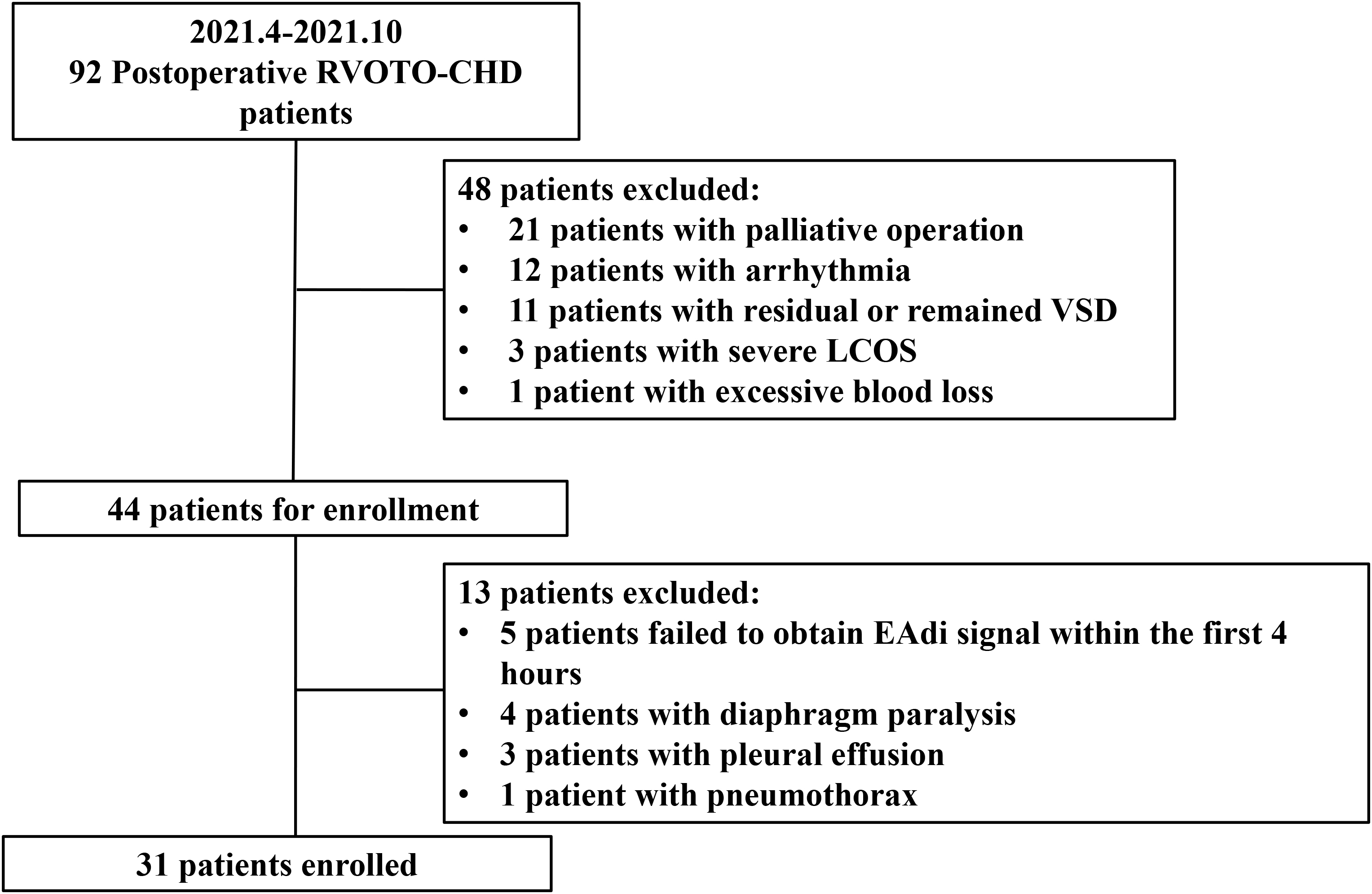 Fig. 1.
Patient flowchart. RVOTO-CHD, right ventricle outflow tract obstructive congenital heart disease; VSD, ventricular septal defect; LCOS, low cardiac output syndrome; EAdi, electrical activity of diaphragm.
Patient characteristics and perioperative outcomes of the enrolled patients are presented in Table 1. Remarkably, the median age of the enrolled patients was 5 months. Most patients were diagnosed with TOF (83.87%) and underwent corrective surgery. The median cardiopulmonary bypass time was 85 minutes, whereas the mean aortic clamp time was 48 minutes. In addition, transannular patch repair was performed in all patients during the reconstruction of the RVOT. Notably, only one patient required noninvasive ventilation support after weaning from the invasive ventilator because of dyspnea caused by laryngeal edema. No deaths were reported in this study.
| Variables (n = 31) | Value |
|---|---|
| Gender Male, n (%) | 18 (58.6) |
| Age, months | 5 (4, 7) |
| Height, cm | 66.27 |
| Weight, kg | 7.27 |
| BSA, m |
0.36 |
| Diagnosis, n (%) | |
| 26 (84) | |
| 2 (6) | |
| 3 (10) | |
| Cardiopulmonary Bypass time, min | 85 (64, 100) |
| Aortic clamping time, min | 48 (40, 58) |
| Invasive ventilation time, hours | 48.2 (43.4, 65.5) |
| ICU LOS, d | 5 (4, 5) |
| In-hospital LOS, d | 13 (12, 17) |
| P-SOFA score | 4.32 |
| NIV, n (%) | 1 (3.23) |
| Mortality, n (%) | 0 (0) |
| Note. Continuous data are presented as mean (SD) or median (IQR).
Categorical data are presented as numbers (%).
BSA, body surface area; DORV/PS, double outlet of right ventricle with subaortic ventricular septal defect and pulmonary stenosis; LOS, length of stay; NIV, noninvasive ventilation; P-SOFA, pediatric sequential organ failure assess; PA/VSD, pulmonary atresia with ventricular septal defect; TOF, tetralogy of Fallot. | |
Cardiac function parameters measured via echocardiography are shown in Table 2.
Compared with right ventricle (RV) hemodynamics during the use of the other two
modes, the hemodynamics during the use of the NAVA mode was significantly
improved. RV cardiac output improved by 10.72% and 19.75% during the use of
NAVA mode compared with cardiac output during the PSV and PRVC modes,
respectively. Notably, S’—a measurement of RV systolic function—also improved
during the use of the NAVA mode compared with that in the other two modes (7.94
| PRVC | PSV | NAVA | p value | |||
| PRVC vs. PSV | PSV vs. NAVA | PRVC vs. NAVA | ||||
| RVOTD, mm | 10.04 |
10.04 |
10.30 |
0.98 | 0.07 | 0.100 |
| RV-VTI, cm | 9.46 |
10.32 |
11.68 |
0.001 | 0.000 | 0.000 |
| RV-CI, L/min/m |
3.19 |
3.45 |
3.82 |
0.003 | 0.564 | 0.035 |
| IVCi, cm | 0.60 |
0.54 |
0.55 |
0.003 | 0.564 | 0.035 |
| IVCe, cm | 0.77 |
0.74 |
0.75 |
0.123 | 0.543 | 0.444 |
| IVCv | 0.22 |
0.27 |
0.27 |
0.036 | 0.98 | 0.067 |
| S’, cm/s | 6.58 |
7.03 |
7.94 |
0.003 | 0.000 | 0.000 |
| TAPSE, cm | 0.56 (0.46, 0.64) | 0.57 (0.51, 0.71) | 0.64 (0.54, 0.78) | 0.303 | 0.019 | 0.000 |
| LVDD, cm | 2.06 |
2.07 |
2.11 |
0.924 | 0.317 | 0.401 |
| LVDS, cm | 1.37 |
1.32 |
1.33 |
0.341 | 0.745 | 0.537 |
| LVEF, % | 66.15 |
67.98 |
67.97 |
0.167 | 0.985 | 0.225 |
| E/e’ | 11.71 (9.86, 13.83) | 11.9 (9.63, 14.00) | 11.63 (9.08, 12.88) | 0.959 | 0.179 | 0.357 |
| Note. Continuous data are presented as mean (SD) or median (IQR). Categorical data are presented as numbers (%). E/e’, the ratio of early diastolic mitral inflow to average mitral annular tissue velocity; IVCe, diameter of inferior vena cava during expiration phase; IVCi, diameter of inferior vena cava during inspiration phase; IVCv, variation of inferior vena cava; LVDD, left ventricle diastolic diameter; LVDS, left ventricle systolic diameter; LVEF, left ventricle ejection fraction; RV-CI, cardiac index of right ventricle; RV-VTI, velocity-time integral of right ventricle; RVOTD, diameter of right ventricular outflow tract; S’, tricuspid annular peak systolic velocity; TAPSE, tricuspid annular plane systolic excursion. | ||||||
Patients had higher systolic blood pressure during the use of NAVA mode (101.61
| PRVC | PSV | NAVA | p value | |||
| PRVC vs. PSV | PSV vs. NAVA | PRVC vs. NAVA | ||||
| HR, beats/minute | 144.84 |
145.48 |
146.48 |
0.714 | 0.403 | 0.318 |
| ABPs, mmHg | 96.35 |
100.55 |
101.61 |
0.088 | 0.547 | 0.022 |
| ABPd, mmHg | 54.65 |
56.74 |
56.97 |
0.164 | 0.869 | 0.244 |
| ABPm, mmHg | 71.00 |
74.10 |
74.65 |
0.058 | 0.726 | 0.112 |
| CVP, mmHg | 11.65 |
12.03 |
12.03 |
0.28 | 1.000 | 0.296 |
| VIS | 15 (13, 17.38) | 15 (12.5, 15.88) | 15 (13, 16.63) | 0.285 | 0.655 | 0.317 |
| CI-PiCCO, L/minute/m |
2.92 |
3.04 |
3.20 |
0.096 | 0.005 | 0.001 |
| SVI, mL/m |
20.38 |
21.23 |
22.00 |
0.089 | 0.036 | 0.002 |
| GEDI, mL/m |
295.74 |
307.26 |
323.74 |
0.225 | 0.043 | 0.026 |
| SVRI, dyn |
1654 (1496, 1900) | 1538 (1412, 1841) | 1458 (1325, 1777) | 0.367 | 0.055 | 0.061 |
| GEF, % | 27.37 |
28.07 |
28.20 |
0.120 | 0.775 | 0.104 |
| DPmx, mmHg/s | 833.5 |
924.5 |
913.53 |
0.007 | 0.644 | 0.156 |
| ELWI, mL/kg | 16.42 |
15.42 |
14.4 |
0.150 | 0.030 | 0.039 |
| NT-proBNP, pg/mL | 17,000 (9850, 27,500) | 17,000 (6350, 25,500) | 16,000 (7445, 22,000) | 0.338 | 0.032 | 0.001 |
| Note. Continuous data are presented as mean (SD) or median (IQR). Categorical data are presented as counts (%). ABPs, systolic arterial blood pressure; ABPd, diastolic arterial blood pressure; CI-PiCCO, cardiac index measured by PiCCO system; CVP, central venous pressure; DPmx, left ventricle myocardial contractility index; ELWI, extravascular lung water index; GEDI, global end diastolic volume index; GEF, global ejection fraction; HR, heart rate; SVI, stroke volume index; SVRI, systemic vascular resistance index; VIS, vasoactive inotropic score. | ||||||
Table 4 shows the respiratory mechanics parameters, blood gas parameters, and NT-proBNP for the three ventilation modes. The respiratory parameters, such as PIP, MAP, DP, and OI, were the lowest during the use of NAVA mode, whereas Vt and Crs were the highest during the use of NAVA mode.
| PRVC | PSV | NAVA | p value | |||
| PRVC vs. PSV | PSV vs. NAVA | PRVC vs. NAVA | ||||
| Freq, breaths/min | 26.5 (22.8, 30.5) | 27 (24, 31.3) | 26.5 (25, 31.3) | 0.059 | 0.837 | 0.087 |
| PIP, cmH |
15 (13, 17) | 13 (12,15.3) | 12 (10, 12.3) | 0.000 | 0.000 | 0.000 |
| MAP, cmH |
7.5 (6, 9) | 7 (6, 8.3) | 6 (5, 7) | 0.003 | 0.000 | 0.000 |
| PEEP, cmH |
4.5 (4, 5) | 4.5 (4, 5) | 4.5 (4, 5) | 1.000 | 1.000 | 1.000 |
| Ti, s | 0.66 (0.6, 0.7) | 0.61 (0.49, 0.7) | 0.76 (0.67, 0.83) | 0.043 | 0.000 | 0.000 |
| Vt, mL | 59.1 |
54.7 |
53.6 |
0.001 | 0.535 | 0.013 |
| Vt, mL/kg | 8.2 |
7.6 |
7.5 |
0.002 | 0.595 | 0.011 |
| DP, cmH |
10.9 |
9.2 |
6.9 |
0.000 | 0.000 | 0.000 |
| Crs, mL/cmH |
0.8 |
0.9 |
1.1 |
0.012 | 0.000 | 0.000 |
| EAdi-P, uV | 3.4 (1.8, 6.2) | 3.9 (2.6, 5.0) | 4.6 (3.1, 5.1) | 0.462 | 0.551 | 0.183 |
| EAdi-m, uV | 0.3 (0.2, 0.8) | 0.4 (0.2, 0.7) | 0.5 (0.3, 0.7) | 0.502 | 0.387 | 0.592 |
| FiO |
0.4 (0.39, 0.43) | 0.4 (0.39, 0.43) | 0.4 (0.39, 0.43) | 1.000 | 1.000 | 1.000 |
| pH | 7.43 |
7.43 |
7.43 |
0.080 | 0.588 | 0.254 |
| PaCO |
40.9 |
39.9 |
41.0 |
0.349 | 0.075 | 0.786 |
| PaO |
117.1 |
114.9 |
119.9 |
0.597 | 0.156 | 0.579 |
| PaO |
288.3 |
283.1 |
292.8 |
0.630 | 0.207 | 0.668 |
| OI | 2.6 (2.0, 3.8) | 2.6 (1.9, 3.9) | 2.1 (1.9, 2.7) | 0.367 | 0.000 | 0.000 |
| SaO |
99 (97.7, 99.5) | 98.4 (97.3, 99.0) | 98.8 (97.9, 99.4) | 0.629 | 0.130 | 0.327 |
| BE | 2.3 |
2.3 |
2.8 |
0.890 | 0.114 | 0.152 |
| PcvCO |
48.5 |
48.4 |
49 |
0.971 | 0.249 | 0.367 |
| ScvO |
68.1 |
68.7 |
67.9 |
0.688 | 0.473 | 0.864 |
| SaO |
30.1 |
29.4 |
30.7 |
0.639 | 0.222 | 0.585 |
| PcvCO |
7.6 |
8.5 |
7.9 |
0.343 | 0.319 | 0.671 |
| LAC, mmol/L | 0.8 (0.7, 1.4) | 0.8 (0.7, 1.0) | 0.8 (0.6, 1.2) | 0.036 | 0.260 | 0.280 |
| Note. Continuous data are presented as mean (SD) or median (IQR).
Categorical data are presented as counts (%). BE, base excess; Crs, compliance
of respiratory system; DP, driving pressure; EAdi-m, minimal voltage of
electrical activity of diaphragm; EAdi-p, peak voltage of electrical activity of
diaphragm; FiO | ||||||
To the best of our knowledge, information on the cardiopulmonary interaction affected by the ventilation mode using different hemodynamic monitoring methods is insufficient in children with CHD. This study found that the use of spontaneous breathing mode, especially the NAVA mode, could increase pulmonary blood flow measured via echocardiography, and the benefit of the spontaneous breathing mode could also be seen using the thermodilution technique. In addition, the respiratory mechanics were found to be improved in the PSV and NAVA modes.
According to the results of echocardiography, after switching to PSV and NAVA mode, the variation rate of the inferior vena cava in children with reduced intrathoracic pressure and increased intraabdominal pressure caused by diaphragm movement during spontaneous breathing increased significantly, and the indices of right ventricular contractility, such as TAPSE and S’, improved. We found that RV-VTI and RV-CI increased, and this finding was similar to the results of a study by Becker et al. [10]. Furthermore, the use of PiCCO catheter with thermodilution technique and pulse waveform analysis technique revealed that GEDI increased with the increased venous return due to spontaneous respiration as well as the cardiac output. Liet et al. [12] reported that cardiac output increased in children with low cardiac output after congenital heart disease surgery during the use of NAVA mode for 30 minutes. In the present study, right ventricular cardiac output measured by ultrasound (RV-CI) differed from that measured by the thermodilution method (CI-PiCCO) owing to the overestimation of RV-CI caused by pulmonary insufficiency in children with right ventricular transannular patch enlargement or by the use of different methods, such as that reported by Wetterslev et al. [13].
After switching to a spontaneous breathing mode, especially the NAVA mode, some respiratory mechanics also changed significantly due to decreased intrathoracic pressure during inspiration, resulting in a decrease in PIP, MAP, and DP and an improvement in Crs. These results are consistent with our previous study in children with single ventricles [14] and many other studies [12, 15, 16, 17]. In some studies of the NAVA mode, children ventilated with the NAVA mode had lower airway pressures than children ventilated with the continuous positive airway pressure mode, suggesting that the NAVA mode used before weaning may be more appropriate than the other modes [18, 19].
Some children may develop weaning-induced pulmonary edema (WIPO) after weaning from mechanical ventilation, mainly due to increased venous return and consequent improvement in right ventricular output as well as a displacement of the intraventricular septum to the left ventricle after ventilator discontinuation [20]. Notably, an increase in NT-proBNP and ELWI after spontaneous breathing can be an important predictor of WIPO [21]. In our study, NT-proBNP, ELWI, and echocardiographic parameters representing left ventricular filling pressure (E/e’) were also measured after switching the mechanical ventilation modes. We found that these indices did not significantly increase in the spontaneous breathing mode. NT-proBNP was lower in the NAVA mode compared with PRVC and PSV, and this was possibly related to the decrease in right ventricular pressure. The decrease in ELWI and NT-proBNP in the NAVA ventilation mode suggests that these children are less likely to develop WIPO.
This study had several limitations. First, no control group was included, and this was only a self-controlled study. Thus, whether hemodynamics and respiratory mechanics changed after surgery in children who did not use the NAVA or PSV mode remains to be investigated. Second, right ventricular pressure data were not collected in this study. Most children had mild tricuspid regurgitation; therefore, echocardiography may underestimate the right ventricular pressure. The patients included in this study were children aged 4–7 months; thus, the placement of a floating pulmonary artery catheter was difficult. Therefore, no right ventricular pressure measurements were performed in this study.
Utilizing spontaneous ventilator modes, especially the NAVA mode, after corrective surgery in patients with RVOTO-CHD could improve their right heart hemodynamics and respiratory mechanics. Further randomized controlled trials should be conducted to verify the advantages of spontaneous ventilation modes in such patients.
The datasets used and/or analyzed during the current study are available from the corresponding author on reasonable request.
(I) Conception and design—XG, LZ, ZX, JZ; (II) Administrative support—ZX, JZ; (III) Provision of study materials or patients—XG, LZ, MZ, YL, CL, ZX, JZ; (IV) Collection and assembly of data—XG, LZ, MZ, YL, CL; (V) Data analysis and interpretation—XG, LZ; (VI) Manuscript writing—XG, LZ, MZ, YL, CL, ZX, JZ; (VII) Final approval of manuscript—XG, LZ, MZ, YL, CL, ZX, JZ.
This study was conducted in accordance with the Declaration of Helsinki (as revised in 2013) and approved by the Institutional Health Research Ethics Board of Shanghai Children’s Medical Center, Shanghai Jiao Tong University School of Medicine (number: SCMC-CHC2021006). Informed consent was obtained from guardians of the patients for participation in this study.
Not applicable.
This work was supported by “The Biomedical and Engineering (Science) Interdisciplinary Study Fund of Shanghai Jiaotong University” (No: YG2019QNA04 to Xiaolei Gong and No: YG2019ZDA03 to Zhuoming Xu).
The authors declare no conflict of interest.
References
Publisher’s Note: IMR Press stays neutral with regard to jurisdictional claims in published maps and institutional affiliations.