IMR Press / FBS / Volume 14 / Issue 2 / DOI: 10.31083/j.fbs1402013
Open Access Original Research
The Effects of Novel Formulations of Edaravone and Curcumin in the Mouse Intrastriatal Lipopolysaccharide Model of Parkinson’s Disease
Show Less
1 Health and Biomedical Innovation, Clinical and Health Sciences, University of South Australia, Adelaide, 5000 South Australia, Australia
2 Centre for Pharmaceutical Innovation, Clinical and Health Sciences, University of South Australia, Adelaide, 5000 South Australia, Australia
*Correspondence: Larisa.Bobrovskaya@unisa.edu.au (Larisa Bobrovskaya)
Academic Editor: Govindan Dayanithi
Front. Biosci. (Schol Ed) 2022, 14(2), 13; https://doi.org/10.31083/j.fbs1402013
Submitted: 7 February 2022 | Revised: 6 April 2022 | Accepted: 8 April 2022 | Published: 7 May 2022
Copyright: © 2022 The Author(s). Published by IMR Press.
This is an open access article under the CC BY 4.0 license.
Abstract

The major hallmark of Parkinson’s disease (PD) is the degeneration of dopaminergic neurons in the substantia nigra (SN), which is responsible for the core motor symptoms of PD. Currently, there is no cure for PD, and its prevalence is increasing, prompting the search for novel neuroprotective treatments. Neuroinflammation is a core pathological process in PD, evident by increased inflammatory biomarkers in the SN and cerebrospinal fluid. Interestingly, epidemiological studies have reported a reduced risk of PD in users of non-steroidal anti-inflammatory drugs compared to non-users, suggesting the neuroprotective potential of anti-inflammatory drugs. Therefore, this study aimed to: (1) test the efficacy of novel oral formulations of edaravone (EDR) and curcumin (CUR) (which possess anti-inflammatory and anti-oxidative properties) to alleviate motor and non-motor symptoms, and associated pathology in the intrastriatal lipopolysaccharide (LPS) model of PD; (2) investigate the expression of proteins linked to familial PD and markers of autophagy in the intrastriatal LPS model treated with EDR and CUR. Fifty-two C57BL/6 mice were divided into 4 groups, namely; (1) control + vehicle; (2) LPS + vehicle; (3) LPS + EDR (made in vehicle) and (4) LPS + CUR (made in vehicle). 10 μg of LPS was administered stereotaxically into the right striatum, and EDR and CUR treatments were initiated 2-weeks after the LPS injections. Behavioural tests were carried out at 4- and 8-weeks after LPS injection followed by tissue collection at 8-weeks. Intrastriatal administration of LPS induced motor deficits and anxiety-like behaviours at 4- and 8-weeks, which were accompanied by astroglial activation, increased protein expression of α-synuclein, heat shock cognate protein of 70 kDa (HSC-70) and Rab-10, and reduced levels of tyrosine hydroxylase (TH) protein in the striatum. Additionally, LPS induced astroglial activation in the olfactory bulb, along with changes in the protein expression of HSC-70. The changes associated with EDR and CUR in the striatum and olfactory bulb were not statistically significant compared to the LPS group. Intrastriatal administration of LPS induced pathological changes of PD such as motor deficits, reduced expression of TH protein and increased α-synuclein protein, as well as some alterations in proteins linked to familial PD and autophagy in the olfactory bulb and striatum, without pronounced therapeutic effects of EDR and CUR. Our results may suggest that EDR and CUR lack therapeutic effects when administered after the disease process was already initiated. Thus, our treatment regimen or the physicochemical properties of EDR and CUR could be further refined to elevate the therapeutic effects of these formulations.

Keywords
Parkinson's disease
intrastriatal lipopolysaccharide
striatum
olfactory bulb
edaravone
curcumin
neuroinflammation
autophagy
1. Introduction

Parkinson’s disease (PD) is characterised by the cardinal motor features such as bradykinesia, rigidity, resting tremors, and postural/gait disorders. These motor features are largely associated with the degeneration of dopaminergic neurons in the substantia nigra pars compacta (SNpc), which results in the depletion of striatal dopamine, a neurotransmitter essential for the control of movement [1]. PD is classically a motor disorder; however, it has a non-motor aspect characterised by olfactory dysfunction, gastrointestinal dysfunction, anxiety, depression, autonomic disturbance, and cognitive dysfunction that is now well recognised, and evident to precede the onset of motor symptoms [2, 3]. Due to the lack of treatments, it is the non-motor symptoms of PD that have a significant effect on the patients’ quality of life [2]. Additionally, PD is characterised by the presence of Lewy bodies/neurites in the surviving dopaminergic neurons of the SN, and these are cytoplasmic inclusions consisting of abnormal aggregates of α-synuclein protein. α-Synuclein protein is highly expressed in the synaptic terminals, and although its function is not explicitly understood, it is implicated in dopamine biosynthesis, vesicle trafficking, and neurotransmission [4, 5, 6]. The majority of PD cases have no known cause. Nonetheless, animal models of PD have been paramount in understanding the deleterious effects of pathological processes such as neuroinflammation, oxidative stress, mitochondrial dysfunction, defective protein clearance, and neurotrophic insufficiency [1].

Currently, there are no disease-modifying therapies for PD, and the available treatments such as levodopa, dopamine agonists, monoamine oxidase B inhibitors, catechol-O-methyltransferase inhibitors, and deep brain stimulation, only provide relief from motor symptoms [7]. Unfortunately, the beneficial effects of these treatments decrease with time due to the progressive nature of PD, prompting the search for novel treatment approaches [2, 7]. The current PD treatments focus on preserving the levels of endogenous dopamine and do not target the core pathological processes implicated in PD. Therefore, targeting the prominent pathological mechanisms of PD, in combination with the available dopamine therapy could have disease-modifying effects in patients. Neuroinflammation, oxidative stress, and defective protein clearance are crucial events in the aetiology of PD, and for this reason, we aimed to further explore these pathways in this study. Neuroinflammation is a well-recognised phenomenon in PD patients, evident by the presence of activated microglia cells in the SN of post-mortem PD brains, and pro-inflammatory cytokines in the serum and cerebrospinal fluid [8, 9, 10]. These findings are supported by epidemiological studies indicating a decreased risk of PD in users of non-steroidal anti-inflammatory drugs compared to non-users, emphasizing the importance of controlling inflammation in PD [11]. Additionally, a multitude of genetic loci have been associated with the monogenic form of PD which has Mendelian inheritance, and these include genes encoding for α-synuclein, leucine-rich repeat kinase 2 (LRRK2), PTEN-induced putative kinase 1 (PINK1), parkin and deglycase DJ-1 protein (DJ-1) [12]. Mutations in the aforementioned proteins impair autophagy which is important for cellular clearance of α-synuclein, and damaged mitochondria [12, 13]. In return, impaired autophagy results in the accumulation of α-synuclein protein, damaged mitochondria and oxidative stress, which are detrimental to the nigral dopaminergic neurons due to their high energy demands, increased sensitivity to inflammation and low antioxidant capacity [12, 14, 15, 16, 17]. Autophagy is divided into three types: macroautophagy; microautophagy and chaperone-mediated autophagy (CMA), all of which converge in degradation at the lysosome [18]. Briefly, in macroautophagy, a double membrane known as an autophagosome (marked by microtubule-associated protein 1A/1B-light chain 3 (LC3)) sequesters the cellular cargo, and it is then fused with the lysosome. Secondly, microautophagy involves a direct uptake of the cellular cargo via invagination or protrusion of the lysosomal membrane. Lastly, CMA involves the identification of protein cargo by molecular chaperones, which are transported across the lysosomal membrane [18]. Along with the common mutations in PD, it has also been reported that the components of the autophagic pathways are compromised in PD patients [19, 20]. The proteins linked to hereditary PD and autophagy are not well explored in PD and future studies are needed.

In our recent work, we characterised the intrastriatal LPS model of PD which is beneficial for understanding the neuroinflammatory aspect of the disease [21], and for investigating the efficacy of antioxidants and anti-inflammatory compounds. For this study, we selected to further explore edaravone and curcumin which have been shown to have neuroprotective effects in-vivo due to their antioxidant and anti-inflammatory properties [22, 23, 24]. These compounds can be easily repurposed for clinical treatment of PD if they have disease-modifying effects. Edaravone has already been approved by the Food and Drug Administration for the treatment of amyotrophic lateral sclerosis in the USA, and by authorities in Asian countries for the treatment of ischemic stroke. However, the drug is administered twice a day intravenously due to its poor oral bioavailability which could be inconvenient and distressful for patients [25]. In contrast, curcumin is an active compound of turmeric, commonly used in Indian species with great neuroprotective potential. Nonetheless, its progress to clinical use has been impeded by its poor oral bioavailability associated with its physiochemical properties [26]. Previously, Parikh and colleagues from our laboratory developed novel oral formulations of edaravone (EDR) and curcumin (CUR) using soluplus, a polymer as a drug carrier [27, 28]. These novel formulations had improved physical properties and oral bioavailability, and were shown to have protective effects in animal models of Alzheimer’s disease (AD) [29, 30]. Therefore for this study, we aimed to investigate the therapeutic effects of EDR and CUR on motor and non-motor symptoms of PD, and the associated pathological changes such as reduced tyrosine hydroxylase (TH), increased α-synuclein protein, and activation of astroglial cells in the intrastriatal LPS mouse model of PD. Secondly, we aimed to further explore the expression of proteins linked to familial PD such as LRRK2, PINK1 and parkin as well as markers of autophagy such as LC3, heat shock cognate protein of 70 kDa (HSC-70) and lysosomal-associated membrane protein 1 (LAMP-1) in LPS model treated with EDR and CUR.

2. Materials and Methods
2.1 Preparation of Novel Edaravone (EDR) and Curcumin (CUR) Formulations

Curcumin was purchased from Chem-Supply Pty Ltd. (South Australia, Australia), soluplus was a gift from BASF Australia Ltd, and EDR was obtained from Suzhou Auzone Biotech, China. The novel oral formulation of CUR was prepared by combining soluplus and neat curcumin in a ratio of 1:10 and then dissolved in absolute ethanol [27]. The ethanol was evaporated using a rotary evaporator, and the novel formulation of CUR was left to dry in the desiccator. After drying, the CUR formulation was pulverised with mortar and pestle and dissolved in acidified drinking water.

2.1.1 Animals

All the animal experiments were approved by the Animal Ethics Committee of the University of South Australia. Fifty-two C57BL/6 mice were purchased from the Animal Resources Centre (Western Australia, Australia) when they were 9-weeks old, and housed at the Core Animal Facility at the University of South Australia for the duration of the experiment. The mice were housed in a pathogen-free environment with a 12 h alternating light/dark cycle and had an unlimited supply of food and water. LPS (E. coli 0111.4) was purchased from Sigma-Aldrich (USA) and diluted to 5 mg/mL in sterile water and stored at –80 °C. C57BL/6 mice were trained for the buried food-seeking test and rotarod test before the stereotaxic injections to ensure that they were accustomed to these tests. Subsequently, the mice were randomly divided into 4 groups, namely: control + vehicle (soluplus) (n = 12), LPS + vehicle (n = 15), LPS + EDR (made in vehicle) (n = 13), and LPS + CUR (made in vehicle) (n = 12). A total of 10 μg of LPS was administered unilaterally into the right striatum when the mice were about 12-weeks old. The mice were then given either soluplus (120 mg/kg), EDR (48 mg/kg), or CUR (48 mg/kg) orally in drinking water starting at 2-weeks after LPS injection until the end of the study (8-weeks in total). The formulations were replaced every 5-days. EDR and CUR formulations were made in soluplus to improve their physicochemical properties, and the doses were selected based on the response studies performed by Parikh and colleagues [27, 28, 29, 30]. Moreover, a series of behavioural tests were performed at 4- and 8-weeks after LPS injection to investigate olfactory function, anxiety-like behaviour, and motor function. The mice were humanely killed after the last behavioural test at 8-weeks via intraperitoneal injection of sodium pentobarbitone, and all tissues of interest were collected. The experimental design and timeline are shown in Fig. 1.

Fig. 1.

The timeline of the study. C57BL/6 mice were injected intrastriatally with 10 μg of LPS and then treated with EDR and CUR starting at 2-weeks post-LPS until the end of the study period. A behavioural test battery was conducted at 4- and 8-weeks, followed by tissue collection after the last behaviour test.

2.1.2 Stereotaxic Surgery

All the surgical procedures were conducted according to recent studies [21]. Briefly, the mice were deeply anaesthetised via inhalation of isoflurane and mounted onto the stereotaxic frame (motorized stereotaxic apparatus, Stoelting, USA). Once the mice were stable under anaesthesia, the skin on the cranium was prepared with 2% chlorhexidine/70% ethanol, and an incision was made on the scalp from the lambda and extended anteriorly to between the eyes. The bregma point was identified by applying 3% hydrogen peroxide to the exposed cranium and the two injection sites of the right striatum were identified using the following coordinates from the bregma point (point A: +1.2 mm anterior-posterior, +1.5 mm medial-lateral, –3.5 mm deep, and point B: –0.34 mm anterior-posterior, +2.5 mm medial-lateral, and –3.2 mm deep) [31]. Subsequently, a micro-drill was used to drill a hole at each of the injection sites, and a 10 μL Hamilton syringe coupled with a quintessential stereotaxic injector was lowered to the ventral coordinates. The quintessential stereotaxic injector was then used to infuse 1 μL of LPS (5 μg) per an injection site at the rate of 0.5 μL/min. Once the infusion was completed, the needle was left in place for 5 min to limit the reflux of the injected solution, and then it was slowly withdrawn. A local analgesic mixture of lignocaine and bupivacaine was applied to the surgical wound, and the two ends of the scalp were glued together with surgical glue. The mice were then given 0.5–0.8 mL of subcutaneous saline and placed in a recovery cage.

2.2 Behavioural Testing

Behavioural testing was conducted at 4- and 8-weeks after intrastriatal injection of LPS to determine if the animals display olfactory deficits (assessed by buried-food seeking test), anxiety-like behaviour (assessed by open field test), and motor deficits (assessed by rotarod test). These behavioural tests are described in more detail below.

2.2.1 Buried Food-Seeking Test

The buried food-seeking test was used to assess olfactory function, and it was performed according to previous studies [21]. Briefly, mice fasted overnight for 14–18 h, were placed into a clean individual home cage. They were given up to 10 min to locate a standard chow pellet hidden underneath the bedding of the home cage. The time it took for the mice to find the hidden food was recorded as the latency time.

2.2.2 Open Field Test

The open-field test was used to assess anxiety-like behaviour, and it was performed according to recent studies [21]. Firstly, the mice were placed in an open field arena that was divided into a peripheral and a central zone. Subsequently, they were allowed to explore the open field arena for 5 min, and their movement was tracked with ANY-maze, a video tracking software (Stoelting, USA).

2.2.3 Rotarod Test

The rotarod test assesses balance, endurance, and motor coordination, and it was used to assess motor deficits in LPS injected mice [21, 32]. Briefly, the mice were placed on a rotarod apparatus (Mouse RotaRod NG, Ugo Basile) set to accelerate from 5–30 rpm within 5 min. The latency time was recorded for each mouse, and this refers to the time it took for the mouse to fall off the rotating rod or hang on the rotating rod and swung 360. The test was repeated three times for each mouse, and the latency time was the averaged of the three attempts. The mice were subjected to the same behavioural conditions prior to LPS injection in order to acclimatise to the test.

2.3 Tissue Collection

After the last behavioural test at 8-weeks, the mice were humanely killed via intraperitoneal injection of sodium pentobarbitone (60 mg/kg). The brain tissues were collected fresh, sliced into different areas of interest such as the olfactory bulb and the striatum using brain matrix, and stored at –80 °C.

2.4 Tissue Homogenisation and Total Protein Estimation

Fresh brain tissues (e.g., olfactory bulb and striatum) were homogenised in RIPA buffer (50 mM tris, 150 mM sodium chloride, 1 mM ethylenediaminetetraacetic acid, 0.5% Triton X-100, 0.5% sodium deoxycholate, pH 7.4) plus cocktail protease inhibitor using Precellys 24 Homogeniser (Bertin Technologies, Montigny-le-Bretonneux, France). The protein concentration of the homogenates was measured with a Micro-BCATM protein assay kit (Thermo-scientific, Rockford, IL, USA) according to the manufacturer’s instructions.

2.5 Western Blotting

Each of the homogenates was diluted in a sample buffer in preparation for gel electrophoresis. Western blotting was carried out as outlined in our previous study [21]. Briefly, the proteins were electrophoretically separated on a sodium dodecyl sulfate polyacrylamide gel for 90 min. The proteins on the gel were transferred onto a nitrocellulose membrane for 90 min at 0.6 or 0.8 amps, and the membrane was air-dried for 1 h to allow better adhesion of the proteins. The nitrocellulose membrane was then blocked for 1 h with 5% milk or 5% BSA to reduce non-specific binding and incubated overnight with specific primary antibodies (Table 1). Subsequently, the nitrocellulose membrane was incubated for 1 h at room temperature with corresponding secondary antibodies for near-infrared western blot detection (Table 1). The proteins on the nitrocellulose membrane were visualized with Odyssey CLx imaging system (LI-COR Biosciences, USA), and quantified with Image Studio Lite 5.2 (LI-COR Biosciences, Los Angeles, LA, USA) using β-actin as a loading control.

Table 1.A list of antibodies used in this study.
Antibodies Company Dilution
Primary antibodies
Mouse anti-heat shock cognate protein of 70 kDa (HSC-70) Santa Cruz Biotechnology 5% BSA/TBST (1:1000)
Mouse anti-lysosomal-associated membrane protein 1 (LAMP-1) Santa Cruz Biotechnology 5% BSA/TBST (1:500)
Mouse anti- microtubule-associated protein 1A/1B-light chain 3 b (LC3) Santa Cruz Biotechnology 5% BSA/TBST (1:250)
Mouse anti-parkin Santa Cruz Biotechnology 5% BSA/TBST (1:500)
Mouse anti-PINK1 Santa Cruz Biotechnology 5% BSA/TBST (1:1000)
Mouse anti-tyrosine hydroxylase (TH) Sigma 5% BSA/TBST (1:7000)
Mouse anti-β-actin Abcam TBST (1:15000)
Rabbit anti-glial fibrillary acidic protein (GFAP) Dako 5% milk/TBST (1:2000)
Rabbit anti-LRRK2 Abcam 5% BSA/TBST (1:500)
Rabbit anti-Rab-10 Abcam 5% BSA/TBST (1:1000)
Rabbit anti-Rab-10 (phospho-T73) Abcam 5% BSA/TBST (1:250)
Rabbit anti- α-synuclein C-20 Santa Cruz Biotechnology 5% BSA/TBST (1:300)
Secondary antibodies
Donkey anti-rabbit IgG Li-Cor Biosciences TBST (1:20,000)
Goat anti-mouse IgG Li-Cor Biosciences TBST (1:20,000)
2.6 Statistical Analyses

Using GraphPad Prism 8 software (San Diego, CA, USA), we analysed the behavioural data with 2-way ANOVA test, followed by Tukey’s multiple comparisons test. In addition, the western blot data were analysed with the Kruskal-Wallis test followed by Dunn’s multiple comparisons test. All the data are presented as mean ± standard error of the mean (SEM). Statistical significance was achieved when p 0.05.

3. Results
3.1 The Effects of EDR and CUR on Motor and Olfactory Function after Intrastriatal Administration of LPS

Intrastriatal administration of LPS induced motor impairment at 4-weeks (p = 0.0360) and 8-weeks (p = 0.0023) in LPS injected mice versus controls, indicated by a reduction in latency time in the rotarod test (Fig. 2A). The impairment in motor function was not rescued by EDR or CUR. Secondly, the olfactory function was not altered by intrastriatal administration of LPS according to the buried food-seeking test (Fig. 2B).
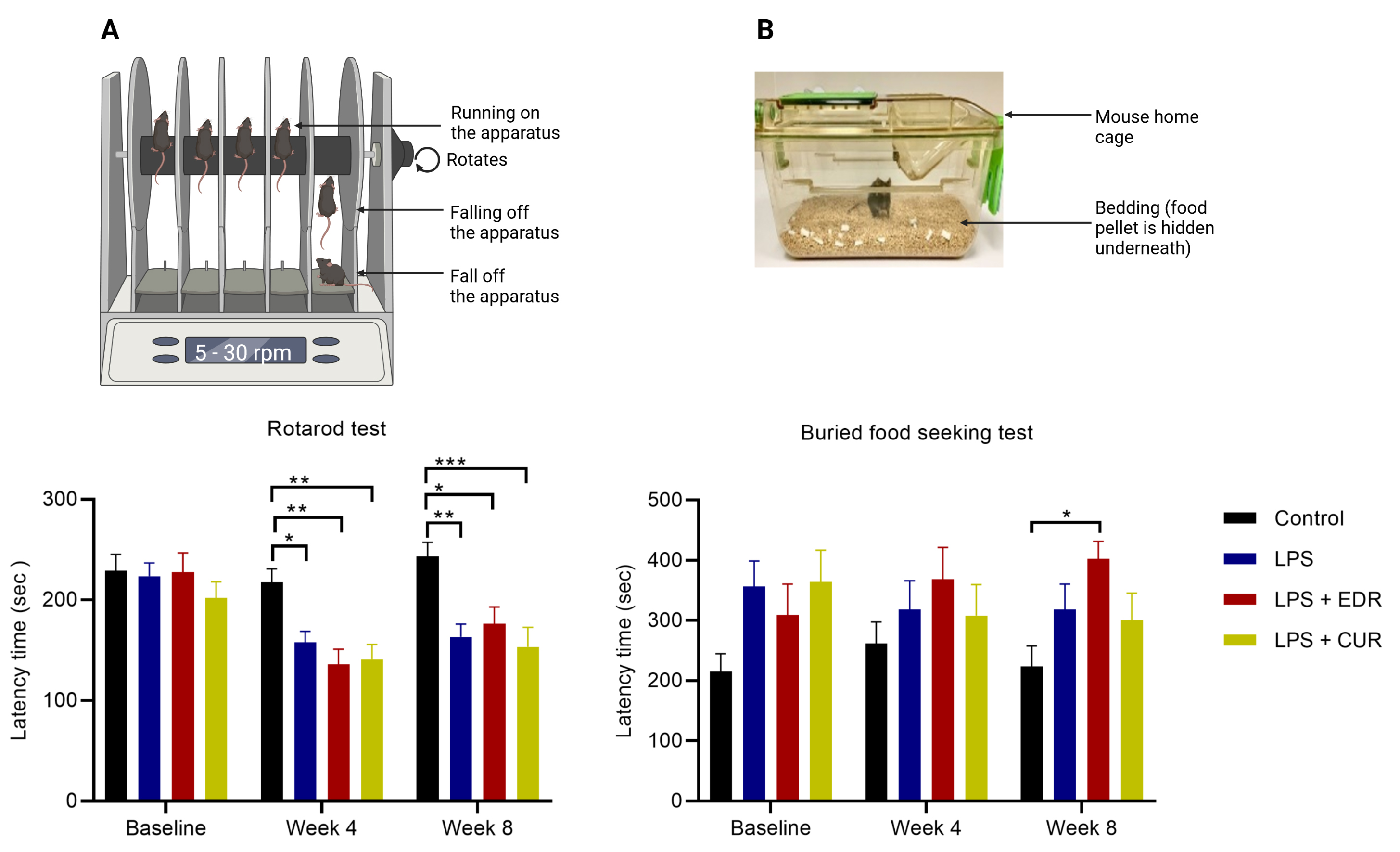
Fig. 2.

The effects of EDR and CUR on motor and olfactory function following intrastriatal administration of LPS. (A) The effects of EDR and CUR on motor function assessed with the rotarod test (latency time in sec). (B) effects on olfactory function assessed with buried food-seeking test (latency time in sec). The results are presented as mean ± SEM (control n = 12; LPS n = 15; LPS + EDR n = 13; LPS + CUR n = 12). Statistical analyses were performed with a 2-way ANOVA test, followed by Tukey’s multiple comparisons test; p < 0.05 (*), p < 0.01 (**), p < 0.001 (***). The figure was created with BioRender.com.

3.2 The Effects of EDR and CUR on LPS Induced Anxiety-like Behaviour

The open-field test was used to examine the effects of LPS on voluntary movement and anxiety-like behaviour. Intrastriatal LPS in C57BL/6 mice did not induce any significant changes in the voluntary movement, illustrated by the total distance travelled in the open field test (Fig. 3B). Also, there was a trend towards reduction in the number of central zone entries in the open field test at 4-weeks (p = 0.0660) and a significant reduction at 8-weeks (p = 0.0229) (Fig. 3C), accompanied by reduced central zone time at 8-week (p = 0.0005) (Fig. 3D) in LPS group compared to the control mice. Unlike EDR, the number of central zone entries was not significantly different in controls versus the CUR group at 4-weeks (p > 0.9999) and 8-weeks (p = 0.1390).

Fig. 3.

The effects of EDR and CUR on voluntary movement and anxiety-like behaviour following intrastriatal administration of LPS. (A) Schematic of the open field arena. (B) Total distance travelled in the open field (in metres denoted as m). (C) Number of central zone entries. (D) Central zone time (sec). The results are presented as mean ± SEM (control n = 12; LPS n = 15; LPS + EDR n = 13; LPS + CUR n = 12). Statistical analyses were performed with 2-way ANOVA test, followed by Tukey’s multiple comparisons test; p < 0.05 (*), p < 0.01 (**), p < 0.001 (***). The figure was created with BioRender.com.

3.3 The Effects of EDR and CUR on the Protein Expression of TH, α-Synuclein, and GFAP in the Striatum Following Intrastriatal Administration of LPS

The major neuropathological hallmarks of PD are the degeneration of the nigrostriatal pathway, constituted by dopaminergic neurons of the SN and their neuronal projections to the striatum, and the presence of Lewy bodies made up predominately of α-synuclein protein. These pathological hallmarks are examined in-vivo via the detection of TH and α-synuclein protein, respectively. Intrastriatal LPS significantly reduced TH expression (p = 0.0476) (Fig. 4B) in the striatum versus the control group. Also, these findings were accompanied by increased protein expression of striatal α-synuclein (p = 0.0057) (Fig. 4C) and GFAP (p = 0.0009) (Fig. 4D), a marker of astroglial activation compared to the controls. The protein expression of α-synuclein was not significantly different in the CUR group versus controls (p = 0.9907). These findings were also true regarding the effects of EDR (controls versus EDR, p = 0.1166) and CUR (controls versus CUR, p = 0.1317) on the expression of GFAP protein.

Fig. 4.

The effects of EDR and CUR on the protein expression of TH, α-synuclein, and GFAP in the striatum following intrastriatal administration of LPS. (A) Representative immunoblots for the protein expression of TH, α-synuclein, and GFAP. (B) Densitometric analysis of TH. (C) Densitometric analysis of α-synuclein. (D) Densitometric analysis of GFAP. The results are presented as mean ± SEM (control n = 6–10; LPS n = 6–10; LPS + EDR n = 6; LPS + CUR n = 6–10). The statistical analyses were performed with the Kruskal-Wallis test, followed by Dunn’s multiple comparisons test for all the experimental groups; p < 0.05 (*), p < 0.01 (**), p < 0.001 (***).

3.4 The Effects of EDR and CUR on the Protein Expression of LAMP-1, HSC-70, and LC3 in the Striatum Following Intrastriatal Administration of LPS

LAMP-1, HSC-70, and LC3 are involved in autophagy; therefore, we scrutinised the effects of EDR and CUR on these proteins in LPS treated mice. Intrastriatal administration of LPS did not alter the expression of LAMP-1 protein (p = 0.8499) (Fig. 5B) and LC3 (p
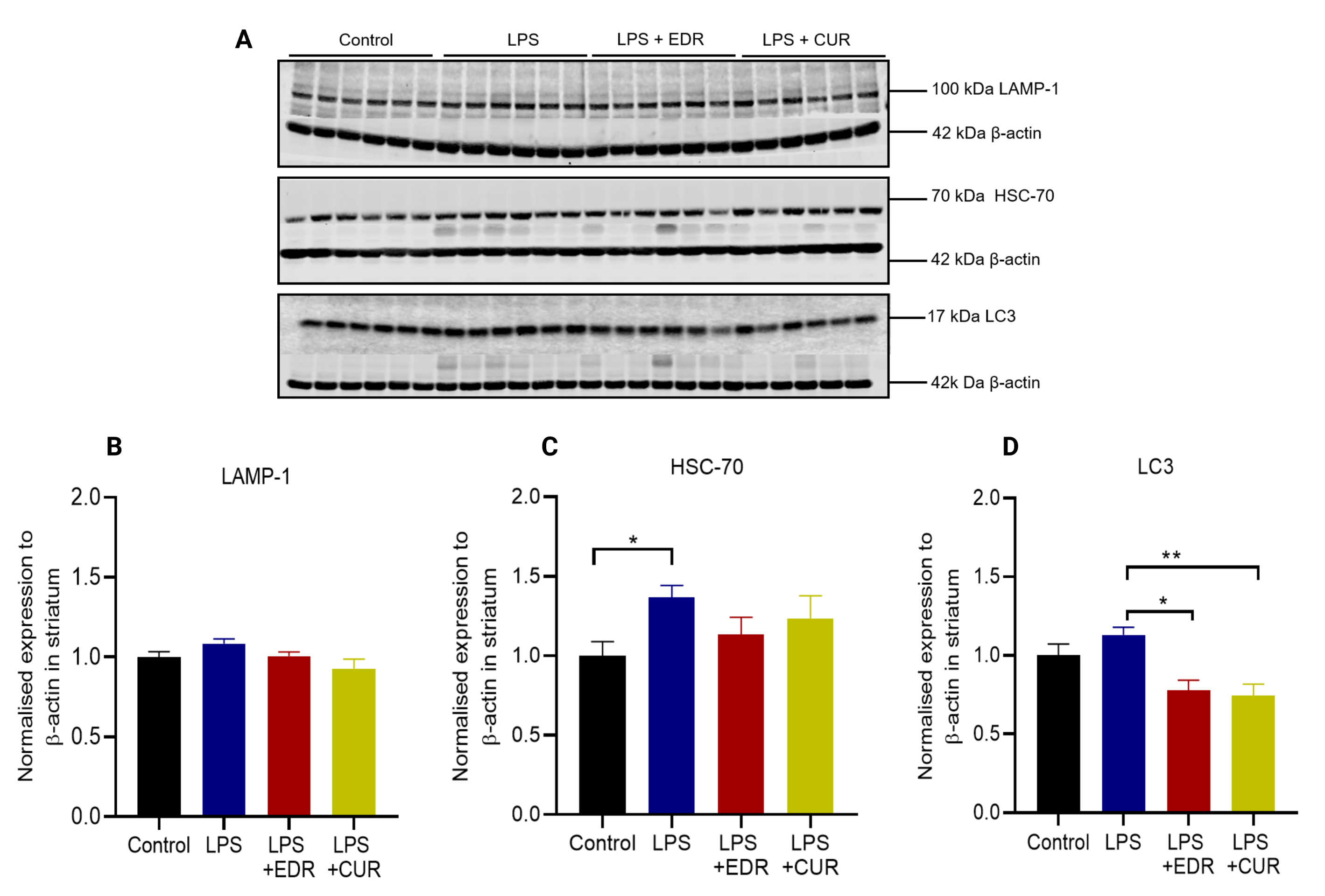
 ˃ 0.9999) (Fig. 5D) relative to controls; however, there was a significant reduction in LC3 protein in EDR (p = 0.0173) and CUR groups (p = 0.0076) compared to LPS group. Furthermore, there was a significant increase in the expression of HSC-70 protein (p = 0.0226) (Fig. 5C) in the control versus LPS treated mice, and this was not affected by EDR and CUR.

Fig. 5.

The effects of EDR and CUR on the protein expression of LAMP-1, HSC-70, and LC3 in the striatum following intrastriatal administration of LPS. (A) Representative immunoblots for the protein expression of LAMP-1, HSC-70, and LC3. (B) Densitometric analysis of LAMP-1. (C) Densitometric analysis of HSC-70. (D) Densitometric analysis of LC3. The results are presented as mean ± SEM (control n = 6–10; LPS n = 6–10; LPS + EDR n = 6; LPS + CUR n = 6–10). The statistical analyses were performed with the Kruskal-Wallis test, followed by Dunn’s multiple comparisons test for all the experimental groups; p < 0.05 (*), p < 0.01 (**).

3.5 The Effects of EDR and CUR on the Protein Expression of PINK1 and Parkin in the Striatum Following Intrastriatal Administration of LPS

Mutations in the genes for PINK-1 and parkin are among the common causes of genetic PD, and the encoded proteins are essential for defence against oxidative stress, and clearance of defective mitochondria via autophagy. Intrastriatal administration of LPS induced a trend towards increased protein expression of PINK1 (p = 0.0736) (Fig. 6B) and parkin (p = 0.0858) (Fig. 6C). Interestingly, there was a significant reduction in the expression of parkin protein in the CUR group compared to the LPS group (p = 0. 0225).
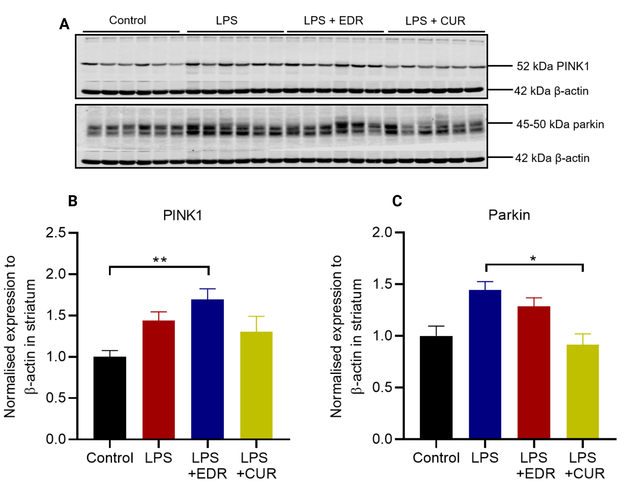
Fig. 6.

The effects of EDR and CUR on the protein expression of PINK1 and parkin in the striatum following intrastriatal administration of LPS. (A) Representative immunoblots for the protein expression of PINK1 and parkin. (B) Densitometric analysis of PINK1. (C) Densitometric analysis of parkin. The results are presented as mean ± SEM (control n = 6–10; LPS n = 6–10; LPS + EDR n = 6; LPS + CUR n = 6–10). The statistical analyses were performed with the Kruskal-Wallis test, followed by Dunn’s multiple comparisons test for all the experimental groups; p < 0.05 (*), p < 0.01 (**).

3.6 The Effects of EDR and CUR on the Protein Expression of LRRK2, Rab-10, and Phospho-Rab-10 in the Striatum Following Intrastriatal Administration of LPS

Mutations in LRRK2, a serine/threonine kinase are the most common cause of autosomal dominant PD [12]. LRRK2 is involved in autophagy via the modulation of Rab GTPases [23]. As a result, we examined the protein expression of LRRK2, Rab-10, and phospho-Rab-10 in the striatum. Intrastriatal LPS did not alter the expression of LRRK2 protein (p = 0.5650) (Fig. 7B) compared to controls. Additionally, there was a significant increase in Rab-10 protein (p = 0.0493) (Fig. 7C) with no alteration in its phosphorylation (p = 0.2026) (Fig. 7D) compared to the LPS group. In contrast, the phosphorylation of Rab-10 was significantly reduced in EDR (p = 0.0197) and CUR group (p = 0.0010) compared to controls. Although, there was a reduced phosphorylation of Rab-10 in EDR and CUR groups, this did not differ compared to the LPS group.
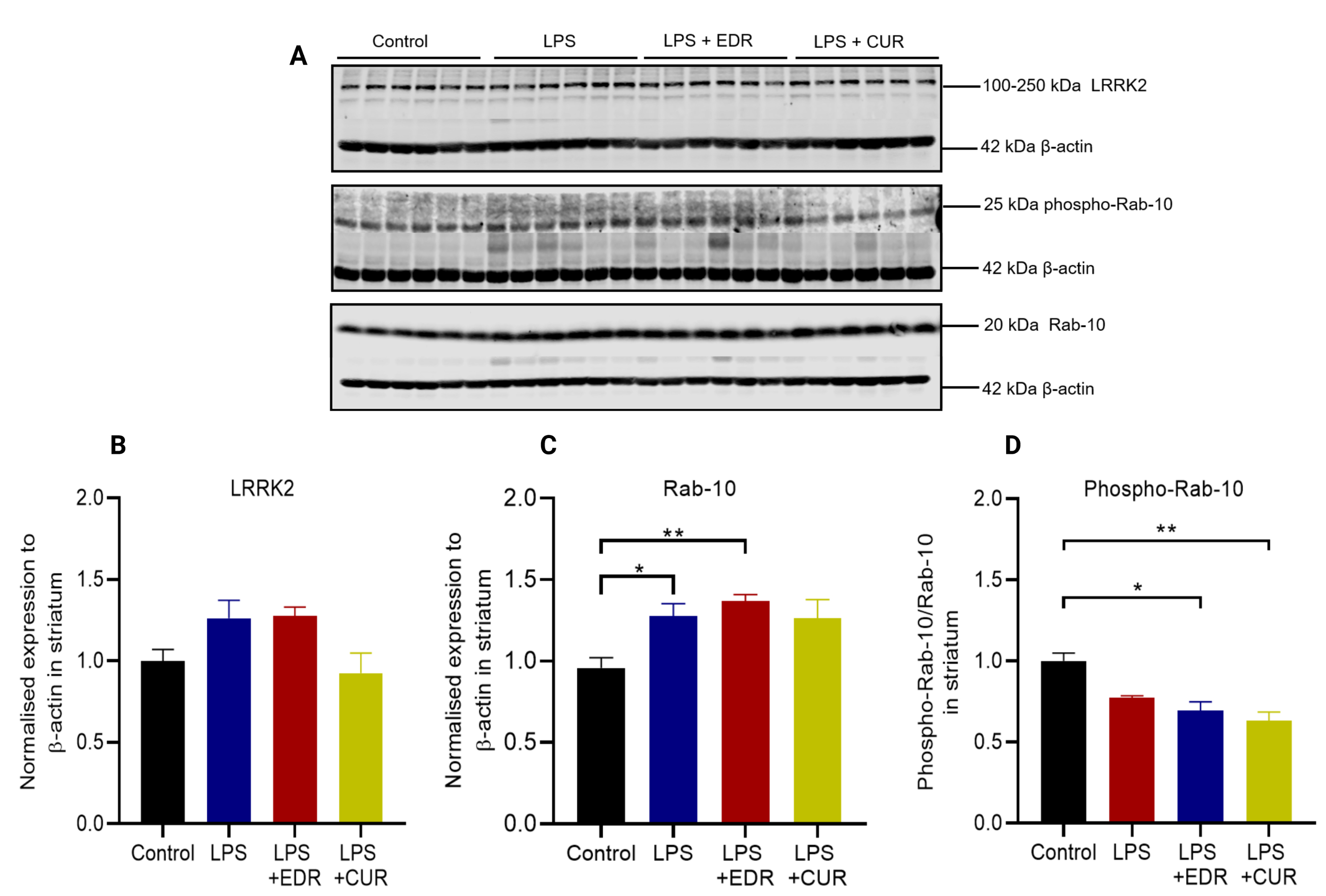
Fig. 7.

The effects of EDR and CUR on the protein expression of LRRK2, Rab-10, and phospho-Rab-10 in the striatum following intrastriatal administration of LPS. (A) Representative immunoblots for the protein expression of LRRK2, Rab-10, and phospho-Rab-10. (B) Densitometric analysis of LRRK2. (C) Densitometric analysis of Rab-10. (D) Densitometric analysis of phospho-Rab-10. The results are presented as mean ± SEM (control n = 6–10; LPS n = 6–10; LPS + EDR n = 6; LPS + CUR n = 6–10). The statistical analyses were performed with the Kruskal-Wallis test, followed by Dunn’s multiple comparisons test for all the experimental groups; p < 0.05 (*), p < 0.01 (**).

3.7 The Effects of EDR and CUR on the Protein Expression of TH and GFAP in the Olfactory Bulb Following Intrastriatal Administration of LPS

Intrastriatal LPS did not alter the expression of TH protein in the olfactory bulb compared to controls (p = 0.6668) (Fig. 8B). However, it induced astroglial activation indicated by increase expression of GFAP protein (p = 0.0155) (Fig. 8C). The administration of EDR or CUR did not alter the expression of GFAP protein in the olfactory bulb.

Fig. 8.

The effects of EDR and CUR on the protein expression of TH and GFAP in the olfactory bulb following intrastriatal administration of LPS. (A) Representative immunoblots for the protein expression of TH and GFAP. (B) Densitometric analysis of TH. (C) Densitometric analysis of GFAP. The results are presented as mean ± SEM (control n = 6–10; LPS n = 6–10; LPS + EDR n = 6; LPS + CUR n = 6–10). The statistical analyses were performed with the Kruskal-Wallis test, followed by Dunn’s multiple comparisons test for all the experimental groups; p < 0.05 (*).

3.8 The Effects of EDR and CUR on the Protein Expression of HSC-70 and LC3 in the Olfactory Bulb Following Intrastriatal Administration of LPS

Intrastriatal LPS increased the expression of HSC-70 protein in the olfactory bulb compared to the control group (p = 0.0014) (Fig. 9B). Interestingly, its expression was returned to the control level by oral CUR (p = 0.0422) but not EDR. Additionally, intrastriatal LPS did not alter the expression of LC3 protein in the olfactory bulb (p = 0.3301) (Fig. 9C), with no effects associated with EDR and CUR.
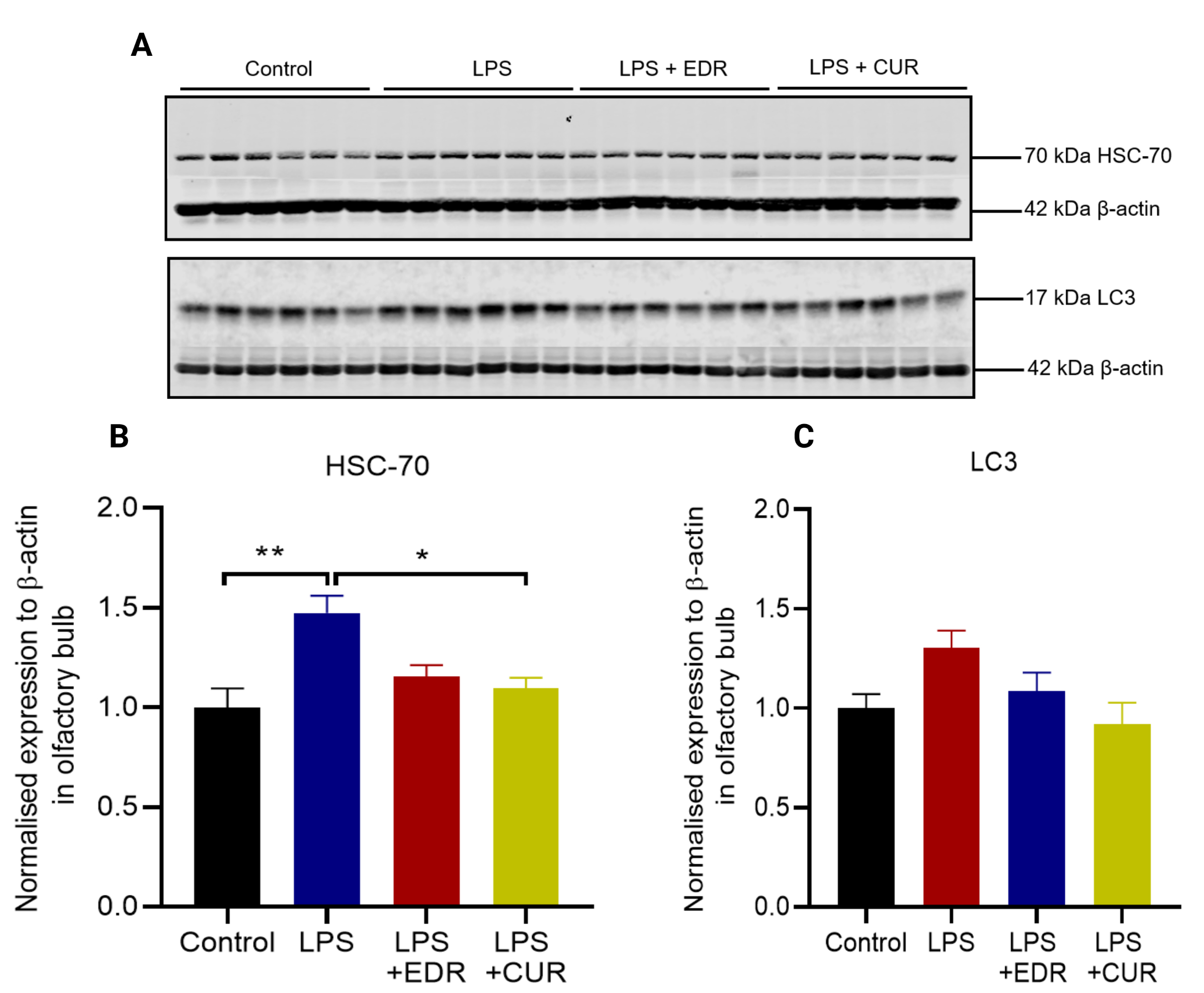
Fig. 9.

The effects of EDR and CUR on the protein expression of HSC-70 and LC3 protein in the olfactory bulb following intrastriatal administration of LPS. (A) Representative immunoblots for the protein expression of HSC-70 and LC3. (B) Densitometric analysis of HSC-70. (C) Densitometric analysis of LC3. The results are presented as mean ± SEM (control n = 6–10; LPS n = 6–10; LPS + EDR n = 6; LPS + CUR n = 6–10). The statistical analyses were performed with the Kruskal-Wallis test, followed by Dunn’s multiple comparisons test for all the experimental groups; p < 0.05 (*), p < 0.01 (**).

3.9 The Effects of Intrastriatal LPS on the Protein Expression of PINK1 and Parkin in the Olfactory Bulb Following Intrastriatal Administration of LPS

Intrastriatal LPS did not alter the expression of PINK1 protein (p = 0.3367) (Fig. 10B) and parkin protein (p = 0.3972) (Fig. 10C) compared to the controls. In contrast, CUR significantly reduced the expression of PINK1 protein compared to the LPS group (p = 0.0269).

Fig. 10.

The effects of EDR and CUR on the protein expression of PINK1 and parkin in the olfactory bulb following intrastriatal administration of LPS. (A) Representative immunoblots for the protein expression of PINK1 and parkin. (B) Densitometric analysis PINK1. (C) Densitometric analysis of parkin. The results are presented as mean ± SEM (control n = 6–10; LPS n = 6–10; LPS + EDR n = 6; LPS + CUR n = 6–10). The statistical analyses were performed with the Kruskal-Wallis test, followed by Dunn’s multiple comparisons test for all the experimental groups; p < 0.05 (*).

3.10 The Effects of EDR and CUR on the Protein Expression of LRRK2, Rab-10 and Phospho-Rab-10 in the Olfactory Bulb Following Intrastriatal Administration of LPS

LPS itself (LPS versus control) did not alter the expression of LRRK2 (p = 0.4591) (Fig. 11B) and phosphorylation of Rab-10 (p = 0.7527) (Fig. 11D), but there was a trend towards increased expression of Rab-10 protein (p = 0.0657) (Fig. 11C) in the olfactory bulb. On the other hand, CUR significantly decreased the protein expression of LRRK2 (p = 0. 0007) and returned the phosphorylation of Rab-10 (p = 0.0021) to the control levels when compared to the LPS group. Additionally, both EDR (p = 0.0253) and CUR (p = 0.0003) significantly reduced the expression of Rab-10 protein compared to LPS group.

Fig. 11.

The effects of EDR and CUR on the protein expression of LRRK2, Rab-10, and phospho-Rab-10 in the olfactory bulb following intrastriatal administration of LPS. (A) Representative immunoblots for the protein expression of LRRK2, Rab-10, and phospho-Rab 10. (B) Densitometric analysis of LRRK2. (C) Densitometric analysis of Rab-10. (D) Densitometric analysis of phopsho-Rab-10. The results are presented as mean ± SEM (control n = 6–10; LPS n = 6–10; LPS + EDR n = 6; LPS + CUR n = 6–10). The statistical analyses were performed with the Kruskal-Wallis test, followed by Dunn’s multiple comparisons test for all the experimental groups; p < 0.05 (*), p < 0.01 (**), p < 0.001 (***).

4. Discussion

This study focused on investigating the effects of EDR and CUR in LPS treated mice. The main findings include: (1) Intrastriatal administration of LPS impaired motor function, induced anxiety-like behavior and caused a reduction in TH protein and increases in α-synuclein and GFAP proteins in the striatum; (2) There were significant increases in the expression of Rab-10 and HSC-70 proteins in the striatum, and HSC-70 protein in the olfactory bulb; (3) EDR and CUR did not alleviate motor or anxiety behaviours, and did not significantly affect the expression of proteins altered by LPS treatment; (4) EDR and CUR formulations restored the expression of some proteins such as LRRK2, Rab-10, phospho-Rab-10, HSC-70, LC3, parkin, and PINK1 in the striatum and olfactory bulb to that of control mice when compared to the LPS group. Thus, such findings may suggest some restorative effects of EDR and CUR on the components of autophagy.

4.1 The Effects of EDR and CUR on LPS-induced Motor Dysfunction and Striatal Pathology

Inflammation is a complex process mediated by multiple cell types and markers. Intrastriatal administration of LPS increased the protein expression of GFAP, an astrocytic marker in the striatum at 8-weeks after LPS injection. It is important to note that reactive astrocytes have both pro- and anti- inflammatory profiles which can exert both neuroprotective and neurotoxic effects [10]. Increased GFAP in this study is consistent with our previous study [21] whereby we used western blotting, and immunohistochemistry to illustrate that LPS induced activation of both astrocytes and microglia in the striatum, suggesting the involvement of inflammatory processes. The inflammation could have increased the protein expression of α-synuclein and reduced TH, a marker of dopaminergic neurons in the striatum, all of which could have contributed to motor dysfunction observed in the LPS mice. These findings were consistent with our previous study whereby we characterised the effects of LPS on the striatum [21]. Moreover, there was a trend towards reduced protein expression of α-synuclein and GFAP after 6-weeks of oral EDR and CUR. These findings suggest that the formulations may have restorative effects on astroglial activation and the expression of α-synuclein protein, although not sufficient to alter the expression of TH protein in the striatum, and motor deficits.

Abnormal secretion of α-synuclein protein is a classical feature of clinical PD. To maintain homeostasis, this protein is degraded by the ubiquitin-proteasome pathway or macroautophagy (normally referred to as autophagy) and CMA [33]. We focused our investigation on the markers of autophagy and CMA for this study. Autophagy involves the sequestration of cytoplasmic proteins, macromolecules and organelles by a double membrane autophagosome [33]. A common marker for autophagosomes is LC3, which is further processed into LC3-I and LC3-II [34]. Increased in LC3 and it processed form LC3-II, have been reported in post-mortem nigral samples of PD patients, which indicate accumulation of autophagosomes associated with impaired fusion between autophagosomes and lysosomes [23, 35]. Based on our study, intrastriatal injection of LPS slightly increased the expression of LC3 but not significantly, suggesting intact autophagy. Of note, there was a decreased expression of LC3 in mice that received oral EDR and CUR, suggesting that these formulations may facilitate autophagic clearance. It is important to note that LC3-II better correlates with the number of autophagosomes compared to LC3 and that at a certain time, LC3 does not indicate autophagic flux. To our knowledge, LC3 and LC3-II have not been examined in intrastriatal models of PD; however, systemic administration of 5 mg/kg of LPS increased the expression of LC3-II in the midbrain in C57BL/6 mice at 5-months [36], consistent with the findings in rotenone, 6-OHDA and MPTP models [35, 37, 38].

The clearance of α-synuclein protein via CMA involves HSC-70 and lysosomal-associated membrane protein 2A (LAMP2A) [33]. Indeed, the expression of HSC-70 and LAMP2A is reduced in the SN of PD patients, which is believed to augment α-synuclein pathology in PD [19, 20, 39, 40]. Based on our findings, intrastriatal administration of LPS induced the activation of CMA in the striatum, indicated by increased expression of HSC-70 protein. These findings correlate with increased α-synuclein protein in the striatum of LPS treated mice. Increased HSC-70 and LAMP2A in response to increased α-synuclein protein has been reported in paraquat and 6-OHDA models of PD [38, 39, 41]. However, the reduction of HSC-70 and LAMP2A in the SN of PD patients could be due to the progressive reduction of dopaminergic neurons. Also, we examined the expression of LAMP1 protein, which is crucial in the maintenance of lysosomal integrity and acidification [42]. LAMP-1 protein was not altered in the striatum by LPS injection indicating intact lysosomal integrity. Our findings differ with a study by Burgaz and colleagues which showed increase LAMP-1 in the SN following intrastriatal injection of LPS [43]. In contrast, LAMP-1 protein is significantly reduced in nigral neurons in PD patients compared to age-matched controls, which signifies lysosomal dysfunction in clinical PD, and these findings are consistent with intraperitoneal MPTP mice models of PD [35].

We showed in this study, and in our previous publication [21] that intrastriatal LPS induces inflammation and oxidative stress in the striatum, which may potentially cause mitochondrial dysfunction, a critical aspect in the pathogenesis of PD. Mutations in PINK1, parkin and LRRK2 are implicated in mitochondrial dysfunction in the nigrostriatal pathway; therefore, we examined their expression in non-genetic models of PD. The autophagic clearance of damaged mitochondria (referred to as mitophagy) is essential for mitochondrial quality control and reduction in reactive oxygen species (ROS) production. Mitophagy can be initiated in a PINK1/parkin-dependent manner, and deletion in these proteins in mice results in impaired mitochondrial biogenesis and respiration, increased sensitivity to oxidative stress, and reduced synaptic excitability [44, 45, 46]. We found that intrastriatal administration of LPS induced a trend towards increased expression of PINK1 and parkin in the striatum. Oral EDR and CUR did not have any noticeable effects on the expression of these proteins. PINK1 and parkin are not well studied in non-genetic models of PD, and additional studies are needed to further understand their role in sporadic PD. According to our findings intrastriatal injection of LPS did not have significant effects on mitochondrial proteins such as PINK1 or parkin in the striatum which may suggest intact mitochondrial integrity. However, it is important to note that we did not directly examine mitochondrial morphology or its function, and additional investigation is required to truly understand the effects of our LPS treatment regime on mitochondrial morphology/mitochondrial function as illustrated in LPS rat models of PD [47, 48, 49].

Lastly, LRRK2 protein acts through the Rab GTPases to modulate vesicular trafficking and autophagy [33]. PD mutations such as G2019S in the LRRK2 gene, increase LRRK2 kinase activity, which impairs PINK1/parkin-dependent mitophagy by increasing the phosphorylation of Rab-10 at threonine 73 [50, 51]. Rab-10 protein works downstream in PINK1/parkin-dependent mitophagy to recruit mitochondria to the autophagosome, which is fused with lysosome for degradation. This process is disrupted when Rab-10 protein is aberrantly phosphorylated by LRRK2 [50, 52, 53]. The inhibition or knockout of LRRK2 proteins which reduces its kinase activity, increases mitophagy, suggesting a therapeutic potential of targeting the LRRK2 pathway in PD [51, 54]. Based on the current study, we found that intrastriatal injection of LPS did not alter LRRK2 protein in the striatum. However, it increased Rab-10 protein with a reduction in its phosphorylation at threonine 73 suggesting that LPS may have also activated protein serine/threonine phosphatases. The increased Rab-10 protein in response to intrastriatal LPS could be a protective mechanism to enhance PINK1/parkin-dependent mitophagy. This doctrine is consistent with a study by Wauters and colleagues which demonstrated that increased expression of Rab-10 protein in LRRK2 mutant cells enhances mitophagy in a PINK1/parkin-dependent manner [53]. Of note, our finding for Rab-10 differs compared to a study by Rocha and colleagues [51] which showed that rotenone increased the phosphorylation of Rab-10 in the dopaminergic neurons, which may suggest variability in the pathways targeted in neurotoxin models. Therefore, a reduction of TH protein in the striatum, which may correlate with dopaminergic axonal degeneration, is likely associated with glial-induced inflammation which was not sufficiently suppressed by EDR and CUR. One could examine the levels of inflammatory cytokines, chemokines, and free radicals in the striatum, as well as the upstream pathways responsible for such response to ascertain the pathological mechanisms in the striatum. It was a limitation of our study that we did not examine the aforementioned parameters.

4.2 The Effects of EDR and CUR on Olfactory Function and Olfactory Bulb Pathology, Following Intrastriatal Administration of LPS

The presence of Lewy bodies (inclusions mostly composed of α-synuclein protein) in PD patients is firstly observed in regions of the olfactory system such as the olfactory bulb. Such findings suggest that olfactory bulb dysfunction could have a role in hyposmia which is evident in approximately 90% of PD patients [55]. In our previous study [21], we showed that intrastriatal administration of LPS induces inflammation, oxidative stress and reduction in synaptic proteins in the olfactory bulb, with no functional changes. We found that LPS-induced astroglial activation, a sign of inflammation in the olfactory bulb. Similar to the striatum, there was no statistical significance of the effects of EDR and CUR on astroglial activation in the olfactory bulb. Thus, some other inflammatory biomarkers may need to be assessed in the future studies in both the olfactory bulb and striatum to establish the efficacy of EDR and CUR on inflammation.

Next, we examined the effects of intrastriatal LPS on the expression of autophagic markers in the olfactory bulb that may be affected in non-genetic PD, and to our knowledge they have not been examined in PD models. Firstly, intrastriatal LPS did not alter the expression of LC3, which might suggest intact autophagy. In contrast, there was increased expression of HSC-70 protein in the olfactory bulb which indicates the activation of CMA. HSC-70 mediates the clearance of α-synuclein protein via CMA, and in our previous study [21] we showed that α-synuclein protein in the olfactory bulb was not altered by intrastriatal injection of LPS. So, it is likely that HSC-70 protein in the olfactory bulb was not increased as a result of aberrant α-synuclein protein. Of note, HSC-70 protein is induced by stress, and an increase in its expression could be due to LPS-induced inflammation in the olfactory bulb. Interestingly, oral CUR reduced the expression of HSC-70 protein to control levels in the olfactory bulb compared to the LPS group. These findings suggest that CUR may reverse the LPS-induced pathology; although the mechanisms are not clear since α-synuclein protein was not altered in the olfactory bulb. Secondly, LPS itself did not have any significant effects on the protein expression of PINK1, parkin, LRRK2 and Rab-10 in the olfactory bulb which are all involved in PINK1/parkin-dependent mitophagy. However, there was a trend towards increased expression of these proteins. Oral administration of EDR and CUR did not have any effects on the expression of parkin, but they reduced the expression of LRRK2, Rab-10 and phospho-Rab-10 to control level. These findings suggest that these formulations may have restorative effects on autophagy in the olfactory bulb. Of critical importance, the role of the aforementioned proteins in the olfactory bulb is still not well explored, and a more mechanistic approach is needed to further elucidate their function in olfactory dysfunction in PD. Based on the above findings, the effects of intrastriatal LPS on the olfactory bulb were mild and may explain the lack of changes in the olfactory function.

4.3 The Effects of EDR and CUR on LPS Induced Anxiety-like Behaviour

Anxiety has not been widely examined in LPS and other models of PD. Nonetheless, it has been shown in animal models of PD via the open field test and the elevated plus-maze test that lesions to the nigrostriatal pathway induce anxiety-like behaviour [56, 57]. Congruent with these findings, we showed that intrastriatal injection of LPS reduced central zone entries and time at 4 and/or 8-weeks which are indicative of anxiety-like behaviour. This result, however, is not consistent with our previous study when we did not find anxiety-like behaviours after LPS injections [21]. This discrepancy may potentially be explained by the fact that in the current study the control and LPS groups were receiving soluplus (120 mg/kg) orally in drinking water to match the treatment conditions of EDR and CUR groups between weeks 3 and 8 of the study. Unlike EDR, the number of central zone entries was not significantly different in the CUR group compared to controls, suggesting that CUR may have some therapeutic effects on anxiety-like behaviour, although, this was not significant compared to the LPS group. Regions of the brain such as the amygdala, prefrontal cortex, and hippocampus are vital in the modulation of anxiety [58]. We were unable to examine the pathological effects of intrastriatal LPS on these regions, and this would be an insightful addition for future studies.

5. Conclusions

In summary, intrastriatal LPS induced pathological changes of PD such as reduced expression of TH protein and increased α-synuclein protein in the striatum, which were accompanied by motor impairment and anxiety-like behaviour. In addition, intrastriatal LPS induced astroglial activation in the striatum and olfactory bulb along with the differential expression of Rab-10 and HSC-70 proteins in these regions. In-vivo models of PD and other neurodegenerative disorders commonly administer EDR and CUR intraperitoneally or intravenously [22, 23, 24]. Indeed, it has been reported in the aforementioned models that EDR and CUR ameliorate neuronal degeneration, neuroinflammation, oxidative stress, and abnormal behavioural changes. Ideally, oral administration is the most preferable route for clinical application to mitigate patient distress and inconvenience associated with intravenous injections. Oral administration is a challenge for EDR and CUR due to their low oral bioavailability associated with physicochemical properties. As a result, Parikh and colleagues developed novel oral formulations of EDR and CUR with improved physical properties. These formulations were able to attenuate cognitive deficits in AD transgenic mice [27, 28, 29, 30]. Neurodegenerative disorders such as AD and PD are proteinopathies, and although it is yet unclear, they may have similar neuropathological processes. Thus, we chose to examine the effects of oral novel formulations of EDR and CUR in PD. We administered 48 mg/kg of the respective compounds, which have therapeutic effects according to Parikh and colleagues [29, 30]. Our data suggest that oral administration of EDR and CUR may have some effects on anxiety-like behaviour, astroglial activation and protein expression of α-synuclein, hereditary PD related proteins and autophagic markers in a manner that may be beneficial. However, additional investigation is required to truly understand the significance of these findings. Additional refinements to our treatment regime are required to elevate the therapeutic effects of EDR and CUR. For example, we administered the EDR and CUR formulations for 6-weeks which was a relatively short time compared to the study by Parikh and colleagues (3-months), who developed the oral formulations that we used [29, 30]. Therefore, the lack of robust therapeutic effects in our study could be due to insufficiency in duration of administration of EDR and CUR. Commonly, preclinical studies for PD utilize the preventative approach when conducting therapeutic studies, whereby the compounds are administered before the induction of the disease. Nonetheless, it is important to examine if EDR and CUR can halt or ameliorate the disease after its initiation. Thus, we administered the formulations 2-weeks after the disease induction, and the lack of robust therapeutic effects of EDR and CUR could suggest that the formulations were initiated too late. Lastly, it could very well be that the oral bioavailability of novel formulations is not yet sufficient to induce therapeutic effects in intrastriatal PD models. Collectively, the PD features in our intrastriatal model were moderate which may constrain the therapeutic potential of EDR and CUR formulations. Therefore, future studies could administer the formulations earlier and extend the duration of treatments using an in-vivo model with robust PD features. Subsequently, studies could modify the doses and re-examine the oral bioavailability of the formulations and physicochemical properties to make suitable adjustments.

Author Contributions

ID performed all the experiments and wrote the manuscript. LB and XFZ were involved in conceptualization, methodology, supervision, and editing of the manuscript. SG was involved in student supervision and contributed to the critical review of the manuscript. All authors read and approved the final manuscript.

Ethics Approval and Consent to Participate

All the animal experiments reported in this manuscript were approved by the Animal Ethics Committee of the University of South Australia under the animal ethics U12-21.

Acknowledgment

Isaac Deng is a recipient of University of South Australia Postgraduate Award, Australia.

Funding

This research received no external funding.

Conflict of Interest

The authors declare no conflict of interest.

References
[1]
Stoker TB, Greenland JC. Parkinson’s disease: pathogenesis and clinical aspects [internet]. Codon Publications: Brisbane (AU). 2018.
[2]
Obeso JA, Rodriguez-Oroz MC, Goetz CG, Marin C, Kordower JH, Rodriguez M, et al. Missing pieces in the Parkinson’s disease puzzle. Nature Medicine. 2010; 16: 653–661.
[3]
Sveinbjornsdottir S. The clinical symptoms of Parkinson’s disease. Journal of Neurochemistry. 2016; 139: 318–324.
[4]
Dawson TM, Dawson VL. Molecular pathways of neurodegeneration in Parkinson’s disease. Science. 2003; 302: 819–822.
[5]
Yasuda T, Nakata Y, Choong C, Mochizuki H. Neurodegenerative changes initiated by presynaptic dysfunction. Translational Neurodegeneration. 2013; 2: 16.
[6]
Agliardi C, Meloni M, Guerini FR, Zanzottera M, Bolognesi E, Baglio F, et al. Oligomeric α-Syn and SNARE complex proteins in peripheral extracellular vesicles of neural origin are biomarkers for Parkinson’s disease. Neurobiology of Disease. 2021; 148: 105185.
[7]
Zahoor I, Shafi A, Haq E. Pharmacological treatment of Parkinson’s disease. Exon Publications. 2018; 129–144.
[8]
Mogi M, Harada M, Kondo T, Riederer P, Inagaki H, Minami M, et al. Interleukin-1β, interleukin-6, epidermal growth factor and transforming growth factor-α are elevated in the brain from parkinsonian patients. Neuroscience Letters. 1994; 180: 147–150.
[9]
Mogi M, Harada M, Riederer P, Narabayashi H, Fujita K, Nagatsu T. Tumor necrosis factor-α (TNF-α) increases both in the brain and in the cerebrospinal fluid from parkinsonian patients. Neuroscience Letters. 1994; 165: 208–210.
[10]
McGeer PL, Itagaki S, Boyes BE, McGeer EG. Reactive microglia are positive for HLA-DR in the substantia nigra of Parkinson’s and Alzheimer’s disease brains. Neurology. 1988; 38: 1285–1291.
[11]
Chen H, Zhang SM, Hernán MA, Schwarzschild MA, Willett WC, Colditz GA, et al. Nonsteroidal anti-inflammatory drugs and the risk of Parkinson disease. Archives of Neurology. 2003; 60: 1059–1064.
[12]
Kouli A, Torsney KM, Kuan W-L. Parkinson’s disease: etiology, neuropathology, and pathogenesis. Exon Publications. 2018; 3–26.
[13]
Wang B, Abraham N, Gao G, Yang Q. Dysregulation of autophagy and mitochondrial function in Parkinson’s disease. Translational Neurodegeneration. 2016; 5: 19.
[14]
Kim WG, Mohney RP, Wilson B, Jeohn GH, Liu B, Hong JS. Regional difference in susceptibility to lipopolysaccharide-induced neurotoxicity in the rat brain: role of microglia. The Journal of Neuroscience. 2000; 20: 6309–6316.
[15]
Saijo K, Winner B, Carson CT, Collier JG, Boyer L, Rosenfeld MG, et al. A Nurr1/CoREST pathway in microglia and astrocytes protects dopaminergic neurons from inflammation-induced death. Cell. 2009; 137: 47–59.
[16]
Park J, Davis RL, Sue CM. Mitochondrial Dysfunction in Parkinson’s Disease: New Mechanistic Insights and Therapeutic Perspectives. Current Neurology and Neuroscience Reports. 2018; 18: 21.
[17]
Villacé P, Mella RM, Kortazar D. Mitochondria in the context of Parkinson’s disease. Neural Regeneration Research. 2017; 12: 214–215.
[18]
Parzych KR, Klionsky DJ. An overview of autophagy: morphology, mechanism, and regulation. Antioxidants & Redox Signaling. 2014; 20: 460–473.
[19]
Alvarez-Erviti L, Rodriguez-Oroz MC, Cooper JM, Caballero C, Ferrer I, Obeso JA, et al. Chaperone-mediated autophagy markers in Parkinson disease brains. Archives of Neurology. 2010; 67: 1464–1472.
[20]
Chu Y, Dodiya H, Aebischer P, Olanow CW, Kordower JH. Alterations in lysosomal and proteasomal markers in Parkinson’s disease: relationship to alpha-synuclein inclusions. Neurobiology of Disease. 2009; 35: 385–398.
[21]
Deng I, Corrigan F, Garg S, Zhou X-F, Bobrovskaya L. Further characterization of intrastriatal lipopolysaccharide model of Parkinson’s disease in C57BL/6 mice. International Journal of Molecular Sciences. 2021; 22: 7380.
[22]
Xiong N, Xiong J, Khare G, Chen C, Huang J, Zhao Y, et al. Edaravone guards dopamine neurons in a rotenone model for Parkinson’s disease. PLoS ONE. 2011; 6: e20677.
[23]
Wu G, Wang X, Feng X, Zhang A, Li J, Gu K, et al. Altered expression of autophagic genes in the peripheral leukocytes of patients with sporadic Parkinson’s disease. Brain Research. 2011; 1394: 105–111.
[24]
Wang X, Zhang Z, Zhang M, Sun M, Wang W, Xie C. Neuroprotective properties of curcumin in toxin-base animal models of Parkinson’s disease: a systematic experiment literatures review. BMC Complementary and Alternative Medicine. 2017; 17: 412.
[25]
Watanabe T, Tahara M, Todo S. The novel antioxidant edaravone: from bench to bedside. Cardiovascular Therapeutics. 2008; 26: 101–114.
[26]
Omosa L, Midiwo J, Kuete V. Curcuma longa. Medicinal Spices and Vegetables (pp. 425–435). Elsevier: Africa. 2017.
[27]
Parikh A, Kathawala K, Song Y, Zhou X, Garg S. Curcumin-loaded self-nanomicellizing solid dispersion system: part i: development, optimization, characterization, and oral bioavailability. Drug Delivery and Translational Research. 2018; 8: 1389–1405.
[28]
Parikh A, Kathawala K, Tan CC, Garg S, Zhou X. Self-nanomicellizing solid dispersion of edaravone: part i - oral bioavailability improvement. Drug Design, Development and Therapy. 2018; 12: 2051–2069.
[29]
Parikh A, Kathawala K, Li J, Chen C, Shan Z, Cao X, et al. Curcumin-loaded self-nanomicellizing solid dispersion system: part II: in vivo safety and efficacy assessment against behavior deficit in Alzheimer disease. Drug Delivery and Translational Research. 2018; 8: 1406–1420.
[30]
Parikh A, Kathawala K, Li J, Chen C, Shan Z, Cao X, et al. Self-nanomicellizing solid dispersion of edaravone: part II: in vivo assessment of efficacy against behavior deficits and safety in Alzheimer’s disease model. Drug Design, Development and Therapy. 2018; 12: 2111–2128.
[31]
Hunter RL, Cheng B, Choi D, Liu M, Liu S, Cass WA, et al. Intrastriatal lipopolysaccharide injection induces parkinsonism in C57/B6 mice. Journal of Neuroscience Research. 2009; 87: 1913–1921.
[32]
Thornton E, Vink R. Treatment with a substance P receptor antagonist is neuroprotective in the intrastriatal 6-hydroxydopamine model of early Parkinson’s disease. PLoS ONE. 2012; 7: e34138.
[33]
Karabiyik C, Lee MJ, Rubinsztein DC. Autophagy impairment in Parkinson’s disease. Essays in Biochemistry. 2017; 61: 711–720.
[34]
Lu J, Wu M, Yue Z. Autophagy and Parkinson’s Disease. Autophagy: Biology and Diseases. 2020; 21: 21–51.
[35]
Dehay B, Bové J, Rodríguez-Muela N, Perier C, Recasens A, Boya P, et al. Pathogenic lysosomal depletion in Parkinson’s disease. The Journal of Neuroscience. 2010; 30: 12535–12544.
[36]
Zheng H, Yang Y, Hu L, Wang M, Wang F, Cao L, et al. Autophagic impairment contributes to systemic inflammation-induced dopaminergic neuron loss in the midbrain. PLoS ONE. 2013; 8: e70472.
[37]
Chen L, Huang Y, Yu X, Lu J, Jia W, Song J, et al. Corynoxine Protects Dopaminergic Neurons Through Inducing Autophagy and Diminishing Neuroinflammation in Rotenone-Induced Animal Models of Parkinson’s Disease. Frontiers in Pharmacology. 2021; 12.
[38]
Marin C, Aguilar E. In vivo 6-OHDA-induced neurodegeneration and nigral autophagic markers expression. Neurochemistry International. 2011; 58: 521–526.
[39]
Sala G, Marinig D, Arosio A, Ferrarese C. Role of Chaperone-Mediated Autophagy Dysfunctions in the Pathogenesis of Parkinson’s Disease. Frontiers in Molecular Neuroscience. 2016; 9: 157.
[40]
Wu F, Xu H, Guan J, Hou Y, Gu J, Zhen X, et al. Rotenone impairs autophagic flux and lysosomal functions in Parkinson’s disease. Neuroscience. 2015; 284: 900–911.
[41]
Mak SK, McCormack AL, Manning-Boğ AB, Cuervo AM, Di Monte DA. Lysosomal Degradation of α-Synuclein in Vivo. Journal of Biological Chemistry. 2010; 285: 13621–13629.
[42]
Mamais A, Manzoni C, Nazish I, Arber C, Sonustun B, Wray S, et al. Analysis of macroautophagy related proteins in G2019S LRRK2 Parkinson’s disease brains with Lewy body pathology. Brain Research. 2018; 1701: 75–84.
[43]
Burgaz S, García C, Gómez-Cañas M, Muñoz E, Fernández-Ruiz J. Development of an oral treatment with the PPAR-γ-acting cannabinoid VCE-003.2 against the inflammation-driven neuronal deterioration in experimental Parkinson’s disease. Molecules. 2019; 24: 2702.
[44]
Gautier CA, Kitada T, Shen J. Loss of PINK1 causes mitochondrial functional defects and increased sensitivity to oxidative stress. Proceedings of the National Academy of Sciences of the United States of America. 2008; 105: 11364–11369.
[45]
Goldberg MS, Fleming SM, Palacino JJ, Cepeda C, Lam HA, Bhatnagar A, et al. Parkin-deficient mice exhibit nigrostriatal deficits but not loss of dopaminergic neurons. The Journal of Biological Chemistry. 2003; 278: 43628–43635.
[46]
McCoy MK, Cookson MR. DJ-1 regulation of mitochondrial function and autophagy through oxidative stress. Autophagy. 2011; 7: 531–532.
[47]
Choi D-Y, Liu M, Hunter RL, Cass WA, Pandya JD, Sullivan PG, et al. Striatal neuroinflammation promotes Parkinsonism in rats. PLoS ONE. 2009; 4: e5482.
[48]
Deng I, Corrigan F, Zhai G, Zhou X, Bobrovskaya L. Lipopolysaccharide animal models of Parkinson’s disease: Recent progress and relevance to clinical disease. Brain, Behavior, & Immunity - Health. 2020; 4: 100060.
[49]
Hunter R, Ojha U, Bhurtel S, Bing G, Choi D. Lipopolysaccharide-induced functional and structural injury of the mitochondria in the nigrostriatal pathway. Neuroscience Research. 2017; 114: 62–69.
[50]
Di Maio R, Hoffman EK, Rocha EM, Keeney MT, Sanders LH, De Miranda BR, et al. LRRK2 activation in idiopathic Parkinson’s disease. Science Translational Medicine. 2018; 10: eaar5429.
[51]
Rocha EM, De Miranda BR, Castro S, Drolet R, Hatcher NG, Yao L, et al. LRRK2 inhibition prevents endolysosomal deficits seen in human Parkinson’s disease. Neurobiology of Disease. 2020; 134: 104626.
[52]
Manzoni C, Lewis PA. LRRK2 and Autophagy. Advances in Neurobiology. 2017; 14: 89–105.
[53]
Wauters F, Cornelissen T, Imberechts D, Martin S, Koentjoro B, Sue C, et al. LRRK2 mutations impair depolarization-induced mitophagy through inhibition of mitochondrial accumulation of RAB10. Autophagy. 2020; 16: 203–222.
[54]
Singh F, Prescott AR, Rosewell P, Ball G, Reith AD, Ganley IG. Pharmacological rescue of impaired mitophagy in Parkinson’s disease-related LRRK2 G2019S knock-in mice. Elife. 2021;10: e67604.
[55]
Hawkes CH, Del Tredici K, Braak H. Parkinson’s disease: a dual-hit hypothesis. Neuropathology and Applied Neurobiology. 2007; 33: 599–614.
[56]
Hritcu L, Gorgan LD. Intranigral lipopolysaccharide induced anxiety and depression by altered BDNF mRNA expression in rat hippocampus. Progress in Neuro-Psychopharmacology & Biological Psychiatry. 2014; 51: 126–132.
[57]
Bonito-Oliva A, Masini D, Fisone G. A mouse model of non-motor symptoms in Parkinson’s disease: focus on pharmacological interventions targeting affective dysfunctions. Frontiers in Behavioral Neuroscience. 2014; 8: 290.
[58]
Zarrindast M-R, Khakpai F. The modulatory role of dopamine in anxiety-like behavior. Archives of Iranian Medicine. 2015; 18: 591–603.
Share
Back to top