IMR Press / CEOG / Volume 52 / Issue 2 / DOI: 10.31083/CEOG26194
Open Access Review
The Accumulation of Visceral Fat in Postmenopausal Women: The Combined Impact of Prenatal Genetics, Epigenetics, and Fat Depot Heterogeneity—A Descriptive Review
Show Less
Affiliation
1 Zhang Zhongjing College of Chinese Medicine, Nanyang Institute of Technology, 473004 Nanyang, Henan, China
2 School of Medicine, Zhengzhou University of Industrial Technology, 451150 Xinzheng, Henan, China
*Correspondence: zhongmingzhang@hotmail.com (Zhongming Zhang); 1048362628@qq.com (Xiaomen Wei)
Clin. Exp. Obstet. Gynecol. 2025, 52(2), 26194; https://doi.org/10.31083/CEOG26194
Submitted: 20 August 2024 | Revised: 18 December 2024 | Accepted: 31 December 2024 | Published: 18 February 2025
Copyright: © 2025 The Author(s). Published by IMR Press.
This is an open access article under the CC BY 4.0 license.
Abstract
Objective:

This review aims to provide an overview of the factors contributing to central obesity, particularly in postmenopausal women, who are affected at a global rate of 26%. It emphasizes the heterogeneity of adipocytes, the impact of prenatal genetic factors, and the role of estrogenic neuroendocrine regulation. Additionally, the review explores the paradoxical functions of visceral fat and identifies the primary depots that may contribute to its overall function.

Mechanism:

Estrogen deficiency is a key factor in central adiposity among postmenopausal women, leading to a reduction in subcutaneous adipose tissue (SAT) and an increase in visceral adipose tissue (VAT) compared to premenopausal women. This deficiency deactivates pro-opiomelanocortin (POMC) neurons and steroidogenic factor-1 (SF1) neurons via estrogen receptor alpha (ERα), desensitizes vagal cholecystokinin-A (CCK-A) receptors, and hyperactivates the hypothalamic-pituitary-ovary (HPO) axis, resulting in increased food intake and decreased energy expenditure. The differences between VAT and SAT, such as expandability, anatomic location, free fatty acid (FFA) mobility, facilitate energy transfer from SAT to VAT, thereby contributing to central obesity. VAT also compensates for estrogen deficiency by releasing estradiol, inflammatory and anti-inflammatory adipocytokines, and increasing 11β-hydroxysteroid dehydrogenase 1 (11β-HSD1) activity, which potentiates glucocorticoid functions and ultimately leading to the development of metabolic syndromes. VAT is heterogeneous, including distinct depots such as mesenteric, gonadal, and perirenal fat. Mesenteric fat may play a significant role in body weight regulation and insulin resistance, while other fat depots interact more closely with surrounding organs to regulate various physiological functions. Understanding VAT heterogeneity is crucial for identifying adiposopathy markers associated with various metabolic syndromes. This knowledge can inform holistic, personalized therapeutic and bodybuilding approaches, helping patients to mitigate the risks associated with current hormone therapies.

Findings in Brief:

The ratio of SAT to VAT is shaped by a combination of prenatal genetics, neuroendocrine regulation, and postnatal epigenetic factors influenced by environmental energy availability and estrogen deficiency. VAT accumulation exhibits paradoxical roles, aiding adaption to energy surplus stress while simultaneously contributing to postmenopausal syndromes. Within VAT, heterogeneity exists, with mesenteric fat depots playing a key role in its overall function. Long-term protective strategies during the perimenopausal and menopausal periods may include energy restriction and the maintenance of normal estrogen levels. Personalized diets and estrogen supplementation hold promise in alleviating associated syndromes. Further exploration of the relationship between mesenteric fat, VAT accumulation, and menopausal syndromes could help clarify existing contradictory evidence and position mesenteric fat as a potential target for effective interventions aimed at alleviating postmenopausal symptoms with fewer side effects.

Conclusions:

Visceral fat accumulation in postmenopausal women is a consequence of energy stress due to estrogen deficiency, followed by the energy transfer from SAT to VAT. The heterogeneity of VAT suggests that its components may have different roles in body weight regulation. Mesenteric fat may play a major role among the depots.

Keywords
visceral fat
subcutaneous fat
heterogeneity
central adiposity
menopause
1. Subcutaneous Adipose Tissue (SAT) vs. Subcutaneous Adipose Tissue (VAT): Understanding White Adipose Tissue (WAT) as a Heterogeneous and Non-Contiguous Organ Distributed Throughout the Mammalian Body

Body fat can be classified into essential fat and storage fat. Storage fat refers to the fat accumulated in adipose tissue and is primarily composed of triacylglycerols [1]. Essential fat, on the other hand, is found in lipid-rich tissues, including the liver, heart, lungs, central nervous system in both men and women. In women, additional essential fat in found in the mammary glands and pelvic region. Reference values indicate that men have approximately 12% storage fat and 3% essential fat, whereas women generally have around 15% storage fat and 12% essential fat [2]. Storage fat constitutes around 86% of total body fat and is the most variable component among individuals [1]. Body fat can also be categorized based on its location, such as trunk fat and appendicular fat, the latter being found in the arms and legs [3].

Functionally, mammals, including humans, possess different types of adipocytes—white, beige, and brown adipocytes—distributed throughout various regions of the body. WAT is the predominant form of fat in adults [4]. It can be further divided into three main anatomical categories: SAT, VAT, and ectopic fat [5, 6]. Ectopic fat, which accumulates in organs such as muscles, pancreas, liver, and heart [7], is typically present in small quantities and is beyond the scope of this review. This manuscript will primarily focus on SAT and VAT. Additionally, WAT can be further subdivided into three groups based on its depth relative to the skin: superficial SAT, deep SAT, and VAT [8]. Additionally, specific terminology is often used to describe fat in particular anatomical regions, such as abdominal SAT [9].

VAT surrounds intra-abdominal organs, including abdominal and intra-abdominal fat [10, 11]. This encompasses fat surrounding internal organs, such as epicardial fat, perivascular fat, and fat surrounding gastrointestinal structures [6, 7]. VAT is a component of android fat, which also includes abdominal SAT.

Excessive accumulation of VAT is now recognized as adiposopathy due to its strong association with increased risks of insulin resistance, type 2 diabetes, and cardiovascular diseases [5, 12, 13, 14, 15, 16, 17, 18, 19]. For example, individuals with Crohn’s disease or intestinal tuberculosis exhibit significantly higher ratios of VAT to SAT or total fat [20]. VAT plays a key role in the secretion of pro-inflammatory cytokines, including interleukin-6 (IL-6), as well as hormones like leptin and adiponectin, particularly in the context of systemic inflammation [21, 22]. Reducing VAT through interventions such as fasting has been shown to alleviate low-grade chronic inflammation linked to excessive visceral adiposity [23]. Additionally, VAT is closely associated with conditions such as sleep apnea, certain tumors, and other disorders [11].

SAT lies just beneath the skin and is distributed across various body regions, including the thighs, hips, and gluteal region. It is further classified into deep and superficial SAT. SAT distribution varies by anatomical site, age, and sex [24, 25, 26]. Unlike other types of fat, SAT plays protective roles by shielding delicate organs (such as the eye) and cushioning body areas exposed to high mechanical stress (such as the heel and toe pads) [13]. Additionally, it contributes to the sexually dimorphic appearance of human faces [27]. Prior to menopause, women tend to accumulate more SAT, which may offer protection against the adverse effects of obesity and metabolic syndrome [12]. However, with aging, SAT can infiltrate the dermal layer, impairing skin elasticity and promoting wrinkle formation [28]. Additionally, SAT can adapt to changes in nutrient status, age, and sex [26, 29]. As a result, SAT is often considered a beneficial tissue, with its subcutaneous accumulation generally being less harmful, particularly in female [6].

However, SAT is not always beneficial. Excessive SAT, for instance, can contribute to biomechanical disorders associated with increased fat mass, such as ulcers, immobility, and sleep apnea. Moreover, when caloric intake exceeds the storage capacity of SAT, excess energy may be redirected toward VAT accumulation [11]. The benefits of SAT can also vary by anatomical location. In humans, upper-body SAT is responsible for most systemic free fatty acids (FFAs) and is more insulin-resistant than lower-body SAT [6, 15].

Emerging research suggests that, rather than acting as a passive storage site for excess energy, WAT is considered a dynamic and diverse organ, dispersed throughout the body and capable of sensing and responding to various physiological signals from its microenvironment. It, in turn, functionally and morphologically adapts to changes in physiological status. The plasticity of WAT is further evident in the sexual dimorphism of fat mass between females and males [12, 30, 31, 32], as well as in the differences in fat distribution observed between perimenopausal and postmenopausal women [33]. Therefore, WAT is now considered as a heterogeneous organ with a remarkable degree of plasticity [34, 35]. Understanding the factors that drive WAT heterogeneity, including its prenatal and postnatal genetic influences, hormonal status, and regional variations, is essential for gaining deeper insights into the characteristics of this newly recognized organ, as well as the pathological consequences of postmenopausal obesity.

2. Genetics and Epigenetics of Sexual Dimorphism in Fat Mass and Distribution
2.1 Effect of the X Chromosome on Fat Mass and Distribution in Humans and Mouse Models

Men aged 20–24 years typically have a body fat percentage of around 15%, while women of the same age have about 27% body fat, with a higher proportion of SAT [1]. The primary factor driving the differences in fat mass and distribution between sexes is the presence and dosage of the X chromosome, as observed in individuals with Klinefelter syndrome (KS) (47, XXY; mosaicism 47, XXY/46, XY; 48, XXXY; 49, XXXXY) [36, 37]. The supernumerary X chromosome significantly influences VAT, as indicated by measures like epicardial fat thickness and truncal body fat [38, 39, 40]. However, KS patients treated with testosterone tend to exhibit lower abdominal fat [41].

Similarly, mouse models with various combinations of gonads and sex chromosomes (XX female, XX male, XY female, and XY male) demonstrate that the presence of two X chromosomes leads to increased adiposity compared to XY mice, regardless of the effects of ovaries or testes [42]. In both intact and gonadectomized animals, XX mice consistently exhibit higher levels of high-density lipoprotein cholesterol (HDL-C) than XY mice, regardless of sex [43]. Additionally, a quantitative trait locus (QTL) on X chromosome, near D10Mit174 (DXMit174, a microsatellite marker), has been associated with adiposity, body weight, and the size of individual fat depots [44]. The connection between the X chromosome and central obesity is further supported by studies indicating that low-birth-weight males tend to have lower overall and central adiposity, while low-birth-weight females more frequently show increased central fat accumulation [45]. The X-inactive specific transcript (Xist) is expressed specifically in female adipose tissue, which may help explain the observed sex dimorphism in fat accumulation [46]. Moreover, sex differences in adipose tissue gene expression and development may be modulated by differential miRNA expression [47].

2.2 Effect of Autosomal Genes on Fat Mass and Distribution

In addition to sex chromosomes, autosomes also play a significant role in the regulation of fat mass and distribution. Quantitative genetic analyses have revealed that the traits of VAT and SAT are heritable [48]. For example, two single nucleotide polymorphisms (SNPs) in the fat mass and obesity-associated protein (FTO) gene, located on chromosome 16, have been linked to SAT accumulation in humans [49]. Several QTLs on mouse chromosome 9 contribute to strain-specific differences in overall fatness, with at least four regions at or near adipocyte fat-associated gene 5 (Adip5) exerting a stronger influence on specific fat depots, such as the gonadal fat depot weight [50, 51].

Interestingly, gene expression in adipocytes may exhibit sex-specific differences. For example, DEAD-box Y RNA helicase (DBY) and eukaryotic initiation factor 2 gamma (eIF2γ) are exclusively expressed in male adipose tissue, while Xist is solely expressed in female adipose tissue. Fat depot-specific gene expression has also been observed: myosin heavy chain 2B (Myh2b) and phosphoglycerate mutase muscle-specific subunit (Pgam2) are specifically expressed in SAT, while uroplakin IIIb (UPK3b) is specifically expressed in VAT [52].

2.3 Estrogenic Effects on Fat Distribution Specificity

Estrogens play a central role in regulating and exerting sex-biased effects on specific genes, such as reprimo, a putative p53-dependent tumor suppressor gene. This regulation occurs in the ventromedial hypothalamus (VMH), a key regulatory node for energy expenditure that demonstrates sexual dimorphism. This regulation contributes to higher core temperatures in male mice compared to females [53]. Furthermore, estrogen-mediated transcriptional and epigenetic regulation in adipocytes plays a crucial role in the sexual dimorphisms observed in body fat distribution and the associated risk of obesity-related diseases. This topic has been extensively reviewed by Bjune et al. [54].

Furthermore, estrogens regulate gene expression in a site-specific manner, influencing the expression of genes such as monocyte-chemoattractant protein-1 (MCP-1), androgen receptor (AR), estrogen receptor α (ERα), and ERβ. This suggests that estrogenic and androgenic signaling in adipose tissue can influence site-specific fat mass accumulation [46, 55]. Notably, VAT depots exhibit higher mRNA levels of ERα, ERβ, and AR compared to their subcutaneous counterparts [55].

The aforementioned genetic factors collectively establish the prenatal and premenopausal set points for balanced body weight through various pathways, contributing to sex-based differences in fat distribution and related traits [56, 57, 58]. Hormonal fluctuations, environmental chemicals, drugs, and diets can modify these homeostatic metabolic set points through epigenetic mechanisms, thereby influencing individual fat redistribution.

3. Distinction between SAT and VAT as a Key Factor in Fat Redistribution, Particularly in Postmenopausal Women
3.1 Central Obesity in Postmenopausal Women: Regional Variations Between VAT and SAT

Postmenopausal women typically have a higher percentage of body fat and lower lean tissue mass compared to premenopausal and perimenopausal women, with 40% of postmenopausal women experiencing visceral obesity [33, 59, 60, 61]. Excessive VAT accumulation is particularly prevalent in perimenopausal women [33, 62, 63]. Both visceral and gynoid (gluteal-femoral regions) fat increase during the menopause transition, with yearly changes of 6.24% and 2.03%, respectively (both p < 0.05) [64]. Postmenopausal women exhibit 36% more trunk fat (p < 0.01), 49% more intra-abdominal fat area (p < 0.01), and 22% more abdominal SAT (p < 0.05). Notably, the menopause-related increase in intra-abdominal fat persists (p < 0.05) even after statistical adjustment for age and total fat mass, while no differences are observed in trunk or abdominal SAT following adjustment [10].

3.2 Priority of VAT Accumulation under Estrogen Deficiency: Consequences of Cellular and Physiological Heterogeneity between VAT and SAT

VAT is particularly metabolically active, more sensitive to lipolysis, and more insulin-resistant than SAT, releasing FFAs from lipolysis directly into the liver through the portal circulation [5]. As shown in Table 1 (Ref. [5, 18, 65, 66, 67, 68, 69, 70, 71, 72, 73, 74, 75, 76, 77, 78, 79, 80, 81, 82, 83, 84, 85, 86, 87, 88, 89]), visceral adipocytes have a lower capacity for preadipocyte differentiation and a higher percentage of larger adipocytes, which promotes adipocyte expansion [5, 65]. This contributes to central abdominal fat accumulation when there is an excess of energy or low estrogen levels. In other words, estrogen deficiency is a key causative factor driving significant central and VAT redistribution in postmenopausal women [90]. Additionally, VAT depots are responsive to various stimuli during the menopause transition, including aging, positive energy balance [90], physical inactivity, stress [65], and local hormonal influences such as cortisol production [66]. Comparing the cellular characteristics of VAT and SAT can offer valuable insights into the mechanisms underlying fat redistribution (Table 1).

Table 1. Differences between VAT and SAT.
SAT VAT Consequence Reference
Cell division and differentiation ++ + More SAT cells [67]
Expandability capacity + ++ Larger VAT cells [68]
Absorption of FFA + ++ SAT: energy storage VAT: lipid mobilization [5]
Absorption of TG + ++ [69]
Lipolytic activity + ++ VAT is more metabolically active; gluconeogenesis; insulin clearance [5]
Glucose uptake + ++ [70]
Insulin resistant + ++ [65]
Plasma lipoprotein-lipid + ++
Plasma triglyceride + ++ [70]
11β-HSD activity + ++ Cortisol response to stress; VAT mass [71]
11β-HSD expression + Unchanged or decreased Compensation for central obesity; lipid mobilization [71, 72, 73]
GR Fructose or/and stress activate glucocorticoid signaling with corticosterone, 11β-HSD1, but GR expression Response to glucocorticoids and stress; corticosterone; VAT lipolysis [5, 65, 74, 75]
AR Adipocyte AR KO increases susceptibility to visceral obesity Fat mass in male mice [76]
Testosterone low Visceral obesity and metabolic syndrome in males Waist circumference; BMI [66]
Testosterone Testosterone, VAT mass in postmenopausal women Metabolic syndrome [77, 78]
ERα ++ + Lipid storage [65]
ERβ + ++ Anti-ERα, VAT dysfunction [79, 80]
Growth hormone response + ++ Lipolysis; VAT [81, 82]
β3 receptor + ++ Lipolytic sensitivity [83]
α2 receptor ++ + Lipolytic sensitivity
Capillary density + ++ VAT hypoxia dysfunction proinflammatory cytokines [18, 84, 85]
Proinflammatory cytokines + ++ (mostly mesenteric fat) Adipocytokines [18, 86]
Adiponectin content ++ + VAT cell size with division [87]
VAT-resident Treg cells + ++ VAT caused inflammation in male mice [88]
Macrophage + (least infiltration) ++ (mostly omental fat) Cytokines release [89]

+: normal level; ++: higher level. VAT, visceral adipose tissue; SAT, subcutaneous adipose tissue; FFA, free fatty acid; TG, triglyceride; 11β-HSD, 11β-hydroxysteroid dehydrogenase; GR, glucocorticoid receptor; AR, androgen receptor; KO, knockout; BMI, body mass index; ER, estrogen receptor; , increased; , decreased.

In summary, during menopause, characterized by an altered ERα/ERß ratio and subsequent changes in metabolic signaling [91], the primary factors contributing to the increase in VAT mass include: (1) anatomical connection to the liver: VAT is anatomically connected to the liver through the portal circulation, allowing it to receive nutrient-rich blood and promote lipogenesis [5]. (2) Cell specificity of VAT: VAT differs from the SAT by presenting a lower capacity for preadipocyte differentiation capacity and a higher proportion of large adipocytes. This cellular specificity enables VAT to store more energy derived from SAT. (3) Enhanced uptake and lipolysis of FFAs: VAT has a greater ability to uptake FFAs and undergoes lipolysis, contributing to its expansion [70]. (4) Increased inflammation in SAT: during menopause, increased inflammation in SAT can promote the growth of VAT [59]. (5) Elevated 11β-hydroxysteroid dehydrogenase 1 (11β-HSD1) activity: increased activity of 11β-HSD1, an enzyme that converts of cortisone to cortisol in VAT, results in hypercortisolism, which contributes to central adiposity and the development of metabolic syndrome [92, 93]. Together, these factors may contribute to the expansion of VAT and central abdominal fat, particularly in the presence of an energy surplus or/and low estrogen levels [5].

4. Is VAT Simply a Marker of Adiposopathy in Postmenopausal Syndromes?

Beyond fat redistribution, it is important to recognize that the characteristics of VAT, as outlined in Table 1, may reflect its roles extending beyond merely being a marker of adiposopathy or an indicator of obesity-related metabolic complications. Adiposopathy, defined as the pathological dysfunction of adipose tissue, is often a primary cause of metabolic syndrome. In fact, these characteristics are often oversimplified to the idea that “obesity protects obesity” [57, 58, 94]. This concept is particularly evident in the expansion of VAT.

VAT has a higher density of glucocorticoid and ARs compared to SAT. Moreover, VAT adipocytes exhibit increased activity of 11β-HSD1, an enzyme that converts cortisone to cortisol within the tissue [71]. This process enhances local glucocorticoid exposure, influencing a wide array of metabolic, anti-inflammatory, and immunosuppressive mechanisms, thus supporting both normal VAT and systemic processes [56, 95]. For example, increased glucocorticoid-signaling enables VAT adipocytes to respond more sensitively to environmental stressors by mobilizing FFAs and becoming more responsive to adrenergic stimulation. While this local amplification of glucocorticoid signaling aid in adaptation to stress, it also contributes to side effects, such as the development of insulin resistance [71, 96].

Another key factor is that VAT is considered a primary driver of adiposopathy, largely due to the release of adipokines that regulate both adaptive and acquired immune responses, contributing to the development of metabolic and immune diseases [11, 19]. For instance, increased leptin secretion from adipose tissue suppresses appetite and promotes energy expenditure, thereby contributing to the maintenance of energy balance and the mitigation of lipodystrophy. Paradoxically, leptin also stimulates the renin-angiotensin system (RAS), which plays a role in regulating fluid balance, but can lead to hypertension [97], systemic sclerosis, and other disorders [98].

In addition, adipose tissue secretes a combination of pro-inflammatory cytokines (such as leptin, interleukin-1 beta (IL-1β), IL-2, IL-4, IL-6, IL-8, IL-10, IL-12, IL-18, tumor necrosis factor α (TNFα), and resistin) and anti-inflammatory cytokines (such as adiponectin) [99]. This complex cytokine profile is part of the “obesity paradox”, where the same biological processes can exert both protective and harmful effects. For example, the altered ratio of ERs (ERα/ERβ) in the context of estrogen deficiency further complicates the immune and metabolic responses, thereby contributing to the multifaceted health impacts of obesity [91, 99, 100].

Another notable paradox is that, during menopause, adipose tissue becomes the primary source of estrogen, with VAT often referred to as a “third ovary”—a concept central to the “obesity paradox” [94]. Although the total amount of estrogen produced after menopause is significantly lower than during a woman’s reproductive years [12], this shift has crucial implications. Estrogen derived from adipose tissue can help mitigate certain cardiovascular risks, but it also promotes further adipose tissue expansion, thereby contributing to obesity. This, in turn, increases the risk of developing estrogen-sensitive breast cancer, highlighting the complex role of adipose-derived estrogen in both protective and harmful processes [101, 102].

Consistent evidence suggests that VAT may not be the primary cause of metabolic complications [5, 15, 103]. Several loci on human chromosomes have been identified that paradoxically exert beneficial cardiometabolic effects, either by promoting favorable fat distribution or by having no adverse cardiometabolic impacts at all [104]. Additionally, individuals with metabolic syndrome often exhibit a borderline negative correlation with abdominal SAT mass [105]. The paradox may stem from the underlying fat mass—whether it is present in appropriate amount or in excess.

5. Clinical Implications of VAT Heterogeneity in Postmenopausal Syndromes

Emerging clinic evidence has shown that the percentage of VAT is inversely associated with total bone mineral density and osteoporosis [106, 107], as well as with lower levels of HDL-C [108]. In contrast, in postmenopausal women with normal lean mass, VAT does not appear to correlate with bone mineral density or markers of bone formation and resorption. VAT, rather than age, is strongly linked to an increased risk of breast cancer [109], elevated fasting glucose, insulin levels, and cardiovascular risk factors such as triglycerides, apolipoprotein B [108], and blood pressure in postmenopausal women [110, 111, 112]. Notably, peri-coronary epicardial adipose tissue is strongly associated with vascular risk factors and coronary calcification [113]. In contrast, diet-induced weight loss has been shown to significantly improve fasting insulin levels and glucose metabolism [114]. Supplementation with Equol, an ER agonist, can notably alleviate postmenopausal syndromes and reduce VAT mass in postmenopausal women following three months of treatment [115].

Paradoxically, higher adiposity has also been associated with fewer physiological hot flashes among older women experiencing this common postmenopausal symptom, as indicated by age-adjusted models [116]. Furthermore, subcutaneous abdominal fat, but not VAT, appears to be associated with an increased likelihood of hot flashes [117]. These findings suggest that VAT may be a more reliable indicator of metabolic syndrome in menopausal or postmenopausal women, compared to other symptoms such as hot flashes.

6. Exploring the Heterogeneity of VAT: Is Mesenteric Fat the Key Regulator?

VAT, also known as abdominal fat, refers to the fat that surrounds internal organs [7]. Commonly referred to as VAT, it includes a range of fat depots, such as mesenteric, omental, retroperitoneal, intraperitoneal, gonadal, peri-renal, epididymal fat, and others, depending on their anatomical locations [118, 119]. As such, visceral fat is not a homogeneous tissue; it surrounds various intra-abdominal organs and may perform distinct functions depending on the specific abdominal microenvironments. Gene expression studies have also identified validated markers for these fat depots, suggesting a relationship between anatomical location, both adipose tissue identity, and function [120].

VAT depots may differentially contribute to various physiological processes. Regression analyses examining body weight changes in relation to the weights of three visceral fat depots—peri-renal fat, gonadal fat, and mesenteric fat—in C57 mice and nerve-specific receptor-activity modifying protein 1 (RAMP1) overexpressing lean mouse models [121] reveal that mesenteric fat exhibits the largest coefficients compared to the other two fat pads in both strains (unpublished data). Moreover, mesenteric adipocyte size has been associated with a 79% higher likelihood of developing metabolic syndrome compared to other fat depots [18]. Visceral fat, particularly due to its distinct anatomical characteristics and its circulation draining into the portal vein and liver, has been implicated in the development of insulin resistance. Consistent with this, mesenteric fat has been shown to exert more than a two-fold greater influence on improvements in insulin sensitivity in young rats [122].

Other VAT depots also exhibit distinct characteristics, with some being more responsive to dietary changes. For instance, very low energy diets significantly reduce VAT mass, particularly mesorectal fat, while having a lesser impact on total pelvic fat [123]. Among the visceral depots, mesenteric and omental depots contain the fewest macrophages and adipokines [18]. On the other hand, pancreatic fat mass, has been negatively associated with cognition and brain volumes in middle-aged individuals [124]. In contrast, periaortic adipose tissue is characterized by the smallest adipocytes, the highest capillary density, and an elevated secretion of adipokines [18]. There may also be heterogeneity within individual fat depot. For example, in mice, epididymal fat, a component of visceral fat, is divided into distal and proximal regions, each with distinct histochemical and gene expression profiles [125]. This variability may serve an adaptive function, allowing each depot to fulfill more specialized roles in response to subtle nutrient differences within the microenvironment.

The observations outlined above are primarily based on studies in humans and commonly used animal models. To our knowledge, however, no comparative analysis of depot-specific characteristics has been conducted specifically in the context of peri- or post-menopause. Such research could identify the specific fat depots responsible for central adiposity, enhance our understanding of body shape changes during this life stage, and guide the development of targeted therapeutic strategies for related metabolic syndromes.

7. Estrogen Deficiency: A Key Factor in Fat Redistribution during Menopause

Fat redistribution due to estrogen deficiency is supported by comparisons of plasma estrogen levels, which range from 100–250 pg/mL in premenopausal women to approximately 10 pg/mL in postmenopausal women [126]. Total estradiol levels are inversely associated with body shape, with the lowest levels observed in postmenopausal women exhibiting an “apple” phenotype and the highest levels found in women with a “pear” phenotype, including both premenopausal women and postmenopausal women using hormone replacement therapy (HRT) [127].

The role of estrogen in regulating fat distribution is further supported by observations in individuals undergoing cross-sex hormonal therapy. Trans women undergoing estrogen therapy exhibit a gynoid pattern of body fat distribution and a lower waist-to-hip ratio, while trans men receiving testosterone display an android pattern of body fat distribution with a reduced hip circumference [128]. These findings suggest that, in the context of estrogen deficiency, fat deposition shifts toward the visceral depot, contributing to the development of an android body shape [12].

The impact of estrogen on energy metabolism is further demonstrated by studies using ER genetic mouse models, in which activation of ERα leads to a decreased body weight, while activation of ERβ results in an increased body weight [129]. During estrogen deficiency, there is a relative increase in ERβ expression, accompanied by a corresponding decrease in the ERα/ERβ ratio. Estrogen plays a key role in coordinating central and peripheral metabolism, influencing appetite, satiety, and energy expenditure through central nuclei, such as the arcuate nucleus (ARC), ventromedial nucleus (VMN), and nucleus tractus solitarius (NTS), as well as peripheral organs and mitochondrial function in various cell types [130].

7.1 Estrogens Reduce Appetite and Suppress Food Intake by Activating ERα in the ARC of the Hypothalamus

Estrogens reduce appetite and suppress food intake by activating ERα in arcuate pro-opiomelanocortin (POMC) neurons within the ARC. POMC neurons are key regulators of both metabolism and reproduction. Estrogen enhances excitatory synapses on POMC neurons by rapidly uncoupling gamma-aminobutyric acid type B (GABAB) receptors from their G protein-coupled inwardly rectifying K+ channels, thereby inhibiting food intake [131]. Additionally, POMC neurons release α-melanocyte-stimulating hormone (αMSH), a proteolytic product of POMC, which suppresses food intake by activating its receptors, melanocortin-3 receptor (MC3R) and melanocortin-4 receptor (MC4R) [132]. Furthermore, estrogen can suppress neuropeptide Y (NPY) production in hypothalamic neurons through ERα and its associated signaling pathways [133]. In contrast, ERβ expression is significantly lower than that of ERα and is less effective at mediating the inhibitory effects on food intake [134].

7.2 Estrogens Enhance Energy Expenditure by Activating ERα in the VMN of the Hypothalamus

Estrogens promote weight loss and reduce visceral fat deposition through ERα activation in steroidogenic factor-1 (SF1) neurons within the VMN of the hypothalamus [135]. SF1 is a transcription factor uniquely expressed in VMN neurons. Although the VMN was once thought to regulate both food intake and energy expenditure, it is now understood to primarily increase energy expenditure through ERα activation and subsequent phosphatidylinositol 3-kinase (PI3K) signaling [136, 137]. Activation of ERα-positive VMN neurons stimulates heat generation by conditionally expressing ERα-targeting genes, such as reprimo, tachykinin 1 (TAC1), and prodynorphin (PDYN) in both male and female mice [138].

7.3 Estrogens Suppress Food Intake by Sensitizing Satiety Signals in the NTS, Triggered by Cholecystokinin (CCK), through ERα Activation

Estrogens suppress food intake and potentiate CCK-induced satiety by increasing the activity of NTS neurons in the brainstem via ERα activation [130]. They enhance the potency of CCK by increasing the sensitivity of vagal CCK-A receptors [139]. CCK, synthesized in the upper intestine, is released to activate subdiaphragmatic vagal afferent neurons [139]. Estrogens amplify CCK-induced satiety signals in the NTS through ERα signaling, which slows gastric emptying and intestinal motility contributing to their anorexigenic effects [80, 140].

7.4 Estrogen Deficiency Hyperactivates the Hypothalamic-Pituitary-Ovary (HPO) Axis, Leading to Increased VAT Accumulation

Clinical diagnostics for menopausal women typically measure six hormones: estradiol, follicle-stimulating hormone (FSH), luteinizing hormone (LH), testosterone, progesterone, and prolactin. As ovarian aging progresses, declining levels of ovarian steroids and peptides result in a 15-fold increase in FSH, signaling the cessation of reproduction at menopause, and a 10-fold increase in LH in postmenopausal women [87, 141].

Estrogen deficiency leads to hyperactivation of the HPO axis due to the loss of estrogen’s negative feedback inhibition. This enhanced neuroendocrine pathway, characterized by elevated FSH levels, coincides with increased visceral fat accumulation, higher cellular mitochondrial density, and enhanced thermogenesis [87]. Additionally, progesterone has been shown to be inversely correlated with premenstrual food cravings in both humans and rodents [142].

Furthermore, estrogen deficiency desensitizes the satiety centers in the brain, leading to increased food intake, reduced energy expenditure, and a positive energy balance. This energy imbalance further stimulates the hypothalamic-pituitary-adrenal (HPA) axis, resulting in excess glucocorticoid exposure and promoting visceral obesity [143]. The HPA axis not only orchestrates the body’s stress response but also plays critical role in the endocrine regulation of appetite, particularly in the regulation of energy metabolism during the peri- and postmenopausal periods.

7.5 Estrogens Integrate Nuclear and Mitochondrial Signals to Downregulate Biosynthesis and Enhance β-Oxidation

At the cellular level, estrogens regulate a variety of functions by binding to cytosolic ERα and ERβ or the membrane-bound G-protein coupled estrogen receptor (GPER1). In the presence of estrogens, ERα and ERβ can directly bind to mitochondrial DNA (mtDNA) through mitochondrial estrogen receptor elements (EREs) [144]. This binding modulates the expression of nuclear and mitochondrial factors involved in energy expenditure, including nuclear respiratory factor-1 (NRF-1), estrogen receptor-related receptors (ERR1), peroxisome proliferator-activated receptor α (PPARα) and δ (PPARδ), as well as PPAR gamma coactivator 1 alpha (PGC1α) and PGC1β, which regulate nuclear-encoded mitochondrial genes in various cell types, such as adipocytes, muscle cells, and hepatocytes [69].

Estrogens also downregulate enzymes involved in fatty acid and triglyceride synthesis in adipocytes, such as acetyl-CoA carboxylase (ACC1), fatty acid synthase (FAS), and diacylglycerol acyltransferase (DGAT1 and DGAT2) [145]. Simultaneously, they upregulate enzymes that promote fatty acid β-oxidation, such as medium-chain acyl-coenzyme A dehydrogenase (MCAD) and acetyl-CoA oxidase (ACO) [145]. Additionally, estrogens influence the expression of certain mtDNA-encoded genes involved in mitochondrial respiratory chain complexes [144]. In brain mitochondria, this regulation is primarily initiated by ERβ binding to EREs [146], although ERα may also play a role in other tissues [144].

Estrogens further modulate cholesterol metabolism by regulating the expression of 3-Hydroxy-3-Methylglutaryl-Coenzyme A (HMG-CoA) reductase, which is anchored in the endoplasmic reticulum membrane [147]. Moreover, estrogens interact indirectly with peripheral mediators, such as glucagon-like peptide-1 (GLP-1), in the hypothalamus to cooperatively regulate energy homeostasis [148]. In this way, estrogens coordinate brain and body metabolism, orchestrating both endocrine and paracrine effects. Consistently, during menopause, the decline in plasma estrogen levels coincides with a reduction in bioenergetic function across various tissues and organs [149], contributing to postmenopausal phenotypes. The underlying mechanism and its consequences in the context of postmenopause are summarized as Fig. 1.
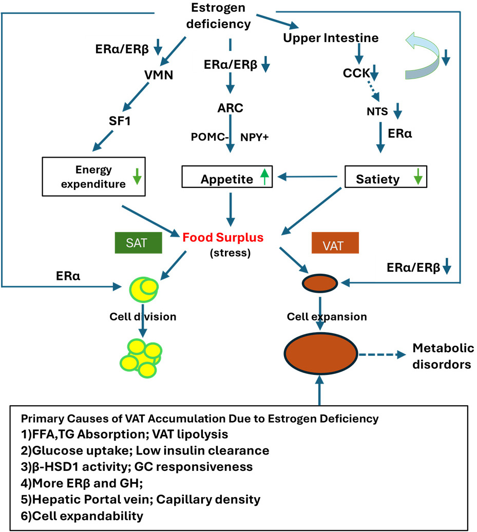
Fig. 1.

Impact of estrogen deficiency on central energy regulation, appetite, and adipose tissue remodeling. Estrogen deficiency reduces the expression of ERα and the ERα/ERβ ratio, leading to decreased SF1 expression and impaired PI3K signaling in the VMN, resulting in a reduction in energy expenditure. In the ARC, estrogen deficiency decreases POMC expression and increases NPY expression, promoting increased appetite. Additionally, estrogen deficiency downregulates CCK signaling, which diminishes CCK-induced satiety by inhibiting ERα-mediated signaling in the nucleus of the NTS. These disruptions in energy homeostasis create a state of energy surplus, contributing to the remodeling of SAT and VAT. This remodeling is influenced by regional and cellular characteristics, leading to increased VAT expansion and a shift toward an android body shape. ARC, arcuate nucleus; CCK, cholecystokinin; ER, estrogen receptor; HSD1, Hydroxysteroid Dehydrogenase 1; GH, growth hormone; TG, triglyceride; GC, glucocorticoids; NPY, neuropeptide Y; NTS, nucleus tractus solitaries; POMC, pro-opiomelanocortin; SAT, subcutaneous adipose tissue; SF1, steroidogenic factor-1; VAT, visceral adipose tissue; VMN, ventromedial nucleus; FFA, free fatty acid.

8. Conclusions and Perspectives

Emerging evidence highlights the heterogeneity of adipose tissue in terms of its composition, distribution, and function. These characteristics are prenatally determined by sex chromosomes and SNPs in autosomes and are further influenced by epigenetic factors such as estrogen and energy intake. SAT and VAT are two key representatives that define sex dimorphism in adipose tissue distribution and function.

In the context of menopause, estrogen deficiency leads to a positive energy balance by increasing appetite and food intake through the inhibition of POMC neurons in the ARC, decreasing energy expenditure by inhibiting SF1 neurons in the VMN of the hypothalamus, and reducing satiety while promoting food intake through the NTS in the brainstem. These regulatory processes are primarily mediated by ERα activity. Additionally, the hyperactivation of the HPO axis contributes to the accumulation of VAT.

The distinct characteristics of VAT, including high expandability, elevated FFA mobility, and increased high 11β-HSD1 activity, make it more susceptible to hypertrophy. VAT accumulation, in turn, compensates for declining estrogen levels by releasing more estradiol and adipocytokines, which exert both inflammatory and anti-inflammatory effects, helping the body adapt to energy stress. However, this “obesity paradox” ultimately contributes to the development of postmenopausal phenotypes.

Although VAT is often termed the “sick fat” associated with postmenopausal syndromes, it is heterogeneous, comprising various anatomically distinct depots such as mesenteric, peri-renal, omental, retroperitoneal, intraperitoneal, and gonadal fat. Recent studies suggest that these depots differ morphologically and functionally [18, 48, 123]. For instance, mesenteric fat within VAT may play a significant role in regulating body weight, insulin resistance, and inflammation.

Further investigation into the specific characteristics of VAT depots in the context of menopause is crucial for a deeper understanding of postmenopausal syndromes. For instance, examining SNPs in autosomes, glucocorticoid receptors (GRs), HSD enzymes, and adipocyte-specific expression of glucose transporter type 4 (GLUT4), along with associated phenotypes in animal models, could shed light on the mechanisms driving VAT accumulation after menopause. Additionally, personalized treatments such as hormone replacement therapies (including phytoestrogens and estrogen-progesterone combinations), non-hormonal medications, and lifestyle interventions (such as regular exercise, healthy diets, and caffeine to boost plasma brain-derived neurotrophic factor (BDNF) levels) could offer tailored therapeutic approaches for patients [150]. In contrast, behaviors such as high-fat diets, smoking, and alcohol consumption, which may exacerbate postmenopausal symptoms, should be avoided.

9. Systematic Literature Search and Selection Criteria

To conduct this study, a systematic search of electronic databases, including PubMed and ResearchGate, was performed in June 2024 and updated in November 2024. Studies in English published before these dates were considered. Screening was performed using predefined search terms such as obesity and X chromosome (225 results), postmenopausal and SAT (91), postmenopausal and VAT (109), postmenopausal, VAT, and SAT (41), postmenopausal, VAT, SAT, and metabolism (35), and VAT, SAT, and metabolism (845). After removing duplicates, 912 publications were identified. Of these, 150 were included in the references based on their relevance to the manuscript and its sections, as well as their status as classic works or recent impactful studies. Publications not meeting these criteria were excluded.

Author Contributions

The research study was designed by all authors, with ZZ responsible for manuscript writing, and ZH, HY, DL, PD, XW contributing to editorial revision. All authors contributed to the manuscript’s editorial changes, read and approved the final version, and are accountable for all aspects of the work.

Ethics Approval and Consent to Participate

Not applicable.

Acknowledgment

The authors would like to express their gratitude to Stephanie Zhang at UCLA for her editorial assistance with this manuscript. They also extend their thanks to all the peer reviewers for their valuable feedback and suggestions.

Funding

This work was supported by grants from Henan Natural Science Foundation (242300421302).

Conflict of Interest

The authors declare no conflict of interest.

References
[1]
Luke A, Schoeller DA. Basal metabolic rate, fat-free mass, and body cell mass during energy restriction. Metabolism: Clinical and Experimental. 1992; 41: 450–456. https://doi.org/10.1016/0026-0495(92)90083-m.
[2]
Ko SH, Jung Y. Energy Metabolism Changes and Dysregulated Lipid Metabolism in Postmenopausal Women. Nutrients. 2021; 13: 4556. https://doi.org/10.3390/nu13124556.
[3]
Kouda K, Nakamura H, Fujita Y, Ohara K, Iki M. Increased ratio of trunk to appendicular fat and increased blood pressure: study of a general population of Hamamatsu children. Circulation Journal: Official Journal of the Japanese Circulation Society. 2012; 76: 2848–2854. https://doi.org/10.1253/circj.cj-12-0417.
[4]
Sakers A, De Siqueira MK, Seale P, Villanueva CJ. Adipose-tissue plasticity in health and disease. Cell. 2022; 185: 419–446. https://doi.org/10.1016/j.cell.2021.12.016.
[5]
Ibrahim MM. Subcutaneous and visceral adipose tissue: structural and functional differences. Obesity Reviews: an Official Journal of the International Association for the Study of Obesity. 2010; 11: 11–18. https://doi.org/10.1111/j.1467-789X.2009.00623.x.
[6]
Chait A, den Hartigh LJ. Adipose Tissue Distribution, Inflammation and Its Metabolic Consequences, Including Diabetes and Cardiovascular Disease. Frontiers in Cardiovascular Medicine. 2020; 7: 22. https://doi.org/10.3389/fcvm.2020.00022.
[7]
Muzurović EM, Vujošević S, Mikhailidis DP. Can We Decrease Epicardial and Pericardial Fat in Patients With Diabetes? Journal of Cardiovascular Pharmacology and Therapeutics. 2021; 26: 415–436. https://doi.org/10.1177/10742484211006997.
[8]
Sniderman AD, Bhopal R, Prabhakaran D, Sarrafzadegan N, Tchernof A. Why might South Asians be so susceptible to central obesity and its atherogenic consequences? The adipose tissue overflow hypothesis. International Journal of Epidemiology. 2007; 36: 220–225. https://doi.org/10.1093/ije/dyl245.
[9]
Drolet R, Richard C, Sniderman AD, Mailloux J, Fortier M, Huot C, et al. Hypertrophy and hyperplasia of abdominal adipose tissues in women. International Journal of Obesity (2005). 2008; 32: 283–291. https://doi.org/10.1038/sj.ijo.0803708.
[10]
Toth MJ, Tchernof A, Sites CK, Poehlman ET. Menopause-related changes in body fat distribution. Annals of the New York Academy of Sciences. 2000; 904: 502–506. https://doi.org/10.1111/j.1749-6632.2000.tb06506.x.
[11]
Bays HE. Evaluation and Practical Management of Increased Visceral Fat: Should Cardiologists Lose Sleep Over It? Journal of the American College of Cardiology. 2022; 79: 1266–1269. https://doi.org/10.1016/j.jacc.2022.01.039.
[12]
Palmer BF, Clegg DJ. The sexual dimorphism of obesity. Molecular and Cellular Endocrinology. 2015; 402: 113–119. https://doi.org/10.1016/j.mce.2014.11.029.
[13]
Rosen ED, Spiegelman BM. What we talk about when we talk about fat. Cell. 2014; 156: 20–44. https://doi.org/10.1016/j.cell.2013.12.012.
[14]
Smith U, Kahn BB. Adipose tissue regulates insulin sensitivity: role of adipogenesis, de novo lipogenesis and novel lipids. Journal of Internal Medicine. 2016; 280: 465–475. https://doi.org/10.1111/joim.12540.
[15]
Jensen MD. Visceral Fat: Culprit or Canary? Endocrinology and Metabolism Clinics of North America. 2020; 49: 229–237. https://doi.org/10.1016/j.ecl.2020.02.002.
[16]
Garg UK, Mathur N, Sahlot R, Tiwari P, Sharma B, Saxena A, et al. Abdominal fat depots and their association with insulin resistance in patients with type 2 diabetes. PloS One. 2023; 18: e0295492. https://doi.org/10.1371/journal.pone.0295492.
[17]
Rendell M, Hulthén UL, Törnquist C, Groop L, Mattiasson I. Relationship between abdominal fat compartments and glucose and lipid metabolism in early postmenopausal women. The Journal of Clinical Endocrinology and Metabolism. 2001; 86: 744–749. https://doi.org/10.1210/jcem.86.2.7260.
[18]
Kranendonk MEG, van Herwaarden JA, Stupkova T, de Jager W, Vink A, Moll FL, et al. Inflammatory characteristics of distinct abdominal adipose tissue depots relate differently to metabolic risk factors for cardiovascular disease: distinct fat depots and vascular risk factors. Atherosclerosis. 2015; 239: 419–427. https://doi.org/10.1016/j.atherosclerosis.2015.01.035.
[19]
Bays HE, González-Campoy JM, Henry RR, Bergman DA, Kitabchi AE, Schorr AB, et al. Is adiposopathy (sick fat) an endocrine disease? International Journal of Clinical Practice. 2008; 62: 1474–1483. https://doi.org/10.1111/j.1742-1241.2008.01848.x.
[20]
Seetharaman J, Srivastava A, Yadav RR, Singh SK, Mishra P, Sen Sarma M, et al. Visceral Fat Indices: Do They Help Differentiate Crohn’s Disease and Intestinal Tuberculosis in Children? Journal of Crohn’s & Colitis. 2023; 17: 2026–2032. https://doi.org/10.1093/ecco-jcc/jjad109.
[21]
Fontana L, Eagon JC, Trujillo ME, Scherer PE, Klein S. Visceral fat adipokine secretion is associated with systemic inflammation in obese humans. Diabetes. 2007; 56: 1010–1013. https://doi.org/10.2337/db06-1656.
[22]
Song SO, Han SJ, Kahn SE, Leonetti DL, Fujimoto WY, Boyko EJ. Leptin and Adiponectin Concentrations Independently Predict Future Accumulation of Visceral Fat in Nondiabetic Japanese Americans. Obesity (Silver Spring, Md.). 2021; 29: 233–239. https://doi.org/10.1002/oby.23035.
[23]
Al Lami Z, Kurtca M, Atique MU, Opekun AR, Siam MS, Jalal PK, et al. Dawn-to-dusk dry fasting decreases circulating inflammatory cytokines in subjects with increased body mass index. Metabolism Open. 2024; 21: 100274. https://doi.org/10.1016/j.metop.2024.100274.
[24]
Cassisa A. Pathophysiology of subcutaneous fat. Giornale Italiano Di Dermatologia E Venereologia: Organo Ufficiale, Societa Italiana Di Dermatologia E Sifilografia. 2013; 148: 315–323.
[25]
Rohrich RJ, Pessa JE. The fat compartments of the face: anatomy and clinical implications for cosmetic surgery. Plastic and Reconstructive Surgery. 2007; 119: 2219–2227. https://doi.org/10.1097/01.prs.0000265403.66886.54.
[26]
Siervogel RM, Roche AF, Himes JH, Chumlea WC, McCammon R. Subcutaneous fat distribution in males and females from 1 to 39 years of age. The American Journal of Clinical Nutrition. 1982; 36: 162–171. https://doi.org/10.1093/ajcn/36.1.162.
[27]
Burke D, Sulikowski D. A new viewpoint on the evolution of sexually dimorphic human faces. Evolutionary Psychology: an International Journal of Evolutionary Approaches to Psychology and Behavior. 2010; 8: 573–585. https://doi.org/10.1177/147470491000800404.
[28]
Ezure T. Subcutaneous fat infiltration into the dermal layer induces wrinkle formation. Skin Research and Technology. 2023; 29: e13296. https://doi.org/10.1111/srt.13296.
[29]
Graybeal AJ, Brandner CF, Tinsley GM, Haynes H, Stavres J. Associations between visceral adipose tissue estimates produced by near-infrared spectroscopy, mobile anthropometrics, and traditional body composition assessments and estimates derived from dual-energy X-ray absorptiometry. The British Journal of Nutrition. 2023; 130: 525–535. https://doi.org/10.1017/S0007114522003488.
[30]
Bond ST, Calkin AC, Drew BG. Sex differences in white adipose tissue expansion: emerging molecular mechanisms. Clinical Science (London, England: 1979). 2021; 135: 2691–2708. https://doi.org/10.1042/CS20210086.
[31]
Bredella MA. Sex Differences in Body Composition. Advances in Experimental Medicine and Biology. 2017; 1043: 9–27. https://doi.org/10.1007/978-3-319-70178-3_2.
[32]
MacCannell ADV, Futers TS, Whitehead A, Moran A, Witte KK, Roberts LD. Sexual dimorphism in adipose tissue mitochondrial function and metabolic flexibility in obesity. International Journal of Obesity (2005). 2021; 45: 1773–1781. https://doi.org/10.1038/s41366-021-00843-0.
[33]
Dmitruk A, Czeczelewski J, Czeczelewska E, Golach J, Parnicka U. Body composition and fatty tissue distribution in women with various menstrual status. Roczniki Panstwowego Zakladu Higieny. 2018; 69: 95–101.
[34]
Berry DC, Stenesen D, Zeve D, Graff JM. The developmental origins of adipose tissue. Development (Cambridge, England). 2013; 140: 3939–3949. https://doi.org/10.1242/dev.080549.
[35]
Samuel O O. Review on multifaceted involvement of perivascular adipose tissue in vascular pathology. Cardiovascular Pathology: the Official Journal of the Society for Cardiovascular Pathology. 2020; 49: 107259. https://doi.org/10.1016/j.carpath.2020.107259.
[36]
Zhang B, Li F, Huang C, Xu L, Cao Z, Kang Y, et al. The correlation between clinical features and ultrastructure of testis of non-mosaic Klinefelter’s syndrome patients with hypogonadism and androgen deficiency: A case report. Heliyon. 2023; 9: e19940. https://doi.org/10.1016/j.heliyon.2023.e19940.
[37]
Spaziani M, Radicioni AF. Metabolic and cardiovascular risk factors in Klinefelter syndrome. American Journal of Medical Genetics. Part C, Seminars in Medical Genetics. 2020; 184: 334–343. https://doi.org/10.1002/ajmg.c.31792.
[38]
Granato S, Barbaro G, Di Giorgio MR, Rossi FM, Marzano C, Impronta F, et al. Epicardial fat: the role of testosterone and lipid metabolism in a cohort of patients with Klinefelter syndrome. Metabolism: Clinical and Experimental. 2019; 95: 21–26. https://doi.org/10.1016/j.metabol.2019.03.002.
[39]
Zore T, Palafox M, Reue K. Sex differences in obesity, lipid metabolism, and inflammation-A role for the sex chromosomes? Molecular Metabolism. 2018; 15: 35–44. https://doi.org/10.1016/j.molmet.2018.04.003.
[40]
Reue K. Sex differences in obesity: X chromosome dosage as a risk factor for increased food intake, adiposity and co-morbidities. Physiology & Behavior. 2017; 176: 174–182. https://doi.org/10.1016/j.physbeh.2017.02.040.
[41]
Øzdemir CM, Ridder LO, Chang S, Fedder J, Just J, Gravholt CH, et al. Mild liver dysfunction in Klinefelter syndrome is associated with abdominal obesity and elevated lipids but not testosterone treatment. Journal of Endocrinological Investigation. 2024; 47: 3057–3066. https://doi.org/10.1007/s40618-024-02394-3.
[42]
Chen X, McClusky R, Chen J, Beaven SW, Tontonoz P, Arnold AP, et al. The number of x chromosomes causes sex differences in adiposity in mice. PLoS Genetics. 2012; 8: e1002709. https://doi.org/10.1371/journal.pgen.1002709.
[43]
Link JC, Chen X, Prien C, Borja MS, Hammerson B, Oda MN, et al. Increased high-density lipoprotein cholesterol levels in mice with XX versus XY sex chromosomes. Arteriosclerosis, Thrombosis, and Vascular Biology. 2015; 35: 1778–1786. https://doi.org/10.1161/ATVBAHA.115.305460.
[44]
York B, Lei K, West DB. Inherited non-autosomal effects on body fat in F2 mice derived from an AKR/J x SWR/J cross. Mammalian Genome: Official Journal of the International Mammalian Genome Society. 1997; 8: 726–730. https://doi.org/10.1007/s003359900554.
[45]
Rockenbach G, Luft VC, Mueller NT, Duncan BB, Stein MC, Vigo Á, et al. Sex-specific associations of birth weight with measures of adiposity in mid-to-late adulthood: the Brazilian Longitudinal Study of Adult Health (ELSA-Brasil). International Journal of Obesity (2005). 2016; 40: 1286–1291. https://doi.org/10.1038/ijo.2016.76.
[46]
Frank AP, de Souza Santos R, Palmer BF, Clegg DJ. Determinants of body fat distribution in humans may provide insight about obesity-related health risks. Journal of Lipid Research. 2019; 60: 1710–1719. https://doi.org/10.1194/jlr.R086975.
[47]
Link JC, Hasin-Brumshtein Y, Cantor RM, Chen X, Arnold AP, Lusis AJ, et al. Diet, gonadal sex, and sex chromosome complement influence white adipose tissue miRNA expression. BMC Genomics. 2017; 18: 89. https://doi.org/10.1186/s12864-017-3484-1.
[48]
Agrawal S, Wang M, Klarqvist MDR, Smith K, Shin J, Dashti H, et al. Inherited basis of visceral, abdominal subcutaneous and gluteofemoral fat depots. Nature Communications. 2022; 13: 3771. https://doi.org/10.1038/s41467-022-30931-2.
[49]
Hotta K, Kitamoto A, Kitamoto T, Mizusawa S, Teranishi H, So R, et al. Association between type 2 diabetes genetic susceptibility loci and visceral and subcutaneous fat area as determined by computed tomography. Journal of Human Genetics. 2012; 57: 305–310. https://doi.org/10.1038/jhg.2012.21.
[50]
Lin C, Fesi BD, Marquis M, Bosak NP, Lysenko A, Koshnevisan MA, et al. Adiposity QTL Adip20 decomposes into at least four loci when dissected using congenic strains. PloS One. 2017; 12: e0188972. https://doi.org/10.1371/journal.pone.0188972.
[51]
McDaniel AH, Li X, Tordoff MG, Bachmanov AA, Reed DR. A locus on mouse Chromosome 9 (Adip5) affects the relative weight of the gonadal but not retroperitoneal adipose depot. Mammalian Genome: Official Journal of the International Mammalian Genome Society. 2006; 17: 1078–1092. https://doi.org/10.1007/s00335-006-0055-1.
[52]
Shinozaki S, Chiba T, Kokame K, Miyata T, Ai M, Kawakami A, et al. Site-specific effect of estradiol on gene expression in the adipose tissue of ob/ob mice. Hormone and Metabolic Research. 2007; 39: 192–196. https://doi.org/10.1055/s-2007-970417.
[53]
Sanchez-Alavez M, Alboni S, Conti B. Sex- and age-specific differences in core body temperature of C57Bl/6 mice. Age (Dordrecht, Netherlands). 2011; 33: 89–99. https://doi.org/10.1007/s11357-010-9164-6.
[54]
Bjune JI, Strømland PP, Jersin RÅ, Mellgren G, Dankel SN. Metabolic and Epigenetic Regulation by Estrogen in Adipocytes. Frontiers in Endocrinology. 2022; 13: 828780. https://doi.org/10.3389/fendo.2022.828780.
[55]
Rodriguez-Cuenca S, Monjo M, Proenza AM, Roca P. Depot differences in steroid receptor expression in adipose tissue: possible role of the local steroid milieu. American Journal of Physiology. Endocrinology and Metabolism. 2005; 288: E200–E207. https://doi.org/10.1152/ajpendo.00270.2004.
[56]
Yang X, Liu S, Zhang Z. Differences Among Sexes in Blood Pressure: A Combinatorial Consequence of the Differences between RAAS Components, Sex Hormones, and Time Course. Current Hypertension Reviews. 2022; 18: 11–16. https://doi.org/10.2174/1573402117666210511011444.
[57]
Garvey WT. Is Obesity or Adiposity-Based Chronic Disease Curable: The Set Point Theory, the Environment, and Second-Generation Medications. Endocrine Practice: Official Journal of the American College of Endocrinology and the American Association of Clinical Endocrinologists. 2022; 28: 214–222. https://doi.org/10.1016/j.eprac.2021.11.082.
[58]
Farias MM, Cuevas AM, Rodriguez F. Set-point theory and obesity. Metabolic Syndrome and Related Disorders. 2011; 9: 85–89. https://doi.org/10.1089/met.2010.0090.
[59]
Abildgaard J, Ploug T, Al-Saoudi E, Wagner T, Thomsen C, Ewertsen C, et al. Changes in abdominal subcutaneous adipose tissue phenotype following menopause is associated with increased visceral fat mass. Scientific Reports. 2021; 11: 14750. https://doi.org/10.1038/s41598-021-94189-2.
[60]
Lizcano F. Roles of estrogens, estrogen-like compounds, and endocrine disruptors in adipocytes. Frontiers in Endocrinology. 2022; 13: 921504. https://doi.org/10.3389/fendo.2022.921504.
[61]
Ambikairajah A, Walsh E, Tabatabaei-Jafari H, Cherbuin N. Fat mass changes during menopause: a metaanalysis. American Journal of Obstetrics and Gynecology. 2019; 221: 393–409.e50. https://doi.org/10.1016/j.ajog.2019.04.023.
[62]
Jeong HG, Park H. Metabolic Disorders in Menopause. Metabolites. 2022; 12: 954. https://doi.org/10.3390/metabo12100954.
[63]
Kosková I, Petrásek R, Vondra K, Skibová J. Weight, body composition and fat distribution changes of Czech women in the different reproductive phases: a longitudinal study. Prague Medical Report. 2007; 108: 226–242.
[64]
Greendale GA, Han W, Finkelstein JS, Burnett-Bowie SAM, Huang M, Martin D, et al. Changes in Regional Fat Distribution and Anthropometric Measures Across the Menopause Transition. The Journal of Clinical Endocrinology and Metabolism. 2021; 106: 2520–2534. https://doi.org/10.1210/clinem/dgab389.
[65]
Björntorp P. Visceral obesity: a “civilization syndrome”. Obesity Research. 1993; 1: 206–222. https://doi.org/10.1002/j.1550-8528.1993.tb00614.x.
[66]
Tchernof A, Després JP. Pathophysiology of human visceral obesity: an update. Physiological Reviews. 2013; 93: 359–404. https://doi.org/10.1152/physrev.00033.2011.
[67]
Liu LF, Craig CM, Tolentino LL, Choi O, Morton J, Rivas H, et al. Adipose tissue macrophages impair preadipocyte differentiation in humans. PloS One. 2017; 12: e0170728. https://doi.org/10.1371/journal.pone.0170728.
[68]
Carobbio S, Pellegrinelli V, Vidal-Puig A. Adipose Tissue Function and Expandability as Determinants of Lipotoxicity and the Metabolic Syndrome. Advances in Experimental Medicine and Biology. 2017; 960: 161–196. https://doi.org/10.1007/978-3-319-48382-5_7.
[69]
Nauli AM, Matin S. Why Do Men Accumulate Abdominal Visceral Fat? Frontiers in Physiology. 2019; 10: 1486. https://doi.org/10.3389/fphys.2019.01486.
[70]
Wajchenberg BL. Subcutaneous and visceral adipose tissue: their relation to the metabolic syndrome. Endocrine Reviews. 2000; 21: 697–738. https://doi.org/10.1210/edrv.21.6.0415.
[71]
Goedecke JH, Wake DJ, Levitt NS, Lambert EV, Collins MR, Morton NM, et al. Glucocorticoid metabolism within superficial subcutaneous rather than visceral adipose tissue is associated with features of the metabolic syndrome in South African women. Clinical Endocrinology. 2006; 65: 81–87. https://doi.org/10.1111/j.1365-2265.2006.02552.x.
[72]
Chedid MF, do Nascimento FV, de Oliveira FS, de Souza BM, Kruel CRP, Gurski RR, et al. Interaction of HSD11B1 and H6PD polymorphisms in subjects with type 2 diabetes are protective factors against obesity: a cross-sectional study. Diabetology & Metabolic Syndrome. 2019; 11: 78. https://doi.org/10.1186/s13098-019-0474-2.
[73]
Ottosson M, Lönnroth P, Björntorp P, Edén S. Effects of cortisol and growth hormone on lipolysis in human adipose tissue. The Journal of Clinical Endocrinology and Metabolism. 2000; 85: 799–803. https://doi.org/10.1210/jcem.85.2.6358.
[74]
Bursać B, Djordjevic A, Veličković N, Milutinović DV, Petrović S, Teofilović A, et al. Involvement of glucocorticoid prereceptor metabolism and signaling in rat visceral adipose tissue lipid metabolism after chronic stress combined with high-fructose diet. Molecular and Cellular Endocrinology. 2018; 476: 110–118. https://doi.org/10.1016/j.mce.2018.04.015.
[75]
Bursać BN, Djordjevic AD, Vasiljević AD, Milutinović DDV, Veličković NA, Nestorović NM, et al. Fructose consumption enhances glucocorticoid action in rat visceral adipose tissue. The Journal of Nutritional Biochemistry. 2013; 24: 1166–1172. https://doi.org/10.1016/j.jnutbio.2012.09.002.
[76]
McInnes KJ, Smith LB, Hunger NI, Saunders PTK, Andrew R, Walker BR. Deletion of the androgen receptor in adipose tissue in male mice elevates retinol binding protein 4 and reveals independent effects on visceral fat mass and on glucose homeostasis. Diabetes. 2012; 61: 1072–1081. https://doi.org/10.2337/db11-1136.
[77]
Janssen I, Powell LH, Kazlauskaite R, Dugan SA. Testosterone and visceral fat in midlife women: the Study of Women’s Health Across the Nation (SWAN) fat patterning study. Obesity (Silver Spring, Md.). 2010; 18: 604–610. https://doi.org/10.1038/oby.2009.251.
[78]
Sander B, Muftah A, Sykes Tottenham L, Grummisch JA, Gordon JL. Testosterone and depressive symptoms during the late menopause transition. Biology of Sex Differences. 2021; 12: 44. https://doi.org/10.1186/s13293-021-00388-x.
[79]
Lv Y, Wang F, Sheng Y, Xia F, Jin Y, Ding G, et al. Estrogen supplementation deteriorates visceral adipose function in aged postmenopausal subjects via Gas5 targeting IGF2BP1. Experimental Gerontology. 2022; 163: 111796. https://doi.org/10.1016/j.exger.2022.111796.
[80]
Zidon TM, Padilla J, Fritsche KL, Welly RJ, McCabe LT, Stricklin OE, et al. Effects of ERβ and ERα on OVX-induced changes in adiposity and insulin resistance. The Journal of Endocrinology. 2020; 245: 165–178. https://doi.org/10.1530/JOE-19-0321.
[81]
Yuen KCJ, Dunger DB. Therapeutic aspects of growth hormone and insulin-like growth factor-I treatment on visceral fat and insulin sensitivity in adults. Diabetes, Obesity & Metabolism. 2007; 9: 11–22. https://doi.org/10.1111/j.1463-1326.2006.00591.x.
[82]
Liu ZT, Yang GW, Zhao X, Dong SH, Jiao Y, Ge Z, et al. Growth hormone improves insulin resistance in visceral adipose tissue after duodenal-jejunal bypass by regulating adiponectin secretion. World Journal of Diabetes. 2024; 15: 1340–1352. https://doi.org/10.4239/wjd.v15.i6.1340.
[83]
Lönnqvist F, Thörne A, Large V, Arner P. Sex differences in visceral fat lipolysis and metabolic complications of obesity. Arteriosclerosis, Thrombosis, and Vascular Biology. 1997; 17: 1472–1480. https://doi.org/10.1161/01.atv.17.7.1472.
[84]
Vieira ADC, Medeiros EB, Zabot GC, Pereira NDS, do Nascimento NB, Lidio AV, et al. Neuroprotective effects of combined therapy with memantine, donepezil, and vitamin D in ovariectomized female mice subjected to dementia model. Progress in Neuro-psychopharmacology & Biological Psychiatry. 2023; 122: 110653. https://doi.org/10.1016/j.pnpbp.2022.110653.
[85]
O’Rourke RW, White AE, Metcalf MD, Olivas AS, Mitra P, Larison WG, et al. Hypoxia-induced inflammatory cytokine secretion in human adipose tissue stromovascular cells. Diabetologia. 2011; 54: 1480–1490. https://doi.org/10.1007/s00125-011-2103-y.
[86]
Kahn D, Macias E, Zarini S, Garfield A, Zemski Berry K, MacLean P, et al. Exploring Visceral and Subcutaneous Adipose Tissue Secretomes in Human Obesity: Implications for Metabolic Disease. Endocrinology. 2022; 163: bqac140. https://doi.org/10.1210/endocr/bqac140.
[87]
Liu P, Ji Y, Yuen T, Rendina-Ruedy E, DeMambro VE, Dhawan S, et al. Blocking FSH induces thermogenic adipose tissue and reduces body fat. Nature. 2017; 546: 107–112. https://doi.org/10.1038/nature22342.
[88]
Vasanthakumar A, Chisanga D, Blume J, Gloury R, Britt K, Henstridge DC, et al. Sex-specific adipose tissue imprinting of regulatory T cells. Nature. 2020; 579: 581–585. https://doi.org/10.1038/s41586-020-2040-3.
[89]
Harman-Boehm I, Blüher M, Redel H, Sion-Vardy N, Ovadia S, Avinoach E, et al. Macrophage infiltration into omental versus subcutaneous fat across different populations: effect of regional adiposity and the comorbidities of obesity. The Journal of Clinical Endocrinology and Metabolism. 2007; 92: 2240–2247. https://doi.org/10.1210/jc.2006-1811.
[90]
Fenton A, Smart C, Goldschmidt L, Price V, Scott J. Fat mass, weight and body shape changes at menopause - causes and consequences: a narrative review. Climacteric: the Journal of the International Menopause Society. 2023; 26: 381–387. https://doi.org/10.1080/13697137.2023.2178892.
[91]
Katzer K, Hill JL, McIver KB, Foster MT. Lipedema and the Potential Role of Estrogen in Excessive Adipose Tissue Accumulation. International Journal of Molecular Sciences. 2021; 22: 11720. https://doi.org/10.3390/ijms222111720.
[92]
Peeke PM, Chrousos GP. Hypercortisolism and obesity. Annals of the New York Academy of Sciences. 1995; 771: 665–676. https://doi.org/10.1111/j.1749-6632.1995.tb44719.x.
[93]
Kupczyk D, Bilski R, Kozakiewicz M, Studzińska R, Kędziora-Kornatowska K, Kosmalski T, et al. 11β-HSD as a New Target in Pharmacotherapy of Metabolic Diseases. International Journal of Molecular Sciences. 2022; 23: 8984. https://doi.org/10.3390/ijms23168984.
[94]
Ganipisetti VM, Bollimunta P. Obesity and Set-Point Theory. StatPearls: USA. 2023.
[95]
Scherholz ML, Schlesinger N, Androulakis IP. Chronopharmacology of glucocorticoids. Advanced Drug Delivery Reviews. 2019; 151-152: 245–261. https://doi.org/10.1016/j.addr.2019.02.004.
[96]
Gao L, Zhang C, Li Q, Peng X, Shima G, Cao H, et al. Network Pharmacology and Experimental Analyses of the Mechanism of Analgesic and Glucose Intolerance Through Glucocorticoid Signaling in C57 Mice Treated with Water Extract of Prunella vulgaris L. Spica. Natural Product Communications. 2022; 17: 1934578X221111032. https://doi.org/10.1177/1934578X221111032.
[97]
Xue B, Yu Y, Zhang Z, Guo F, Beltz TG, Thunhorst RL, et al. Leptin Mediates High-Fat Diet Sensitization of Angiotensin II-Elicited Hypertension by Upregulating the Brain Renin-Angiotensin System and Inflammation. Hypertension (Dallas, Tex.: 1979). 2016; 67: 970–976. https://doi.org/10.1161/HYPERTENSIONAHA.115.06736.
[98]
Maximus PS, Al Achkar Z, Hamid PF, Hasnain SS, Peralta CA. Adipocytokines: Are they the Theory of Everything? Cytokine. 2020; 133: 155144. https://doi.org/10.1016/j.cyto.2020.155144.
[99]
Clemente-Suárez VJ, Redondo-Flórez L, Beltrán-Velasco AI, Martín-Rodríguez A, Martínez-Guardado I, Navarro-Jiménez E, et al. The Role of Adipokines in Health and Disease. Biomedicines. 2023; 11: 1290. https://doi.org/10.3390/biomedicines11051290.
[100]
Bays HE. Adiposopathy is “sick fat” a cardiovascular disease? Journal of the American College of Cardiology. 2011; 57: 2461–2473. https://doi.org/10.1016/j.jacc.2011.02.038.
[101]
Gérard C, Brown KA. Obesity and breast cancer - Role of estrogens and the molecular underpinnings of aromatase regulation in breast adipose tissue. Molecular and Cellular Endocrinology. 2018; 466: 15–30. https://doi.org/10.1016/j.mce.2017.09.014.
[102]
Hetemäki N, Savolainen-Peltonen H, Tikkanen MJ, Wang F, Paatela H, Hämäläinen E, et al. Estrogen Metabolism in Abdominal Subcutaneous and Visceral Adipose Tissue in Postmenopausal Women. The Journal of Clinical Endocrinology and Metabolism. 2017; 102: 4588–4595. https://doi.org/10.1210/jc.2017-01474.
[103]
Cioffi CE, Narayan KMV, Liu K, Uppal K, Jones DP, Tran V, et al. Hepatic fat is a stronger correlate of key clinical and molecular abnormalities than visceral and abdominal subcutaneous fat in youth. BMJ Open Diabetes Research & Care. 2020; 8: e001126. https://doi.org/10.1136/bmjdrc-2019-001126.
[104]
Roshandel D, Lu T, Paterson AD, Dash S. Beyond apples and pears: sex-specific genetics of body fat percentage. Frontiers in Endocrinology. 2023; 14: 1274791. https://doi.org/10.3389/fendo.2023.1274791.
[105]
Garza AL, Lee M, Blangero J, Bauer CX, Czerwinski SA, Choh AC. Genetic correlations between liver fat content, metabolic health, and adiposity distribution in the Fels Longitudinal Study. Nutrition, Metabolism, and Cardiovascular Diseases: NMCD. 2024; 34: 1610–1618. https://doi.org/10.1016/j.numecd.2024.03.002.
[106]
Bihun H, Abdullah N, Abdul Murad NA, Chin SF, Arifin ASK, Khuzaimi AN, et al. Body fat distribution and bone mineral density in a multi-ethnic sample of postmenopausal women in The Malaysian Cohort. Archives of Osteoporosis. 2024; 19: 73. https://doi.org/10.1007/s11657-024-01435-x.
[107]
Oliveira MC, Vullings J, van de Loo FAJ. Osteoporosis and osteoarthritis are two sides of the same coin paid for obesity. Nutrition (Burbank, Los Angeles County, Calif.). 2020; 70: 110486. https://doi.org/10.1016/j.nut.2019.04.001.
[108]
Hernández-Ono A, Monter-Carreola G, Zamora-González J, Cardoso-Saldaña G, Posadas-Sánchez R, Torres-Tamayo M, et al. Association of visceral fat with coronary risk factors in a population-based sample of postmenopausal women. International Journal of Obesity and Related Metabolic Disorders: Journal of the International Association for the Study of Obesity. 2002; 26: 33–39. https://doi.org/10.1038/sj.ijo.0801842.
[109]
Le Marchand L, Wilkens LR, Castelfranco AM, Monroe KR, Kristal BS, Cheng I, et al. Circulating Biomarker Score for Visceral Fat and Risks of Incident Colorectal and Postmenopausal Breast Cancer: The Multiethnic Cohort Adiposity Phenotype Study. Cancer Epidemiology, Biomarkers & Prevention. 2020; 29: 966–973. https://doi.org/10.1158/1055-9965.EPI-19-1469.
[110]
Faria AN, Ribeiro Filho FF, Gouveia Ferreira SR, Zanella MT. Impact of visceral fat on blood pressure and insulin sensitivity in hypertensive obese women. Obesity Research. 2002; 10: 1203–1206. https://doi.org/10.1038/oby.2002.164.
[111]
Nistor IM, Fica S, Martin SC, Mustata T, Oprea TE, Sirbu AE, et al. DXA Android-to-Gynoid Ratio and Cardiovascular Risk Assessment in Age and BMI Propensity-Matched Early Postmenopausal Women. Medicina (Kaunas, Lithuania). 2024; 60: 1096. https://doi.org/10.3390/medicina60071096.
[112]
Thu WPP, Sundström-Poromaa I, Logan S, Kramer MS, Yong EL. Blood pressure and adiposity in midlife Singaporean women. Hypertension Research: Official Journal of the Japanese Society of Hypertension. 2021; 44: 561–570. https://doi.org/10.1038/s41440-020-00600-2.
[113]
de Vos AM, Prokop M, Roos CJ, Meijs MFL, van der Schouw YT, Rutten A, et al. Peri-coronary epicardial adipose tissue is related to cardiovascular risk factors and coronary artery calcification in post-menopausal women. European Heart Journal. 2008; 29: 777–783. https://doi.org/10.1093/eurheartj/ehm564.
[114]
Normandin E, Doucet E, Rabasa-Lhoret R, Brochu M. Effects of a weight loss program on body composition and the metabolic profile in obese postmenopausal women displaying various obesity phenotypes: a MONET group study. Applied Physiology, Nutrition, and Metabolism = Physiologie Appliquee, Nutrition et Metabolisme. 2015; 40: 695–702. https://doi.org/10.1139/apnm-2014-0451.
[115]
Yoshikata R, Myint KZY, Ohta H, Ishigaki Y. Effects of an equol-containing supplement on advanced glycation end products, visceral fat and climacteric symptoms in postmenopausal women: A randomized controlled trial. PloS One. 2021; 16: e0257332. https://doi.org/10.1371/journal.pone.0257332.
[116]
Thurston RC, Santoro N, Matthews KA. Adiposity and hot flashes in midlife women: a modifying role of age. The Journal of Clinical Endocrinology and Metabolism. 2011; 96: E1588–E1595. https://doi.org/10.1210/jc.2011-1082.
[117]
Thurston RC, Sowers MR, Sutton-Tyrrell K, Everson-Rose SA, Lewis TT, Edmundowicz D, et al. Abdominal adiposity and hot flashes among midlife women. Menopause (New York, N.Y.). 2008; 15: 429–434. https://doi.org/10.1097/gme.0b013e31815879cf.
[118]
Chusyd DE, Wang D, Huffman DM, Nagy TR. Relationships between Rodent White Adipose Fat Pads and Human White Adipose Fat Depots. Frontiers in Nutrition. 2016; 3: 10. https://doi.org/10.3389/fnut.2016.00010.
[119]
Bagchi DP, MacDougald OA. Identification and Dissection of Diverse Mouse Adipose Depots. Journal of Visualized Experiments: JoVE. 2019; e59499. https://doi.org/10.3791/59499.
[120]
de Jong JMA, Larsson O, Cannon B, Nedergaard J. A stringent validation of mouse adipose tissue identity markers. American Journal of Physiology. Endocrinology and Metabolism. 2015; 308: E1085–E1105. https://doi.org/10.1152/ajpendo.00023.2015.
[121]
Zhang Z, Liu X, Morgan DA, Kuburas A, Thedens DR, Russo AF, et al. Neuronal receptor activity-modifying protein 1 promotes energy expenditure in mice. Diabetes. 2011; 60: 1063–1071. https://doi.org/10.2337/db10-0692.
[122]
Catalano KJ, Stefanovski D, Bergman RN. Critical role of the mesenteric depot versus other intra-abdominal adipose depots in the development of insulin resistance in young rats. Diabetes. 2010; 59: 1416–1423. https://doi.org/10.2337/db08-0675.
[123]
Bell S, Malouf P, Johnson N, Wale R, Peng Q, Nottle P, et al. Pelvic fat volume reduction with preoperative very low energy diet (VLED): implications for rectal cancer surgery in the obese. Techniques in Coloproctology. 2019; 23: 887–892. https://doi.org/10.1007/s10151-019-02074-y.
[124]
Golan Shekhtman S, Boccara E, Ravona-Springer R, Inbar Y, Zelicha H, Livny A, et al. Abdominal fat depots are related to lower cognitive functioning and brain volumes in middle-aged males at high Alzheimer’s risk. Obesity (Silver Spring, Md.). 2024; 32: 1009–1022. https://doi.org/10.1002/oby.24004.
[125]
Lee KH, Kim NH. Differential Expression of Adipocyte-Related Molecules in the Distal Epididymal Fat of Mouse during Postnatal Period. Development & Reproduction. 2019; 23: 213–221. https://doi.org/10.12717/DR.2019.23.3.213.
[126]
Ko SH, Kim HS. Menopause-Associated Lipid Metabolic Disorders and Foods Beneficial for Postmenopausal Women. Nutrients. 2020; 12: 202. https://doi.org/10.3390/nu12010202.
[127]
Christakoudi S, Riboli E, Evangelou E, Tsilidis KK. Associations of body shape phenotypes with sex steroids and their binding proteins in the UK Biobank cohort. Scientific Reports. 2022; 12: 10774. https://doi.org/10.1038/s41598-022-14439-9.
[128]
Klaver M, de Blok CJM, Wiepjes CM, Nota NM, Dekker MJHJ, de Mutsert R, et al. Changes in regional body fat, lean body mass and body shape in trans persons using cross-sex hormonal therapy: results from a multicenter prospective study. European Journal of Endocrinology. 2018; 178: 163–171. https://doi.org/10.1530/EJE-17-0496.
[129]
Naaz A, Zakroczymski M, Heine P, Taylor J, Saunders P, Lubahn D, et al. Effect of ovariectomy on adipose tissue of mice in the absence of estrogen receptor alpha (ERalpha): a potential role for estrogen receptor beta (ERbeta). Hormone and Metabolic Research. 2002; 34: 758–763. https://doi.org/10.1055/s-2002-38259.
[130]
Mauvais-Jarvis F, Clegg DJ, Hevener AL. The role of estrogens in control of energy balance and glucose homeostasis. Endocrine Reviews. 2013; 34: 309–338. https://doi.org/10.1210/er.2012-1055.
[131]
Malyala A, Zhang C, Bryant DN, Kelly MJ, Rønnekleiv OK. PI3K signaling effects in hypothalamic neurons mediated by estrogen. The Journal of Comparative Neurology. 2008; 506: 895–911. https://doi.org/10.1002/cne.21584.
[132]
Chen AS, Marsh DJ, Trumbauer ME, Frazier EG, Guan XM, Yu H, et al. Inactivation of the mouse melanocortin-3 receptor results in increased fat mass and reduced lean body mass. Nature Genetics. 2000; 26: 97–102. https://doi.org/10.1038/79254.
[133]
Dhillon SS, Belsham DD. Estrogen inhibits NPY secretion through membrane-associated estrogen receptor (ER)-α in clonal, immortalized hypothalamic neurons. International Journal of Obesity (2005). 2011; 35: 198–207. https://doi.org/10.1038/ijo.2010.124.
[134]
Ohlsson C, Hellberg N, Parini P, Vidal O, Bohlooly-Y M, Rudling M, et al. Obesity and disturbed lipoprotein profile in estrogen receptor-alpha-deficient male mice. Biochemical and Biophysical Research Communications. 2000; 278: 640–645. https://doi.org/10.1006/bbrc.2000.3827.
[135]
Xu Y, Nedungadi TP, Zhu L, Sobhani N, Irani BG, Davis KE, et al. Distinct Hypothalamic Neurons Mediate Estrogenic Effects on Energy Homeostasis and Reproduction. Cell Metabolism. 2019; 29: 1232. https://doi.org/10.1016/j.cmet.2019.04.006.
[136]
Musatov S, Chen W, Pfaff DW, Mobbs CV, Yang XJ, Clegg DJ, et al. Silencing of estrogen receptor alpha in the ventromedial nucleus of hypothalamus leads to metabolic syndrome. Proceedings of the National Academy of Sciences of the United States of America. 2007; 104: 2501–2506. https://doi.org/10.1073/pnas.0610787104.
[137]
Saito K, He Y, Yang Y, Zhu L, Wang C, Xu P, et al. PI3K in the ventromedial hypothalamic nucleus mediates estrogenic actions on energy expenditure in female mice. Scientific Reports. 2016; 6: 23459. https://doi.org/10.1038/srep23459.
[138]
van Veen JE, Kammel LG, Bunda PC, Shum M, Reid MS, Massa MG, et al. Hypothalamic estrogen receptor alpha establishes a sexually dimorphic regulatory node of energy expenditure. Nature Metabolism. 2020; 2: 351–363. https://doi.org/10.1038/s42255-020-0189-6.
[139]
Degen L, Matzinger D, Drewe J, Beglinger C. The effect of cholecystokinin in controlling appetite and food intake in humans. Peptides. 2001; 22: 1265–1269. https://doi.org/10.1016/s0196-9781(01)00450-8.
[140]
Thammacharoen S, Lutz TA, Geary N, Asarian L. Hindbrain administration of estradiol inhibits feeding and activates estrogen receptor-alpha-expressing cells in the nucleus tractus solitarius of ovariectomized rats. Endocrinology. 2008; 149: 1609–1617. https://doi.org/10.1210/en.2007-0340.
[141]
Hall JE. Neuroendocrine physiology of the early and late menopause. Endocrinology and Metabolism Clinics of North America. 2004; 33: 637–659. https://doi.org/10.1016/j.ecl.2004.08.002.
[142]
Hamidovic A, Soumare F, Naveed A, Davis J. Mid-Luteal Progesterone Is Inversely Associated with Premenstrual Food Cravings. Nutrients. 2023; 15: 1097. https://doi.org/10.3390/nu15051097.
[143]
Adam TC, Epel ES. Stress, eating and the reward system. Physiology & Behavior. 2007; 91: 449–458. https://doi.org/10.1016/j.physbeh.2007.04.011.
[144]
Chen JQ, Delannoy M, Cooke C, Yager JD. Mitochondrial localization of ERalpha and ERbeta in human MCF7 cells. American Journal of Physiology. Endocrinology and Metabolism. 2004; 286: E1011–E1022. https://doi.org/10.1152/ajpendo.00508.2003.
[145]
Kamei Y, Suzuki M, Miyazaki H, Tsuboyama-Kasaoka N, Wu J, Ishimi Y, et al. Ovariectomy in mice decreases lipid metabolism-related gene expression in adipose tissue and skeletal muscle with increased body fat. Journal of Nutritional Science and Vitaminology. 2005; 51: 110–117. https://doi.org/10.3177/jnsv.51.110.
[146]
Yang SH, Liu R, Perez EJ, Wen Y, Stevens SM, Jr, Valencia T, et al. Mitochondrial localization of estrogen receptor beta. Proceedings of the National Academy of Sciences of the United States of America. 2004; 101: 4130–4135. https://doi.org/10.1073/pnas.0306948101.
[147]
Wagner BK, Kitami T, Gilbert TJ, Peck D, Ramanathan A, Schreiber SL, et al. Large-scale chemical dissection of mitochondrial function. Nature Biotechnology. 2008; 26: 343–351. https://doi.org/10.1038/nbt1387.
[148]
Vigil P, Meléndez J, Petkovic G, Del Río JP. The importance of estradiol for body weight regulation in women. Frontiers in Endocrinology. 2022; 13: 951186. https://doi.org/10.3389/fendo.2022.951186.
[149]
Rettberg JR, Yao J, Brinton RD. Estrogen: a master regulator of bioenergetic systems in the brain and body. Frontiers in Neuroendocrinology. 2014; 35: 8–30. https://doi.org/10.1016/j.yfrne.2013.08.001.
[150]
Zhang Z, He Z, Pan J, Yuan M, Lang Y, Wei X, et al. The interaction of BDNF with estrogen in the development of hypertension and obesity, particularly during menopause. Frontiers in Endocrinology. 2024; 15: 1384159. https://doi.org/10.3389/fendo.2024.1384159.

Publisher’s Note: IMR Press stays neutral with regard to jurisdictional claims in published maps and institutional affiliations.

Share
Back to top