


 , A. Minami 2, N. Morita 1, S. Shimizu 1, H. Kanazawa 3, T. Suzuki 2, K. Watanabe 1, A. Wakatsuki 1
1 Department of Obstetrics and Gynecology, School of Medicine, Aichi Medical University, Nagakute, Aichi, Japan
2 Department of Biochemistry, School of Pharmaceutical Sciences, University of Shizuoka, Shizuoka, Japan
3 Department of Functional Anatomy, School of Nursing, University of Shizuoka, Shizuoka, Japan
Abstract
Purpose: This animal experiment investigated how sesame seed (Sesamum indicum; SSI) extract affects bone metabolism in ovariectomized (Ovx) rats. Materials and methods: Female Wistar rats (12 weeks old) were randomly assigned to the Baseline control (BC), Sham, Ovx, and Ovx + SSI groups (n = 6-10). Animals in the BC group were sacrificed immediately. Rats in the other groups underwent sham surgery (Sham) or bilateral ovariectomy (Ovx and Ovx + SSI). Rats in the Ovx + SSI group were given custom diets, and BMD and bone histomorphometry were evaluated 13 weeks post-surgery. Results: The femur BMD in the Ovx + SSI group was lower than in the Ovx group, as were the measurements for bone formation rate and mineralizing surface. Conclusion: Administration of a methanol extract of sesame seeds decreased femur BMD in Ovx rats.
Keywords
- Bone mineral density
- Bone histomorphometry
- Menopause
- Ovariectomy
- Rat
- Sesame
Osteoporosis is a common age-related disease that increases the risk of fracture by compromising bone density [1]. In Japan, the prevalence of osteoporosis in women over 40 years is approximately 3.3 times higher than in similarly aged men, and the difference becomes greater during and after menopause [2]. Although osteoporosis is a multifactorial disorder, deprivation of estrogen after menopause is a major cause of accelerated bone loss due to a net increase in resorption over formation of bone [3]. Hormone replacement therapy (HRT) appears to be a reasonable therapeutic strategy for the prevention of bone loss observed after menopause [4, 5], as it reduces the risk of osteoporosis and fractures [6-10]; however, a large randomized controlled clinical trial demonstrated that HRT increases the risks of cardiovascular disorders, breast cancer, and venous thromboembolism, outweighing the beneficial effect of HRT on bone density [11]. As a result, bisphosphonates and selective estrogen receptor modulators are used as first-line anti-osteoporotic agents as alternatives to HRT, although these drugs are also associated with drug-specific side effects [12]. Thus, the modification of lifestyle factors, including diet and exercise, has recently garnered interest for reducing an individual’s risk of postmenopausal osteoporosis.
Since ancient times, sesame (Sesamum indicum L.; SSI) has been considered a health food to promote energy and prevent aging in Asian cultures. Sesame is usually consumed as a semiliquid paste, confectionery, or in desserts, and oil extracted from sesame seeds is commonly used as a cooking oil [13]. Sesame is rich in lignans [14, 15], such as sesamin, sesamolin, sesamol, and sesaminol [16-18], comprising up to 1.5% of the weight of sesame seed or oil [13]. These lignans are converted metabolically by bacteria in the colon to mammalian lignans, enterodiol and enterolactone, which are structurally similar to human estrogen [17, 19]. Such sesame lignans might therefore alleviate vasomotor symptoms, improve the metabolic lipid profile, and mitigate bone loss in women after menopause.
To assess whether dietary intake of sesame seeds might prevent bone loss after menopause, the present study was performed to examine how sesame seed extract affects bone metabolism in ovariectomized (Ovx) rats.
Black sesame seeds (8 kg) were purchased from Kadoya Sesame Mills Inc. (Tokyo, Japan). Then, ground sesame was dissolved in 8 L of methanol (99.7%) and shaken at 50 °C for 24 h. The resulting extracts were filtered through vacuum filtration, and the residues were extracted again in 8 L of methanol. The first and second methanol extractions were mixed together and then vacuum-evaporated, yielding a solid extract of 690.9 g, which was stored at -20 ºC until use.
The Animal Ethics Committee of the University of Shizuoka, Japan, approved the protocol of this investigation. This experiment was performed alongside with our previous study [20] to minimize the number of rats utilized as control groups. Female rats of the Wistar strain (eight weeks old) were maintained on a 12-hr light and 12-hr dark cycle with free access to commercial rat chow (MF; Oriental Yeast Co., Ltd., Tokyo, Japan) and distilled water. After acclimation for four weeks, rats were randomly divided into four groups: Baseline Control (BC; n = 6), Sham (n = 10), Ovx (n = 10), and Ovx + SSI (n = 10). Rats in the BC group were anesthetized and then sacrificed immediately as previously described [20], and provided the baseline data on skeletal mass, structure, and dynamics [21]. The anesthetized animals in the other three groups underwent bilateral ovariectomy (Ovx and Ovx + SSI) or a sham operation (Sham group) via a dorsal approach. After surgery, rats in the Sham and Ovx groups were fed a standard rat chow (MF), and rats in the Ovx + SSI group were fed a custom pellet diet (12 g/kg MF; Oriental Yeast) containing the sesame seed extract. The rats were weighed weekly, and at 13 weeks post-surgery, the rats were anesthetized and sacrificed. The animals were injected fluorochrome markers, tetracycline-HCl (25 mg/kg body weight [b.w.]) and calcein (10 mg/kg b.w.) intraperitoneally, two and six days before sacrifice to examine bone dynamics by histomorphometry. At sacrifice, the femurs, tibias, and uterus were collected from all rats. The uterus weights were recorded to confirm whether the Ovx had been performed successfully. The right femurs were wrapped in saline-soaked gauze and frozen at -20 °C for subsequent measurement of BMD. The right tibiae were trimmed of soft tissues and fixed in 70% ethanol for bone histomorphometry.
Bone densitometry was measured as previously described [20]. In addition, after dividing the femur longitudinally into three equal parts by length, BMDs in the proximal, middle, and distal thirds were also evaluated. The coefficients of variation for scans and standards were < 1.0%.
Undecalcified specimens from the proximal right tibia were prepared, and histomorphometric measurements were conducted at the cancellous bone in the proximal metaphysis as previously described in detail [20]. Measurements, terminology, and units used herein complied with the report of the ASBMR Histomorphometry Nomenclature Committee [22].
All data are expressed as mean ± standard error of the mean. Analyses were done with JMP statistical software JMP (version 12.2.0) as described previously [20].
As mentioned above, this experiment was performed alongside another study to minimize the number of rats utilized as control group (BC, Sham, and Ovx groups). Therefore, results reported for these groups were taken from that study [20].
The uterine weight of rats in the Ovx (0.059 ± 0.006 g) group was lower than those in the Sham (0.553 ± 0.057 g) group (P < 0.05), confirming that the Ovx was performed successfully. Administration of SSI to the Ovx rats had no apparent effect on uterus weight (0.066 ± 0.005 g). As reported previously [20], body weight of rats in the Ovx group increased early after ovariectomy. Body weight differed significantly between the Ovx and Sham groups from four weeks post-surgery to the study end. Of note, body weights of rats in the Ovx and Ovx + SSI groups were not significantly different during the study period (not shown).
BMD values for whole femurs were significantly lower in the Ovx group than in the Sham group due to Ovx. Administration of sesame extract to the Ovx rats resulted in a further significant decrease (Figure 1). There were significant differences BMD of the proximal and distal thirds of femur, regions that have high proportions of cancellous bone, but not at the middle third, a region that has a high proportion of cortical bone. In addition, although BMD values in the distal and middle thirds were significantly lower in the Ovx + SSI group than in the Ovx group, the BMD value of the proximal site was not significantly different between the groups (Figure 1).
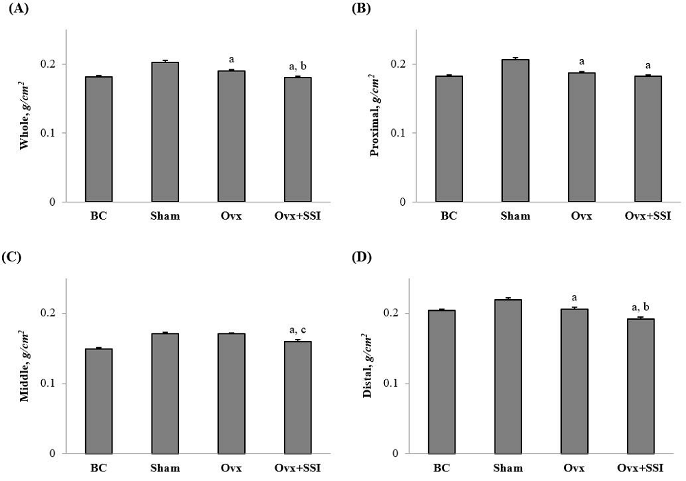 Figure 1.
— BMD of the whole femur (A) and the proximal (B), middle (C) and distal (D) thirds of the femur of the BC, Sham, Ovx and Ovx + SSI group. Values are expressed as the mean ± standard error the mean. Data for BC, Sham, and Ovx groups are taken from Morita et al. [20]. aP < 0.01 vs. Sham group, and bP < 0.05 and cP < 0.01 vs. Ovx group (one-way ANCOVA with Tukey’s honest significant difference test). BMD, bone mineral density; BC, baseline control; SSI, Sesamum indicum; ANCOVA, analysis of covariance.
Table 1 summarizes the structural indices of bone histomorphometry for the proximal tibial metaphyseal cancellous bone. As a result of Ovx, bone volume and trabecular number were significantly lower in the Ovx group than in the Sham group. In addition, bone formation indices, such as osteoid surface, osteoid volume, and osteoblast surface, were significantly higher in the Ovx group than in the Sham group. Bone resorption indices, such as eroded surface and osteoclast surface, were also higher in the Ovx group compared with the Sham group, although these differences did not reach statistical significance. There were no significant effects of dietary addition of sesame seed extract on these parameters. Changes of the histomorphometric dynamic indices are shown in Figure 2. Ovx resulted in the significantly higher measurements for mineralizing surface and bone formation rate in the Ovx group compared with the Sham group. However, supplementation with sesame extract had a deleterious effect on bone remodelling, with the Ovx + SSI group showing significantly lower mineralizing surface and bone formation rate compared with the Ovx group.
| BCa | Shama | Ovxa | Ovx + SSI | |
|---|---|---|---|---|
| Structural indices | ||||
| Bone volume, % | 21.7 ± 0.9 | 29.7 ± 1.7 | 8.0 ± 0.8c | 8.9 ± 0.9c |
| Trabecular thickness, μm | 58.6 ± 1.8 | 69.7 ± 3.0 | 61.8 ± 1.4 | 63.2 ± 1.6 |
| Trabecular number, /mm | 3.69 ± 0.09 | 4.25 ± 0.12 | 1.29 ± 0.10c | 1.40 ± 0.13c |
| Osteoid surface, % | 23.1 ± 2.1 | 18.5 ± 2.1 | 31.8 ± 2.3c | 28.6 ± 2.0c |
| Osteoid volume, % | 3.00 ± 0.34 | 2.11 ± 0.33 | 4.28 ± 0.49c | 3.32 ± 0.27b |
| Osteoblast surface, % | 3.47 ± 0.44 | 1.13 ± 0.25 | 3.15 ± 0.65c | 3.17 ± 0.52c |
| Eroded surface, % | 7.51 ± 1.02 | 3.32 ± 0.16 | 4.04 ± 0.35 | 5.13 ± 0.77 |
| Osteoclast surface, % | 4.64 ± 0.69 | 0.65 ± 0.11 | 1.29 ± 0.12 | 1.86 ± 0.28c |
Values are expressed as mean ± SEM. n = 6-10.
aData for the BC, Sham, and Ovx groups taken from Morita et al. [20].
bP < 0.05 and cP < 0.01 vs. Sham group (one-way ANCOVA with Tukey's HSD test).
BC, baseline control; Ovx, bilateral ovariectomy; SSI, Sesamum indicum; ANCOVA, analysis of covariance; HSD, honest significant difference.
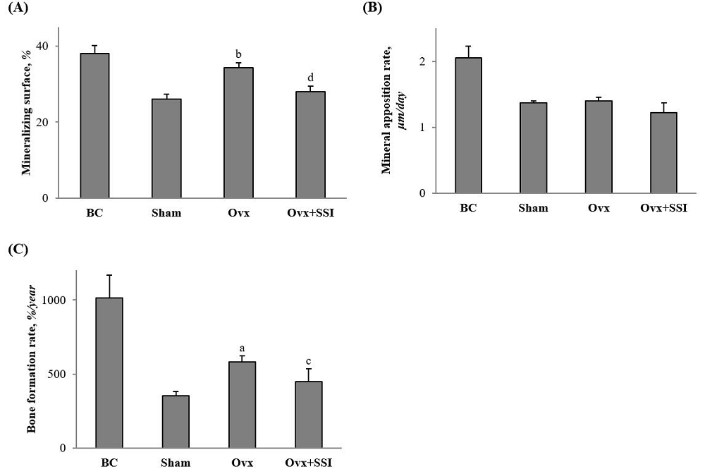 Figure 2.
— Dynamic indices of cancellous bone at the proximal tibiae of the BC, Sham, Ovx and Ovx + SSI group. Values are expressed as the mean ± standard error the mean. Data for BC, Sham, and Ovx groups are taken from Morita et al. [20]. aP < 0.05 and bP < 0.01 vs. Sham group, and cP < 0.05 and dP < 0.01 compared vs. Ovx group (one-way ANCOVA with Tukey’s honest significant difference test). BC, baseline control; SSI, Sesamum indicum; ANCOVA, analysis of covariance.
Postmenopausal bone loss is caused by a disproportionate increase in bone resorption compared to bone formation, resulting in net bone loss [23, 24]. Herein, we demonstrated that ovariectomy in rats resulted in loss of femur BMD based on DXA measurement, with the loss most pronounced at sites rich in cancellous bone (proximal and distal thirds). The loss was characterized by increased bone formation and resorption indices in bone histomorphometry. Dietary supplementation with methanol extracts of sesame seeds in the ovariectomized rats further lowered femur BMD compared to control-fed Ovx rats. In addition, bone histomorphometry revealed that such supplementation diminished the compensatory increases in bone formation following Ovx, such as mineralizing surface and bone formation rate. These results suggest that the consumption of sesame seeds might not alleviate, but instead accelerate, the bone loss observed in postmenopausal women.
Investigations on the potential benefits of sesame on bone metabolism in animal models of postmenopausal osteoporosis have been limited. In one study, Boulbaroud et al. [25] showed that 3-month-old Ovx rats given a diet containing 10% sesame oil for four weeks had moderately thicker and elongated trabeculae, and narrowed inter-trabecular spaces, compared to the Ovx control, based on histological examination of the distal femur epiphyseal cancellous bone. The authors also demonstrated significantly lower bone formation (alkaline phosphatase) and resorption (tartrate-resistant acid phosphatase) markers in the rats fed sesame oil compared to the Ovx control rats. Hassan et al. [26] showed that 3-month-old Ovx rats fed a diet containing 10% sesame oil for two months had significantly higher femur BMD compared with Ovx controls. Why the present experiment did not corroborate the findings of these two earlier ones is unknown, although the form of sesame used in the diet supplementation and the resultant fat content is one possible factor. In the present study, an extract mixed with the basal diet at a 13.9% weight ratio of sesame seeds/diet was used. Sesame seed is an oil seed that contains 50% fat [27], and the weight ratio of oil/diet in the present study was estimated to be approximately 7%. Boulbaroud et al. [25] found the favorable effect of sesame oil at an oil/diet ratio of 10%, but not at 7%. In a different study, where a diet containing 10% freshly ground sesame seeds was fed to 4-5-week-old Ovx athymic mice for eight weeks, Sacco et al. also found no increase in the BMD in the femur and L1-3 vertebra [27]. Further studies are required to clarify these apparent discrepancies.
The present findings that methanol extracts of sesame seeds diminished femur BMD and bone formation indices in Ovx rats was, indeed, unexpected, and we speculate that vitamin E could also be a factor responsible, in part, for the effects observed in the present study. Vitamin E is lipid-soluble, and tocopherols and tocotrienols are major vitamin E derivatives. These two compounds have four distinct isoforms (α, β, γ, and δ), and α-tocopherol has the highest bioavailability among them [28, 29]. Yamashita et al. [30, 31] showed that sesame or its lignan elevated α- and γ-tocopherol concentrations in rats, even in those fed a low α-tocopherol diet. Additionally, Sontag and Parker [32, 33] showed that sesame lignan inhibited tocopherol-ω-hydroxylase activity exhibited by cytochrome P450 enzymes, resulting in elevated tocopherol levels. Some clinical studies also indicated elevated blood concentrations of α- and γ-tocopherol in response to sesame lignan intake [34-37]. In a separate, cross-sectional population-based study, postmenopausal women who used vitamin E (α-tocopherol) supplements had significantly lower bone-specific alkaline phosphatase, a biomarker of bone formation [38]. In contrast, Fujita et al. [39] showed that excessive vitamin E might have adverse effects on bone health, because it reduced bone mass by promoting the fusion of mononuclear preosteoclasts into multinucleated osteoclasts in rodents [39]. Based on these findings, we speculate that the administration of sesame extract increased the concentration of tocopherols, resulting in decreased BMD, although a recent investigation by Ikegami et al. [40] demonstrated that vitamin E intake did not result in bone loss in Ovx mice. Therefore, it would be of great benefit to fully characterize the effect of sesame seeds on bone metabolism.
In conclusion, administering methanol extracts of sesame seeds to Ovx rats resulted in decreased bone mass based on bone densitometry and bone formation indices under the experimental conditions employed here. Further studies are needed to conclude whether consumption of sesame seeds could be beneficial for mitigating bone loss observed in postmenopausal women.
This study was supported in part by a JMWH Bayer Grant from the Japan Society for Menopause and Women's Health to A.M.
The authors declare no conflict of interest.