
Cornual pregnancy is a challenging situation which can threaten the life of the patient if underestimated. The authors present a case of a 19-week angular pregnancy with hemoperitoneum caused by placenta percreta and uterine rupture.
Cornual pregnancy is defined as embryo implanted in the lateral angle of the uterine cavity, which is medial to the uterotubal junction and round ligament. It represents 1% of ectopic pregnancies whose occurrence rate ranges from 1/2,500 to 1/5,000 live births. Maternal mortality rates attributed to cornual pregnancy are estimated at 2.5% to 4.0%, with the vast majority due to rupture and extensive hemorrhage [1]. The authors present a case of a 19-week angular pregnancy with hemoperitoneum caused by placenta percreta and uterine rupture.
A 28-year-old woman arrived at the emergency department presenting 19 weeks of amenorrhea with severe abdominal pain for two hours. She complained of lower right abdominal pain progressed during the last two weeks; she also mentioned dizziness, but denied syncopy. She had a previous history of an abortion. The heart rate was 120 beats per minute, blood pressure was 111/76 mmHg, and temperature was 37.4°C. Her abdomen presented a generalized tenderness, guarding, rigidity, and rebound tenderness, and no vaginal bleeding was observed. Emergency bedside ultrasonography revealed intrauterine gestation without detectable fetal heart pulsation and moderate amount of free fluids in the Douglas cul-de-sac. Non-clotting blood was obtained from transabdominal puncture. Furthermore, an emergency abdominal and pelvic non-enhanced CT scan was performed and demonstrated an empty uterine cavity bent left laterally and compressed by an amniotic sac adjacent to the right cornal region of the uterus (Figure 1). Her hemoglobin level was 7.4 g/L. During laparotomy, it was observed that the right uterine angle was enlarged by approximately 20 cm, as well as cystic gestational swelling, which was displacing the reflection of the round ligament laterally. Partial surface of the swelling with active bleeding appeared black and fragile, and uncovered with myometrium. Placenta located at the right uterine horn, firmly attached to the uterine wall and extended through the entire myometrial layer (Figure 2). Placenta histopathology also confirmed placenta percreta subsequently. Both ovaries and fallopian tubes were normal. As the patient was with no offspring, partial cornuectomy and uterine reconstruction was performed for fertility preservation after discussion with the family about rupture risk for the subsequent pregnancy. The postoperative course was uneventful and the patient was discharged on postoperative day 8. No complications were detected during the follow-up period.
 Figure 1.
— Non-enhanced CT using multiplanar reconstructed technique revealed an empty uterine cavity (blue arrow) and the amniotic sac (red arrow) on axial (A), saggital (B) and coronal (C) plane of the uterus. Note the sac layed adjacent to the cornal region of the uterus, pushing the uterine cavity latterly (C).
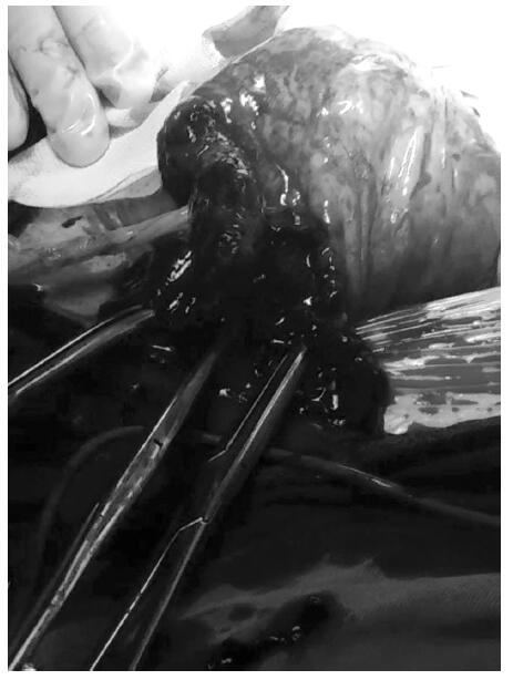 Figure 2.
— Placenta at the right uterine horn, firmly attached to the uterine wall and extended through the entire myometrial layer
Cornual pregnancy is a challenging situation which may lead to massive hemoperitoneum and rupture of uterus. Cornual pregnancy can now be diagnosed early in many cases with transvaginal sonography and serum β-hCG assays, but definitive diagnosis could also be challenging. In this case, the patient was diagnosed with normal intrauterine pregnancy in the early stage, as well as with emergency bedside ultrasonography. MRI is an optional modality to improve the diagnosis, but limited to such emergency cases, CT was the preferred method to confirm the diagnosis and excluded hemorrhage of abdominal organs. 3D reconstruction techniques, such as multiplanar reconstruction, facilitate displaying the shape of uterine cavity and its relationship with amniotic sac. Although the present case did not apply contrast enhancement, iodine medium could be used under restricted indication to increase the contrast between the cavity and wall of uterus, and aid the imaging recognition and interpretation.
Cornual pregnancy may cause persistent uterine pain and bleeding, spontaneous abortion, ruptured uterus during pregnancy. Deferential diagnosis in ultrasonography or radiography image should consider interstitial and Müllerian anomaly pregnancies. Interstitial pregnancies usually rupture following eight to 16 weeks of amenorrhea, while cornual pregnancies can sometimes be carried to term but within increased risk of abnormal placentation, and its consequence [2]. Laparoscopy or laparotomy may confirm the diagnosis. It is treated by cornual resection or even hysterectomy by laparotomy traditionally because of the delayed diagnosis; if future pregnancy is desired, fertility preservation surgery should be considered, while early diagnosis is crucial.
Placenta percreta defines villi that penetrate through the myometrium and to or through the serosa. It is encountered in an approximate ratio of 5% in placenta accreta syndromes [2], which are usually seen in women with a prior cesarean delivery and an associated previa. It will also lead to uterine rupture and could be life threatening. In the present case, placenta percreta may due to the previous induced abortion and abnormal placentation. There is evidence that women with accreta syndromes have an increased risk for recurrence, uterine rupture, hysterectomy, and previa in subsequent pregnancy [3]. The present authors totally resected the lesion before uterine construction to decrease the risk. When placenta percreta is the suspicious diagnosis by ultrasonography in the routine antenatal examination, MRI should also be applied for confirmation.