IMR Press / RCM / Volume 22 / Issue 2 / DOI: 10.31083/j.rcm2202051
Open Access Original Research
Long-term results of endovascular intervention with unibody bifurcation endograft for elective abdominal aortic aneurysm management
Show Less
1 Department of Cardiovascular Surgery, Ankara City Hospital, 06800 Ankara, Turkey
2 Department of Cardiovascular Surgery, Kartal Kosuyolu Hospital, 34145 İstanbul, Turkey
3 Department of Cardiovascular Surgery, Gülhane Training and Research Hospital, Gülhane Faculty of Medicine, University of Health Sciences, 06010 Ankara, Turkey
4 Department of Cardiovascular Surgery, Erol Olçok Training and Research Hospital, Hitit University, 19030 Corum, Turkey
*Correspondence: drzaferiscan@gmail.com (Hakkı Zafer İşcan)
Academic Editor: Peter A. McCullough
Rev. Cardiovasc. Med. 2021, 22(2), 453–459; https://doi.org/10.31083/j.rcm2202051
Submitted: 17 February 2021 | Revised: 2 April 2021 | Accepted: 6 April 2021 | Published: 30 June 2021
Copyright: © 2021 The Author(s). Published by IMR Press.
This is an open access article under the CC BY 4.0 license (https://creativecommons.org/licenses/by/4.0/).
Abstract

Unibody bifurcated endografts have the advantage of reducing the operative time, avoiding migration and iliac limb dislocation in patient with abdominal aortic aneurysm (AAA). We report our long-term experience in patients who underwent endovascular aortic repair (EVAR) due to infrarenal AAA with Endologix AFX® endograft system. Between January 2013–December 2018, 68 patients with infrarenal AAA had EVAR procedure with Endologix AFX® endograft system. Mean follow-up was 40.4 ± 19.5 months, and all patients had computed tomography periodically, with colored Doppler ultrasonography (CDUS) every six months. Mean age was 68.5 ± 7.1 years and, 63 (92.6%) patients were male. Early mortality, renal complications, stent-graft migration and cardiac complications were not seen in early post-operative period. There was no early mortality in the group and no conversion to open repair. In long-term follow-up 12 patients (17.6%) had endoleak (5 with type II, 7 with type III). Overall survival estimated by Kaplan-Meier analysis was 94.1% at 1 year, 85.2% at 2 years, 74.1% at 3 years and 54.0% at 5 years. Freedom from second intervention and conversion was 98.4% at 1 year, 95.3% at 2 years, 93.3% at 3 years and 87.4% at 5 years. Endologix AFX® endograft provides a fast, safe and effective endograft for the early period, therefore it seems more suitable for elderly and comorbid patients. This endograft has low reintervention rates however according to our results, aneurysms larger than 6 cm may have more sideway displacement possibility and by this way type III endoleak. Proper patient selection and sufficient overlap are the key issues. Close monitoring is mandatory at the follow-up period.

Keywords
Abdominal aortic aneurysm
Endovascular procedures
Cumulative survival rate
Endoleaks
1. Introduction

Endovascular aneurysm repair (EVAR) has gained wide acceptance as the preferred method of treating aortic aneurysms with suitable anatomy. The technique is associated with lower early mortality, morbidity, faster discharge and patient turnover by offering small incisions with minimal invasive nature [1, 2, 3, 4]. The adverse events in the endovascular procedures may be due to endograft, challenging aortic anatomy or iatrogenic. Endografts are dynamically in evolution according to the early and late results of the randomized studies to reduce the adverse events related to structural impairments.

Unibody bifurcated endografts have the advantage of reducing the operative time and avoiding iliac limb dislocation in the long term. The Endologix AFX® (Endologix, Inc., Irvine, CA, USA) endograft system for endovascular abdominal aortic aneurysm repair is the only graft with anatomical fixation to the aortic bifurcation in comparison to most other endografts that use suprarenal neck as the main fixation point. In short and mid-term outcomes, the Endologix AFX® endograft had a low-rate of device and procedure-related complication but there is not yet enough study that showed long-term results [5].

United States Food and Drug Administration (FDA) approval was originally granted for Endologix Powerlink system in 2004 [6], a modification to this device was introduced in 2011 as the AFX endograft, which used a lower profile 17F delivery system. A proximal and distal landing zone length of 15 mm, neck diameter between 18–32 mm and infrarenal angulation to the aneurysm of 60 degrees were the recommended instructions for use [7, 8]. A change in the original IFU in 2013 recommended a minimum aortic component overlap of 30 mm to 40 mm which was followed in 2014 by introduction of the change of expanded polytetrafluoroethylene (e-PTFE) material from Strata to Duraply referring to a highly dense, helical wrap providing longitudinal strength. Finally, the instructors regarding the minimum length of overlap were further revised in 2016 as follows: overlap length equals half the maximum aortic aneurysm diameter plus 20 mm [9].

Endoleaks are defined by persistent blood flow within the aneurysm sac intraoperatively or following EVAR. During the follow-up, in a meta-analysis, type II, III or IV endoleak could be seen in 13.7% of patients [10]. Late type III endoleak is an uncommon but serious complication caused by fabric tears, disruptions or junctional separation of the endograft components.

We report our long-term experience of patients who underwent endovascular infrarenal abdominal aortic aneurysm electively with Endologix AFX® endograft system.

2. Methods
2.1 Study design

In our Cardiovascular Surgery Department, during January 2013–December 2018, 68 patients with elective infrarenal abdominal aortic aneurysm had EVAR procedure with Endologix AFX® endograft system by the same surgical team. Urgent procedures, ruptured aneurysms and procedures including hybrid operations, simultaneous coronary bypass or other cardiovascular operations were excluded from the study. The study protocol was approved by local institutional review board (Turkey Yuksek Ihtisas Hospital, 29620911-929-1303). The study was conducted in accordance with the principles of Declaration of Helsinki. No informed consent was obtained because of the retrospective nature of data collection from hospital records in the study.

The patients were predominantly American Society of Anesthesiologists (ASA) class II–IV, with a high prevalence of cardiopulmonary comorbidities. 30 patients (44.1%) were symptomatic because of the aneurysm, mostly umbilical pain. All patients had undergone EVAR procedure in elective manners.

2.2 Operative procedure

Preoperatively each patient had a customized plan upon multislice contrast enhanced computerized tomography (CT) for aneurysm anatomy, dimensions, length and endograft sizes. Graft diameter was determined by oversizing the graft 10–20% with respect to measured neck diameter and anatomy. Graft length was chosen to preserve at least one hypogastric artery. Measurement of the AAA centerline or greater curvature length is currently preferred, not the traditional straight-line method, for EVAR planning purposes.

The reporting standards according to the guidelines from the Society for Vascular Surgery were used as indications [1]. Almost all procedures were performed with unilateral femoral artery exposure for main body and percutaneously for the aortic extension for access. EVAR was performed under general, local, or regional anesthesia in the hybrid room equipped with “Siemens Artees Zee” fluoroscopy. Access was carried out through an ipsilateral open exposure and contralateral percutaneous access for all patients. When performing aortic extension, at least 3 cm of overlap was performed in all patients. A completion angiogram was performed to document the status after endograft implantation. Technical success was defined a successful deployment of the endograft and completion of the procedure with no type I or III endoleaks and without the need for a secondary intervention or open repair within the first 24 hours.

2.3 Surveillance

Postoperative surveillance protocol included physical examination, blood samples and imaging control with abdominal aortic Doppler and/or multislice contrast CT. In the follow up period, all patients had a CT between the first three months and after that all patients were evaluated with abdominal aortic Doppler ultrasound every six-month period and CT annually. For patients with a shrinking aneurysm sac and/or >3 years of stability, a non contrast enhanced CT and CDUS was preferred. No patient had CT control before discharge after the procedure, however, this should be performed if there was any suspicion at the completion angiogram, a hostile anatomy or ongoing symptomatology.

2.4 The technical features of the Endologix AFX® endograft

The Endologix AFX® device consists of a main bifurcated unibody and a proximal aortic extension, resting on the aortic bifurcation to provide anatomic fixation and the proximal extension in order to provide sealing at the aortic neck. Endologix AFX® represents the second generation of the Endologix endograft system and uses novel strata with high density e-PTFE. This device is commercially available since 2011. The skeleton of the endograft is made of cobalt-chromium alloy in a multilinked self-expanding unibody. The stent is located inside the endograft, so the stent is not directly related to the aneurysm sac, facilitating conformity. However, this is almost always a technical difficulty for endovascular reinterventions. Unique to this device graft material is externally mounted. It is attached only to the proximal and distal ends at the proximal aortic extension and allows e-PTFE facilitating to seal to challenging aortic necks. During the course of the study, Endologix AFX® bifurcated device was available in diameters 22, 25 mm and 28 mm; lengths ranging from 40–120 mm for body and 30–55 mm for limbs. The proximal extension was available in suprarenal manner. The device is delivered with 17 French introducer system ipsilaterally and 9-French sheath contralaterally. Instructions for use requires an aortic neck and iliac seal zone length of 15 mm, neck diameter between 18–32 mm and infrarenal angulation to the aneurysm of 60 degrees.

2.5 Statistical analysis

The variables were investigated using visual (histograms, probability plots) and analytical methods (Kolmogorov-Smirnov/Shapiro-Wilk test) to determine the normality of their distribution. Normally distributed continuous variables were expresses as mean ± standard deviation (SD) or median values with range if not normally distributed. Categorical variables were expressed as number and percentages. Aneurysm diameter was compared using the Mann-Whitney U test. Kaplan-Meier analysis was conducted to demonstrate freedom from all-cause mortality and freedom from secondary interventions. A p value of <0.05 was considered to be statistically significant, and all statistical analyses were performed using the SPSS for Windows version 15.0 statistical software program (SPSS Inc., Chicago, IL, USA).

3. Results
3.1 Patients and procedural data

Baseline characteristics of patients are listed in Table 1. In our series, mean age was 68.5 ± 7.1 years and 63 (92.6%) patients were male. Most seen comorbidities were hypertension (64.7%) and coronary artery disease (CAD) (54.4%). Mean abdominal aorta diameter was 60.4 ± 8.7 mm. Local anesthesia was used for 36 (52.9%) patients, spinal anesthesia for 4 (5.9%) and general anesthesia for 28 (41.2%). Mean intervention duration was 132.4 ± 25.4 min, total fluoroscopy duration was 17.2 ± 4.1 min and mean volume of 66.3 ± 16.9 mL contrast agent was used (Table 2). Mean radiation exposure was 3.2 ± 0.9 mGym2. In two patients, thoracic endovascular aortic aneurysm repair was performed simultaneously. 151 components were used for 68 patients (2.2 ± 0.5 components for each patient). Early mortality (30-day or in-hospital mortality), renal complications, stent migration and cardiac complications were not seen in early post-operative period. Among the periprocedural complications, access site problems were experienced by eight patients resolved with embolectomy in four and with graft interposition for the rest. Mean hospital stay was 3.1 ± 2.1 days after intervention.

Table 1.Demographics of patients.
Features n (%) or mean ± SD
Male gender 63 (92.6)
Age, years 68.5 ± 7.1
ASA grade
2 25 (36.8)
3 29 (42.6)
4 14 (20.6)
Comorbidities
Diabetes mellitus 15 (22.1)
Hypertension 44 (64.7)
Hyperlipidemia 18 (26.5)
Chronic obstructive pulmonary disease 25 (36.8)
Renal disease 12 (17.6)
Peripheral artery disease 6 (8.8)
Coronary artery disease 37 (54.4)
Heart failure 6 (8.8)
Smoking habit 26 (38.2)
History of cerebrovascular accident 3 (4.4)
Malignancy 9 (13.2)
Aneurysm diameter, mm 60.4 ± 8.7
Left ventricle ejection fraction, % 52.0 ± 10.2
SD, Standard deviation.
Table 2.Peri-operative data.
Features n (%) or mean ± SD
Endovascular aneurysm repair 68 (100)
Thoracic endovascular aneurysm repair 2 (3)
Iliac extension 13 (19.1)
Anesthesia
Local 36 (52.9)
Spinal 4 (5.9)
General 28 (41.2)
Embolectomy 4 (5.9)
Graft interposition 4 (5.9)
Mean intervention duration, minute 132.4 ± 25.4
Total fluoroscopy duration, minute 17.2 ± 4.1
Amount of contrast agent (mL) 66.3 ± 16.9
SD, Standard deviation.
3.2 Follow-up period

Mean follow-up was 45.5 ± 21.4 months (range 1 to 85 months) and, 12 patients (17.6%) had endoleak (5 with type II, 7 with type III). Patients with type II endoleaks which was detected in the completion angiogram had no growth in aneurysm diameter at follow-up. Additionally, there was no detected new type II endoleak in the follow-up period. Three of these type II endoleaks (60.0%) were resolved spontaneously at the first year.

Three patients with type III endoleak did not accept any intervention who were ASA class IV (Fig. 1). These patients all died because of aneurysm rupture at 12–18 months after the diagnosis of type III endoleak. One patient referred to hospital with ruptured aneurysm and died intraoperatively. Other two patients had also symptomatic contained ruptured aneurysm and firstly endovascular intervention was attempted, however both failed and, open surgical repair was performed and discharged (Fig. 2). One of these patients experienced fabric tear close to bifurcation after a blunt trauma [11]. The last patient was discharged after successful endovascular intervention (Fig. 3). All type III endoleaks were symptomatic and 5 patients were already diagnosed with CT. The other two patients were referred to our outpatient clinic and type III endoleak was detected firstly with CDUS and then confirmed by CT. The mean time interval between the primary EVAR procedure and type III diagnosis was 24.7 ± 9.8 months (range 12–37 months). Patients with type III endoleaks are listed with aortic diameters, aortic size indexes (ASI) as a summary in Table 3. All patients have an aortic diameter >60 mm, and ASI >3. 0.1 unit increase in ASI was found to be a predictor for type III endoleak (OR: 2.37, 95.0% CI: 1.14–4.90, p = 0.020). On the other hand, the preoperative diameter of aneurysm was significantly higher for the patients with type III endoleak (67.4 mm vs 59.6 mm; p = 0.010).

Fig. 1.

Preoperative, 6th and 14th month CTA and first month overlap measurements of a type III endoleak of patient #4 (IFU compatible). Although there was an adequate overlap at first month scans, the separationand kinking of the endografts during the follow-up can be seen.
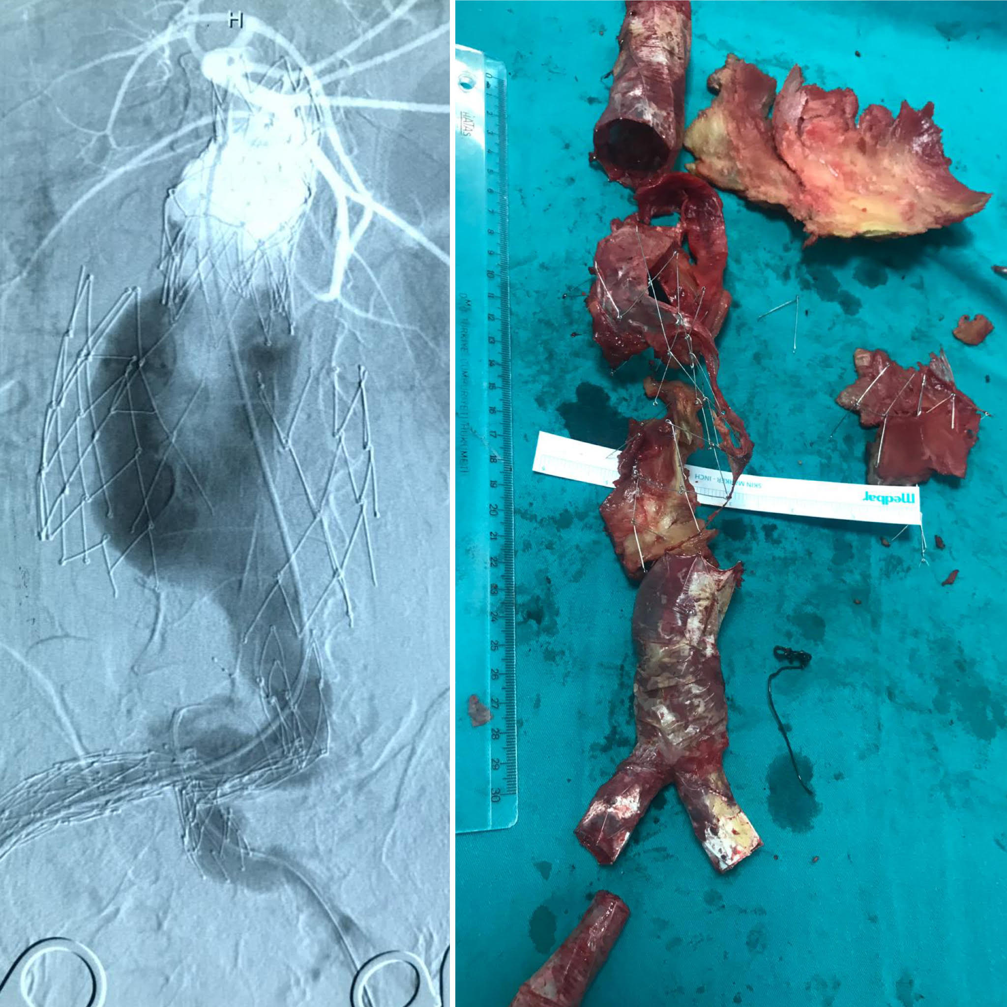
Fig. 2.

The enlargement of the endoskeleton and the demonstrative main body deployment problem over the guidewire, the endovascular attempt converted to a successful open repair (patient #1). Structural deterioration and the separation of endografts can be seen on angiographic views. Endovascular rescue for this patient was unsuccessful and the extreme deterioration of the endograft is presented on the right side.
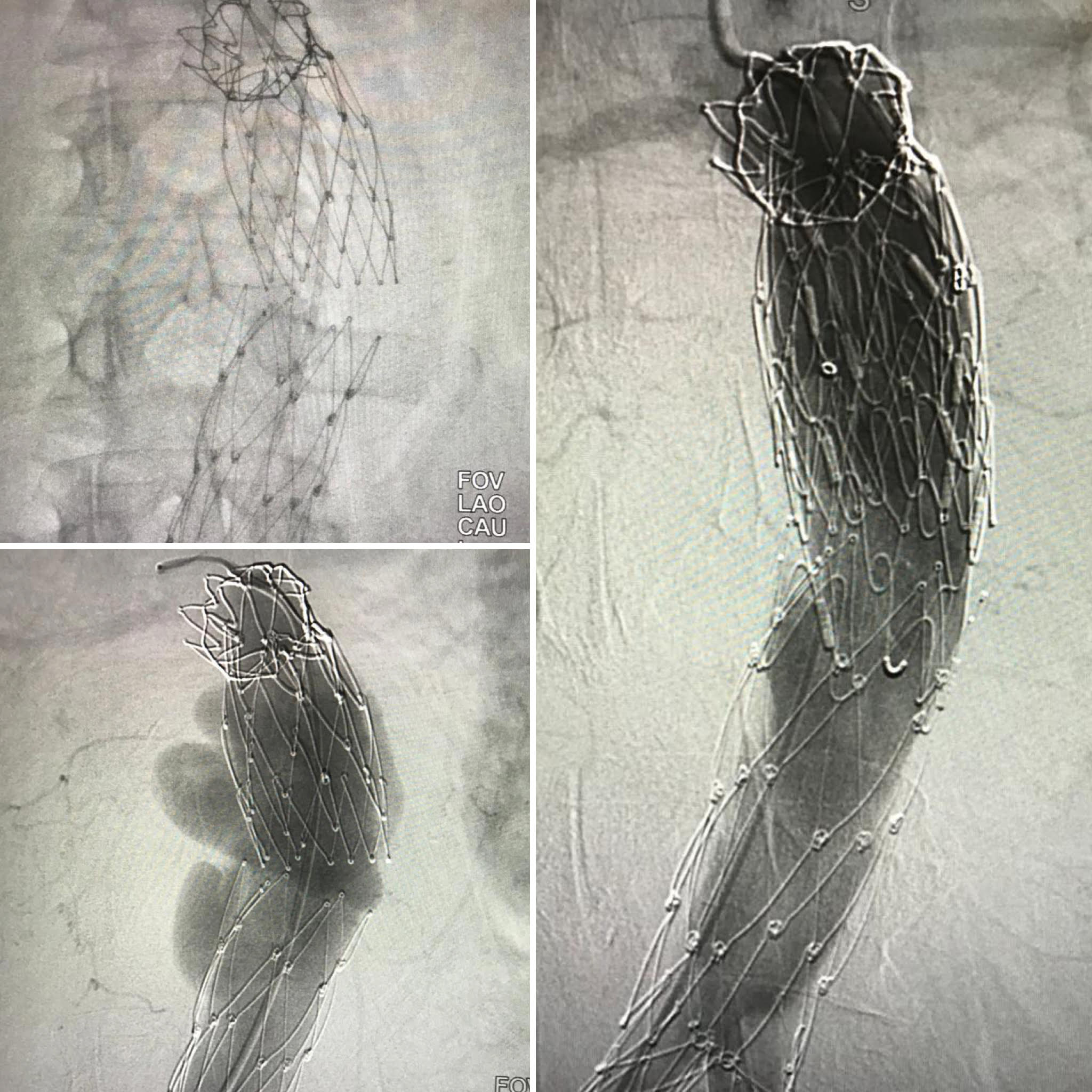
Fig. 3.

Successful endovascular reintervention for type III endoleak (patient #5). The patient was admitted for ruptured aneurysm. The separation of the endografts and the endoleak between the endografts can be seen. A bridging tubular endograft was deployed between the previous endograft.

Table 3.Detailed description of patients with type III endoleak.
Patient IFU compatible AA diameter (mm) BSA ASI Outcome
1 Negative 85 1.99 4.26 Survived (OS)
2 Negative 70 1.66 4.22 Death (No intervention)
3 Positive 60 1.66 3.6 Death (No intervention)
4 Positive 82 2.10 3.9 Death (Rupture)
5 Positive 67 1.94 3.5 Survived (Endovascular)
6 Positive 62 2.00 3.1 Death (No intervention)
7 Negative 72 2.00 3.6 Survived (OS)
IFU, Instructions for use; AA, Aortic aneurysm diameter (initial); BSA, Body surface area; ASI, Aortic size index; OS, Open surgery.

There was no early mortality in the group and no conversion to open repair. Technical success was achieved in all patients. Overall survival estimated by Kaplan-Meier analysis was 94.1% at 1 year, 85.2% at 2 years, 74.1% at 3 years and 54.0% at 5 years (Fig. 4A). Freedom from second intervention and conversion was 98.4% at 1 year, 95.3% at 2 years, 93.3% at 3 years and 87.4% at 5 years (Fig. 4B). On the other hand, actual mortality was 36.8% at our patient cohort. Aneurysm related mortality occurred in six patients, and the other causes of death were cardiac problems in 12 patients, malignancies in six patients and renal failure in one patient.

No graft infection, migration and post-implantation syndrome were found during follow-up period.
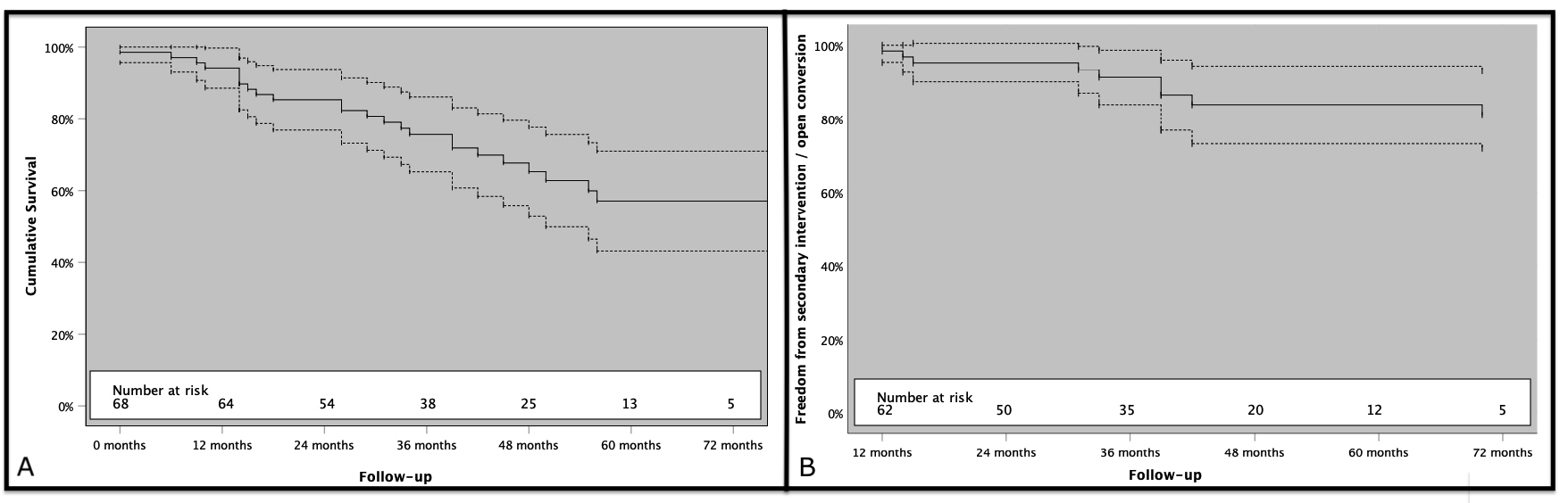
Fig. 4.

Kaplan-Meier survival analysis. (A) Cumulative survival of patients. (B) Freedom from secondary interventions (dashed lines show 95.0% confidence intervals).

4. Discussion

EVAR is widely accepted treatment of choice for AAA. The superiority of EVAR over open surgery was seen especially in short-term outcomes [1, 2, 3, 4]. The use of the Endologix AFX® endograft was associated with a low rate of device and procedure-related complications in this period [12, 13, 14, 15]. The Endologix AFX® endograft system is a single-body bifurcation graft that is different from other grafts. Anatomic fixation in native bifurcation has the potential to reduce the risk of distal migration in long term. This advantage was also seen in our series. There was no migration.

In contrast to all other endografts, AFX stent frames are located inside to facilitate the aortic conformability. However, this endoskeleton can be challenging at reintervention times, especially for guidewire entrapment or the passage for the endograft bodies.

The texture is e-PTFE, located outside the skeleton, and only proximal and distal endings are sutured. Therefore, it provides flexibility and better adaptation to the irregular aortic neck in the proximal sitting area since the graft material can move freely in an “active seal” manner [16, 17]. This independent movement of the graft material from the stent may be a factor for uncoupling as the columnar strength may diminish when fixation is at the aortic bifurcation.

Besides long-term outcomes with abdominal aortic aneurysm (AAA) treated with unibody bifurcated endograft, our study demonstrated excellent short-term results, with no intra-procedural open surgical conversion and no 30-day mortality encountered. The first finding was related to the long-term prognosis of treatment of AAA with minimally invasive procedure. Cumulative survival was 54.0% at 5th year in this patient cohort. Because of the presence of CAD in 54.4% of our patient population, one can think that all-cause mortality was high in the long term due to diseases such as CAD and other vascular system disorders. Increasing malignancy rates with advanced age may also have caused this high mortality rate. Moreover, patients who are at high risk for surgery and older patients are now treated with endovascular intervention; in the long term, death due to non-aneurysm causes is more common. In Bahia et al. [18] meta-analysis, they showed no improvement in mortality, with a mean 69.0% rate, of elderly patients over the years. There is long-term follow-up of different endografts in the literature. In a recent study, overall survival at 7 years was 50.3% to 61.4% [19]. Therefore, this high mortality in long-term; it should be kept in mind that it is more dependent on additional issues like cardiac morbidity or malignity, not aneurysm related. In order to avoid extra nephrotoxic agent, we did not perform routine coronary angiography (CAG) on patients without cardiac symptoms. We routinely used transthoracic echocardiography and electrocardiogram. In cases where surgical stress was low and no cross clamp, we did not experience cardiac morbidities in the early period. In our series with high cardiac comorbidities, long-term cardiac mortality may have caused a significant reduction in survival.

As all designs carry their own pros and cons; there seems a component uncoupling problem, as the main body and aortic extension disconnection, tried to dissolve by the manufacturer’s suggestion as to maximize the overlap and some changes over the material used. Manufacturers’ material exchange does not seem to be sufficient for eliminating the type III endoleak, so proper patient selection, sufficient overlap are the key issues.

Endoleak type III due to fabric tears or disintegration of the endograft components has been a concern in EVAR. From study 701 EVAR patients treated with the Endologix Powerlink and Endologix AFX® endograft, Skibba et al. [16] reported type III endoleak in 17 patients (2.4%). Welborn et al. [13] showed a similar incidence (2.3%) in 108 patients treated with the AFX endografts. Even though, the 30 mm and more overlap requirement was always applied in our clinic, six of the patients in our series had an endoleak type IIIa and one type IIIb, during long-term follow-up. It was 10.3% and close to 7.3% of Lemmon et al. [5]. Among the type III endoleak patients, five were already diagnosed with CT however the other two patients were detected firstly with CDUS and than confirmed by CT. This correlation leads us to another study concerning the primary tool for EVAR surveillance [20] where we found high correlation of type I and III endoleak detection with CDUS and CT.

The treatment of type III endoleak often employs an endovascular approach as first line. In case of failure open late conversion and repair are mandatory. Meticulous preoperative planning and proper choice of endograft for a sufficient overlap arevery important.

To avoid type III endoleaks, maximizing component overlap or more proper patient selection may be a choice. Proper patient selection for Endologix AFX® endograft should include saccular aneurysms, infrarenal AAA with narrow aortic bifurcations and with short infrarenal-bifurcation lengths where modular endografts have technical disadvantages. Also older patients with ASA IV status necessitating a faster procedure may be proper candidates.

It is well established that the dominant forces on modular EVAR components are directed sideways and that such force increases along the greater curvature with aortic or endograft angulation [21, 22]. The resulting sideways displacement of the endograft is more prevalent in larger aneurysms with sufficient volume in the aneurysm sac to accommodate this conformational change and is associated with an increased incidence of type I and III endoleaks [23]. Long term integrity of the central overlap zone between the two modular components thus depends on appropriate outward radial force from the proximal extension, the sealing mechanism of the expanded PTFE endograft covering, adequacy of the overlap zone between the two aortic components and the degree of angulation.

The probability of type III endoleak was increased two-fold by 0.1 unit increase at ASI. Moreover, the interpretation that AAA larger than 60 mm and ASI over 3 had the possibility of type III endoleak may be revealed as the all patients with type III endoleaks were adjusted to these criteria.

Type II endoleaks were detected in the completion angiogram in 5 patients (7.4%). They were all followed up and three of them (60.0%) disappeared in the first year while the others were closely followed for the aneurysm sac enlargement. No aneurysm sac enlargement was seen and therefore no reintervention was necessitated.

No graft infection and post-implantation syndrome were seen. There are studies suggesting e-PTFE grafts are associated with a lower probability of postoperative fever, however there is no randomized clinical support [24].

Clinical reports confirm low limb occlusion rates [14, 18]. Stent grafts each have different variations in graft material, stent material and configuration. These differences are made for providing the perfect adaptation of graft and iliac artery anatomy, especially with angulated, calcified or nonuniform atherosclerotic landing zones. Clinical studies confirm low limb occlusion rates [9, 14, 18, 25]. Patients treated with the Endologix Powerlink and Endologix AFX® endografts during an 8-year period, reported only three (0.4% of all patients) graft thrombosis that needed reintervention [16]. Limb thrombosis has low prevalence in patients with AFX endografts. In our long-term follow-up, the patients did not need to reintervention for limb issues. We only had periprocedural access site problems, resolved with femoral embolectomy and/or graft interposition.

Study limitations are the retrospective design, a single center experience with a relatively small patient population that may prevent statistical significance. There was no randomization as only Endologix AFX® endograft for elective AAA were chosen. There is no actual comparison with other endografts.

5. Conclusions

Endologix AFX® endograft does not require contralateral limb cannulation, therefore it enables a relatively fast procedure. Predisposition of performing with local anesthesia, it is more suitable for elderly and comorbid patients. This endograft has low reintervention rate even though all mandates reintervention. According to our results, aneurysms larger than 6 cm has more sideway displacement possibility and by this way type III endoleak. Especially in terms of type III endoleak, it should be closely monitored at follow-up. Manufacturers material exchange not seems to be sufficient for eliminating the type III endoleak, so proper patient selection, sufficient overlap are the key issues.

Author contributions

HZİ, MK, BBA and EUÜ conceived and designed the analysis; BBA, VB, GA, EK, BA collected the data; HZİ, BBA, GA, EK and EUÜ analyzed the data; HZİ, MK, BBA, VB, GA and EUÜ wrote the paper.

Ethics approval and consent to participate

No informed consent was obtained because of the retrospective nature of data collection from hospital records in the study. The institutional review board of the Turkey Yuksek Ihtisas Hospital approved, code 29620911-929-1303.

Acknowledgment

We thank three anonymous reviewers for excellent criticism of the article.

Funding

This research received no external funding.

Conflict of interest

The authors declare no conflict of interest.

References
[1]
Chaikof EL, Dalman RL, Eskandari MK, Jackson BM, Lee WA, Mansour MA, et al. The Society for Vascular Surgery practice guidelines on the care of patients with an abdominal aortic aneurysm. Journal of Vascular Surgery. 2018; 67: 2–77.e2.
[2]
Wanhainen A, Verzini F, Van Herzeele I, Allaire E, Bown M, Cohnert T, Dick F, et al. Editor’s choice: European society for vascular surgery (ESVS) 2019 clinical practice guidelines on the management of abdomin l aorto-iliac artery aneurysms. European Journal of Vascular and Endovascular Surgery. 2019; 57: 8–93.
[3]
İşcan HZ, Ünal EU, Sarıcaoğlu MC, Aytekin B, Türkcan B, Akkaya BB, et al. Our clinical approach to the last five-year elective infrarenal abdominal aortic aneurysm: short-term results. Damar Cerrahi Dergisi. 2018; 27: 1–7.
[4]
Powell JT, Sweeting MJ, Ulug P, Blankensteijn JD, Lederle FA, Becquemin JP, et al. Meta-analysis of individual-patient data from EVAR-1, DREAM, OVER and ACE trials comparing outcomes of endovascular or open repair for abdominal aortic aneurysm over 5 years. British Journal of Surgery. 2017; 104: 166–178.
[5]
Lemmon GW, Motaganahalli RL, Chang T, Slaven J, Aumiller B, Kim BJ, et al. Failure mode analysis of the Endologix endograft. Journal of Vascular Surgery. 2016; 64: 571–576.
[6]
Carpenter JP, Endologix Investigators. Midterm results of the multicenter trial of the powerlink bifurcated system for endovascular aortic aneurysm repair. Journal of Vascular Surgery. 2004; 40: 849–859.
[7]
Harlin SA, Beasley RE, Feldman RL, Thompson CS, Williams JB. Endovascular abdominal aortic aneurysm repair using an anatomical fixation technique and concomitant suprarenal orientation: results of a prospective, multicenter trial. Annals of Vascular Surgery. 2010; 24: 921–929.
[8]
Carpenter JP, Garcia MJ, Harlin SA, Jordan WD, Jung MT, Krajcer Z, et al. Contemporary results of endovascular repair of abdominal aortic aneurysms: effect of anatomical fixation on outcomes. Journal of Endovascular Therapy. 2010; 17: 153–162.
[9]
Georgakarakos E, Ioannidis G, Koutsoumpelis A, Papatheodorou N, Argyriou C, Spanos K, et al. Τhe AFX unibody bifurcated unibody aortic endograft for the treatment of abdominal aortic aneurysms: current evidence and future perspectives. Expert Review of Medical Devices. 2020; 17: 5–15.
[10]
Franks SC, Sutton AJ, Bown MJ, Sayers RD. Systematic review and meta-analysis of 12 years of endovascular abdominal aortic aneurysm repair. Journal of Vascular Surgery. 2007; 45: 154–171.
[11]
Bahar IG, Tutun U, Iscan Z, Ayabakan N, Ozdemir M. Aneurysm rupture from blunt abdominal trauma after endovascular repair. Vascular. 2016; 24: 668–670.
[12]
HZ İşcan, EU Ünal, Aytekin B, Sarıcaoğlu MC, Karahasanoğlu O, Kiriş E, et al. Short term results of endovascular intervention with bifurcation graft in elective infrarenal AAA. Turkish Journal of Vascular Surgery. 2018; 27: 78–84.
[13]
Welborn MB 3rd, McDaniel HB, Johnson RC, Kennedy RE, Knott A, Mundinger GH, et al. Clinical outcome of an extended proximal seal zone with the AFX endovascular aortic aneurysm system. Journal of Vascular Surgery. 2014; 60: 876–883.
[14]
Kouvelos GN, Nana P, Bouris V, Peroulis M, Drakou A, Rousas N, et al. Initial clinical experience with the Endologix AFX unibody stent graft system for treating patients with abdominal aortic aneurysms: a case controlled comparative study. Vascular Specialist International. 2017; 33: 16–21.
[15]
Skibba AA, Evans JR, Greenfield DT, Yoon HR, Katras T, Ouriel K, et al. Management of late main-body aortic endograft component uncoupling and type IIIa endoleak encountered with the Endologix Powerlink and AFX platforms. Journal of Vascular Surgery. 2015; 62: 868–875.
[16]
Diethrich EB. Novel sealing concept in the Endologix AFX unibody stent-graft. The Journal of Cardiovascular Surgery. 2014; 55: 93–102.
[17]
Melas N, Stavridis K, Saratzis A, Lazarides J, Gitas C, Saratzis N. Active proximal sealing in the endovascular repair of abdominal aortic aneurysms. Journal of Endovascular Therapy. 2015; 22: 174–178.
[18]
Bahia SS, Holt PJE, Jackson D, Patterson BO, Hinchliffe RJ, Thompson MM, et al. Systematic review and meta-analysis of long-term survival after elective infrarenal abdominal aortic aneurysm repair 1969–2011: 5 year survival remains poor despite advances in medical care and treatment strategies. European Journal of Vascular and Endovascular Surgery. 2015; 50: 320–330.
[19]
Oliveira-Pinto J, Oliveira NFG, Bastos-Gonçalves FM, Hoeks S, Rijn MJV, Raa ST, et al. Long-term results after standard endovascular aneurysm repair with the endurant and excluder stent grafts. Journal of Vascular Surgery. 2020; 71: 64–74.
[20]
Iscan HZ, Unal EU, Akkaya B, Daglı M, Karahan M, Civelek I, et al. Color Doppler ultrasound for surveillance following EVAR as the primary tool. Journal of Cardiac Surgery. 2021; 36: 111–117.
[21]
Figueroa CA, Taylor CA, Yeh V, Chiou AJ, Zarins CK. Effect of curvature on displacement forces acting on aortic endografts: a 3-dimensional computational analysis. Journal of Endovascular Therapy. 2009; 16: 284–294.
[22]
Molony DS, Kavanagh EG, Madhavan P, Walsh MT, McGloughlin TM. A computational study of the magnitude and direction of migration forces in patient-specific abdominal aortic aneurysm stent-grafts. European Journal of Vascular and Endovascular Surgery. 2010; 40: 332–339.
[23]
Waasdorp EJ, Gorrepati ML, Rafii BY, de Vries JPM, Zarins CK. Sideways displacement of the endograft within the aneurysm sac is associated with late adverse events after endovascular aneurysm repair. Journal of Vascular Surgery. 2012; 55: 947–955.
[24]
Ito E, Toya N, Fukushima S, Nishie R, Akiba T, Ohki T. Polyester grafts are a risk factor for post implantation syndrome after abdominal endovascular aneurysm repair: retrospective analysis for polyester Graft, Excluder(R), and Endologix Powerlink(R)/AFX(R). Annals of Vascular Diseases. 2018; 11: 520–524.
[25]
Verhoeven EL, Katsargyris A, Bachoo P, Larzon T, Fisher R, Ettles D, et al. Real-world performance of the new C3 Gore Excluder stent-graft: 1-year results from the European C3 module of the Global Registry for Endovascular Aortic Treatment (GREAT). European Journal of Vascular and Endovascular Surgery. 2014; 48: 131–137.
Share
Back to top