IMR Press / RCM / Volume 21 / Issue 4 / DOI: 10.31083/j.rcm.2020.04.154
Open Access Review
Epidemiology and pathogenesis of heart failure with preserved ejection fraction
Show Less
1 Division of Cardiology, Department of Medicine, Texas Tech Health Sciences Center, Lubbock, 79430, TX, USA
*Correspondence: Nandini.nair@gmail.com (Nandini Nair)
Rev. Cardiovasc. Med. 2020, 21(4), 531–540; https://doi.org/10.31083/j.rcm.2020.04.154
Submitted: 28 July 2020 | Revised: 25 October 2020 | Accepted: 27 October 2020 | Published: 30 December 2020
Copyright: © 2020 Nair Published by IMR Press.
This is an open access article under the CC BY 4.0 license (https://creativecommons.org/licenses/by/4.0/).
Abstract

Heart failure (HF) is a complex syndrome that affects approximately 6.5 million adults in the United States. About half of the 6.5 million adults with HF are estimated to be individuals with heart failure with preserved ejection fraction (HFpEF). It is a common cause for poor quality of life, increased health-care resource utilization, and early mortality. HF incidence has risen to epidemic proportions in the recent years. This review attempts to address the epidemiology and pathophysiology of HFpEF. The incidence of HFpEF increased from 48% to 57% from 2000 to 2007 with a slight decrease in 2010 to 52%. The temporal trends in heart failure show an overall stable incidence of HF over the last two decades with increasing incidence of HFpEF and decreasing HFrEF incidence. Many etiologies contribute to the development of HFpEF which makes the treatment very challenging. Pathophysiology of HFpEF is multifaceted stemming from several disease-specific aspects of inflammation and endothelial function, cardiomyocyte hypertrophy and fibrosis, ventricular-vascular uncoupling, pulmonary hypertension and chronotropic incompetence. Hence identifying the risk factors and etiologies is imperative to achieve optimal outcomes in this population. Newer insights into myocardial remodeling have led to an interesting finding of abnormal fibroblasts in HFpEF which are apoptosis resistant and initiate the development of an abnormal myocardial matrix resulting in initiation and progression of the disease. Upregulation of ROS has also been implicated in HFpEF. Further investigation could provide new avenues to target therapeutics specifically to stop initiation and progression of fibrosis.

Keywords
HFpEF
diastolic dysfunction
pathophysiology
oxidative stress
myocardial remodeling
1. Introduction

Heart failure (HF) is a syndrome which includes multi organ failure. About 6.5 million adults in the United States have heart failure (HF). HF caused 1 in 8 deaths in 2017. Nationally, HF care costs were estimated at $31 billion in 2012 which included costs for health care services, heart failure medications, and missed days of work. Approximately 50% of the 6.5 million adults with HF are estimated to be individuals with heart failure with preserved ejection fraction (HFpEF) as per the Heart and Stroke statistics -2020 update (Virani et al., 2020). HF is a common cause for poor quality of life, increased health-care resource utilization, and early mortality. These sequelae to HF exacerbation episodes are secondary to high frequency of readmissions and repeated hospitalizations. HF has risen to epidemic proportions in the recent years and will steadily increase over the next decades due to aging and longer life spans. As risk factors such as diabetes and obesity increase, HF incidence will also increase thus becoming an eternal burden on the healthcare system.

2. Epidemiology

The diagnosis of HFpEF can be challenging due to its multiple etiologies and the lack of a single diagnostic test. Such challenges in precise diagnosis make estimation of incidence and prevalence more difficult. Currently a wide variety of approaches have been used for diagnosis of HFpEF. Many epidemiological studies use the Diagnosis-Related Code (DRG) and the International Classification of Disease Code [ICD] in addition to ejection fraction (EF) (Dunlay et al., 2017). Other epidemiological criteria proposed are the Framingham, Gothenburg, Boston and the European Society of Cardiology Criteria (Carlson et al., 1985; Eriksson et al., 1987; McKee et al., 1971; Ponikowski et al., 2016). However, these criteria have their own pitfalls as they are based largely on clinical signs and symptoms. It would be worthwhile to combine the criteria to produce one standard set of criteria as they all have overlapping signs and symptoms. Such an approach would produce more uniformity in assessing the epidemiology of HFpEF. More recently natural language processing (NLP) to probe electronic medical records (EMR) is being used. Using artificial intelligence (AI) driven machine-learning algorithms may improve the diagnostic capability of existing systems especially if used to probe EMR in hospitals and cardiovascular imaging centers (Bielinski et al., 2015; Blecker et al., 2016; Shafiq et al., 2017).

3. Incidence

Although the overall incidence of HF has reduced approximately 38% in the past decade (2000 to 2010) the incidence of HFpEF increased from 48% to 57% from 2000 to 2007 with a slight decrease in 2010 to 52%. A recent study analyzed results from three longitudinal epidemiological cohorts the Framingham Heart Study (FHS), Prevention of Renal and Vascular End-Stage Disease (PREVEND) study, and the Cardiovascular Health Study (CHS) (Ho et al., 2016). All of these studies had cohorts with different baseline ages. Consistent with the existing literature that the incidence of HFpEF increases with age, the cumulative incidence of HF (EF 45%) was the highest in the CHS cohort with mean age of 73 years and the lowest in the PREVEND cohort with a mean age of 49 years. The incidence of HFpEF annually is approximately 250,000 to 300,000 cases. In a recent study of temporal trends from 1990 to 2009 The temporal trends in heart failure show an overall stable incidence of HF over the last two decades with increasing incidence of HFpEF and decreasing HFrEF incidence (Tsao et al., 2018).

4. Prevalence

The prevalence of HFpEF has been studied extensively. The two consistent findings have been that the prevalence of HFpEF is higher in women and about 50% of all heart failure patients have preserved ejection fraction (Bursi et al., 2006). From the different studies that currently exist in literature HFpEF prevalence ranges from 31-55% (Bhatia et al., 2006; Brouwers et al., 2013; Ceia et al., 2002; Gerber et al., 2015; Yancy et al., 2005). Such variation has been largely attributed to the differences in EF used to diagnose HFpEF and inherent differences in the study population (van Riet et al., 2016). The prevalence of HFpEF is 2.4 to 3.4 million as estimated in the United States (Vasan et al., 2018).

5. Risk factors and outcomes

Risk factors such as coronary artery disease/ischemia, obesity, diabetes, chronic kidney disease as well as aging contribute to HFpEF. Four clinical phenotypes such as - aging, obesity, pulmonary hypertension (PH) and coronary artery disease (CAD) phenotypes have been described based on the risk factors. This classification describes the heterogeneity in etiology and the need to target and individualize the treatments to achieve optimum results. Another classification uses the biological phenogroups. These phenogroups put forth using machine learning techniques may be better for risk stratification and targeting therapies. The three phenogroups include the natriuretic peptide deficiency syndrome group which comprises of younger subjects with moderate diastolic dysfunction and relatively low to normal levels of the natriuretic peptides; the extreme cardiometabolic syndrome group consisting of obese diabetics with a high prevalence of obstructive sleep apnea; and the right ventricle-cardio-abdomino-renal syndrome group hosting older individuals with significant chronic kidney disease and cardiopulmonary comorbidities. In terms of outcomes phenogroups 2 and 3 had the poorest outcomes as compared to phenogroup 1. Cardiovascular outcomes such as cardiovascular death, heart failure hospitalization and aborted cardiac arrest were worse in phenogroup 3 as compared to 1. All cause mortality was worse in phenogroups 2 and 3 as compared to phenogroup 1 (Adamczak et al., 2020; Cohen et al., 2020; Samson et al., 2016; Shah et al., 2015, 2017) The H2FPEF score appears to have good discriminatory power to distinguish HfpEf from other causes of dyspnea with a c statistic of 0.886 (Paulus, 2018; Reddy et al., 2018). The other score HFA-PEFF put forth uses functional, morphological and biomarker characteristics of HFpEF to aid diagnosis. The score is derived using a defined algorithm. A score of 5 or greater is said to be diagnostic of HFpEF while a score of 1 or lower will suggest that HFpEF is unlikely. The score is attractive but needs further research to refine its ability to diagnose and classify HFpEF into subgroups (Pieske et al., 2019).

6. Pathophysiology

The pathophysiology of HFpEF is highly complex due to its multiple etiologies. Initially diastolic dysfunction was the main focus of research. Many other mechanisms have since been identified to play important roles. Some of these include endothelial dysfunction, abnormal ventricular-vascular coupling, abnormal exercise-induced and flow mediated vasodilation, chronotropic incompetence, oxidative stress and PH.

HFpEF is a complex syndrome with multiple etiologies as shown in Fig. 1. Obesity, CAD, hypertension, diabetes, dyslipidemia and chronic renal insufficiency increase systemic inflammation possibly via CRP (C-Reactive protein), GDF-15 (Growth Differentiation Factor-15), sST2 (Soluble suppression of tumorigenesis-2). Systemic inflammation contributes to other pathology such as PH, increased collagen and other matrix protein deposition in turn leading to cardiomyofibrosis and left ventricular hypertrophy (DuBrock et al., 2018; Putko et al., 2014; Zach et al., 2020). Increased fibrosis can lead to atrial and ventricular remodeling to cause arrhythmias such as atrial fibrillation. Such remodeling and pathology eventually leads to decreased exercise tolerance (Fukuta et al., 2019). Obesity is an independent risk factor for sleep apnea which in turn can cause chronic pulmonary vascular remodeling and PH (Ayinapudi et al., 2018; Farr et al., 2016). The cause and effect relationship between several of these factors need to be further investigated due to the complexity of the pathophysiology of HFpEF.
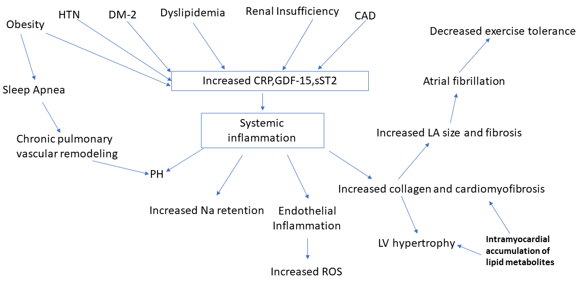
Fig. 1.

Shows the complex pathophysiology underlying heart failure with preserved ejection fraction. This figure depicts how systemic inflammation is caused by multiple factors which leads to pathology at the molecular and cellular levels. Risk factors such as hypertension, diabetes, dyslipidemia, atherosclerosis and renal insufficiency create a milieu for upregulation of biomarkers for inflammation such as CRP, GDF-15 and sST2 signaling systemic inflammation. Obesity increases systemic inflammation via molecular signaling and also remains one of the most prominent etiological factors for inducing sleep apnea which in turn leads to chronic pulmonary vascular remodeling and PH. PH also results from systemic inflammation making it an important pathology noted in HFpEF. Intramyocardial accumulation of lipid metabolites in diabetes lead to increased collagen deposition and cardiomyofibrosis resulting in LV hypertrophy as well as fibrosis of atrial walls leading to atrial fibrillation and exercise intolerance.

7. Endothelial dysfunction

The endothelium has more recently been recognized not just as a barrier between blood and the extravascular tissues but that it is composed of dynamic, highly interactive cells involved in regulating function, physiology and homeostasis of blood vessels. The endothelium prevents platelet and leukocyte adhesion/aggregation, inhibits smooth muscle proliferation, and regulates vascular tone through release of vasoactive substances required for organ perfusion. Nitric oxide (NO) produced from L-arginine by endothelial NO synthase (eNOS) in response to stimuli such as shear stress, cytokines, and platelet-derived factors is an important vasodilatory molecule NO reduces vascular inflammation and atherosclerosis. NO diffuses into platelets and vascular smooth muscle cells and stimulates the soluble guanylate cyclase and activates the cyclic GMP pathway which prevents platelet aggregation and also produces vasodilation. NO diffuses into cardiomyocytes from adjacent coronary microvasculature and modulates cardiac function. Another important function of NO is to mobilize stem cells and progenitor cells responsible for vascular homeostasis and repair. An inverse relationship exists between NO and endothelin (ET). ET is a potent vasoconstrictor and a fine balance of the 2 factors dictate vascular homeostasis (Brutsaert, 2003; Bruyndonckx et al., 2016; Pacher et al., 2007)Cardiovascular risk factors such as smoking, aging, hypercholesterolemia, hypertension, hyperglycemia, and obesity affect the endothelium. Reactive oxygen species (ROS) combine with NO to form peroxynitrite therefore reducing NO availability and tipping the balance to vasoconstriction and a proinflammatory/prothrombotic phenotype. Such a predisposition to vasoconstriction, inflammation and thrombosis disrupts the vascular homeostasis leading to endothelial dysfunction.

Endothelial dysfunction has been implicated in the development of HFpEF (Paulus and Tschöpe, 2013). The comorbidities observed in HFpEF are possibly secondary to systemic inflammation resulting in coronary microvascular endothelial dysfunction and elevated levels of inflammatory cytokines (Bishu et al., 2012; Franssen et al., 2016). Inflammatory cytokines predict the onset of HFpEF. Inflammation of the coronary microvascular endothelium and reduced endothelium-dependent vasodilation have been noted in HFpEF. Clinical indices of endothelial function such as the Flow-mediated dilation (FMD) and Reactive Hyperemic Index (RHI) are both reduced in HFpEF patients (Borlaug et al., 2010b). Another aspect of importance is that endothelial dysfunction is directly related to exercise intolerance which is measured by cardiopulmonary exercise testing and determination of VO2 max. The VO2 max is correlated with adverse prognosis and remains one of the strongest predictors of mortality in HFpEF (Shafiq et al., 2016).

Reduced NO signaling from dysfunctional endothelium influences adjacent cardiomyocytes and cardiac fibroblasts via the sGC-cGMP-PKG pathway resulting in functional and structural cardiac changes such as delayed myocardial relaxation, increased cardiomyocyte stiffness, cardiac hypertrophy, and interstitial fibrosis in patients with HFpEF.

The interaction between HFpEF and endothelial dysfunction results in a vicious cycle further impairing endothelial function. HFpEF causes a systemic inflammatory state with high levels of circulating proinflammatory cytokines and increased production of ROS. This in turn exerts deleterious effects on eNOS expression. Additionally, neurohormonal upregulation in HFpEF leads to increased oxidative stress and upregulation of collagen synthesis. HFpEF worsens systemic endothelial dysfunction leading to progressive heart failure.

Microvascular dysfunction as the cause of HFpEF is a mechanism that can pave the pathway for therapeutic targets like NO, sarcomeric titin, transforming growth factor beta (TGF-β), adenosine receptors and other immunomodulatory pathways to treat endothelial dysfunction (Lam et al., 2018).

8. Diastolic dysfunction of the left ventricle

Diastolic dysfunction (DD) is a result of myocardial stiffness in the absence of endocardial and pericardial disease. Myocardial stiffness is influenced by the extracellular matrix and the cardiomyocytes. Change in stiffness within the cardiomyocytes is transmitted to the extracellular matrix via matrix proteins. The total amount of collagen type 1 and the extent of collagen cross-linking determines the stiffness of the extracellular matrix. In HFpEF patients, increased deposition of collagen type I results from an imbalance between increased synthesis and decreased degradation (Weber et al., 1993). Collagen synthesis and degradation involve a multistep process in which the procollagen is processed to the collagen type I by proteinases and lysyl oxidase to collagen type I. The degradation is catalyzed by collagenases 9MATRIX metalloproteinases (MMP-1, MMP-8 and MMP-13) and gelatinases (MMP-2 and MMP-9). In HFpEF patients with hypertension or aortic stenosis, a decrease in matrix degradation because of downregulation of matrix metalloproteinases (MMPs) and upregulation of tissue inhibitors of matrix metalloproteinases (TIMPs) has been noted. TIMP-1 appears to be a potential biomarker of HFpEF development in patients with arterial hypertension. Cardiomyocyte stiffness is influenced by titin the cytoskeletal protein (Ahmed et al., 2006; Heymans et al., 2005).

Titin is a giant elastic protein that resides in the cardiomyocytes in two isoforms, N2B (stiffer spring) and N2BA (compliant spring) (Bang et al., 2001) It has been noted in literature that the N2BA : N2B isoform expression ratio is increased in eccentrically remodeled explanted hearts from dilated cardiomyopathy (Makarenko et al., 2004; Nagueh et al., 2004). Such switching of isoforms influences myocardial passive stiffness. Additionally, alterations in the phosphorylation state of titin or formation of disulfide bridges within the titin molecule due to oxidative stress can all induce myocardial stiffness (Borbely et al., 2009; Grutzner et al., 2009; Hidalgo et al., 2009).

HFpEF is characterized by slow LV relaxation reducing LV stroke volume, as heart rate increases. Cross-bridge detachment and sarcoplasmic reticular Ca2+ reuptake affect LV relaxation. Nitric oxide (NO) and cyclic guanosine monophosphate (cGMP) reduces myofilamentary Ca2+ sensitivity and thereby facilitates cross-bridge detachment. Interestingly correlation of dimethylarginine and diastolic LV dysfunction in failing human hearts (Wilson Tang et al., 2008; Bronzwaer et al., 2008) has been noted due to uncoupling of NO synthase-1 inducing HFpEF in a mouse model (Silberman et al., 2010). As cross-bridge detachment is an energy-consuming process, slow LV relaxation is possibly due to deficit in myocardial energy reserve. Myocardial phosphorus magnetic resonance spectroscopy indeed showed lower myocardial creatine phosphate/adenosine triphosphate ratio in HFpEF patients compared with normal controls, suggestive of reduced myocardial energy reserve (Paulus et al., 1994; Phan et al., 2009; Sohn et al., 2007; Wilson Tang et al., 2008).

Cardiomyocyte hypertrophy and survival is regulated by matricellular proteins which affects fibroblast function (Schellings et al., 2004). They have been known to improve the quality of the matrix and cardiomyocyte function by binding to collagen, cell surface receptors, and MMPs (Schroen et al., 2004). Their role in the pathophysiology of HFpEF needs further investigation.

9. Pulmonary hypertension

PH at rest in HFpEF patients occurs up to 83%. HFpEF patients also show an exaggerated increase in pulmonary artery pressures during exercise. Such an increase in afterload on the right ventricle (RV) in the presence of other risk factors possibly explains the high prevalence of RV dysfunction in HFpEF increasing the morbidity and mortality in this population. Impaired NO-dependent pulmonary vasodilation seen in HFpEF patients is also manifested as reduced exercise-induced pulmonary vasodilation in HFpEF (Andersen et al., 2015; Borlaug et al., 2010a, 2016; Lam et al., 2009; Mohammed et al., 2014).

Pulmonary arterial endothelial dysfunction with higher pulmonary artery pressures were also noted in an animal infarct model of HFpEF in the setting of normal aortic endothelial function and intracardiac pressures (Driss et al., 2000). This suggests that pulmonary vascular endothelial dysfunction precedes systemic endothelial dysfunction in HFpEF. This may be best explained by the fact that the pulmonary circulation is primarily flow-driven versus the pressure-driven systemic circulation and is therefore more susceptible to the shear stress and endothelial dysfunction. In a murine PH model of obese/hypertensive HFpEF rats in which vascular endothelial growth factor receptors were blocked nitrite given orally served as a NO donor preventing the development of PH but unfortunately could not reverse established PH suggesting that long standing PH is fixed and irreversible (Lai et al., 2016; Lam and Brutsaert, 2012).

In a small study of HFpEF patients with PH and severe macrovascular endothelial dysfunction with abnormal FMD an inverse correlation was noted between FMD and pulmonary vascular resistance (PVR) while no correlation was noted when compared with pulmonary capillary wedge pressure (PCWP). This could suggest that longstanding HFpEF is associated with severe endothelial dysfunction in the systemic and pulmonary vasculature (Farrero et al., 2014).

PH can also occur due to reactive pulmonary vasoconstriction and vascular remodeling, which is predominantly mediated by NO, as pulmonary vascular reactivity is maintained by continuous local synthesis of NO. A systemic reduction in NO as noted in HFpEF would lead to pulmonary vascular smooth muscle dysfunction and generate elevated pulmonary pressures and PH (Cooper et al., 1996; Segers et al., 2012).

Pulmonary dysfunction noted in HFpEF patients adds another dimension to the problem. As pulmonary impairment increases with increase in symptoms it results in pulmonary edema. However, this finding could be also due to diaphragmatic dysfunction. Changes in skeletal muscle structure and physiology has been noted in the diaphragm of HFpEF patients. Fiber Atrophy of myofibers, decreased oxidative capacity, mitochondrial dysfunction, and increased fati Olson gability seen in a rat model could explain the effect (Bowen et al., 2015) suggesting a link between skeletal muscle dysfunction and respiratory abnormalities. Pulmonary gas exchange is impaired in HFpEF patients, showing O2 diffusion limitation in the systemic and pulmonary microcirculatory circuits (Andrea et al., 2014; Olson et al., 2016).

Vascular remodeling, reactive pulmonary vasoconstriction due to reduced systemic NO bioavailability, impaired diaphragm function, and decreased pulmonary diffusion capacity noted in HFpEF patients contribute to the adverse pathophysiology noted in this population with PH.

10. Ventricular-vascular uncoupling

Ventricular and vascular stiffening increase with age/hypertension/diabetes and are abnormally elevated in patients with HFpEF (Melenovsky et al., 2007; Paulus and van Ballegoij, 2010) and is strongly associated with impaired exercise capacity (Hundley et al., 2001). Arterial elastance (Ea) and Ees are elevated in HFpEF, resulting in labile blood pressure swings commonly seen in HFpEF, due to exaggerated blood pressure changes seen in preload or afterload changes (Borlaug and Kass, 2008; Kawaguchi et al., 2003). Acute afterload increase in the setting of ventricular-arterial stiffening leads to increases in blood pressure worsening diastolic relaxation and higher filling pressures during stress resulting in exercise intolerance in HFpEF. If ventricular-arterial stiffening is reduced exercise capacity improves in these patients (Borlaug et al., 2007; Chantler et al., 2008; Chen et al., 1999). It should be noted that such ventricular -vascular stiffening increases with age, hypertension and diabetes suggesting that a manifestation of several of the cardiovascular risk factors / comorbidities present in these patients.

11. Chronotropic incompetence

Abnormalities in cardiovascular reserve function with exercise stress has been implicated in HFpEF. due to abnormal venous return, contractility, heart rate, and peripheral vasodilation. contribute to the pathophysiology. Exaggerated decrease in chronotropic reserve in HFpEF, is possibly due to deficits in β-adrenergic stimulation secondary to increased plasma catecholamines with exercise. Additionally, autonomic dysfunction may contribute to chronotropic incompetence, as baroreflex sensitivity is reduced in the setting of impaired heart rate recovery in HFpEF (Borlaug et al., 2006; Phan et al., 2010).

12. Lessons learned from exercise training

The beneficial effects of exercise in HFpEF patients is slowly emerging. Current literature supports significant benefit from training in HFpEF patients (Edelmann et al., 2011). Exercise training in HFpEF increases VO2peak and physical function scores and improved diastolic function (Kitzman et al., 2010, 2016; Pandey et al., 2015). Peripheral endothelial function and muscle metabolism are improved by exercise. Exercise upregulates eNOS through increased shear stress and vascular endothelial growth factor 2 release and reduces oxidative stress by downregulating angiotensin receptors and nicotinamide adenine dinucleotide phosphate oxidase (NADP(H)oxidase). Exercise promotes endothelial function via enhancing anti-inflammatory processes (Adams et al., 2005; Suchy et al., 2014).

Ex-DHF trial secondary analysis showed that inflammatory cytokines (interleukins 1ß, 6, and 10 and tumor necrosis factor alpha) showed no change with exercise but growth hormone releasing peptide ghrelin, which inhibits cardiomyocyte and endothelial cell apoptosis in vitro, increased. Molecular mechanisms underlying exercise induced benefits in HFpEF need further definitive investigation (Conraads et al., 2013; Trippel et al., 2017).

13. New insights from myocardial remodeling

Risk factors for pressure induced hypertrophy (PIH) include hypertension, advanced age and valvular disease. Longstanding overload initiates mechanical and neurohormonal upregulation resulting in adaptive changes in the myocardium in the form of hypertrophy of cardiomyocytes. In the early stages the adaptive response remains beneficial for maintaining cardiac output but over time it becomes detrimental leading to hypertrophy of the entire ventricle. At the cellular level cardiomyocytes increase structural proteins and how abnormal Ca2+ handling and energy metabolism. Additionally, fibroblasts cause increased accumulation of collagen and other matrix proteins, resulting in myocardial stiffness and impaired relaxation leading to suboptimal filling signifying diastolic dysfunction. inadequate LV filling, the hallmark of diastolic dysfunction. Chronic changes compromise cardiac output generating HFpEF (Creemers and Pinto, 2011; Frohlich and Susic, 2012; Spinale, 2007).

Early trans differentiation of fibroblast in pressure overload seem to interesting have a strange similarity to fibroblasts associated with cancer. The PIH and cancer associated fibroblasts differ from the reactive fibroblasts noted in physiological healing. These abnormal fibroblasts replace the regular fibroblasts slowly leading to progression of HFpEF with myocardial stiffening. Such abnormal processes also cause solid tumor progression.

Interestingly the PIH fibroblasts and the cancer associated ones share a molecular signature of expressing α-smooth muscle actin (αSMA) which is also expressed in fibroblasts in physiological wound healing. Increases in expression other cellular and matrix protein markers such as fibroblast-specific protein 1 (FSP1), platelet-derived growth factor receptor (PDGFR), fibroblast activation protein (FAP) periostin, tenascin C and the extra domain A variant present in fibronectin. This molecular the unique molecular marker signature found in both PIH and cancer associated fibroblasts directly are also implicated in malignant fibroblast expansion (Fan et al., 2013; Moore-Morris et al., 2015; Shimojo, 2015).

The abnormal fibroblasts cause matrix remodeling by upregulating the matrix metalloproteinases (MMPs). MMP2, MMP9 and MMP14 have been associated with ventricular dysfunction (Chen et al., 2013; Polyakova et al., 2004; Zile et al., 2011). FAP has gelatinase activity and is implicated in degradation of matrix products (Tillmanns et al., 2015). Once the matrix is degraded it is replaced with fibrillary collagens I and III increasing myocardial stiffness (Creemers and Pinto, 2011; Frohlich and Susic, 2012; Spinale, 2007). Domain A fibronectin activates TGF beta resulting in cardiac hypertrophy. Periostin and Tenascin C cause increased fibrosis and progression of HFpEF (Shimojo, 2015; Wu et al., 2016) Impaired diastolic dysfunction in HFpEF can in turn cause activated latent TGF beta expression further increasing cardiac hypertrophy.

The abnormal fibroblasts upregulate the production and secretion of inflammatory cytokines and growth factors such as IL-1, IL-6, TGFβ and basic fibroblast growth factor (bFGF) resulting in hypertrophy and impaired Ca2+ handling. (He et al., 2005; Porter and Turner, 2009; Stewart et al., 2010). Growth factors and inflammatory cytokines promote the development and proliferation of the abnormal fibroblasts and such autocrine and paracrine signaling modifies the local microenvironment leading to disease progression from a positive feedback loop. Inhibition of TGF beta pathways in a rat model showed decreased abnormal fibroblast proliferation, collagen deposition and fibrosis with improvement in cardiac function (Kuwahara et al., 2005). TGF beta and bFGF seem to contribute significantly to fibroblast proliferation, fibrosis and fibrillary collagen deposition in the setting of inflammation (He et al., 2005).

Cellular and molecular studies have now paved the way to an interesting finding of abnormal fibroblasts in HFpEF which are apoptosis resistant and initiate the development of an abnormal myocardial matrix resulting in initiation and progression of the disease (Oatmen et al., 2019). The fact that it shares molecular markers with cancer associated fibroblasts has opened another therapeutic avenue of using cancer chemotherapeutics to control the fibrosis and its progression (Oatmen et al., 2019).

14. Diabetes and HFpEF

In 45% of HFpEF patients diabetes exists as a co-morbidity. The characteristics of this population is poorly understood. It is important to understand the pathophysiology when both these conditions exist together to develop personalized medicine. It is interesting that inflammation exists in both conditions but treatments that target inflammation and endothelial dysfunction, such as statins, renin-angiotensin system inhibitors, and phosphodiesterase-5 inhibitors do not sem to be effective suggesting that the pathophysiology that exists in DM is different from those operating in HFpEF (Parikh et al., 2018).

Cardiomyopathy secondary to lipid toxicity is unique to diabetic cardiomyopathy. Dyslipidemia, increased body mass index and insulin resistance lead to mitochondrial dysfunction and altered energy metabolism. At the biochemical level toxic lipid accumulation, membrane lipid remodeling of the cardiac myocytes, abnormal Ca2+handling, ROS production, inflammation and fibrosis set in (Nakamura and Sadoshima, 2020). Advanced glycation end products (AGE) accumulate and crosslink the extracellular matrix therefore forming the molecular basis of fibrosis in this type of cardiomyopathy via ROS generation or through activation of the receptor for AGEs (RAGE) mediated pathways. Activation of transforming growth factor pathways causes deposition of structural proteins contributing to fibrosis. Cardiomyocytes and endothelial cells may secrete growth factors modulating the fibroblast phenotype. Additionally, endothelial cells/pericytes transdifferentiate into fibroblasts. Endothelin-1 and the renin-angiotensin system have also been implicated in the generation of diabetic cardiomyopathy (Russo and Frangogiannis, 2016).

Lipid metabolites such as triacylglycerols and ceramides accumulate in the intramyocardial space in obesity and diabetes. This leads to augmented cardiac myocyte apoptosis, fibrosis, impaired contractility and poor diastolic filling. One of the etiologies proposed for intramyocardial accumulation is the downregulation of peroxisome proliferator activated receptor alpha and downregulation of beta oxidation of fatty acids noted in heart failure patients with obesity and diabetes. Further investigation is needed in this area to define clinical relevance for identifying therapeutic targets (Fukushima and Lopaschuk, 2016; Lopaschuk et al., 2010).

Diabetes and HFpEF together cause greater morbidity and mortality. In this population the GWTG-HF registry shows worse in-hospital/post-discharge morbidity, longer length of stay, and higher 30-day all-cause/HF readmissions (McHugh et al., 2019). The pathophysiology of DM in HFpEF patients possibly occurs via increased sodium retention, neurohumoral activation, volume overload, and upregulation of the sodium-glucose cotransporter-2 (SGT2) mechanisms because SGT2 inhibitors decrease volume overload and reduce readmissions (McHugh et al., 2018). DM propagates systemic inflammation in HFpEF patients via multiple pathways such as fatty acid oxidation, decreased nitric oxide availability, and increased AGE. Therapies that target these pathophysiological mechanisms using antihyperglycemic drugs may decrease the progression of remodeling and improve mortality. Graded exercise regimens and targeted therapies may improve skeletal muscle oxygen utilization/exercise tolerance/quality of life (Gandhi et al., 2016). A combination of therapies targeted at different aspects of both the co-morbidities may improve outcomes in this population. Further research is required in this area.

15. Estrogen and HFpEF

The role of estrogens may be multifold in attenuating progression of HFpEF. It has an important role in reducing oxidative stress/free radical production, endothelial dysfunction, inflammation, regulating the renin-angiotensin aldosterone pathways, and decreasing fibrosis and hypertrophy via upregulation of the atrial and brain natriuretic peptide levels. E2 levels decrease following menopause and is accompanied by changes in body fat, blood pressure, lipid levels influencing cardiovascular risk in postmenopausal women (Matthews et al., 2009). Therefore, strategies targeting E2 could be beneficial in risk factor modification. However, the risks of hormone therapy differ depending on the drug type, dose, duration, administration route, and initiation time, pre-existing cardiac disease and genetic variations. Considering that estrogen has a versatile action on all the cardiovascular risk factors it may have a preventive role in development of HFpEF (Khalil, 2013). Prospective clinical trials and future studies to balance the risk versus benefit to avoid potentially unwanted effects or abnormal hormone-receptor interactions will help. AI using silico models (Cui et al., 2018) also may help in predicting responses. Since HFpEF is more prevalent in women than their male counterparts, role of estrogen in the pathophysiology of HFpEF should be an active area of investigation (Sabbatini and Kararigas, 2020).

16. Conclusions

HFpEF is increasing in prevalence and relatively high mortality. Many etiologies contribute to the development of HFpEF which makes treatment very challenging. Pathophysiology of HFpEF is multifaceted, stemming from several disease-specific aspects of inflammation and endothelial function, cardiomyocyte hypertrophy and fibrosis. Hence identifying the risk factors and etiologies is imperative to achieve optimal outcomes in this population. Some comorbidities such as sleep disordered breathing is common in HFpEF patients and is associated with worsening diastolic dysfunction. However, a cause and effect relationship and appropriate treatment has not been definitively established (Khattak et al., 2018). As further research into cellular and molecular basis of fibrosis and inflammation became more lucid, targeted therapies for HFpEF would become more apparent. Additionally, role of comorbidities such as diabetes is an important area of research in this population. The role of estrogen in post-menopausal women needs to be better understood to prevent progression of HFpEF in this population.

Authors’ contributions

Nandini Nair was responsible for the conception of ideas presented, writing and the entire preparation of this manuscript.

Acknowledgements

No funding was obtained for this work.

Conflict of Interest

The author declares no conflicts of interests.

References
[1]
Adamczak, D. M., Oduah, M., Kiebalo, T., Nartowicz, S., Bęben, M., Pochylski, M., Ciepłucha, A., Gwizdała, A., Lesiak, M. and Ewa Straburzyńska-Migaj, E. (2020) Heart failure with preserved ejection fraction-a concise review. Current Cardiology Reports 22, 82-94.
[2]
Adams, V., Linke, A., Kränkel, N., Erbs, S., Gielen, S., Mobius-Winkler, S., Gummert, J. F., Mohr, F. W., Schuler, G. and Hambrecht, R. (2005) Impact of regular physical activity on the NAD(P)H oxidase and angiotensin receptor system in patients with coronary artery disease. Circulation 111, 555-562.
[3]
Ahmed, S. H., Clark, L. L., Pennington, W. R., Webb, C. S., Bonnema, D. D., Leonardi, A. H., McClure, C. D., Spinale, F. G. and Zile, M. R. (2006) Matrix metalloproteinases/tissue inhibitors of metalloproteinases: relationship between changes in proteolytic determinants of matrix composition and structural, functional, and clinical manifestations of hypertensive heart disease. Circulation 113, 2089-2096.
[4]
Andersen, M. J., Hwang, S. J., Kane, G. C., Melenovsky, V., Olson, T. P., Fetterly, K. and Borlaug, B. A. (2015) Enhanced pulmonary vasodilator reserve and abnormal right ventricular: pulmonary artery coupling in heart failure with preserved ejection fraction. Circulation: Heart Failure 8, 542-550.
[5]
Andrea, R., López-Giraldo, A., Falces, C., Sobradillo, P., Snchis, L., Gistau, C., Heras, M., Sabate, M., Brugada, J. and Agusti, A. (2014) Lung function abnormalities are highly frequent in patients with heart failure and preserved ejection fraction. Heart, Lung & Circulation 23, 273-279.
[6]
Ayinapudi, K., Singh, T., Motwani, A., Le Jemtel, T. H. and Oparil, S. (2018) Obesity and Pulmonary Hypertension. Current Hypertension Reports 20, 99.
[7]
Bang, M. L., Centner, T., Fornoff, F., Geach, A. J., Gotthardt, M., McNabb, M., Witt, C. C., Labeit, D., Gregorio, C. C., Granzier, H. and Labeit, S. (2001) The complete gene sequence of titin, expression of an unusual approximately 700-kDa titin isoform, and its interaction with obscurin identify a novel Z-line to I-band linking system. Circulation Research 89, 1065-1107.
[8]
Bhatia, R. S., V Tu, J. V., Lee, D. S., Austin, P. C., Fang, J., Haouzi, A., Gong, Y. and Liu, P. P. (2006) Outcome of heart failure with preserved ejection fraction in a population-based study. New England Journal of Medicine 355, 260-269.
[9]
Bielinski, S. J., Pathak, J., Carrell, D. S., Takahashi, P. Y., Olson, J. E., Larson, N. B., Liu, H., Sohn, S., Wells, Q. S., Denny, J. S., Rasmussen-Torvik, L. J., Pacheco, J. A., Jackson, K. L., Lesnick, T. G., Gullerud, R. E., Decker, P. A., Pereira, N. L., Ryu, E., Dart, R. A., Peissig, P., Linneman, J. G., Jarvik, G. P., Larson, E. B., Bock, J. A., Tromp, G. C., de Andrade, M. and Roger, V. L. (2015) A robust e-epidemiology tool in phenotyping heart failure with differentiation for preserved and reduced ejection fraction: the electronic medical records and genomics (eMERGE) network. Journal of Cardiovascular Translational Research 8, 475-483.
[10]
Bishu, K., Deswal, A., Chen, H. H., Le Winter, M. M., Lewis, G. L., Semigram, M. J., Borlaug, B. A., McNulty, S., Hernandez, A. F., Braunwald, E. and Redfield, M. M. (2012) Biomarkers in acutely decompensated heart failure with preserved or reduced ejection fraction. American Heart Journal 164, 763-770.
[11]
Blecker, S., Katz, S. D., Horwitz, L. I., Kuperman, G., Park, H., Gold, A. and Sontag, D. (2016) Comparison of approaches for heart failure case identification from electronic health record data. JAMA Cardiology 1, 1014-1020.
[12]
Borbely, A., Falcao-Pires, I., van Heerebeek, L., Hamdani, N., Edes, I., Gavina, C., Leite-Moreira, A. F., Bronzwaer, J. G., Papp, Z., van der Velden, J., Stienen, G. J. and Paulus, W. J. (2009). Hypophosphorylation of the stiff N2B titin isoform raises cardiomyocyte resting tension in failing human myocardium. Circulation Research 104, 780-786.
[13]
Borlaug, B. A. and Kass, D. A. (2008) Ventricular-vascular interaction in heart failure. Heart Failure Clinics 4, 23-36.
[14]
Borlaug, B. A., Kane, G. C., Melenovsky, V. and Olson, T. P. (2016) Abnormal right ventricular-pulmonary artery coupling with exercise in heart failure with preserved ejection fraction. European Heart Journal 37, 3293-3302.
[15]
Borlaug, B. A., Melenovsky, V., Redfield, M. M., Kessler, K., Chang, H. J., Abraham, T. P. and Kass, D. A. (2007) Impact of arterial load and loading sequence on left ventricular tissue velocities in humans. Journal of the American College of Cardiology 50, 1570-1577.
[16]
Borlaug, B. A., Melenovsky, V., Russell, S. D., Kessler, K., Pacak, K., Becker, L. C. and Kass, D. A. (2006) Impaired chronotropic and vasodilator reserves limit exercise capacity in patients with heart failure and a preserved ejection fraction. Circulation 114, 2138-2147.
[17]
Borlaug, B. A., Nishimura, R. A., Sorajja, P., Lam, C. S. P. and Redfield, M. M. (2010a) Exercise hemodynamics enhance diagnosis of early heart failure with preserved ejection fraction. Circulation: Heart Failure 3, 588-595.
[18]
Borlaug, B. A., Olson, T. P., Lam, C. S. P., Flood, K. S., Lerman, A., Johnson, B. D. and Redfield, M. M. (2010b) Global cardiovascular reserve dysfunction in heart failure with preserved ejection fraction. Journal of the American College of Cardiology 56, 845-854.
[19]
Bowen, T. S., Rolim, N. P. L., Fischer, T., Baekkerud, F. H., Medeiros, A., Werner, S., Bronstaf, E., Rognmo, O., Mangner, N., Linke, A., Schuler, G., Silva, G. J. J., Wisloff, U. and Adams, V. (2015) Heart failure with preserved ejection fraction induces molecular, mitochondrial, histological, and functional alterations in rat respiratory and limb skeletal muscle. European Journal of Heart Failure 17, 263-272.
[20]
Bronzwaer, J. G., Paulus, W. J. (2008) Nitric oxide: the missing lusitrope in failing myocardium. European Heart Journal 29, 2453-2455.
[21]
Brouwers, F. P., de Boer, R. A., van der Harst, P., Voors, A. A., Gansevoort, R. T., Bakker, S. J., Hillege, H. L., van Veldhuisen, D. J. and van Gilst, W. H. (2013) Incidence and epidemiology of new onset heart failure with preserved vs. reduced ejection fraction in a community-based cohort: 11-year Follow-Up of PREVEND. European Heart Journal 34, 1424-1431.
[22]
Brutsaert, D. L. (2003) Cardiac endothelial-myocardial signaling: its role in cardiac growth, contractile performance, and rhythmicity. Physiological Reviews 83, 59-115.
[23]
Bruyndonckx, L., Hoymans, V. Y., Lemmens, K., Ramet, J. and Vrints, C. J. (2016) Childhood obesity-related endothelial dysfunction: an update on pathophysiological mechanisms and diagnostic advancements. Pediatric Research 79, 831-837.
[24]
Bursi, F., Weston, S. A., Redfield, M. M., Jacobsen, S. J., Pakhomov, S., Nkomo, V. T., Meverden, R. A. and Roger, V. L. (2006) Systolic and diastolic heart failure in the community. Journal of the American Medical Association 296, 2209-2216.
[25]
Carlson, K. J., Lee, D. C., Goroll, A. H., Leahy, M. and Johnson, R. A. (1985) An analysis of physicians’ reasons for prescribing long-term digitalis therapy in outpatients. Journal of Chronic Diseases 38, 733-739.
[26]
Ceia, F., Fonseca, C., Mota, T., Morais, H., Matias, F., de Sousa, A. and Oliveira, A. (2002) Prevalence of chronic heart failure in southwestern Europe: the EPICA study. European Journal of Heart Failure 4, 531-539.
[27]
Chantler, P. D., Lakatta, E. G. and Najjar, S. S. (2008) Arterial-ventricular coupling: mechanistic insights into cardiovascular performance at rest and during exercise. Journal of Applied Physiology 105, 1342-1351.
[28]
Chen, C. H., Nakayama, M., Talbot, M., Nevo, E., Fetics, B., Gerstenblith, G., Becker, L. C. and Kass, D. A. (1999) Verapamil acutely reduces ventricular-vascular stiffening and improves aerobic exercise performance in elderly individuals. Journal of the American College of Cardiology 33, 1602-1609.
[29]
Chen, R., Xue, J. and Xie, M. (2013) Osthole regulates TGF-β1 and MMP-2/9 expressions via activation of PPARα/γ in cultured mouse cardiac fibroblasts stimulated with angiotensin II. Journal of Pharmacy and Pharmaceutical Sciences 16, 732-741.
[30]
Cohen, J. B., Schrauben, S. J., Zhao, L., Basso, M. D., Cvijic, M. E., Li, Z., Yarde, M., Wang, Z., Bhattacharya, P. T., Chirinos, D. A., Prenner, S., Zamani, P., Seiffert, D. A., Car, B. D., Gordon, D. A., Marguilies, K., Cappola, T. and Chirinos, J. A. (2020) Clinical phenogroups in heart failure with preserved ejection fraction: Detailed phenotypes, prognosis and response to spironolactone. JACC: Heart Failure 8, 172-184.
[31]
Conraads, V. M., Van Craenenbroeck, E. M., De Maeyer, C., Van Berendoncks, A. M., Beckers, P. J. and Vrints, C. J. (2013) Unraveling new mechanisms of exercise intolerance in chronic heart failure. Role of exercise training. Heart Failure Reviews 18, 65-77.
[32]
Cooper, C. J., Landzberg, M. J., Anderson, T. J., Charbonneay, F., Creager, M. A., Ganz, P. and Selwyn, A. P. (1996) Role of nitric oxide in the local regulation of pulmonary vascular resistance in humans. Circulation 93, 266-271.
[33]
Creemers, E. E. and Pinto, Y. M. (2011) Molecular mechanisms that control interstitial fibrosis in the pressure-overloaded heart. Cardiovascular. Research 89, 265-272.
[34]
Cui, C., Huang, C., Liu, K., Xu, G., Yang, J., Zhou, Y., Feng, Y., Kararigas, G., Geng, B. and Cui, Q. (2018) Large-scale in silico identification of drugs exerting sex-specific effects in the heart. Journal of Translational Medicine 16, 236.
[35]
Driss, A. B., Devaux, C., Henrion, D., Duriez, M., Thulliez, C., Levy, B. I. and Michel, J. B. (2000) Hemodynamic stresses induce endothelial dysfunction and remodeling of pulmonary artery in experimental compensated heart failure. Circulation 101, 2764-70.
[36]
DuBrock, H. M., AbouEzzeddine, O. F. and Redfield, M. M. (2018) High-sensitivity C-reactive protein in heart failure with preserved ejection fraction. PLoS One 13, e0201836.
[37]
Dunlay, S. M., Roger, V. L. and Redfield, M. M. (2017) Epidemiology of heart failure with preserved ejection fraction. Nature Reviews Cardiology 14, 591-602.
[38]
Edelmann, F., Gelbrich, G., Düngen, H.-D., Frohling, S., Wachter, R., Stahrenburg, R., Binder, L., Topper, A., Lashki, D. J., Schwarz, S., Herrmann-Lingen, C., Loffler, M., Hasenfuss, G., Halle, M. and Pieske, B. (2011) Exercise training improves exercise capacity and diastolic function in patients with heart failure with preserved ejection fraction (ex-DHF) Journal of the American College of Cardiology 58, 1780-1791.
[39]
Eriksson, H., Caidaul, K., Larsson, B., Ohlson, L.-O., Welin, L., Wilhelmsen, L., and Svardsudd, K. (1987) Cardiac and pulmonary causes of dyspnea-validation of a scoring test for clinical-epidemiological use: the study of men born in 1913. European Heart Journal 8, 1007-1014.
[40]
Fan, B., Ma, L., Li, Q., Wang, L., Zhou, J. and Wu, J. (2013) Role of PDGFs/PDGFRs signaling pathway in myocardial fibrosis of DOCA/salt hypertensive rats. International Journal of Clinical and Experimental Pathology 7, 16-27.
[41]
Farr, G., Shah, K., Markley, R., Abbate, A., Salloum, F. N. and Grinnan, D. (2016) Development of pulmonary hypertension in heart failure with preserved ejection fraction. Progress in Cardiovascular Disease 59, 52-58.
[42]
Farrero, M., Blanco, I., Batlle, M., Santiagi, E., Cardona, M., Vidal, B., Castel, M. A., Sitges, M., Barbera, J. A. and Perez Villa, F. (2014) Pulmonary hypertension is related to peripheral endothelial dysfunction in heart failure with preserved ejection fraction. Circulation: Heart Failure 7, 791-798.
[43]
Franssen, C., Chen, S., Unger, A., Korkmaz, H. I., Keulenaer, G. W., Tschöpe, C., Leite-Moreira, A. F., Musters, R., Niessen, H. W. M., Linke, W. A., Paulus, W. J. and Hamdani, N. (2016) Myocardial microvascular inflammatory endothelial activation in heart failure with preserved ejection fraction. JACC: Heart Failure 4, 312-324.
[44]
Frohlich, E. D. and Susic, D. (2012) Pressure overload. Heart Failure Clinics 8, 21-32.
[45]
Fukushima, A. and Lopaschuk, G. D. (2016) Cardiac fatty acid oxidation in heart failure associated with obesity and diabetes. Biochimica Biophysica Acta 1861, 1525-34.
[46]
Fukuta, H., Goto, T., Wakami, K., Kamiya, T. and Ohte, N. (2019) Effects of mineralocorticoid receptor antagonists on left ventricular diastolic function, exercise capacity, and quality of life in heart failure with preserved ejection fraction: a meta-analysis of randomized controlled trials. Heart Vessels 34, 597-606.
[47]
Gandhi, P. U., Gaggin, H. K., Redfield, M. M., Chen, H. H., Stevens, S. R., Anstrom, K. J., Semigran, M. J., Liu, P. and Januzzi, J. L. Jr. (2016) Insulin-like growth factor-binding protein-7 as a biomarker of diastolic dysfunction and functional capacity in heart failure with preserved ejection fraction: results from the RELAX trial. JACC Heart Failure 4, 860-869.
[48]
Gerber, Y., Susan, A., Weston, M. S., Redfield, M. M., Chamberlain, A., Manemann, S. M., Jiang, R., Killian, J. M. and Roger, V. L. (2015) A contemporary appraisal of the heart failure epidemic in olmsted county, minnesota, 2000 to 2010. JAMA Internal Medicine 175, 996-1004.
[49]
Graziani, F., Varone, F., Crea, F. and Richeldi, L. (2018). Treating heart failure with preserved ejection fraction: learning from pulmonary fibrosis. European Journal of Heart Failure 20, 1385-1391.
[50]
Grutzner, A., Garcia-Manyes, S., Kotter, S., Badilla, C. L., Fernandez, J. M. and Linke, W. A. (2009) Modulation of titin-based stiffness by disulfide bonding in the cardiac titin N2-B unique sequence. Biophysical Journal 97, 825-834.
[51]
He, Z. Y., Feng, B., Yang, S. L. and Luo, H. L. (2005) Intracardiac basic fibroblast growth factor and transforming growth factor-beta 1 mRNA and their proteins expression level in patients with pressure or volume-overload right or left ventricular hypertrophy. Acta Cardioliogica 60, 21-25.
[52]
Heymans, S., Schroen, B., Vermeersch, P., Milting, H., Gao, F., Kassner, A., Gillijns, H., Herijgers, P., Flameng, W., Carmeliet, P., Van de Werf, F., Pinto, Y. M. and Janssens, S. (2005). Increased cardiac expression of tissue inhibitor of metalloproteinase-1 and tissue inhibitor of metalloproteinase-2 is related to cardiac fibrosis and dysfunction in the chronic pressure-overloaded human heart. Circulation 112, 1136-1144.
[53]
Hidalgo, C., Hudson, B., Bogomolovas, J., Zhu, Y., Anderson, B., Greaser, M., Labeit, S. and Granzier, H. (2009) PKC phosphorylation of titin’s PEVK element: a novel and conserved pathway for modulating myocardial stiffness. Circulation Research 105, 631-638.
[54]
Ho, J. E., Enserro, D., Brouwers, F. P., Kizer, J. R., Shah, S. J., Psaty, B. M., Bartz, T. M., Santhanakrishnan, R., Lee, D. S., Chan, C., Liu, K., Blaha, M. J., Hillege, H. L., van der Harst, P., van Gilst, W. H., Kop, W. J., Gansevoort, R. T., Vasan, R. S., Gardin, J. M., Levy, D., Gottdiener, J. S., de Boer, R. A. and Larson, M. G. (2016) Predicting heart failure with preserved and reduced ejection fraction: the international collaboration on heart failure subtypes. Circulation Heart Failure 9, 10.1161/CIRCHEARTFAILURE.115.003116 e003116.
[55]
Hundley, W. G., Kitzman, D. W., Morgan, T. M., Hamilton, C. A., Darty, S. N., Stewart, K. P., Herrington, D. M., Link, K. M. and Little, W. C. (2001) Cardiac cycle-dependent changes in aortic area and distensibility are reduced in older patients with isolated diastolic heart failure and correlate with exercise intolerance. Journal of the American College of Cardiology 38, 796-802.
[56]
Kawaguchi, M., Hay, I., Fetics, B. and Kass, D. A. (2003) Combined ventricular systolic and arterial stiffening in patients with heart failure and preserved ejection fraction: implications for systolic and diastolic reserve limitations. Circulation 107, 714-720.
[57]
Khalil, R. A. (2013) Estrogen, vascular estrogen receptor and hormone therapy in postmenopausal vascular disease. Biochemical Pharmacology 86, 1627-1642.
[58]
Khattak, H. K., Hayat, F., Pamboukian, S. V., Hahn, H. S., Schwartz, B. P. and Stein, P. K. (2018) Obstructive sleep apnea in heart failure: review of prevalence, treatment with continuous positive airway pressure, and prognosis. Texas Heart Institute Journal 45, 151-161.
[59]
Kitzman, D. W., Brubaker, P. H., Morgan, T. M., Stewart, K. P. and Little, W. C. (2010) Exercise training in older patients with heart failure and preserved ejection fraction: a randomized, controlled, single-blind trial. Circulation: Heart Failure 3, 659-667.
[60]
Kitzman, D. W., Brubaker, P., Morgan, T., Haykowsky, M., Hundley, G., Kraus, W. E., Eggebeeen, J. and Nicklas, B. J. (2016) Effect of caloric restriction or aerobic exercise training on peak oxygen consumption and quality of life in obese older patients with heart failure with preserved ejection fraction. Journal of the American Medical Association 315, 36-46.
[61]
Kuwahara, F., Kai, H., Tokuda, K., Kai, M., Takeshita, T., Kensuke, E. and Imaizumi, T. (2005) Transforming growth factor-beta function blocking prevents myocardial fibrosis and diastolic dysfunction in pressure-overloaded rats. Circulation 106, 130-135.
[62]
Lai, Y.-C., Tabima, D. M., Dube, J. J., Hughan, K. S., Vandepool, R. R., Goncharov, D. A., St. Croix, C. M., Garcia-Ocaria, A., Goncharova, E. A., Tofovic, S. P., Mora, A. l. and Gladwin, M. T. (2016) SIRT3-AMPK activation by nitrite and metformin improves hyperglycemia and normalizes pulmonary hypertension associated with heart failure with preserved ejection fraction (PH-HFpEF). Circulation 133, 717-731.
[63]
Lam, C. S. P. and Brutsaert, D. L. (2012) Endothelial dysfunction: a pathophysiologic factor in heart failure with preserved ejection fraction. Journal of the American College of Cardiology 60, 1787-1789.
[64]
Lam, C. S. P., Roger, V. L., Rodeheffer, R. J., Borlaug, B., Enders, F. T. and Redfield, M. M. (2009) Pulmonary hypertension in heart failure with preserved ejection fraction: a community-based study. Journal of the American College of Cardiology 53, 1119-1126.
[65]
Lam, C. S. P., Voors, A. A., De Boer, R. A., Solomon, S. D. and Van Veldhuisen, D. J. (2018). Heart failure with preserved ejection fraction: from mechanisms to therapies. European Heart Journal 39, 2780-2792.
[66]
Lopaschuk, G. D., Ussher, J. R., Folmes, C. D., Jaswal, J. S. and Stanley, W. C. (2010) Myocardial fatty acid metabolism in health and disease. Physiological Reviews 90, 207-258.
[67]
Makarenko, I., Opitz, C. A., Leake, M. C., Neagoe, C., Kulke, M., Gwathmey, J. K., del Monte, F., Hajjar, R. J. and Linke, W. A. (2004) Passive stiffness changes caused by upregulation of compliant titin isoforms in human dilated cardiomyopathy hearts. Circulation Research 95, 708-716.
[68]
Matthews, K. A., Crawford, S. L., Chae, C. U., Everson-Rose, S. A., Sowers, M. F., Sternfeld, B. and Sutton-Tyrrell, K. (2009) Are changes in cardiovascular disease risk factors in midlife women due to chronological aging or to the menopausal transition? Journal of the American College of Cardiology 54, 2366-2373.
[69]
McHugh, K. R., De Vore, A. D., Mentz, R. J., Edmonston, D., Green, J. B. and Hernandez, A. F. (2018) The emerging role of novel antihyperglycemic agents in the treatment of heart failure and diabetes: A focus on cardiorenal outcomes. Clinical Cardiology 41, 1259-1267.
[70]
McHugh, K., De Vore, A. D., Wu, J., Matsouaka, R. A., Fonarow, G. C., Heidenreich, P. A., Yancy, C. W., Green, J. B., Altman, N. and Hernandez, A. F. (2019) Heart failure with preserved ejection fraction and diabetes: JACC state-of-the-art review. Journal of the American College of Cardiology 73, 602-611.
[71]
McKee, P. A., Castelli, W. P., McNamara, P. M. and Kannel, W. B. (1971) The natural history of congestive heart failure: The Framingham study. New England. Journal of Medicine 285, 1441-1446.
[72]
Melenovsky, V., Borlaug, B. A., Rosen, B., Hay, I., Ferruci, L., Morell, C. H., Lakatta, E. G., Najjar, S. S. and Kass, D. A. (2007) Cardiovascular features of heart failure with preserved ejection fraction vs. nonfailing hypertensive left ventricular hypertrophy in the urban Baltimore community: the role of atrial remodeling/dysfunction. Journal of the American College of Cardiology 49, 198-207.
[73]
Mohammed, S. F., Hussain, I., Abou Ezzeddine, O. F., Takahama, H., Kwon, S. H., Forfia, P., Roger, V. L. and Redfield, M. M. (2014) Right ventricular function in heart failure with preserved ejection fraction: a community-based study. Circulation 130, 2310-2320.
[74]
Montezano, A. C. and Touyz R. M. (2012) Molecular mechanisms of hypertension-reactive oxygen species and antioxidants: a basic science update for the clinician Canadian Journal Cardiology 28, 288-295.
[75]
Moore-Morris, T., Guimarães-Camboa, N., Yutzey, K. E., Pucéat, M. and Evans, S. M. (2015) Cardiac fibroblasts: from development to heart failure. Journal of Molecular Medicine 93, 823-830.
[76]
Nagueh, S. F., Shah, G., Wu, Y., Torre-Amione, G., King, N. M., Lahmers, S., Witt, C. C., Becker, K., Labeit, S. and Granzier, H. L. (2004). Altered titin expression, myocardial stiffness, and left ventricular function in patients with dilated cardiomyopathy. Circulation 110, 155-162.
[77]
Nakamura, M. and Sadoshima, J. (2020) Cardiomyopathy in obesity, insulin resistance and diabetes. Journal of Physiology 598, 2977-2993.
[78]
Oatmen, K. E., Cull, E. and Spinale, F. G. (2019) Heart failure as interstitial cancer: emergence of a malignant fibroblast phenotype. Nature Review Cardiology 17, 523-531.
[79]
Olson, T. P., Johnson, B. D. and Borlaug, B. A. (2016) Impaired pulmonary diffusion in heart failure with preserved ejection fraction. JACC: Heart Failure 4, 490-498.
[80]
Pacher, P., Beckman, J. S. and Liaudet, L. (2007) Nitric oxide and peroxynitrite in health and disease. Physiological Reviews 87, 315-424.
[81]
Pandey, A., Parashar, A. and Kumbhani, D. J. (2015) Exercise training in patients with heart failure and preserved ejection fraction: meta-analysis of randomized control trials. Circulation: Heart Failure 8, 33-40.
[82]
Parikh, K. S., Sharma, K., Fiuzat, M., Surks, H. K., George, J. T., Honarpour, N., Depre, C., Desvigne-Nickens, P., Nkulikiyinka, R., Lewis, G. D., Gomberg-Maitland, M., O’Connor, C. M., Stockbridge, N., Califf, R. M., Konstam, M. A., Januzzi, J. L. Jr., Solomon, S. D., Borlaug, B. A., Shah, S. J., Redfield, M. M. and Felker, G. M. (2018) Heart failure with preserved ejection fraction expert panel report: current controversies and implications for clinical trials. JACC Heart Failure 6, 619-632.
[83]
Paulus, W. J. (2018) H2FPEF score at last, a properly validated diagnostic algorithm for heart failure with preserved ejection fraction. Circulation 138, 871-873.
[84]
Paulus, W. J. and Tschöpe, C. (2013) A novel paradigm for heart failure with preserved ejection fraction. Journal of the American College of Cardiology 62, 263-271.
[85]
Paulus, W. J. and van Ballegoij, J. J. (2010) Treatment of heart failure with normal ejection fraction: an inconvenient truth! Journal of the American College of Cardiology 55, 526-537.
[86]
Paulus, W. J., Vantrimpont, P. J. and Shah, A. M. (1994) Acute effects of nitric oxide on left ventricular relaxation and diastolic distensibility in humans: assessment by bicoronary sodium nitroprusside infusion. Circulation 89, 2070-2078.
[87]
Phan, T. T., Abozguia, K., Nallur Shivu, G., Mahadevan, G., Ahmed, I., Williams, L., Dwivedi, G., Patel, K., Steendijk, P., Ashrafian, H., Henning, A. and Frenneaux, M. (2009) Heart failure with preserved ejection fraction is characterized by dynamic impairment of active relaxation and contraction of the left ventricle on exercise and associated with myocardial energy deficiency. Journal of the American College of Cardiology 54, 402-409.
[88]
Phan, T. T., Nallur Shivu, G., Abozguia, K., Davies, C., Nassimizadeh, M., Jimenez, D., Weaver, R., Ahmed, I. and Frenneaux, M. (2010) Impaired heart rate recovery and chronotropic incompetence in patients with heart failure with preserved ejection fraction. Circulation Heart Failure 3, 29-34.
[89]
Pieske, B., Tschöpe, C., de Boer, R. A., Fraser, A. G., Anker, S. D., Donal, E., Edelmann, F., Fu, M., Guazzi, M., Lam, C. S. P., Lancellotti, P., Melenovsky, V., Morris, D. A., Nagel, E., Pieske-Kraigher, E., Ponikowski, P., Solomon, S. D., Vasan, R. S., Rutten, F. H., Voors, A. A., Ruschitzka, F., Paulus, W. J., Seferovic, P. and Filippatos, G. (2019) How to diagnose heart failure with preserved ejection fraction: the HFA-PEFF diagnostic algorithm: a consensus recommendation from the heart failure association (HFA) of the European Society of cardiology (ESC). European Heart Journal 40, 3297-3317.
[90]
Polyakova, V., Hein, S., Kostin, S., Ziegelhoeffer, T. and Schaper, J. (2004) Matrix metalloproteinases and their tissue inhibitors in pressure-overloaded human myocardium during heart failure progression. Journal of the American College of Cardiology 44, 1609-1618.
[91]
Ponikowski, P., Voors, A. A., Anker, S. D., Bueno, H., Cleland, J. G. F., Coats, A. J. S., Falk, V., González-Juanatey, J. R., Harjola, V. P., Jankowska, E. A., Jessup, M., Linde, C., Nihoyannopoulos, P., Parissis, J. T., Pieske, B., Riley, J. P., Rosano, G. M. C., Ruilope, L. M., Ruschitzka, F., Rutten, F. H. and van der Meer, P. (2016) 2016 ESC guidelines for the diagnosis and treatment of acute and chronic heart failure: the task force for the diagnosis and treatment of acute and chronic heart failure of the European Society of Cardiology (ESC). Developed with the special contribution of the Heart Failure Association (HFA) of the ESC. European Heart Journal 37, 2129-2200.
[92]
Porter, K. E. and Turner, N. A. (2009) Cardiac fibroblasts: at the heart of myocardial remodeling. Pharmacology and Therapeutics 123, 255-278.
[93]
Putko, B. N., Yogasundaram, H. and Oudit, G. Y. (2014) The harbinger of mortality in heart failure with preserved ejection fraction: Do GDF-15 levels reflect tandem, deterministic effects of fibrosis and inflammation? Canadian Journal of Cardiology 30, 264-266.
[94]
Reddy, Y. N. V., Rickey, E., Carter, R. E., Obokata, M., Redfield, M. M. and Borlaug, B. A. (2018) A simple, evidence-based approach to help guide diagnosis of heart failure with preserved ejection fraction. Circulation 138, 861-870.
[95]
Russo, I. and Frangogiannis, N. G. (2016) Diabetes-associated cardiac fibrosis: Cellular effectors, molecular mechanisms and therapeutic opportunities. Journal of Molecular Cellular Cardiology 90, 84-93.
[96]
Sabbatini, A. R. and Kararigas, G. (2020) Menopause-related estrogen decrease and the pathogenesis of HFpEF. Journal of the American College of Cardiology 75, 1074-1082.
[97]
Samson, R., Jaiswal, A., Ennezat, P. V., Cassidy, M. and Le Jemtel, T. H. (2016) Clinical phenotypes in heart failure with preserved ejection fraction. Journal of the American Heart Association 5, e002477.
[98]
Schellings, M. W., Pinto, Y. M. and Heymans, S. (2004) Matricellular proteins in the heart: possible role during stress and remodeling. Cardiovascular Research 64, 24-31.
[99]
Schroen, B., Heymans, S., Sharma, U., Blankesteijn, W. M., Pokharel, S., Cleutjens, J. P., Porter, J. G., Evelo, C. T., Duisters, R., van Leeuwen, R. E., Janssen, B. J., Debets, J. J., Smits, J. F., Daemen, M. J., Crijns, H. J., Bornstein, P. and Pinto, Y. M. (2004) Thrombospondin-2 is essential for myocardial matrix integrity: increased expression identifies failure-prone cardiac hypertrophy. Circulation Research 95, 515-522
[100]
Segers, V. F. M., Brutsaert, D. L. and De Keulenaer, G. W. (2012) Pulmonary hypertension and right heart failure in heart failure with preserved left ventricular ejection fraction. Current Opinion in Cardiology 27, 273-280.
[101]
Shafiq, A., Brawner C. A., Aldred H. A., Lewis B., Williams C. L., Tita C., Schairer J. R., Ehrman J. K., Velez M., Selecktor Y. and Shah S. J. (2017) Precision medicine for heart failure with preserved ejection fraction: an overview. Journal of Cardiovascular Translational Research 10, 233-244.
[102]
Shafiq, A., Brawner, C. A., Aldred, H. A., Lewis, B., Williams, C. T., Tita, C., Schairer, J. R., Ehrman, J. K., Velez, M., Selektor, Y., Lanfear, D. E. and Keteyian, S. J. (2016) Prognostic value of cardiopulmonary exercise testing in heart failure with preserved ejection fraction. the henry ford hospital cardiopulmonary exercise testing (FIT-CPX) project. American Heart Journal 174, 167-172.
[103]
Shah, K. S., Xu, H., Matsouaka, R. A., Bhatt, D. L., Heidenreich, P. A., Hernandez, A. F., Devore, A. D., Yancy, C. W. and Fonarow, G. C. (2017). Heart failure with preserved, borderline, and reduced ejection fraction: 5-year outcomes. Journal of the American College of Cardiology 70, 2476-2486.
[104]
Shah, S. J., Katz, D. H., Selvaraj, S., Burke, M. A., Yancy, C. W., Gheorghiade, M., Bonow, R. O., Huang, C. and Deo, R. C. (2015) Phenomapping for novel classification of heart failure with preserved ejection fraction. Circulation 131, 269-279.
[105]
Shimojo, N. (2015) Tenascin-C may accelerate cardiac fibrosis by activating macrophages via the integrin αVβ3/nuclear factor-κB/interleukin-6 axis. Hypertension 66, 757-766.
[106]
Silberman, G.A., Fan, T. H., Liu, H., Jiao, Z., Xiao, H. D., Lovelock, J. D., Boulden, B. M., Widder J., Fredd S., Bernstein, K. E., Wolska, B. M., Dikalov, S., Harrison, D. G. and Dudley, S. C., Jr. (2010) Uncoupled cardiac nitric oxide synthase mediates diastolic dysfunction. Circulation 121, 519-528.
[107]
Sohn, D. W., Kim, H. K., Park, J. S., Chang, H. J., Kim, Y. J., Zo, Z. H., Oh, B. H., Park, Y. B. and Choi, Y. S. (2007) Hemodynamic effects of tachycardia in patients with relaxation abnormality: abnormal stroke volume response as an overlooked mechanism of dyspnea associated with tachycardia in diastolic heart failure. Journal of the American Society of Echocardiography 20, 171-176.
[108]
Spinale, F. G. (2007) Myocardial matrix remodeling and the matrix metalloproteinases: influence on cardiac form and function. Physiological Reviews 87, 1285-1342.
[109]
Stewart, J. A. Jr., Massey, E. A., Fix, C., Zhu, J., Goldsmith, E. C. and Carver, W. (2010) Temporal alterations in cardiac fibroblast function following induction of pressure overload. Cell Tissue Research 340, 117-126.
[110]
Suchy, C., Massen, L., Rognmo, O., van Craenenbroeck, E. M., Beckers, P., Kraigher-Krainer, E., Linke, A., Adans, V., Wisleff, U., Pieske, B. and Halle, M. (2014) Optimising exercise training in prevention and treatment of diastolic heart failure (OptimEx-CLIN): rationale and design of a prospective, randomised, controlled trial. European Journal of Preventive Cardiology 21, 18-25.
[111]
Tillmanns, J., Hoffmann, D., Habbaba, Y., Schmitto, J. D., Sedding, D., Fraccarollo, D., Galuppo, P. and Bauersachs, J. (2015) Fibroblast activation protein alpha expression identifies activated fibroblasts after myocardial infarction. Journal of Molecular and Cellular Cardiology 87, 194-203.
[112]
Trippel, T. D., Holzendorf, V., Halle, M., Gelbrich, G., Nolte, K., Duvinage, A., Schwarz, S., Rutscher, T., Wiora, J., Wachter, R., Herman-linger, C., Duengen, H., Hasenfub, G., Pieske, B. and Edelmann, F. (2017) Ghrelin and hormonal markers under exercise training in patients with heart failure with preserved ejection fraction: results from the ex-DHF pilot study. European Society of Cardiology Heart Failure 4, 56-65.
[113]
Tsao, C. W., Lyass, A., Enserro, D., Larson, M. G., Ho, J. E., Kizer, J. R., Gottdiener, J. S., Psaty, B. M. and Vasan, R. S. (2018) Temporal trends in the incidence of and mortality associated with heart failure with preserved and reduced ejection fraction. JACC: Heart Failure 6, 678-685.
[114]
van Riet, E. E. S., Hoes, A. W., Wagenaar, K. P., Limburg, A., Landman, M. A. J. and Rutten, F. H. (2016) Epidemiology of heart failure: the prevalence of heart failure and ventricular dysfunction in older adults over time. A systematic review. European Journal of Heart Failure 18, 242-252.
[115]
Vasan, R. S., Xanthakis, V., Lyass, A., Andersson, C., Tsao, C., Cheng, S., Aragam, J., Benjamin, E. J. and Larson, M. G. (2018) Epidemiology of left ventricular systolic dysfunction and heart failure in the framingham study: an echocardiographic study over 3 decades. JACC Cardiovasc Imaging 11, 1-11.
[116]
Virani, S. S., Alonzo, A., Benjamin, E. J., Bittencourt, M. S., Callaway, C. W., Carson, A. P., Chamberlain, A. M., Chang, A. R., Cheng, S., Delling, F. N., Djousse, L., Elkind, M. S. V., Ferguson, J. F., Fornage, M., Khan, S. S., Kissela, B. M., Knutson, K. L., Kwan, T. W., Lackland, D. T., Lewis, T. T., Lichtman, J. H., Longenecker, C. T., Loop, M. S., Lutsey, P. L., Martin, S. S., Matsushita, K., Moran, A. E., Mussolino, M. E., Perak, A. M., Rosamond, W. D., Roth, G. A., Sampson, U. K. A., Satou, G. M., Schroeder, E. B., Shah, S. H., Shay, C. M., Spartano, N. L., Stokes, A., Tirschwell, D. L., Van Wagner, L. B. and Tsao, C. W. (2020) Heart disease and stroke statistics-2020 update: A report from the american heart association. Circulation 141, e139-e596.
[117]
Weber, K. T., Brilla, C. G. and Janicki, J. S. (1993) Myocardial fibrosis: functional significance and regulatory factors. Cardiovascular Research 27, 341-348.
[118]
Wilson Tang, W. H., Tong, W., Shrestha, K., Wang, Z., Levison, B. S., Delfraino, B., Hu, B., Troughton, R. W., Klein, A. L. and Hazen, S. L. (2008) Differential effects of arginine methylation on diastolic dysfunction and disease progression in patients with chronic systolic heart failure. European Heart Journal 29, 2506-2513.
[119]
Wu, H., Chen, L., Xie, J., Li, R., Li, G., Chen, Q., Zhang, X., Kang, L. and Xu, B. (2016) Periostin expression induced by oxidative stress contributes to myocardial fibrosis in a rat model of high salt-induced hypertension. Molecular Medicine Reports 14, 776-782.
[120]
Yancy, C. W., Lopatin, M., Stevenson, L. W., De Marco, T. and Fonarow, G. C. ADHERE scientific advisory committee and investigators (2005) Clinical presentation, management, and in-hospital outcomes of patients admitted with acute decompensated heart failure with preserved systolic function: A report from the acute decompensated heart failure national registry (ADHERE) database. Journal of the American College of Cardiology 45, 76-84.
[121]
Zach, V., Bahr, F. L. and Edelmann, F. (2020) Suppression of tumourigenicity 2 in heart failure with preserved ejection fraction. Cardiac Failure Review 6, e02.
[122]
Zile, M. R., Desantis, S. M., Bicu, C. F., Stroud, R. E., Thompson, S. B., McClure, C. D., Mehurg, S. M. and Spinale, F. G. (2011) Plasma biomarkers that reflect determinants of matrix composition identify the presence of left ventricular hypertrophy and diastolic heart failure. Circulation Heart Failure 4, 246-256.
Share
Back to top