IMR Press / JIN / Volume 21 / Issue 6 / DOI: 10.31083/j.jin2106167
Open Access Review
Pathophysiological Abnormalities in Migraine Ameliorated by Ketosis: A Proof-of-Concept Review
Show Less
1 Neuropsychology and Behavioral Neurology Unit, Faculty of Psychology and Transfacultary Platform Psychiatry and Psychology, University of Basel, 4055 Basel, Switzerland
*Correspondence: Pasquale.Calabrese@unibas.ch (Pasquale Calabrese)
Academic Editor: Rocco Salvatore Calabrò
J. Integr. Neurosci. 2022, 21(6), 167; https://doi.org/10.31083/j.jin2106167
Submitted: 26 April 2022 | Revised: 30 June 2022 | Accepted: 1 July 2022 | Published: 11 October 2022
(This article belongs to the Special Issue Women in Integrative Neuroscience)
Copyright: © 2022 The Author(s). Published by IMR Press.
This is an open access article under the CC BY 4.0 license.
Abstract

Migraine is a prevalent heterogeneous neurological disorder, enumerated as the eighth most disabling neurological disorder by the World Health Organization. The growing advancement in technology and investigation of various facets of cerebral metabolism in migraine has shed light to metabolic mechanisms in migraine pathophysiology. A growing number of clinical research postulates migraine as a reaction to oxidative stress levels that go beyond antioxidant capacity or cerebral energy deficiency. This has become an extremely attractive subject area and over the past years there has also been a sustained research activity in using ketone bodies (KB) as a novel potential migraine prophylaxis. Not much epidemiological research has been conducted to exhibit the efficacy of ketone bodies in abnormal metabolism in migraine pathophysiology. Therefore, a better understanding of ketone bodies in metabolic migraine may provide novel therapeutic opportunities. The goal of this review is to assess present understanding on potential migraine triggers, as well as how ketogenic interventions support metabolic disability in migraines and address the therapeutic importance of ketones in migraine treatment, accenting clinical studies (including neuroimaging and therapeutic studies). This review is intended to demonstrate existing literature on the effects of ketone bodies on metabolic migraine traits to guide the readership through current concepts and foster a perspective for future research.

Keywords
attacks frequency
cerebral energy metabolism
headache
ketosis
ketone bodies
migraine
metabolism
1. Introduction

Headache affects a large population in the world and the estimated prevalence among adults of current headache disorder is about 50% [1]. This makes headache disorders become the most prevalent neurological conditions, yet a minority of people with headache disorders is diagnosed appropriately by health-care provider [1]. The impact of headache disorders on society is still an underrepresented public health issue, yet it is an exponentially fast-growing research subject because of the globally increasing disability rate caused by headache [2]. Nevertheless, among those estimated 50% of the adults aged 18–65 years in the whole world population, who have experienced headache in the last year, over 30% have reported migraine [1]. Migraine is a common and debilitating primary headache disorder and is regarded as the second most common neurological disorder, that diminishes quality of life [3]. It is characterized by mostly unilateral throbbing head pain accompanied with neurological symptoms including hypersensitivity to light, sound and smell, nausea, and a variety of autonomic, cognitive, emotional, and motor disturbances [4]. An estimate of 17% of woman, 8% of men in Europe and > 15% of the population worldwide are known to be suffering from migraine headache [4]. According to the World Health Organization (WHO), migraine is the eighth most disabling disease, and presents the leading cause of disability in people under 50 years old [5]. While a majority of the incidences happen throughout the most active years of life, migraine is not just responsible for the enormous level of agony but also imposes a large amount of expenses on the society [6]. Given that, about 122$ billion per year just in Europe sum up the total costs when estimations of health-care related expenditures and wastes caused by inclined productivity are considered [6]. Early concepts of migraine were primarily based on vascular pathophysiology [7]. However, bearing in mind that there is a high prevalence of migraine, migraine genotypes might have bestowed some kind of evolutionary advantage to humans [8]. Because over 15% of our population is affected by this condition, it appears improbable that the common gene polymorphisms behind it does not confer to any evolutionary advantage [9]. Given this, having a susceptibility to migraine could be conceivably linked with reproductive or survival advantages, such as the consequences the human species have to endure for possessing a high-performing, highly advanced, and energy-consuming brain [10]. Given that the adult brain needs an excessively big energy reserve, although it only embodies around 2% of our total body weight, it spends an estimated 20% of whole-body energy demands, mainly in the form of glucose [11]. Whereas the brain typically depends on glucose as the main energy source, different substrates might be able to support metabolism also, particularly during times when glucose reserve remains limited or insufficient, especially while on low carbohydrate diets and fasting [12]. After all, ketone bodies (KBs), as well as lactate, represent the prime alternative energy sources for the brain. Ketonemia can be obtained by the intake of supplements such as exogenous ketone esters, salts, or ketogenic medium-chain fatty acids (MCFA), or during non-fasting states such as ketogenic diets [12]. Given that, the question remains whether a metabolic vicious cycle, so-called “energy deficit syndrome”, is a potential cause of migraine? In turn, can a novel metabolic approach be a keystone for future migraine treatment? Therefore, in the scope of this proof-of-concept review, an area of renewed interest in (1) current understanding of migraine pathophysiology, (2) diagnosis of migraine, (3) prevalent migraine triggers in correlation to pathophysiological mechanisms in migraine, (4) the role of metabolism in migraine susceptibility, and (5) potential migraine-protective mechanism and treatment with ketone-bodies will be discussed.

2. Background
2.1 Pathology of Migraine

Migraine evolves through the interplay of a genetically predisposed human in a suboptimal environment, hence considered as a multigenic and multifactorial disease [1]. According to IHS [1], the features of migraine are recurrent moderate to severe, typically throbbing, and unilateral headache attacks that generally last between 4 to 72 hours. These burdensome headache attacks are aggravated by any kind of physical activity and accompanied by either photo-, phono-, or osmophobia, nausea or a combination of these. Migraines occur usually with neurological symptoms during a premonitory phase, which precedes the headache by up to 12 hours and a postdrome phase, which is followed by the dragging migraine headache and can last hours or days [7].

2.2 Current Understanding of Migraine Pathophysiology

Headache carries a history of nearly 600 years and the present conception of migraine was recognized at the very start of the 17th century [13]. In the early days, migraine used to be seen as a solely vascular disorder, in view with vasodilation as the largest cause to migraine headache [7]. Nevertheless, this assumption has meanwhile been refuted [7]. Today it is believed to be a neurogenic and possibly even a neuro-metabolic disorder [10]. The metabolic aspects of migraine were considered only relatively recently [1]. The activation and sensitization of the trigeminal pain pathway is understood as the current origin of the migraine headache, where afferents tightly innervate the meninges and its associated blood vessels [14, 15, 16]. The brain stem and diencephalic nuclei have been observed to control the trigeminovascular system, which involves efferent neurons providing vascular networks and afferent neurons that in turn send information to the trigeminal nucleus caudalis [17, 18, 19]. Given that, headache is considered as vasodilation and meningeal inflammation caused by the activation of these networks [13]. The exact process of how and where exactly it initiates is still unknown [12]. Clinically, the process can be subdivided in four stages: Premonitory, Aura, Headache and Post-dromal. These phases usually occur in sequences, but may overlap also [12]. Migraine occurs in women three times more often than in men, especially during the reproductive years of women [20]. The cause of this gender difference may be linked to the different sexual hormones produced in male and females, such as progesterone and estrogens that play an essential role in the disease [21]. For instance, migraines can be triggered during fluctuations in estrogen levels, and thus higher incidences of migraines can be observed around the time of menses, perimenopause and menopause [20]. Moreover, hormonal headaches during pregnancy seem to happen less often, but increase after delivery. Therefore, hormone replacement therapy and/or oral contraceptives may lead to migraine attacks [20]. Nevertheless, the significant gender difference in frequency, prevalence, and intensity of migraine cannot be explained by hormonal causes only, and thus other biological and behavioural differences should be taken into consideration [20].

Evolutionary aspect of Migraine

Nutrition has changed greatly during the past 10,000 years and furthermore, the agricultural revolution guaranteed the existence of one macronutrient up until the present day: carbohydrate [22]. One study demonstrated that based on genotype, especially mitochondrial (mt) DNA-haplotype, certain diets consisting of excessive carbohydrates, may cause a decreased mitochondrial function and higher levels of oxidative stress [23]. Hence depending on the specific mitochondrial DNA (mtDNA) variations one has, a person may metabolise carbohydrate differently, which could affect a variety of diseases such as migraine [23]. Interestingly, in the past, the correlation between metabolism and migraine has been examined before where migraine was referred as a “hypoglycaemic headache” [24]. Nevertheless, most studies in migraine focused on its vascular origin until Willem Amery restored the concept again how metabolism plays a role in the pathogenesis of migraine [25].

2.3 Diagnosis of Migraine

Despite migraine being known as a very heterogeneous disorder, two major subtypes of migraine are differentiated: (1) migraine without aura and (2) migraine with aura.

2.3.1 Migraine without Aura (MO)

As formulated in the International Classification of Headache Disorders (ICHD-3), migraine without aura (MO), also known as common migraine or hemicrania simplex, is a recurrent headache disorder manifesting in attacks lasting 4 to 72 hours [1]. At least five attacks are required to distinguish a MO from symptomatic migraine-like attacks. The typical characteristics of this headache are unilateral location, moderate or severe intensity, pulsating quality, aggravation by routine physical activity, and association with nausea and/or phonophobia and photophobia [1]. Bilateral migraine headache can be found more often in children and adolescent aged under 18 years than in adults. Unilateral pain generally develops in late adolescence or early adult life.

2.3.2 Migraine with Aura (MA)

According to ICHD-3 [1], one third of migraineurs suffer from migraine with aura (MA). MA is usually known by the term “classic migraine”. MA is a syndrome that is featured by recurrent attacks of reversible focal neurological symptoms, such as visual, sensory, speech and language, motor, brainstem, and retinal symptoms, that generally develop gradually over 5 to 20 minutes and last for less than 60 minutes. Sharing the same criteria as MO, it is followed by headache [1]. Many migraine patients who suffer from MA also experience attacks without aura [1]. Visual aura is considered as the most frequent type of aura, arising in over 90% of migraine patients with MA.

This symptom frequently is observed as a fortification spectrum, such as a zigzag pattern close to the fixation point that continuously spreads from right to left and undertakes a convex shape with an angular sparkling edge, remaining absolute or inconstant degrees of relative scotoma in its wake [1]. In youth, less characteristic bilateral visual symptoms happen that can indicate an aura. A visual aura rating scale with high specificity and sensitivity has already been established and validated [26]. Sensory disturbances are the next common type of aura, arising in the shape of pins and needles moving slowly from the point of origin and influencing a significant part of one side of the body or face. In its wake, patients might experience numbness [1]. Speech disturbances, mostly aphasic, appear less frequent among the migraine patients. Interestingly, if multiple aura symptoms exist, they happen in succession such as starting with visual, then sensory and finally aphasic. Nonetheless, the reverse and different orders have been recorded as well. Most aura symptoms last around one hour, but motor symptoms often last longer [1].

2.3.3 Differentiation between Migraine without Aura and Migraine with Aura

Currently, it is still discussed whether MA and MO belong to the same spectrum of illness or are two distinct disorders [26]. An overview of the diagnosis of MA and MO is given in Table 1 (Ref. [1, 13]) based on the standardized headache identification tool by the International Classification of Headache Disorders (ICHD). It is worth mentioning, that the diagnosis of headache disorders is established mainly on the clinical manifestations [13].

Table 1.The diagnostic criteria for migraine headache as formulated by the international headache society [1].
Migraine without aura Migraine with aura
-Minimum of five headaches within 4–72 h -Minimum of five headaches, among which at least two episodes must be accompanied by an aura
-Pulsation -The headache should begin with, or be within 60 min of, the aura
-Unilateral location The aura must consist of
-reversible dysphasic speech
-Intense pain -unilateral sensory
-homonymous visual symptoms
-Exacerbation of headache with routine activities
These features are accompanied by vomiting or nausea and phonophobia or photophobia. Minimum of one symptom that gradually increases with time and each symptom ranges from 5 to 60 min. Minimum of one symptom that gradually increases with time and each symptom ranges from 5 to 60 min.
Adapted from [13].
2.4 Migraine Triggers and Oxidative Stress

When trying to understand the pathogenesis of migraines, looking at trigger factors seems very helpful [9]. Therefore, it has been suggested that modifications of lifestyle might prevent migraine, which in turn would reduce the burden to migraine patients, and health-related costs [27]. Dieting and fasting, exercising and other physical activities, stress level and coping skills, health-related behaviours, sleeping and resting habits, smoking and drinking, medications and drugs are all part of lifestyle factors [27]. A study with 1207 patients has shown that a migraine attack is often associated with different internal and external triggers, for instance: hormonal imbalance, sensory overload, stress, sleep disorders and skipping meals [28]. Furthermore, a systematic review conducted by Peroutka [29] showed that migraine triggers such as weather changes, dehydration, physical exercise (including sexual activity), and alcohol are also very common [10]. The distinction between trigger factors and premonitory symptoms of migraine attacks is not always evident, as some premonitory symptoms might be misunderstood [30]. Examples for premonitory symptoms are fatigue or the consumption of calorie dense foods like chocolate [31]. In addition, these metabolism-related triggers are directly associated to energy homeostasis, but also noteworthy is, that majority of the triggers have a shared denominator, which is oxidative stress [32]. A few common triggers in correlation to oxidative stress will be addressed in detail in the following paragraphs.

2.4.1 Sensory Trigger

Some researchers believe that migraine is primarily a disorder of sensory processing depending on how migraineurs react to stimuli [18]. However, next to the premonitory symptoms, visual, auditory and olfactory triggers are also very well-known migraine triggers [33]. Moreover, intense stimulation is also known to be associated with higher oxidative stress [34]. Although, the direction of whether sensory trigger factors happen to be the attack generators or arising from stimulus hypersensitivity on account of the premonitory phase stays inconclusive. Few good examples from our daily lives like cigarette smoke, perfume or odorant inhalation have been proved to intensify markers of oxidative stress in healthy subjects after exposure [35, 36, 37]. While a large percentage of the population uses the phone or computer on a daily basis, bright light such as blue light is also a very common migraine trigger and likewise, higher oxidative stress in the retina has been found [38]. Loud noises also appear to increase oxidative stress [39].

2.4.2 Mental Stress vs. Physical Stress

While extreme aerobic exercise and physical effort is often considered as a migraine trigger, hence it might also constitute a candidate to experimentally trigger migraine [28, 40], low levels of aerobic exercise, on the other hand can help preventing migraine [41]. Mental stress is also a migraine trigger [42]. Both physical stress and severe psychological stress produce oxidative stress in the central nervous system [43, 44]. Findings have observed how chronic stress in mice can cause damage to the structure of brain mitochondria due to excessive oxidative stress [45], which is also associated with changes in energy metabolism [46].

2.4.3 Sleep Changes

Circadian disruptions caused by suboptimal sleeping patterns are often a major factor for brain dysfunction as well as affecting other body systems, such as metabolism [47, 48]. A study showed that nurses with different work schedules (day work, two-shift rotation, night work, three-shift rotation) have a greater prevalence of chronic headache, medication overuse headache as well as migraine compared to those who have a regular work schedule [49]. Hence, it can be argued that sleep deprivation uses up metabolic reserves. Other studies have also shown that sleep deprivation causes the glycogen stores in animal models to become drained, whereas on the contrary oxidative stress and inflammation are elevated [50, 51]. Another study conducted in healthy humans has shown how one night of sleep deprivation was already reducing glutathione, cysteine, ATP and homocysteine levels significantly [52]. Together, these results indicate how suboptimal sleep patterns can increase migraine attacks via different metabolic pathways [47, 49, 50, 52].

2.4.4 Fasting/Skipping Meals

Fasting as well as skipping meals are not only amongst the most often cited triggers [29, 33, 53, 54], but also a popular way to experimentally evoke migraine attacks in affected patients [9, 55]. In a study conducted by [55], 12 migraine patients fasted for 19 hours and 9 of the patients had a migraine attack. Additionally, an intriguing observation during Ramadan, where Muslims fast every day from dawn to sunset, showed that migraine attacks happened more often [56]. Another experiment on rats found that repeated cerebral hypoglycaemia results in damaged oxidative phosphorylation distinugished by a lower mitochondrial membrane potential and ATP levels [57].

2.4.5 Dietary Factors

As expected, diet is a core factor for a healthy lifestyle [27]. For this reason, managing an optimal diet plays a significant role in promoting health in the society [27]. On the other hand, because of the complexity of migraine, as a multidimensional disease, and also the difficulty of establishing studies to investigate how dietary factors can influence migraine [58], inconsistency prevails in the literature, ranging from a limited significance of dietary modification for migraine to some promising effects. Dietetic intervention by using very high-fat, low-carbohydrate ketogenic diet has been proposed conceptually in respect to a possible investment to non-pharmaceutical interventions for migraine patients [59]. Moreover, the efficacy of ketogenic diet has been observed within only one week [60]. Nevertheless, it should be considered that improvements might happen later in some patients, and if there are no response to the ketogenic diet within 3 months, a higher ketogenic ratio should be taken into account [60]. Given that, about 50% of the patients who sustain a low-carbohydrate ketogenic diet for over 12 months, benefit from its efficacy persistence even after stopping [60]. Clinical studies [61, 62] in which migraine patients had to report their common headache instigators have shed some light on the prevalence of dietary triggers. The reported dietary triggers were caffeine (14%), alcohol (29% to 35%), and monosodium glutamate (MSG) (12%). Especially high amount of MSG are known to be migraine triggers [13]. A common solution to this issue is an elimination diet [13], by discovering dietetic ingredients that possibly trigger migraines and then eliminating these specific ingredients from one’s usual diet [13]. This personalized approach constitutes a step in the right direction considering the drastic change in food consumption that may trigger headache [13]. It is also worth remarking that migraine triggers vary from person to person. Foods that trigger migraines vary among people with different immunological responses [63]. It is therefore worth stressing the degree of effort to determine specific foods that could cause migraine. On top of all, genetic factors should also be considered as predisposing factors. In this scenario, certain individuals are vulnerable to different food ingredients while others are vulnerable to some sort of drinks [10, 13]. For this reason, evading these potential triggers may support to prevent migraine.

2.5 Relevance of ATP-PCR System in Migraine Patients

Adenosine triphosphate (ATP) is the universal primary carrier of energy in cells, also known as the principal donor of free energy in tissue and is majorly produced in the brain by oxidative phosphorylation of ADP in mitochondria. As shown in Fig. 1 (Ref. [64]), this energy storage molecule is linked to creatine kinase reactions which releases phosphate (Pi) emerging from phosphocreatine (PCr) to ADP to then re-synthesise ATP from ADP in the so-called ATP-PCR system [64]. During metabolic stress, the ATP-PCR system enables rapid mobilization of restricted high-energy phosphates to restore ATP.

Fig. 1.

Demonstration of the ATP-PCR System in Brain of Familial Hemiplegic Migraine (FHM) adapted from [64].

As phosphorus magnetic resonance spectroscopy (P-MRS) offers information about metabolites that play key roles in tissue energy metabolism, including the high-energy phosphates ATP and PCr—a reservoir for ATP generation through the creatine kinase reaction. Hence, P-MRS studies have surprisingly supported theories of mitochondrial dysfunction in migraine [65, 66]. P-MRS can reveal an deterioration of the mitochondrial oxidative phosphorylation in individuals with migraine [10]. In fact, most consistent findings in migraine research were developed with the P-MRS technique suggesting an interictal disturbed brain energy metabolism [65]. PCr content, suggestive of free cellular energy, is significantly lower at rest in the brain of both migraine with and without aura patients [64]. Interestingly, decreased PCr was also observed during migraine attacks in patients with aura [67]. P-MRS findings in migraineurs show a significant disparity between enhanced brain metabolism and reducion in free cellular energy supply, hence hypothesized as a biochemical substrate for headache attack [64]. This arrangement of impaired mitochondrial respiration with low PCr, high Pi and high ADP found in mitochondrial cytopathies indicates how migraine might also possess related aspects of pathology [64, 68]. Additionally, the reduction of brain high-energy phosphates in regular state measurements indicates an asymmetry between ATP generation and ATP usage in migraine patients, and also imply that the brain of a migraineur is threatened to handle certain metabolic stress [65].

2.6 Impaired Glucose Metabolism in the Pathogenesis of Migraine

As mentioned above, the human brain needs a compelling amount of energy supply for standard functions of the brain and comprises about one fifth of the human body’s entire energy usage at rest, in spite of the fact that it only embodies ~2% of the entire body weight [11, 69]. For this reason, majority of the brain’s energy intake is resulting from glucose oxidation and is for the most part utilized to support synaptic transmission [70, 71]. The brain is especially subjected on energy supplies from the circulation due to restricted glycogen reservoirs and therefore is especially susceptible to their scarcity [10]. Given that a restricted amount of glycogen is available in the brain, a sufficient and uninterrupted reservoir of fuel is a condition for basic cellular functions in the brain [72]. For this reason, different clinical symptomatologies are recorded in pathological states especially during times when brain metabolism is challenged, such as glucose transporter type 1 (GLUT-1) deficiency leading to disabled cerebral glucose uptake, which in turn clinical symptoms may emerge as seizures, movement disturbances, cognitive impairments [73]. Furthermore, it is also worth mentioning that migraine takes part in the clinical characteristics of GLUT-1 [74, 75]. Other than that, hypoglycaemia has been associated with migraine headache for nearly 100 years [10, 55]. Comparisons between migraine-​associated symptoms and symptoms of hypoglycaemia report considerably similarities including symptoms such as dizziness, pale skin, cold hands and feet, binge eating and/or sugar cravings, yawning, nausea, low blood pressure, shaking, cognitive difficulties, tiredness, fatigue, visual dysfunction and slurred speech [10, 76]. Interestingly, hypoglycaemia is correlated to age and gender, especially during the productive years of life where various life events require high glucose expenditure [77]. For instance, diabetic women during pregnancy are at a higher risk to develop complications such as experiencing migraines [77]. All of these shared symptoms are suggesting inadequate storage of glucose to the brain or by release of catecholamines due to sympathetic activation [10]. Recent 18-fluorodeoxyglucose positron emission tomography (FDG-PET) studies have become very useful for examining energy dynamics in the brain and showed several areas of altered glucose metabolism in MA and MO [78, 79]. In consequence, there has been an increased interest in exploring the metabolism of glucose in migraine pathology in recent years. FDG-PET imaging exhibits the magnitude of localised cerebral glucose metabolism by using a radiotracer-labelled glucose analogue [64]. Another study observed during interictal periods in episodic migraine patients substantial glucose hypometabolism in various areas associated in central pain processing, in comparison to controls [80]. Considering that, recurrent migraine attacks over time may result in progressive decline in glucose metabolism of central pain processes [80]. In a study where resting cerebral glucose uptake by using 18-fluorodeoxyglucose PET and visual cortical activation with visual evoked potentials were compared to, they were able to show that visual neuronal activation surpassed glucose uptake in visual areas in 90% of patients with interictal migraines without aura, but only in 15% of the healthy controls [78]. Hence, around half of glucose absorbed in the brain goes to the astrocytes, where energy is stored in the central nervous system (CNS). Likewise, findings in functional neuroimaging studies (functional MRI and PET) discovered significant increased metabolism in healthy controls in comparison to migraine patients [78]. These results support the hypothesis that energy supply is significantly lower in migraine patients, meaning that the argument of a disproportion between glucose metabolism and neural activity is supported and could be a keystone of migraine pathophysiology [10, 78]. Also worth mentioning, GLUT1 deficiency syndrome (GLUT1DS) is a distinct metabolic disorder that comes from impaired glucose transport into the central nervous system [81]. Another study suggests that GLUT1DS may also provoke migraines [79].

2.7 Ketosis and (Endogenous) Ketone Bodies

Before breaking down the biological and therapeutical effects of KBs, it is vital to comprehend the fundamental biological and physiological context of KBs. Ketone body metabolism is an essential matter in physiological homeostasis. Ketogenesis is a metabolic pathway that produces ketone bodies (KBs), which deliver an alternative form of energy for the body [82]. As shown in Fig. 2 (Ref. [12]), ketogenesis demands the attendance of at least three enzymes: (1) mitochondrial Acetoacetyl-CoA thiolase, (2) mitochondrial 3-hydroxy-3-methylglutaryl-CoA (HMG-CoA) synthase (HMGCS2), and (3) HMG-CoA lyase, which form the KB, acetoacetate. Given this, KBs are generated as a by-product of the fat metabolism process [83]. The exact synthesis of KBs in the liver will not be further addressed due to the scope of this review. Ketosis can be defined as the metabolic state of increased ketone bodies in the blood (
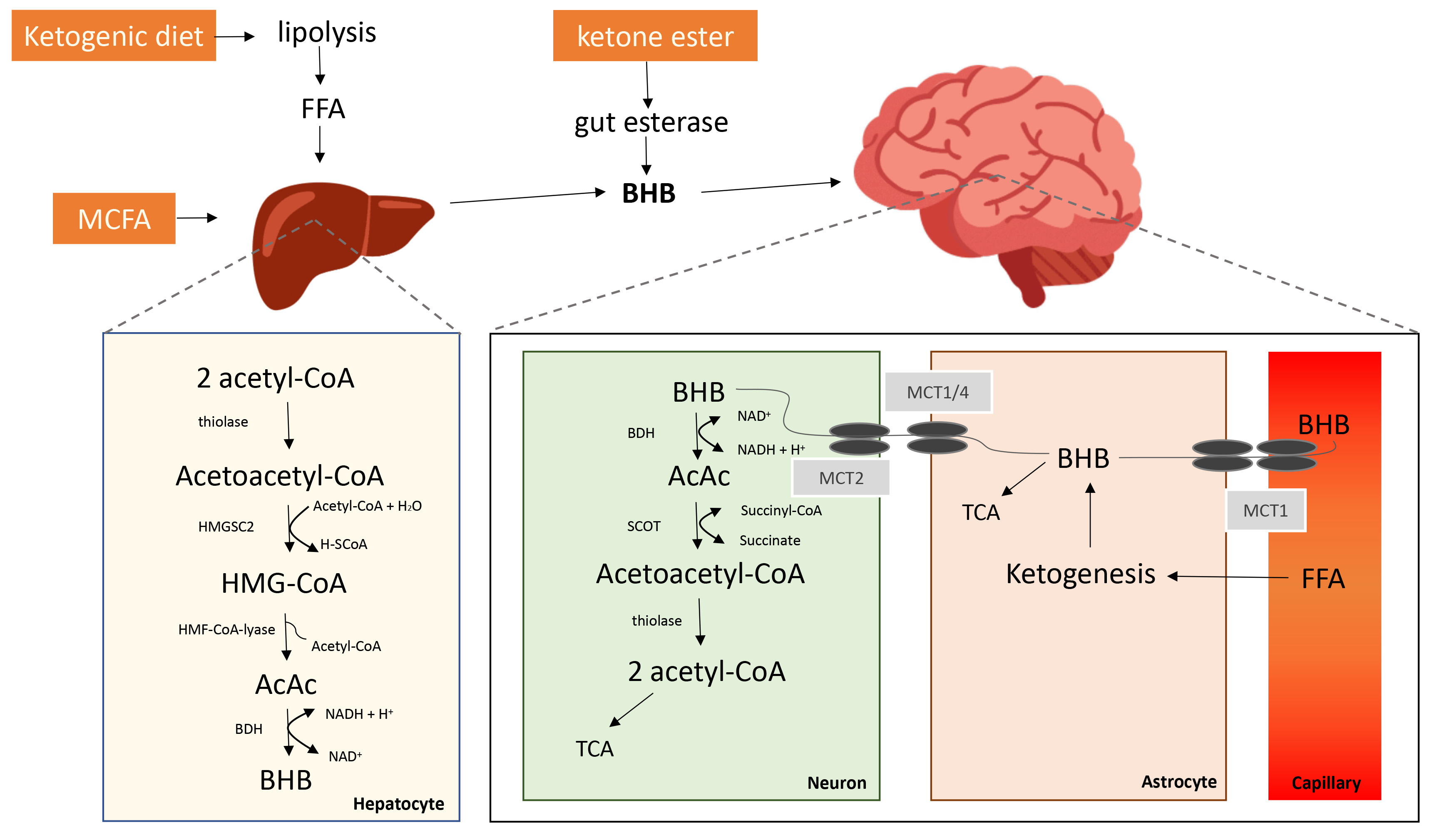
 > 0.5 mmol/L) [84]. Beta-hydroxybutyrate ( β HB) is an endogenous metabolite expressed in a formula as CH3CH(OH)CH2CO2H and one of the main ketone bodies [85]. KBs are a crucial alternative metabolic energy source for all the fields in life such as eukaryota, bacteria, and archaea [86, 87, 88]. During unavailability of glucose and glycogen stores, KBs are produced from fatty acids by astrocytes to serve as energetic substrate in a metabolic pathway known as ketogenesis [89]. In fact, KBs have always been seen as a consequence of fatty acid breakdown that rise during fasting and other reasons of glucose deficiency, which can be physiological, including extenuating exercise, or pathological, such as diabetes [90]. It is important to highlight that their correlation with starvation and fat mobilization has been studied for several decades [91], although their exact physiological function was still unknown at the time. Even so, a study from 1967 has discovered how KBs could replace glucose as the major fuel of the brain in situations of prolonged fasting or glucose deficiency, being able to provide up to 60% of the energy needs of the brain [92]. Moreover, the impact of KBs on brain function were discovered much earlier in the 1920s, as it was observed that a low-carb and high-fat diet was successful in treating paediatric epilepsy [93, 94]. While the human brain mainly relies on glucose as the primary energy source, other substrates might support metabolism also, for the most part when glucose stock is confined or insufficient [95]. The majority of ketone bodies production happens in the liver and is utilized as an energy source especially in times where glucose is absent due to fasting, low-carbohydrate, or average protein diet, which has its own term known as ketogenic diet [96]. Free fatty acids are produced from adipocytes and transported to the liver where they impart to the synthesis of KBs during fasting [97]. The ketogenic diet gained a lot of popularity recently, especially after the discovery that extended fasting has anticonvulsive impacts in epilepsy [98]. The metabolic effect of starvation can be simulated by keeping a high fat, low-carbohydrate, and moderate protein concentration. Also noteworthy, recent research has shown that ketogenic diet may have advantages for a series of neurological diseases via diverse mechanism, as well as improving energy metabolism [99, 100, 101]. The observations also agree with the results reported by [102], that a reduction in migraine attack frequency was greatly reduced during ketosis.

Fig. 2.

Potential Ketone Bodies Agents and its Pathways involved in Synthesis and Catabolism (adapted from [12]).

2.8 Exogenous Ketone Bodies

Apart from fasting, dietary ketogenic supplements and ketogenic diets have the potential to enhance the circulatory pool of KBs [12]. In the recent years exogenous β HB supplements have gained popularity and become accessible around the world. For instance, they usually come in mineral salt form, such as calcium, potassium, magnesium, sodium, or ester form. These can be found in different production sites internationally, the biggest market being in the United States, where these ketone bodies supplements are mostly sold as a sport supplement. Also worth mentioning, exogenous ketone bodies were initially produced for the US army in the past and has been used for over 10 years already [103]. The ketogenic diet may increase ketone bodies high enough to fight common neurological disorders, however, it might not be a potential long-term solution for many sufferers, due to the tremendous effort to execute it in an outpatient setting and patients might have difficulty with adherence. Likewise, ketogenic diets may be tied with adverse effects such as weight reduction, that ideally should be circumvented in neurodegenerative diseases, gastrointestinal problems, hyperlipidaemia, and on top of all, it may also cause lack in mineral and vitamin, because of the rigorous diet scheme [104]. Thus, nutritional supplementation with exogenous ketone bodies could be more viable than ketogenic diet in general patient populations [105]. It could potentially prevent negative effects of unwell managed diet such as reduced vegetable intake and more dairy, highly processed protein, and polyunsaturated fats, all of which could have the consequence of mineral and vitamin deficiency as well as the higher risk of inflammation [105]. Nutritional supplements of MCFA similarly provoke ketosis and it takes place shortly after consumption because this group of fatty acids is absorbed directly into the portal vein, whereas on the contrary to long-chain fatty acids it goes into the lymphatic system and is as a result expeditiously transformed to ketones through beta-oxidation in the liver [12]. The greatest ketogenic MCFA exist in palm kernel oil or coconut oil which are caprylic acid (C8) and capric acid (C10) [11]. Nonetheless, due to the low concentration of lipids in coconut oils, such interventions only increase ketone body levels partially [106]. For that reason, various products with greater levels of caprylic and capric acid have been established [107]. Also remarkable is that neuroprotective effects of MCFA are not associated to ketonemia, because of MCFA’s ability to cross the blood–brain barrier (BBB) and act as substrates for energy metabolism [108]. Additionally, MCFA, especially capric acid, have been observed in several studies to promote mitochondrial function and reduce neuronal hyperactivity [109], which is typically seen in migraine patients by blocking glutamatergic AMPA receptors [110, 111, 112]. Further option to the ketogenic diet is the consumption of exogenous KBs, which greatly raise ketone levels to > 1 mM after intake, with the ketone ester being most influential in increasing prevailing ketones even while maintaining habitual meals [108, 113].

2.9 Potential Mechanism of Exogenously Elevated Beta-Hydroxybutyrate
2.9.1 Oxidative Stress

Common migraine triggers are causing an imbalance of oxidative stress levels through unfavourably influencing energy metabolism or mitochondrial functioning [9, 29, 33]. A protective mechanism of KBs can be demonstrated in the improvement of mitochondrial function because oxidation in the brain decreases as ketone body levels increase [12]. Therefore ketone bodies are also used as protective molecules against refractory epilepsy [114]. It has been proven that β HB protects from neuronal death provoked by intense non-coma hypoglycaemia in the rat in vivo as well as by glucose deprivation in cortical culture [115]. Further animal studies have shown how β HB decreases reactive oxygen species production in specific cortical areas and subregions of the hippocampus and thus effectively prevented neuronal death in the cerebral mantle of hypoglycaemic animals [115]. In vitro findings have shown similar observations of reduced reactive oxygen species levels as well as additional stimulation of ATP production with β HB [116, 117, 118]. All these findings suggest β HB’s protective mechanisms with its metabolic process and also its capability to reduce reactive oxygen species [119]. In a nutshell, KBs has the potential to rise antioxidant protein level and decrease levels of oxidative stress resulting in greater cellular energy productivity [120, 121]. Lastly, findings have shown how ketone levels are increased in failing mice hearts as a compensatory response against oxidative stress [122]. Given that, higher ketone levels during a migraine attack might indicate a parallel reaction in response to elevated oxidative stress [9].

2.9.2 Inflammation

While inflammation is a set response with protective tissues against disease, infection, or injury, involvement of neurogenic inflammation in migraine headache stays inconclusive, and not that migraine is not classified as an inflammatory disease. Various studies have demonstrated that pro-inflammatory peptides or a “sterile neurogenic inflammation” can be exhibited in migraine pain [9, 123, 124]. First and foremost, molecules linked with migraine headache both in animal and human studies have shown the involvement of calcitonin gene related peptide (CGRP), nitric oxide (NO), substance P, vasoactive intestinal peptide (VIP), pituitary adenylate cyclase-activating polypeptide (PACAP) [9, 125, 126]. Cytokines are referred to as pain mediators in neurovascular inflammation, and they could have an effect on the generation of migraine pain [125]. Moreover, cytokines are essential mediators of the inflammatory pathway and are associated with migraine pathogenesis [125]. Similarly, observations of several studies hint that peripheral and central levels of cytokines and immunological changes are present in migraineurs [127, 128, 129] though it should be noted that it is still unclear what might be the triggering factor that activates the inflammatory cascade leading to a migraine attack. A potential reasoning would be that a genetic predisposition exists to have an abnormal inflammatory response to exogenous stimuli (such as high-carb diet or other environmental factors) and could represent a trigger for headache attacks [125]. In fact, twin studies and recent findings based on genetic mutations causing hemiplegic familial migraine hint that genetic background play a significant role in migraine pathogenesis [130]. Finally, many studies on migraine patients have focused on peripheral and central levels of cytokines and the role of inflammation in migraine pathogenesis, despite that data are controversial. Having this mentioned, reduction in inflammation can be seen in prolonged fasting and also caloric restrictions [131, 132]. In another combined animal and cellular study using exogenous KBs has discovered the anti-inflammatory protection of the NLRP3 inflammasome [133]. Thus, NLRP3 inflammasome is prevalent in especially immune and inflammatory cells following activation by inflammatory stimuli [134]. Furthermore, it has been shown that in response to urate crystals, ATP and lipotoxic fatty acids, KBs inhibits activation of the NLRP3 inflammasome [133]. Given the findings above, a decrease in inflammation and pain can be recorded in rats who underwent a ketogenic diet [135, 136]. Finally, the capability of underlying mechanisms of KD efficacy to support mitochondrial energy metabolism and counteract neural inflammation was also demonstrated in an observational study on migraine patients [137].

2.9.3 Hypometabolism/Hypoglycaemia

As mentioned above, the brain is exceptionally relied on energy sources from the circulation that can transcend through the blood-brain barrier and is in particular susceptible to their shortage due to its restricted glycogen sources and high energy demands [9]. Further research has shown that intracellular glucose levels are increased in the presence of β HB supplementation [138]. Additionally, further results have demonstrated the influence of exogenous increment of KBs and observed reduced glucose levels in patients with a four hour β HB infusion compared to treatment with a control solution [139]. The results point to an alternative metabolic substrate through β HB or alternatively a decreased gluconeogenesis. Interestingly, an absolute decrease of 16% in ATP levels in MO patients was recorded by using 31P-MRS [140]. Further findings indicated that hypometabolism was significantly associated with attack frequency [140, 141, 142]. There have been a number of studies that have investigated the effectiveness of KBs and results have proved its ability to counteract negative effects of hypoglycaemia and/or hypometabolism [107, 115, 143]. Experimental results have also demonstrated the concept of inhibition in glycolysis at the occurance of β HB, while ketosis relatively reserves glucose exertion in the human brain [107, 143]. An intriguing finding in 36 h fasted rats is that during adequate concentration to saturate metabolism, KBs backs up the whole fundamental energy demands, as well as around 50% of the of the activity-dependent oxidative needs [144]. These results indicate that β HB is an alternative and presumably more efficient fuel source for the brain. Catabolized β HB produces more ATP per oxygen molecule than other respiratory substrates (such as carbohydrates, proteins, fats and organic acids) during the synthesis of ATP in mitochondria [145]. Interesting observations have been made in initial experimental studies, that glucose tolerance tests after an overnight fast may evoke migraine attacks in patients Interesting observations by initial experimental studies [146, 147]. Yet, the metabolic responses in patients were significantly different between those who experienced a migraine attack and those that did not. KB levels increased significantly in the patients who had a migraine attack, already before headache onset, and anticipated an increase in KB levels despite equivalent food consumption. These observations may be explained as a counter-regulatory reaction to a lack of cerebral energy [9]. Whereas KBs are an efficient alternative energy source for the brain (see Fig. 3, Ref. [12]), during low glucose availability, restoration in brain energy homeostasis is expected after sufficient elevation of KBs in the body [9].

Fig. 3.

Vicious Circle of Energy Crisis in Metabolic Migraine Patients (adapted from [12]).

2.9.4 Cortical Spreading Depression

Cortical spreading depression (CSD) implies a slowly propagated wave of depolarization of the neurons and glial cells across the cerebral cortex followed by depression of cortical activity and has been suggested as a possible underlying pathophysiological mechanism in migraine aura. There is a strong correlation between metabolic factors and CSD susceptibility. A potential CSD trigger is hypoxia and above all, cerebral glucose availability regulates extrinsically induced CSD in both ways [148, 149, 150, 151]. In addition to that, hypoglycaemia greatly extends CSD length while hyperglycaemia also safeguards the tissue from CSD initiation [138]. During a short- and long-term treatment with a middle chain triglyceride enriched ketogenic diet, similar protective effects against CSD were observed in an experimental animal study, where the brain was provided with another energy substrate to glucose [152].

2.9.5 Ketone Bodies in Healthy Individuals

Rises in ketone body availability to the CNS in humans lead to substantial changes in cerebral fuel metabolism [12]. Further results have demonstrated that in healthy adults, an in vivo infusion of β HB effected roughly 14% reduction in cerebral glucose usage while oxygen consumption ended up indifferent [153]. Having this said, when KBs are provided intensely, they may be used instantly as an alternative energy source to glucose [153]. Considering that, an intense cerebral glucose-sparing impact is achieved when ketone accessibility is high. Study participants on ketogenic diets have shown to be one of the other equivalent metabolic adaptations, where ketones replace glucose [95]. During extended fasting, a more prominent change from glucose to ketone body metabolism is exhibited and β HB providing more than half of the human brain’s energy needs [154], consequently substituting glucose as the main fuel supply. In conclusion, a great way to increase the availability of alternative energy reserves for the brain is by utilizing KBs. It should be noted that during resting conditions, KBs substitute other energy sources instead of supplementing them. Provided that, another study have recorded unaltered ATP levels in the brain of healthy participants [155]. When experimental hypoglycaemia reduces glucose availability, further supply of KBs by intake of MCFA or infusion may be able to maintain cognitive functions in individuals, and extends the glycaemic threshold for symptoms [156]. In view of this, KBs are in the position to preserve glucose, besides supporting brain metabolism during energy crises [12]. Hence, treatments that raise prevailing ketones imply to have effects in disorders with compromised glucose metabolism, such as migraine. A comparison of potential pathophysiological mechanisms of exogenously elevated or absence of beta-hydroxybutyrate in healthy/migraine brain is given in Table 2.

Table 2.Systematic comparison of potential mechanism of exogenously elevated beta-hydroxybutyrate in brain vs. absence of beta-hydroxybutyrate in brain.
Ketogenesis ATP level Glucose level CSD Oxidative stress Inflammation Migraine
Healthy Brain Occasionally* Optimal Optimal No No No No
BHB in Healthy Brain Yes Optimal Optimal No No No No
Migraine Brain No Suboptimal Suboptimal Yes Yes Yes Yes
BHB in Migraine Brain Yes Better** Suboptimal Less Less Less Less
Note: In this figure, an overall comparison of healthy brain and a migraine brain with/or without Beta-Hydroxybutyrate ( β HB) and its contribution to migraine related pathophysiological mechanisms such as ketogenesis, ATP levels, glucose levels, cortical spreading depression (CSD), oxidative stress, inflammation are demonstrated.
* e.g., during fasting/skipping meals.
**if enough β HB available for ATP synthesis.
3. Discussion
3.1 Prophylactic Migraine Treatment with Ketone Bodies

Certainly, the aim of intervention for migraine is to diminish the duration and severity of the migraine attack [157]. Other objectives crucial for improvement comprise restoring functioning ability, reducing oxidative stress, increasing antioxidants, and encouraging comprehensive handling with no or little side effects [43, 158]. An interesting finding is, that the most common preventive measure reported for migraine is diet [13]. Diet is a significant element and modifiable aspect of lifestyle, thus dietary behaviours that might act as a trigger for migraine can be strategically identified and implemented to help prevent migraine attacks [159]. More recently, the underlying metabolic and endocrine mechanisms triggering migraine have begun to unfold. Therefore it is of critical importance to consider the benefits from supplying an alternative energy substrate for the human brain with restricted energy stocks [160]. Henceforward, for patients with compromised energy metabolism, an alternative source of fuel for the brain through ketogenic diet and/or use of exogenous ketogenic substances, such as medium-chain triglycerides or exogenous ketone body salts can have beneficial effects and hence supports the role of abnormal metabolism in migraine [137, 161].

3.1.1 Dietary Ketogenesis

“Ketogenic diet” refers to its correlation of increased KBs and acetoacetate (AcAc) in blood and urine due to mimicked fasting by forcing fatty acid breakdown by reducing the intake of carbohydrates [162]. Low carbohydrate diets or ketogenic diets (KD) typically restrict carbohydrates to less than 20 grams per day [96]. Few days into a ketogenic diet, the reserves of glycogen are drained and KBs are generated to maintain energy production within the mitochondria. Immense reduction in migraine frequency (up to 80%) have been noticed after one month on ketogenic diet [137]. Interestingly, ketogenic diets have been primarily utilized to fight obesity in individuals and to treat epilepsy [82, 163, 164]. Furthermore, due to altered tissue excitability in migraine, ketogenic diet might benefit brain metabolism restoration and excitability in migraine pathophysiology [82]. A well-designed study recorded significant improvements through a 3-month ketogenic diet in patients with medication overuse headache [165]. Moreover, ketogenic diet is more advantageous to patients with migraine compared with a low-calorie diet [13]. Furthermore, a low-calorie diet is not recommended for migraine patients [137] and migraine improvement has been reported in those on a ketogenic diet and continued improvement was reported for two months after the ketogenic diet was stopped [102, 166]. In another study where they tried to examine the efficacy of ketogenic diets in migraine patients observed a significant decline in attack frequency as well as the amount of days with headaches during the initial month of ketogenesis [137]. Likewise, additional studies by the same authors reported differences in cortical excitability, and decreased attack incidences and duration after one single month on diet [167]. Further findings also suggest the critical role of KBs particularly in the cerebral cortex, and not subcortically [168]. A key strength of KBs is its independence from GLUT1, as well as ketosis has a variety of other effects that are potentially valuable in understanding migraine pathophysiology, such as increased anti-oxidant capacity, mitochondrial biogenesis, upregulation of GLUT1 and ketone body transporters, increased GABA but inhibition of glutamate transport and, therefore, reduced excitatory synaptic transmission and inflammation [9].

3.1.2 Exogenous Ketone Bodies/Nutraceuticals

As mentioned above, KBs are a productive alternative energy source for the human brain during low glucose reserves, restoration in brain energy homeostasis is expected after a sufficient elevation in KBs, and therefore carry the ability to attenuate some of the abnormalities in glucose metabolism as well as glucose transport discovered in migraine patients. The role of KBs is especially relevant for the management of hypoglycaemia/hypometabolism, as well as for glucose transport [64]. Thus, KBs play a crucial role in the improvement of cerebral metabolism. On top of all, since KBs have the potential to positively impact other pathways and are considered to be involved in migraine pathophysiology, such as cerebral excitability, oxidative stress, CSD, inflammation mitochondrial functioning, hypoglycaemia/hypometabolism and the gut microbiome, KBs may also be considered as potential signalling molecules [120]. Given this, an elevation of KBs through exogenous nutraceuticals has been hypothesized to possibly impact all of the foregoing migraine pathophysiological mechanisms, and might support a comparatively side-effect free solution for a proportion of the migraine population [9]. For instance, high doses of riboflavin (400 mg/day) may improve mitochondrial metabolism in migraine patients with a mutation in mtDNA [169]. The effects of the β HB supplementation on migraine patients without an abrupt dietary alteration is still unknown, and on top of that, the exact mechanism of process of most common medications applied in migraine prophylaxis is still unclear [10]. Most of the medications for migraine prophylaxis dim neural reactivity and thus decrease the energetic needs of the human brain [10]. Interestingly, few prophylactic agents can also have direct metabolic effects. For example, Topiramate which represents a preventive drug with level A evidence for efficacy, and protects against mitochondrial membrane depolarization, oxidative stress and inflammation [170]. Fig. 4 (Ref. [10]) shows that oxidative stress plays a major role in migraine attack generation. The addition of protective medications therefore supports the equipoise between metabolic offers and metabolic needs that is essential in order to keep up cerebral homeostasis [10]. Taking this into consideration, suggested strategies to decrease oxidative stress is as mentioned above, to eliminate or reduce processed food, food with a high glycaemic index as well as alcohol, and promoting the use of green or blue light filtering glasses [171]. Consequently, lifestyle changes such as addition of antioxidants like β -hydroxybutyrate mineral salts to the diet has been reported to be beneficial [114]. Hence, energy-preserving behavioural modifications and reduction in oxidative stress levels as well as increase glucose and ketone body supply for the brain, may assist to recover energy homeostasis [10]. In summation, migraine patients with a greatly compromised energy metabolism can support the brain with an alternative fuel source besides glucose and lactate with the help of exogenous KBs. Nonetheless, the validation of ketogenic therapies in migraine requires further placebo-controlled trials.

Fig. 4.

Migraine Attack Generation and Resolution (adapted from [10]).

3.2 Limitations and Future Perspectives

The present review has some limitations. Firstly, due to insufficient amount of literature on using KBs for migraine prevention, more extensive studies are needed to validate its efficacy and effects. Secondly, due to poor methodologies and weak study designs of existing research, including lack of control groups and small sample sizes, further studies with larger population groups are required to investigate the impact of KBs for migraine treatment. Thirdly, no inferences can be made on gender and age regarding metabolic mechanisms in migraine patients due to limited literature, hence future studies should aim to conduct a well-designed and mechanism-driven longitudinal research to investigate this further as outcomes are generally affected by diverse factors such as age, genetics, gender and environmental factors. Fourthly, to our knowledge, there are no existing literature on the impact of diurnal variations on using exogeneous KBs in migraine prevention, hence should be further investigated since disruptions in circadian rhythms cause impairment in metabolism [1]. Given that, there are limitations associated with the variety of how different mechanisms might lead to the migraine phenotype and hence future research should seek to substantiate these concepts and their potential application for patients. In addition, further clinical research is needed to validate the hypothesized pathophysiological effects of KBs in migraine patients. Finally, other prophylactic medications of migraine were not considered in this review which deserves to be further explored and put into comparison. Future studies should also consider applying KBs in migraine prevention for more extended periods. Moreover, further efforts are required to minimize patient burden during a ketogenic diet, and to improve palatability of the diet would also have to be discussed. Regardless of the limited amount of data, the findings delineated in this review have demonstrated the potential therapeutic implications of KBs. This poses an area for future work. Hence, future research carrying out other ketogenic approaches should aim to reinforce the idea of a ketone-based metabolic progress in migraine patients. Having said that, this review has highlighted the possibility of future studies in elucidating particular mechanisms of glucose metabolism impairment in migraine. Despite that, the modulation of this metabolic pathway in migraine demands further exploration. Several dietary treatments have been proposed for migraine prevention and ketogenic diets that result in elevation of KBs, has demonstrated promising contribution in the prevention of migraines [172, 173]. Nonetheless, prospective research is required to confirm this hypothesis. Above all, it has to be remarked that migraine triggers such as poor sleep patterns, stress, hormonal changes, bright lights and diet-related triggers, including caffeine, hypo-hydration and skipping meals, vary from person to person [59]. Given the fact that, there are gender and age differences in the prevalence of migraine and hypoglycaemia, future research should conduct a longitudinal study to investigate these factors further. Considering the spectrum of potential triggers implies further complexity for developing effective intervention strategies. Consequently, future research should consider how intervention strategies can be personalised to the triggers that are specific to individuals. Additionally, recent research has observed that ketosis may have benefits in the treatment of other conditions such as neurodegenerative diseases like Alzheimer’s disease, Parkinson’s disease, multiple sclerosis [12, 39, 174, 175]. Consequently, neurological and neuropsychiatric diseases, for instance, epilepsy, autism, depression and affective disorders, anxiety, and traumatic brain injury might also benefit from a ketosis treatment [176, 177, 178, 179, 180]. Last but not least, ketosis might also open avenues in the treatment of metabolic disorders like cancer, glycogen storage disease, GLUT1 deficiency syndrome [105, 181, 182]. Additionally, more studies researching the effects of ketone bodies on (cerebral) metabolism, oxidative stress, inflammation would be of great interest. Expanding inclusion criteria to also incorporate those with severe headache, not only migraine, should be addressed in future research. Lastly, neuroimaging methods have excellent translational values and can be widely applied in future studies to identify dietary effects on brain functions in humans.

4. Conclusions

Migraine is a highly complex multifactorial neurological disease that differs in frequency, severity, and its impact on the quality of life. Furthermore, genetic factors must be considered when defining an individual’s susceptibility to migraine. Findings have shown that the migraine pathophysiology emphasizes the aspects of different triggers that may initiate a migraine attack or increase the frequency of the attacks. In this review, metabolic bases of migraine regarding the use of KBs were discussed. With migraine being such a multigenic and complex disease, diverse aspects of the most prevalent migraine triggers were mentioned that have a strong correlation to the pathophysiological mechanisms: (1) sensory trigger, (2) mental stress vs. physical stress, (3) sleep changes, (4) fasting/skipping meals, and (5) dietary factors. While migraine is also a prevalent disease, it is often under-diagnosed. On top of all, it is yet to be perceived adequately by certain migraine patients who are eligible for prophylactic care. In that case, these therapies could increase migraine patients’ quality of life and reduce the physical and functional disabilities of migraine. Furthermore, this work defines migraine as a complex and heterogeneous neurological disease, along with several common genetic polymorphisms and thus abnormal mechanisms resulting in the migraine phenotype. In this review, only two typical and important pathophysiological mechanisms have been discussed in detail and thus, also their possible exploitation through exogenous ketone bodies such as β HB supplementation or ketogenic diet: (1) oxidative stress, (2) inflammation, (3) glucose metabolism, and (4) CSD. Studies have shown that ketone bodies—especially β HB—are more than just metabolites, because as signalling molecules, ketone bodies can potentially influence other pathways, that are part of migraine pathophysiology [120]. Hence, intervention that can consider various potential pathogenic pathways might benefit heterogenous, polygenic and complex disease like migraine [173].

Abbreviations

AcAc, acetoacetate; AcetylCoA, acetyl coenzyme A; BBB, blood–brain barrier; BDH, beta-hydroxybutyrate dehydrogenase; CGRP, calcitonin gene-related peptide; CNS, central nervous system; FDG-PET, 18-fluorodeoxyglucose-PET; FFA, free fatty acids; FHM, familial hemiplegic migraine; GLUT-1, glucose transporter type 1; HMG-CoA, 3-hydroxy-3-methylglutaryl-CoA; HMGCS2, 3-Hydroxy-3-methylglutaryl-CoA Synthase 2; IHS, International Headache Society; KB, ketone body; KD, ketogenic diet; MA, migraine with aura; MCFA, medium-chain fatty acids; MCT, monocarboxylate transporter; MO, migraine without aura; MRI, magnetic resonance imaging; NO, nitric oxide; PACAP, pituitary adenylate cyclase-activating polypeptide; PCr, phosphocreatine; PET, positron-emission tomography; P-MRS, phosphorus magnetic resonance spectroscopy; SCOT, succinyl-CoA,3-ketoacid Coenzyme A transferase; TCA, tricarboxylic acid cycle; VIP, vasoactive intestinal peptide; YLD, years lived with disability; β HB, beta-hydroxybutyrate.

Author Contributions

PH and PC conceived and designed the review; PH performed the literature analysis and overview; PH and PC discussed the results; PC aided in interpreting the results and worked on the manuscript. PH wrote the paper in consultation with PC; All authors read and approved the final manuscript.

Ethics Approval and Consent to Participate

Not applicable.

Acknowledgment

We would like to thank the reviewers for their helpful comments and efforts towards improving our manuscript.

Funding

This research received no external funding.

Conflict of Interest

The authors declare no conflict of interest. PC is serving as one of the Editorial Board members of this journal. We declare that PC had no involvement in the peer review of this article and has no access to information regarding its peer review. Full responsibility for the editorial process for this article was delegated to RSC.

References
[1]
Headache Classification Committee of the International Headache Society (IHS) The International Classification of Headache Disorders, 3rd edition. Cephalalgia. 2018; 38: 1–211.
[2]
de Oliveira AB, Mercante JPP, Benseñor IM, Goulart AC, Peres MFP. Headache disability, lifestyle factors, health perception, and mental disorder symptoms: a cross-sectional analysis of the 2013 National Health Survey in Brazil. Neurological Sciences. 2022; 43: 2723–2734.
[3]
Scuteri D, Bagetta G. Progress in the Treatment of Migraine Attacks: From Traditional Approaches to Eptinezumab. Pharmaceuticals (Basel). 2021; 14: 924.
[4]
Stovner LJ, Hagen K. Prevalence, burden, and cost of headache disorders. Current Opinion in Neurology. 2006; 19: 281–285.
[5]
Steiner TJ, Stovner LJ, Vos T, Jensen R, Katsarava Z. Migraine is first cause of disability in under 50s: will health politicians now take notice? The Journal of Headache and Pain. 2018; 19: 17.
[6]
Linde M, Gustavsson A, Stovner LJ, Steiner TJ, Barré J, Katsarava Z, et al. The cost of headache disorders in Europe: the Eurolight project. European Journal of Neurology. 2012; 19: 703–711.
[7]
Silberstein SD, Lipton RB, Dodick DW. Wolff’s Headache and Other Head Pain. Eighth edn. Oxford University Press: Oxford, New York. 2007.
[8]
Loder E. What is the Evolutionary Advantage of Migraine? Cephalalgia. 2002; 22: 624–632.
[9]
Gross EC, Klement RJ, Schoenen J, D’Agostino DP, Fischer D. Potential Protective Mechanisms of Ketone Bodies in Migraine Prevention. Nutrients. 2019; 11: 811.
[10]
Gross EC, Lisicki M, Fischer D, Sándor PS, Schoenen J. The metabolic face of migraine — from pathophysiology to treatment. Nature Reviews Neurology. 2019; 15: 627–643.
[11]
Cunnane SC, Courchesne-Loyer A, St-Pierre V, Vandenberghe C, Pierotti T, Fortier M, et al. Can ketones compensate for deteriorating brain glucose uptake during aging? Implications for the risk and treatment of Alzheimer’s disease. Annals of the New York Academy of Sciences. 2016; 1367: 12–20.
[12]
Jensen NJ, Wodschow HZ, Nilsson M, Rungby J. Effects of Ketone Bodies on Brain Metabolism and Function in Neurodegenerative Diseases. International Journal of Molecular Sciences. 2020; 21: 8767.
[13]
Khan J, Asoom LIA, Sunni AA, Rafique N, Latif R, Saif SA, et al. Genetics, pathophysiology, diagnosis, treatment, management, and prevention of migraine. Biomedicine & Pharmacotherapy. 2021; 139: 111557.
[14]
Levy D. Migraine pain and nociceptor activation–where do we stand? Headache. 2010; 50: 909–916.
[15]
Olesen J, Burstein R, Ashina M, Tfelt-Hansen P. Origin of pain in migraine: evidence for peripheral sensitisation. The Lancet Neurology. 2009; 8: 679–690.
[16]
Pietrobon D, Striessnig J. Neurobiology of migraine. Nature Reviews Neuroscience. 2003; 4: 386–398.
[17]
Akerman S, Williamson DJ, Kaube H, Goadsby PJ. The effect of anti-migraine compounds on nitric oxide-induced dilation of dural meningeal vessels. European Journal of Pharmacology. 2002; 452: 223–228.
[18]
Goadsby PJ. The vascular theory of migraine—a great story wrecked by the facts. Brain. 2009; 132: 6–7.
[19]
Goadsby PJ, Holland PR, Martins-Oliveira M, Hoffmann J, Schankin C, Akerman S. Pathophysiology of Migraine: a Disorder of Sensory Processing. Physiological Reviews. 2017; 97: 553–622.
[20]
Calabrò RS. Gender effect on migraine: nature or culture? Journal of Integrative Neuroscience. 2022; 21: 018.
[21]
Krause DN, Warfvinge K, Haanes KA, Edvinsson L. Hormonal influences in migraine — interactions of oestrogen, oxytocin and CGRP. Nature Reviews Neurology. 2021; 17: 621–633.
[22]
Montagna P, Pierangeli G, Cortelli P. The Primary Headaches as a Reflection of Genetic Darwinian Adaptive Behavioral Responses. Headache. 2010; 50: 273–289.
[23]
Aw WC, Towarnicki SG, Melvin RG, Youngson NA, Garvin MR, Hu Y, et al. Genotype to phenotype: Diet-by-mitochondrial DNA haplotype interactions drive metabolic flexibility and organismal fitness. PLoS Genetics. 2018; 14: e1007735.
[24]
Gray PA, Burtness HI. Hypoglycemic Headache. Endocrinology. 1935; 19: 549–560.
[25]
Amery WK. Brain Hypoxia: the Turning-Point in the Genesis of the Migraine Attack? Cephalalgia. 1982; 2: 83–109.
[26]
Kincses ZT, Veréb D, Faragó P, Tóth E, Kocsis K, Kincses B, et al. Are Migraine With and Without Aura Really Different Entities? Frontiers in Neurology. 2019; 10: 982.
[27]
Nazari F, Safavi M, Mahmudi M. Migraine and its relation with lifestyle in women. Pain Practice. 2010; 10: 228–234.
[28]
Kelman L. The Triggers or Precipitants of the Acute Migraine Attack. Cephalalgia. 2007; 27: 394–402.
[29]
Peroutka SJ. What Turns on a Migraine? A Systematic Review of Migraine Precipitating Factors. Current Pain and Headache Reports. 2014; 18: 454.
[30]
Schoonman GG, van der Grond J, Kortmann C, van der Geest RJ, Terwindt GM, Ferrari MD. Migraine headache is not associated with cerebral or meningeal vasodilatation—a 3T magnetic resonance angiography study. Brain. 2008; 131: 2192–2200.
[31]
Lucchesi C, Sassi AN, Siciliano G, Gori S. Fatigue is Increased in Episodic Migraine without Aura Patients. Headache. 2013; 53: 1163–1165.
[32]
Borkum JM. Migraine Triggers and Oxidative Stress: a Narrative Review and Synthesis. Headache. 2016; 56: 12–35.
[33]
Pavlovic JM, Buse DC, Sollars CM, Haut S, Lipton RB. Trigger Factors and Premonitory Features of Migraine Attacks: Summary of Studies. Headache. 2014; 54: 1670–1679.
[34]
Schulte LH, Jürgens TP, May A. Photo-, osmo- and phonophobia in the premonitory phase of migraine: mistaking symptoms for triggers? The Journal of Headache and Pain. 2015; 16: 14.
[35]
Amiri A, Turner-Henson A. The Roles of Formaldehyde Exposure and Oxidative Stress in Fetal Growth in the second Trimester. Journal of Obstetric, Gynecologic & Neonatal Nursing. 2017; 46: 51–62.
[36]
Angelucci FL, Silva VV, Dal Pizzol C, Spir LG, Praes CEO, Maibach H. Physiological effect of olfactory stimuli inhalation in humans: an overview. International Journal of Cosmetic Science. 2014; 36: 117–123.
[37]
Duan Y, Wang L, Han L, Wang B, Sun H, Chen L, et al. Exposure to phthalates in patients with diabetes and its association with oxidative stress, adiponectin, and inflammatory cytokines. Environment International. 2017; 109: 53–63.
[38]
Nakamura M, Kuse Y, Tsuruma K, Shimazawa M, Hara H. The Involvement of the Oxidative Stress in Murine Blue LED Light-Induced Retinal Damage Model. Biological & Pharmaceutical Bulletin. 2017; 40: 1219–1225.
[39]
Henderson D, Bielefeld EC, Harris KC, Hu BH. The Role of Oxidative Stress in Noise-Induced Hearing Loss. Ear & Hearing. 2006; 27: 1–19.
[40]
Koppen H, van Veldhoven PL. Migraineurs with exercise-triggered attacks have a distinct migraine. The Journal of Headache and Pain. 2013; 14: 99.
[41]
Amin FM, Aristeidou S, Baraldi C, Czapinska-Ciepiela EK, Ariadni DD, Di Lenola D, et al. The association between migraine and physical exercise. The Journal of Headache and Pain. 2018; 19: 83.
[42]
Lipton RB, Buse DC, Hall CB, Tennen H, Defreitas TA, Borkowski TM, et al. Reduction in perceived stress as a migraine trigger: testing the “let-down headache” hypothesis. Neurology. 2014; 82: 1395–1401.
[43]
Salim S. Oxidative stress: a potential link between emotional wellbeing and immune response. Current Opinion in Pharmacology. 2016; 29: 70–76.
[44]
Schiavone S, Jaquet V, Trabace L, Krause K. Severe Life Stress and Oxidative Stress in the Brain: from Animal Models to Human Pathology. Antioxidants & Redox Signaling. 2013; 18: 1475–1490.
[45]
Gong Y, Chai Y, Ding J, Sun X, Hu G. Chronic mild stress damages mitochondrial ultrastructure and function in mouse brain. Neuroscience Letters. 2011; 488: 76–80.
[46]
Knapman A, Kaltwasser SF, Martins-de-Souza D, Holsboer F, Landgraf R, Turck CW, et al. Increased stress reactivity is associated with reduced hippocampal activity and neuronal integrity along with changes in energy metabolism. European Journal of Neuroscience. 2012; 35: 412–422.
[47]
Musiek ES, Holtzman DM. Mechanisms linking circadian clocks, sleep, and neurodegeneration. Science. 2016; 354: 1004–1008.
[48]
Reinke H, Asher G. Circadian Clock Control of Liver Metabolic Functions. Gastroenterology. 2016; 150: 574–580.
[49]
Bjorvatn B, Pallesen S, Moen BE, Waage S, Kristoffersen ES. Migraine, tension-type headache and medication-overuse headache in a large population of shift working nurses: a cross-sectional study in Norway. BMJ Open. 2018; 8: e022403.
[50]
McEwen BS, Karatsoreos IN. Sleep Deprivation and Circadian Disruption: Stress, Allostasis, and Allostatic Load. Sleep Medicine Clinics. 2022; 17: 253–262.
[51]
Periasamy S, Hsu DZ, Fu YH, Liu MY. Sleep deprivation-induced multi-organ injury: role of oxidative stress and inflammation. EXCLI Journal. 2015; 14: 672–683.
[52]
Trivedi MS, Holger D, Bui AT, Craddock TJA, Tartar JL. Short-term sleep deprivation leads to decreased systemic redox metabolites and altered epigenetic status. PLoS ONE. 2017; 12: e0181978.
[53]
Dalkara T, Kılıç K. How does Fasting Trigger Migraine? A Hypothesis. Current Pain and Headache Reports. 2013; 17: 368.
[54]
Yadav RK, Kalita J, Misra UK. A Study of Triggers of Migraine in India. Pain Medicine. 2010; 11: 44–47.
[55]
Blau JN, Cumings JN. Method of precipitating and preventing some migraine attacks. British Medical Journal. 1966; 2: 1242–1243.
[56]
Abu-Salameh I, Plakht Y, Ifergane G. Migraine exacerbation during Ramadan fasting. The Journal of Headache and Pain. 2010; 11: 513–517.
[57]
Cardoso S, Santos RX, Correia SC, Carvalho C, Santos MS, Baldeiras I, et al. Insulin-induced recurrent hypoglycemia exacerbates diabetic brain mitochondrial dysfunction and oxidative imbalance. Neurobiology of Disease. 2013; 49: 1–12.
[58]
Sun-Edelstein C, Mauskop A. Foods and Supplements in the Management of Migraine Headaches. The Clinical Journal of Pain. 2009; 25: 446–452.
[59]
Haslam RL, Bezzina A, Herbert J, Spratt N, Rollo ME, Collins CE. Can Ketogenic Diet Therapy Improve Migraine Frequency, Severity and Duration? Healthcare. 2021; 9: 1105.
[60]
Di Lorenzo C, Ballerini G, Barbanti P, Bernardini A, D’Arrigo G, Egeo G, et al. Applications of Ketogenic Diets in Patients with Headache: Clinical Recommendations. Nutrients. 2021; 13: 2307.
[61]
Peatfield R, Glover V, Littlewood J, Sandler M, Rose FC. The Prevalence of Diet-Induced Migraine. Cephalalgia. 1984; 4: 179–183.
[62]
Scharff L, Turk DC, Marcus DA. Triggers of Headache Episodes and Coping Responses of Headache Diagnostic Groups. Headache. 1995; 35: 397–403.
[63]
Nowaczewska M, Wiciński M, Kaźmierczak W, Kaźmierczak H. To Eat or Not to Eat: A Review of the Relationship between Chocolate and Migraines. Nutrients. 2020; 12: 608.
[64]
Grech O, Mollan SP, Wakerley BR, Fulton D, Lavery GG, Sinclair AJ. The Role of Metabolism in Migraine Pathophysiology and Susceptibility. Life. 2021; 11: 415.
[65]
Paemeleire K, Schoenen J. (31) P-MRS in migraine: fallen through the cracks. Headache. 2013; 53: 676–678.
[66]
Reyngoudt H, Achten E, Paemeleire K. Magnetic resonance spectroscopy in migraine: what have we learned so far? Cephalalgia. 2012; 32: 845–859.
[67]
Welch KMA, Levine SR, D’Andrea G, Schultz LR, Helpern JA. Preliminary observations on brain energy metabolism in migraine studied by in vivo phosphorus 31 NMR spectroscopy. Neurology. 1989; 39: 538–538.
[68]
Barbiroli B, Montagna P, Martinelli P, Lodi R, Iotti S, Cortelli P, et al. Defective brain energy metabolism shown by in vivo 31P MR spectroscopy in 28 patients with mitochondrial cytopathies. Journal of Cerebral Blood Flow & Metabolism. 1993; 13: 469–474.
[69]
Rolfe DF, Brown GC. Cellular energy utilization and molecular origin of standard metabolic rate in mammals. Physiological Reviews. 1997; 77: 731–758.
[70]
Harris J, Jolivet R, Attwell D. Synaptic Energy Use and Supply. Neuron. 2012; 75: 762–777.
[71]
Oyarzabal A, Marin‐Valencia I. Synaptic energy metabolism and neuronal excitability, in sickness and health. Journal of Inherited Metabolic Disease. 2019; 42: 220–236.
[72]
Bak LK, Walls AB, Schousboe A, Waagepetersen HS. Astrocytic glycogen metabolism in the healthy and diseased brain. Journal of Biological Chemistry. 2018; 293: 7108–7116.
[73]
Pearson TS, Akman C, Hinton VJ, Engelstad K, De Vivo DC. Phenotypic Spectrum of Glucose Transporter Type 1 Deficiency Syndrome (Glut1 DS). Current Neurology and Neuroscience Reports. 2013; 13: 342.
[74]
Gburek‐Augustat J, Heinze A, Abou Jamra R, Merkenschlager A. Hemiplegic Migraine in Glut1 Deficiency Syndrome and Paroxysmal Dyskinesia at Ketogenic Diet Induction: Case Report and Literature Review. Movement Disorders Clinical Practice. 2020; 7: 965–970.
[75]
Urbizu A, Cuenca-León E, Raspall-Chaure M, Gratacòs M, Conill J, Redecillas S, et al. Paroxysmal exercise-induced dyskinesia, writer’s cramp, migraine with aura and absence epilepsy in twin brothers with a novel SLC2a1 missense mutation. Journal of the Neurological Sciences. 2010; 295: 110–113.
[76]
Binder C, Bendtson I. Endocrine emergencies. Hypoglycaemia. Baillière’s Clinical Endocrinology and Metabolism. 1992; 6: 23–39.
[77]
Kennedy ML, Campuzano-Bublitz MA. Carbohydrate Metabolism in Hypoglycemia. In Blood Glucose Levels. IntechOpen: London, UK. 2019.
[78]
Lisicki M, D’Ostilio K, Coppola G, Scholtes F, Maertens de Noordhout A, Parisi V, et al. Evidence of an increased neuronal activation-to-resting glucose uptake ratio in the visual cortex of migraine patients: a study comparing 18FDG-PET and visual evoked potentials. The Journal of Headache and Pain. 2018; 19: 49.
[79]
Rainero I, Roveta F, Vacca A, Noviello C, Rubino E. Migraine pathways and the identification of novel therapeutic targets. Expert Opinion on Therapeutic Targets. 2020; 24: 245–253.
[80]
Kim J, Kim S, Suh S, Koh S, Park K, Oh K. Interictal metabolic changes in episodic migraine: a voxel-based FDG-PET study. Cephalalgia. 2010; 30: 53–61.
[81]
Gras D, Roze E, Caillet S, Méneret A, Doummar D, Billette de Villemeur T, et al. GLUT1 deficiency syndrome: an update. Revue Neurologique. 2014; 170: 91–99.
[82]
Barbanti P, Fofi L, Aurilia C, Egeo G, Caprio M. Ketogenic diet in migraine: rationale, findings and perspectives. Neurological Sciences. 2017; 38: 111–115.
[83]
Prabhakar A, Quach A, Wang D, Zhang H, Terrera M, Jackemeyer D, et al. Breath acetone as biomarker for lipid oxidation and early ketone detection. Global Journal of Obesity, Diabetes and Metabolic Syndrome. 2014; 1: 012–019.
[84]
Saasa V, Beukes M, Lemmer Y, Mwakikunga B. Blood Ketone Bodies and Breath Acetone Analysis and Their Correlations in Type 2 Diabetes Mellitus. Diagnostics. 2019; 9: 224.
[85]
Newman JC, Verdin E. β -Hydroxybutyrate: A Signaling Metabolite. Annual Review of Nutrition. 2017; 37: 51–76.
[86]
Aneja P, Dziak R, Cai GQ, Charles TC. Identification of an acetoacetyl coenzyme A synthetase-dependent pathway for utilization of L-(+)-3-hydroxybutyrate in Sinorhizobium meliloti. Journal of Bacteriology. 2002; 184: 1571–1577.
[87]
Cahill GF. Fuel Metabolism in Starvation. Annual Review of Nutrition. 2006; 26: 1–22.
[88]
Krishnakumar AM, Sliwa D, Endrizzi JA, Boyd ES, Ensign SA, Peters JW. Getting a Handle on the Role of Coenzyme M in Alkene Metabolism. Microbiology and Molecular Biology Reviews. 2008; 72: 445–456.
[89]
Edmond J, Robbins RA, Bergstrom JD, Cole RA, de Vellis J. Capacity for substrate utilization in oxidative metabolism by neurons, astrocytes, and oligodendrocytes from developing brain in primary culture. Journal of Neuroscience Research. 1987; 18: 551–561.
[90]
García-Rodríguez D, Giménez-Cassina A. Ketone Bodies in the Brain Beyond Fuel Metabolism: From Excitability to Gene Expression and Cell Signaling. Frontiers in Molecular Neuroscience. 2021; 14: 732120.
[91]
Edson NL, Leloir LF. Ketogenesis-antiketogenesis: Metabolism of ketone bodies. Biochemical Journal. 1936; 30: 2319–2332.
[92]
Owen OE, Morgan AP, Kemp HG, Sullivan JM, Herrera MG, Cahill GF. Brain Metabolism during Fasting. Journal of Clinical Investigation. 1967; 46: 1589–1595.
[93]
Helmholz HF. Ten Years’ Experience in the Treatment of Epilepsy with Ketogenic Diet. Archives of Neurology and Psychiatry. 1933; 29: 808.
[94]
Mcquarrie I. Epilepsy in children: relationship of variations in the degree of ketonuria to occurrence of convulsions in epileptic children on ketogenic diets. American Journal of Diseases of Children. 1927; 34: 1013.
[95]
Courchesne-Loyer A, Croteau E, Castellano C, St-Pierre V, Hennebelle M, Cunnane SC. Inverse relationship between brain glucose and ketone metabolism in adults during short-term moderate dietary ketosis: a dual tracer quantitative positron emission tomography study. Journal of Cerebral Blood Flow & Metabolism. 2017; 37: 2485–2493.
[96]
Paoli A, Bosco G, Camporesi EM, Mangar D. Ketosis, ketogenic diet and food intake control: a complex relationship. Frontiers in Psychology. 2015; 6: 27.
[97]
Fukao T, Lopaschuk GD, Mitchell GA. Pathways and control of ketone body metabolism: on the fringe of lipid biochemistry. Prostaglandins, Leukotrienes and Essential Fatty Acids. 2004; 70: 243–251.
[98]
Bailey EE, Pfeifer HH, Thiele EA. The use of diet in the treatment of epilepsy. Epilepsy & Behavior. 2005; 6: 4–8.
[99]
Barañano KW, Hartman AL. The ketogenic diet: Uses in epilepsy and other neurologic illnesses. Current Treatment Options in Neurology. 2008; 10: 410–419.
[100]
Danial NN, Hartman AL, Stafstrom CE, Thio LL. How does the Ketogenic Diet Work? Four Potential Mechanisms. Journal of Child Neurology. 2013; 28: 1027–1033.
[101]
Stafstrom CE, Rho JM. The ketogenic diet as a treatment paradigm for diverse neurological disorders. Frontiers in Pharmacology. 2012; 3: 59.
[102]
Strahlman RS. Can Ketosis Help Migraine Sufferers? A Case Report. Headache. 2006; 46: 182–182.
[103]
Veech RL. The therapeutic implications of ketone bodies: the effects of ketone bodies in pathological conditions: ketosis, ketogenic diet, redox states, insulin resistance, and mitochondrial metabolism. Prostaglandins, Leukotrienes and Essential Fatty Acids. 2004; 70: 309–319.
[104]
Rusek M, Pluta R, Ułamek-Kozioł M, Czuczwar SJ. Ketogenic Diet in Alzheimer’s Disease. International Journal of Molecular Sciences. 2019; 20: 3892.
[105]
Valayannopoulos V, Bajolle F, Arnoux J, Dubois S, Sannier N, Baussan C, et al. Successful Treatment of Severe Cardiomyopathy in Glycogen Storage Disease Type III with D,L-3-Hydroxybutyrate, Ketogenic and High-Protein Diet. Pediatric Research. 2011; 70: 638–641.
[106]
Vandenberghe C, St-Pierre V, Pierotti T, Fortier M, Castellano C, Cunnane SC. Tricaprylin alone Increases Plasma Ketone Response more than Coconut Oil or other Medium-Chain Triglycerides: an Acute Crossover Study in Healthy Adults. Current Developments in Nutrition. 2017; 1: e000257.
[107]
Courchesne-Loyer A, Lowry C, St-Pierre V, Vandenberghe C, Fortier M, Castellano C, et al. Emulsification Increases the Acute Ketogenic Effect and Bioavailability of Medium-Chain Triglycerides in Humans. Current Developments in Nutrition. 2017; 1: e000851.
[108]
Augustin K, Khabbush A, Williams S, Eaton S, Orford M, Cross JH, et al. Mechanisms of action for the medium-chain triglyceride ketogenic diet in neurological and metabolic disorders. The Lancet Neurology. 2018; 17: 84–93.
[109]
Roopashree PG, Shetty SS, Suchetha Kumari N. Effect of medium chain fatty acid in human health and disease. Journal of Functional Foods. 2021; 87: 104724.
[110]
Andreou AP, Edvinsson L. Mechanisms of migraine as a chronic evolutive condition. The Journal of Headache and Pain. 2019; 20: 117.
[111]
Viganò A, Toscano M, Puledda F, Di Piero V. Treating Chronic Migraine With Neuromodulation: The Role of Neurophysiological Abnormalities and Maladaptive Plasticity. Frontiers in Pharmacology. 2019; 10: 32.
[112]
Michail Vikelis, Dimos D. Mitsikostas. The Role of Glutamate and its Receptors in Migraine. CNS & Neurological Disorders - Drug Targets. 2007; 6: 251–257.
[113]
Soto-Mota A, Vansant H, Evans RD, Clarke K. Safety and tolerability of sustained exogenous ketosis using ketone monoester drinks for 28 days in healthy adults. Regulatory Toxicology and Pharmacology. 2019; 109: 104506.
[114]
Simeone TA, Simeone KA, Stafstrom CE, Rho JM. Do ketone bodies mediate the anti-seizure effects of the ketogenic diet? Neuropharmacology. 2018; 133: 233–241.
[115]
Julio-Amilpas A, Montiel T, Soto-Tinoco E, Gerónimo-Olvera C, Massieu L. Protection of Hypoglycemia-Induced Neuronal Death by β -Hydroxybutyrate Involves the Preservation of Energy Levels and Decreased Production of Reactive Oxygen Species. Journal of Cerebral Blood Flow & Metabolism. 2015; 35: 851–860.
[116]
Brieger K, Schiavone S, Miller FJ Jr, Krause KH. Reactive oxygen species: from health to disease. Swiss Medical Weekly. 2012; 142: w13659.
[117]
Forrester SJ, Kikuchi DS, Hernandes MS, Xu Q, Griendling KK. Reactive Oxygen Species in Metabolic and Inflammatory Signaling. Circulation Research. 2018; 122: 877–902.
[118]
Nita M, Grzybowski A. The Role of the Reactive Oxygen Species and Oxidative Stress in the Pathomechanism of the Age-Related Ocular Diseases and other Pathologies of the Anterior and Posterior Eye Segments in Adults. Oxidative Medicine and Cellular Longevity. 2016; 2016: 1–23.
[119]
Prins ML, Matsumoto JH. The collective therapeutic potential of cerebral ketone metabolism in traumatic brain injury. Journal of Lipid Research. 2014; 55: 2450–2457.
[120]
Achanta LB, Rae CD. Β-Hydroxybutyrate in the Brain: one Molecule, Multiple Mechanisms. Neurochemical Research. 2017; 42: 35–49.
[121]
Puchalska P, Crawford PA. Multi-dimensional Roles of Ketone Bodies in Fuel Metabolism, Signaling, and Therapeutics. Cell Metabolism. 2017; 25: 262–284.
[122]
Nagao M, Toh R, Irino Y, Mori T, Nakajima H, Hara T, et al. Β-Hydroxybutyrate elevation as a compensatory response against oxidative stress in cardiomyocytes. Biochemical and Biophysical Research Communications. 2016; 475: 322–328.
[123]
Peroutka SJ. Neurogenic inflammation and migraine: implications for the therapeutics. Molecular Interventions. 2005; 5: 304–311.
[124]
Ramachandran R. Neurogenic inflammation and its role in migraine. Seminars in Immunopathology. 2018; 40: 301–314.
[125]
Longoni M, Ferrarese C. Inflammation and excitotoxicity: role in migraine pathogenesis. Neurological Sciences. 2006; 27: s107–s110.
[126]
Lukacs M, Tajti J, Fulop F, Toldi J, Edvinsson L, Vecsei L. Migraine, Neurogenic Inflammation, Drug Development - Pharmacochemical Aspects. Current Medicinal Chemistry. 2017; 24: 3649–3665.
[127]
Empl M, Sostak P, Riedel M, Schwarz M, Müller N, Förderreuther S, et al. Decreased Stnf-Ri in Migraine Patients? Cephalalgia. 2003; 23: 55–58.
[128]
Korn T, Magnus T, Jung S. Autoantigen specific T cells inhibit glutamate uptake in astrocytes by decreasing expression of astrocytic glutamate transporter GLAST: a mechanism mediated by tumor necrosis factor‐ α . The FASEB Journal. 2005; 19: 1878–1880.
[129]
Perini F, D’Andrea G, Galloni E, Pignatelli F, Billo G, Alba S, et al. Plasma Cytokine Levels in Migraineurs and Controls. Headache. 2005; 45: 926–931.
[130]
Lee J, Kim M. Current Issues in Migraine Genetics. Journal of Clinical Neurology. 2005; 1: 8.
[131]
Cotter DG, Schugar RC, Crawford PA. Ketone body metabolism and cardiovascular disease. American Journal of Physiology-Heart and Circulatory Physiology. 2013; 304: H1060–H1076.
[132]
McGettrick AF, O’Neill LAJ. How Metabolism Generates Signals during Innate Immunity and Inflammation. Journal of Biological Chemistry. 2013; 288: 22893–22898.
[133]
Youm Y, Nguyen KY, Grant RW, Goldberg EL, Bodogai M, Kim D, et al. The ketone metabolite β -hydroxybutyrate blocks NLRP3 inflammasome–mediated inflammatory disease. Nature Medicine. 2015; 21: 263–269.
[134]
Shao BZ, Xu ZQ, Han BZ, Su DF, Liu C. NLRP3 inflammasome and its inhibitors: a review. Frontiers in Pharmacology. 2015; 6: 262.
[135]
Masino SA, Ruskin DN. Ketogenic Diets and Pain. Journal of Child Neurology. 2013; 28: 993–1001.
[136]
Ruskin DN, Suter TACS, Ross JL, Masino SA. Ketogenic Diets and Thermal Pain: Dissociation of Hypoalgesia, Elevated Ketones, and Lowered Glucose in Rats. The Journal of Pain. 2013; 14: 467–474.
[137]
Di Lorenzo C, Coppola G, Sirianni G, Di Lorenzo G, Bracaglia M, Di Lenola D, et al. Migraine improvement during short lasting ketogenesis: a proof-of-concept study. European Journal of Neurology. 2015; 22: 170–177.
[138]
Kashiwaya Y, King MT, Veech RL. Substrate Signaling by Insulin: A Ketone Bodies Ratio Mimics Insulin Action in Heart. The American Journal of Cardiology. 1997; 80: 50A–64A.
[139]
Beylot M, Chassard D, Chambrier C, Guiraud M, Odeon M, Beaufrère B, et al. Metabolic effects of a D-beta-hydroxybutyrate infusion in septic patients: inhibition of lipolysis and glucose production but not leucine oxidation. Critical Care Medicine. 1994; 22: 1091–1098.
[140]
Reyngoudt H, Paemeleire K, Descamps B, De Deene Y, Achten E. 31P-MRS demonstrates a reduction in high-energy phosphates in the occipital lobe of migraine without aura patients. Cephalalgia. 2011; 31: 1243–1253.
[141]
Lodi R, Kemp GJ, Pierangeli G, Cortelli P, Iotti S, Radda GK, et al. Quantitative analysis of skeletal muscle bioenergetics and proton efflux in migraine and cluster headache. Journal of the Neurological Sciences. 1997; 146: 73–80.
[142]
Lodi R, Iotti S, Cortelli P, Pierangeli G, Cevoli S, Clementi V, et al. Deficient energy metabolism is associated with low free magnesium in the brains of patients with migraine and cluster headache. Brain Research Bulletin. 2001; 54: 437–441.
[143]
Zhang Y, Kuang Y, Xu K, Harris D, Lee Z, LaManna J, et al. Ketosis Proportionately Spares Glucose Utilization in Brain. Journal of Cerebral Blood Flow & Metabolism. 2013; 33: 1307–1311.
[144]
Chowdhury GM, Jiang L, Rothman DL, Behar KL. The contribution of ketone bodies to basal and activity-dependent neuronal oxidation in vivo. Journal of Cerebral Blood Flow & Metabolism. 2014; 34: 1233–1242.
[145]
Sato K, Kashiwaya Y, Keon CA, Tsuchiya N, King MT, Radda GK, et al. Insulin, ketone bodies, and mitochondrial energy transduction. The FASEB Journal. 1995; 9: 651–658.
[146]
Hockaday J, Williamson DH, Whitty CWM. Blood-glucose levels and fatty-acid metabolism in migraine related to fasting. The Lancet. 1971; 297: 1153–1156.
[147]
Shaw SWJ, Johnson RH, Keogh HJ. Metabolic changes during glucose tolerance tests in migraine attacks. Journal of the Neurological Sciences. 1977; 33: 51–59.
[148]
Gerich FJ, Hepp S, Probst I, Müller M. Mitochondrial Inhibition Prior to Oxygen-Withdrawal Facilitates the Occurrence of Hypoxia-Induced Spreading Depression in Rat Hippocampal Slices. Journal of Neurophysiology. 2006; 96: 492–504.
[149]
Hoffmann U, Sukhotinsky I, Eikermann-Haerter K, Ayata C. Glucose Modulation of Spreading Depression Susceptibility. Journal of Cerebral Blood Flow & Metabolism. 2013; 33: 191–195.
[150]
Kilic K, Karatas H, Dönmez-Demir B, Eren-Kocak E, Gursoy-Ozdemir Y, Can A, et al. Inadequate brain glycogen or sleep increases spreading depression susceptibility. Annals of Neurology. 2018; 83: 61–73.
[151]
Takano T, Tian G, Peng W, Lou N, Lovatt D, Hansen AJ, et al. Cortical spreading depression causes and coincides with tissue hypoxia. Nature Neuroscience. 2007; 10: 754–762.
[152]
de Almeida Rabello Oliveira M, da Rocha Ataíde T, de Oliveira SL, de Melo Lucena AL, de Lira CEPR, Soares AA, et al. Effects of short-term and long-term treatment with medium- and long-chain triglycerides ketogenic diet on cortical spreading depression in young rats. Neuroscience Letters. 2008; 434: 66–70.
[153]
Svart M, Gormsen LC, Hansen J, Zeidler D, Gejl M, Vang K, et al. Regional cerebral effects of ketone body infusion with 3-hydroxybutyrate in humans: Reduced glucose uptake, unchanged oxygen consumption and increased blood flow by positron emission tomography. A randomized, controlled trial. PLoS ONE. 2018; 13: e0190556.
[154]
Morris AAM. Cerebral ketone body metabolism. Journal of Inherited Metabolic Disease. 2005; 28: 109–121.
[155]
Xin L, Ipek Ö, Beaumont M, Shevlyakova M, Christinat N, Masoodi M, Greenberg N, Gruetter R, Cuenoud B. Nutritional Ketosis Increases NAD+/NADH Ratio in Healthy Human Brain: An in Vivo Study by 31P-MRS. Frontiers in Nutrition. 2018; 5: 62.
[156]
Page KA, Williamson A, Yu N, McNay EC, Dzuira J, McCrimmon RJ, et al. Medium-Chain Fatty Acids Improve Cognitive Function in Intensively Treated Type 1 Diabetic Patients and Support in Vitro Synaptic Transmission during Acute Hypoglycemia. Diabetes. 2009; 58: 1237–1244.
[157]
Silberstein SD, Marmura MJ. Acute Migraine Treatment. Headache. 2015; 55: 1–2.
[158]
Silberstein SD. Practice parameter: evidence-based guidelines for migraine headache (an evidence-based review): report of the Quality Standards Subcommittee of the American Academy of Neurology. Neurology. 2000; 55: 754–762.
[159]
Gazerani P. Migraine and Diet. Nutrients. 2020; 12: 1658.
[160]
Chioléro R, Mavrocordatos P, Burnier P, Cayeux MC, Schindler C, Jéquier E, et al. Effects of infused sodium acetate, sodium lactate, and sodium beta-hydroxybutyrate on energy expenditure and substrate oxidation rates in lean humans. The American Journal of Clinical Nutrition. 1993; 58: 608–613.
[161]
Di Lorenzo C, Pinto A, Ienca R, Coppola G, Sirianni G, Di Lorenzo G, et al. A Randomized Double-Blind, Cross-Over Trial of very Low-Calorie Diet in Overweight Migraine Patients: A Possible Role for Ketones? Nutrients. 2019; 11: 1742.
[162]
Thiele EA. Assessing the efficacy of antiepileptic treatments: the ketogenic diet. Epilepsia. 2003; 44 Suppl 7: 26–29.
[163]
Bueno NB, de Melo IS, de Oliveira SL, da Rocha Ataide T. Very-low-carbohydrate ketogenic diet v. low-fat diet for long-term weight loss: a meta-analysis of randomised controlled trials. British Journal of Nutrition. 2013; 110: 1178–1187.
[164]
Keene DL. A Systematic Review of the Use of the Ketogenic Diet in Childhood Epilepsy. Pediatric Neurology. 2006; 35: 1–5.
[165]
Bongiovanni D, Benedetto C, Corvisieri S, Del Favero C, Orlandi F, Allais G, et al. Effectiveness of ketogenic diet in treatment of patients with refractory chronic migraine. Neurological Sciences. 2021; 42: 3865–3870.
[166]
Di Lorenzo C, Currà A, Sirianni G, Coppola G, Bracaglia M, Cardillo A, et al. Diet transiently improves migraine in two twin sisters: possible role of ketogenesis? Functional Neurology. 2013; 28: 305–308.
[167]
Di Lorenzo C, Coppola G, Bracaglia M, Di Lenola D, Evangelista M, Sirianni G, et al. Cortical functional correlates of responsiveness to short-lasting preventive intervention with ketogenic diet in migraine: a multimodal evoked potentials study. The Journal of Headache and Pain. 2016; 17: 58.
[168]
Di Lorenzo C, Coppola G, Bracaglia M, Di Lenola D, Sirianni G, Rossi P, et al. A ketogenic diet normalizes interictal cortical but not subcortical responsivity in migraineurs. BMC Neurology. 2019; 19: 136.
[169]
Coppola G, Di Lorenzo C, Serrao M, Parisi V, Schoenen J, Pierelli F. Pathophysiological targets for non-pharmacological treatment of migraine. Cephalalgia. 2016; 36: 1103–1111.
[170]
Alam HB, Austin B, Koustova E, Rhee P. Resuscitation-Induced Pulmonary Apoptosis and Intracellular Adhesion Molecule-1 Expression in Rats are Attenuated by the Use of Ketone Ringer’s Solution. Journal of the American College of Surgeons. 2001; 193: 255–263.
[171]
Noseda R, Burstein R. Migraine pathophysiology: Anatomy of the trigeminovascular pathway and associated neurological symptoms, cortical spreading depression, sensitization, and modulation of pain. Pain. 2013; 154: S44–S53.
[172]
Poggiogalle E, Jamshed H, Peterson CM. Circadian regulation of glucose, lipid, and energy metabolism in humans. Metabolism. 2018; 84: 11–27.
[173]
Gross E, Putananickal N, Orsini A, Schmidt S, Vogt DR, Cichon S, et al. Efficacy and safety of exogenous ketone bodies for preventive treatment of migraine: a study protocol for a single-centred, randomised, placebo-controlled, double-blind crossover trial. Trials. 2019; 20: 61.
[174]
Storoni M, Plant GT. The Therapeutic Potential of the Ketogenic Diet in Treating Progressive Multiple Sclerosis. Multiple Sclerosis International. 2015; 2015: 1–9.
[175]
Taylor MK, Sullivan DK, Mahnken JD, Burns JM, Swerdlow RH. Feasibility and efficacy data from a ketogenic diet intervention in Alzheimer’s disease. Alzheimer’S & Dementia: Translational Research & Clinical Interventions. 2018; 4: 28–36.
[176]
Ari C, Kovács Z, Juhasz G, Murdun C, Goldhagen CR, Koutnik AP, et al. Exogenous Ketone Supplements Reduce Anxiety-Related Behavior in Sprague-Dawley and Wistar Albino Glaxo/Rijswijk Rats. Frontiers in Molecular Neuroscience. 2016; 9: 137.
[177]
Garcia-Penas JJ. Autism spectrum disorder and epilepsy: the role of ketogenic diet. Revista de Neurologia. 2016: 62: S73–S78.
[178]
Nangia S, Caraballo RH, Kang H, Nordli DR, Scheffer IE. Is the ketogenic diet effective in specific epilepsy syndromes? Epilepsy Research. 2012; 100: 252–257.
[179]
Sussman D, Germann J, Henkelman M. Gestational ketogenic diet programs brain structure and susceptibility to depression & anxiety in the adult mouse offspring. Brain and Behavior. 2015; 5: e00300.
[180]
White H, Venkatesh B. Clinical review: ketones and brain injury. Critical Care. 2011; 15: 219.
[181]
Klepper J, Leiendecker B. Glut1 deficiency syndrome and novel ketogenic diets. Journal of Child Neurology. 2013; 28: 1045–1048.
[182]
Schmidt M, Pfetzer N, Schwab M, Strauss I, Kämmerer U. Effects of a ketogenic diet on the quality of life in 16 patients with advanced cancer: A pilot trial. Nutrition & Metabolism. 2011; 8: 54.
Share
Back to top