IMR Press / JIN / Volume 20 / Issue 3 / DOI: 10.31083/j.jin2003065
Open Access Original Research
Prenatal sevoflurane exposure causes abnormal development of the entorhinal cortex in rat offspring
Show Less
1 Department of Anesthesiology, Guangzhou Women and Children’s Medical Center, Guangzhou Medical University, 510623 Guangzhou, Guangdong, China
2 Department of Anesthesiology, Guangdong Hospital of Traditional Chinese Medicine (The Second Affiliated Hospital of Guangzhou University of Traditional Chinese Medicine), 510120 Guangzhou, Guangdong, China
3 Department of Anesthesiology, Shanghai University of Medicine & Health Sciences Affiliated Zhoupu Hospital, 201318 Shanghai, China
*Correspondence: songxingrong@gwcmc.org (Xingrong Song)
J. Integr. Neurosci. 2021, 20(3), 613–622; https://doi.org/10.31083/j.jin2003065
Submitted: 14 March 2021 | Revised: 26 March 2021 | Accepted: 12 May 2021 | Published: 30 September 2021
Copyright: © 2021 The Author(s). Published by IMR Press.
This is an open access article under the CC BY 4.0 license (https://creativecommons.org/licenses/by/4.0/).
Abstract

As a gamma-aminobutyric acid type A receptor agonist sevoflurane is a common general anesthetic used in anesthesia and affects the neural development in offspring. We hypothesized that sevoflurane could regulate interneurons via the neuregulin-1-epidermal growth factor receptor-4 (NRG1–ErbB4) pathway in the entorhinal cortex (ECT) of the middle pregnancy. Six female rats in middle pregnancy (14.5 days of pregnancy) were randomly and equally divided into sevoflurane (SeV) and control groups. The rats in the SeV group were exposed to 4% sevoflurane for 3 hours. The expression levels of NRG1 and ErbB4, parvalbumin (PV) and glutamic acid decarboxylase (GAD67), and N-methyl-D-aspartate receptor subunit 2A (NR2A) and subunit 2B (NR2B) in offspring were examined through immunohistochemistry. The pyramidal neurons in the ECT were examined via Golgi staining. The levels of NRG1 and ErbB4 were significantly decreased (P < 0.01) and the levels of PV and GAD67 (interneurons) were found to be decreased in the SeV group (P < 0.01). The level of NR2B was found to be increased while the level of NR2A being decreased in the SeV group (P < 0.01). The development of pyramidal neurons was abnormal in the SeV group (P < 0.05). Conclusively, prenatal sevoflurane exposure could lead to the disturbance of the interneurons by activating the NRG1–ErbB4 pathway and subsequently result in abnormal development of pyramidal neurons in middle pregnancy. Prenatal sevoflurane exposure in middle pregnancy could be potentially harmful to the neural development of rat offspring. This study may reveal a novel pathway in the influence mechanism of sevoflurane on rat offspring.

Keywords
Sevoflurane
Interneurons
Middle pregnancy
NRG1–ErbB4
Entorhinal cortex
1. Introduction

About 0.75–2% of pregnant women need non-obstetric surgery every year [1]. Middle pregnancy (namely, second trimester) is considered to be a safe period for surgical anesthesia. With the development of fetal surgery and laparoscopic technology, an increasing number of pregnant women undergo surgery under general anesthesia during middle pregnancy [1]. However, recently, studies have shown that middle pregnancy is a “busy period of neural development” in which neurons proliferate, migrate, and differentiate [2]. Anesthetics commonly used in clinical practice have been found to mainly affect two major neurotransmitter receptors, N-methyl-D-aspartate (NMDA) receptors and gamma-aminobutyric acid (GABA) receptors, during the development of the central nervous system (CNS). Exposure to general anesthetics before and after birth could affect brain development. A warning against prolonged and repeated exposure to general anesthetics during pregnancy was proposed by the American Food and Drug Administration. Sevoflurane, as a GABA type A (GABAA) receptor agonist/enhancer [3] is a common general anesthetic used in anesthesia for pregnant women. Hence, prenatal sevoflurane exposure during middle pregnancy could affect the neural development of offspring.

As an excitatory transmitter in the early stage of neural development, GABA is a key neurotransmitter in the development of the brain that plays an important role in the proliferation and migration of neurons [4, 5]. When neurons migrate to the target cortex, the process of neuronal migration is terminated by contact of GABA receptor. With the development of the brain, the GABAA receptor changes from excitability to inhibition and depolarization to hyperpolarization [4, 5]. As a GABAA receptor agonist, sevoflurane could play an important role in the proliferation and migration of neurons.

The “vulnerability window” of neurotoxicity of general anesthetics is mainly at the peak of brain nerve cell proliferation, migration, or synaptic development (i.e., during the middle or late stage of pregnancy) [2]. In middle pregnancy, the fetus’ brain is extremely sensitive to changes in the environment. Middle pregnancy is a critical period for brain nerve development (neuron proliferation, migration, and the formation of neural connections), and most of these events occur in middle pregnancy [6]. In addition to unavoidable emergency operations in middle pregnancy, selective operations are also mostly carried out in this period. Prenatal sevoflurane exposure in middle pregnancy may affect fetal neural development and cause neural dysfunction in offspring.

Interneurons, located with GABAA receptors, can control and regulate the pyramidal neurons in the entorhinal cortex (ECT). The layers II and III (LII and LIII) pyramidal neurons of the ECT send out perforating fibers to transmit information to the hippocampus, and the hippocampus forms memories by editing and storing information [7]. The hippocampus is the key center of brain association, learning, and memory. Thus, interference with interneurons may lead to learning and memory dysfunction. However, previous related studies have been mainly focused on ultrastructural and functional impairments of pyramidal neurons after sevoflurane exposure [8, 9, 10, 11]. Relatively little attention has been paid to interneurons.

Neuregulin-1 (NRG1), a member of the epidermal growth factor family, plays an important role in promoting the phosphorylation of epidermal growth factor receptor-4 (ErbB4) [12, 13]. ErbB4 receptors have a variety of isomers, including ErbB (1–4). ErbB4 is highly expressed in the brain and has a strong affinity for NRG1. Most ErbB4 receptors are expressed on parvalbumin (PV) interneurons, which account for 40% of interneurons [12, 13, 14]. The NRG1–ErbB4 pathway can regulate and interact with the secretion function of GABAA receptors [15]. The NRG1–ErbB4 pathway plays a great role in the occurrence, migration, and synaptic plasticity of neurons [16, 17, 18] by regulating the composition of interneurons’ subunit and the activity of receptor [19, 20, 21]. Furthermore, alterations of the NRG1–ErbB4 pathway could regulate key receptors connected to learning and memory ability, such as NMDA receptors [22, 23]. Moreover, exposure to general anesthetics can interfere with the key processes of dendritic growth and the development of pyramidal neurons [24]. Li et al. [25] found that disruption of NRG1–ErbB4 signaling in the PV-positive interneurons caused cognitive impairment in rats after exposure to isoflurane. Hence, it is possible that sevoflurane can induce neural dysfunction in offspring by regulating GABAA receptors in the PV interneurons of the ECT through the NRG1–ErbB4 pathway. Consequently, the influenced NRG1–ErbB4 pathway could further regulate interneurons and pyramidal neurons and affect the information transmission function of the ECT.

Herein, we hypothesize that sevoflurane could regulate interneurons via the NRG1–ErbB4 pathway in the ECT of offspring during the middle pregnancy. Gestational day 14.5 in pregnant rats is similar to middle pregnancy in humans [26]. In this study, pregnant mice were exposed to sevoflurane in middle pregnancy. The status of the NRG1–ErbB4 pathway, interneurons, NMDA receptors, and the dendrite morphology of pyramidal neurons were examined to test the hypothesis. Discovering the mechanism of sevoflurane-induced neurotoxicity is of great significance for guiding the standardized clinical use of general anesthetics and research into toxicity prevention and treatment.

2. Methods
2.1 Animals

Six adult female Sprague-Dawley (SD) rats, weighing 180–220 g, were raised with free diet and water intake in polypropylene cages for 7 days. Then the female SD rats were mated with male SD rats with sexual experience at 7:00 PM after adaptive feeding. Vaginal smears were performed the next morning and pregnancy day 0, G0, was defined by sperm detection. The pregnant rats were randomly divided into two groups: a control group (control, n = 3) and a sevoflurane group (SeV, n = 3). The six female SD rats were raised to G14.5 (middle pregnancy).

2.2 Anesthesia

On pregnancy day 14.5, the rats allocated to sevoflurane exposure were put inside a 30 cm × 20 cm × 120 cm box. A mixture of oxygen and sevoflurane (2 L/min with 4% sevoflurane) was delivered through an inlet port connected to a vaporizer, while a gas analyzer installed on a second port allowed monitoring of anesthetic gas concentration. Pregnant rats are more sensitive to sevoflurane and a minimum alveolar concentration (MAC) of 2.4% in healthy adult rats [27], so the concentration of sevoflurane (3%) is equivalent to 1.3 MAC to maintain a surgical level of anesthesia. The rats in the control group inhaled oxygen (2 L/min). However, limited to anesthesia machine conditions that it is not completely airtight, the inhalation concentration of sevoflurane should be 4% in order to reach 1.8 MAC to maintain a surgical level of anesthesia in the SeV group. The inhalation time is 3 hours in the SeV group. During the procedure, the skin color of the rats’ mouths, noses, limbs, and respiratory amplitudes and frequencies were observed to avoid hypoxia respiratory depression. After anesthesia, the rats were sent back to their cages after the righting reflex was recovered. After sevoflurane anesthetization, an arterial blood gas analysis was performed to assess gas exchange and glycemic status in female rats. The site of blood sampling was left heart artery. If there was a significant derangement, e.g., severe hypoxemia, these female rats were no longer involved in the follow-up experiments. No female rats were excluded in the study. Then the rat offspring were reared and delivered naturally.

2.3 Tissue section preparation

Three offspring were randomly selected from each group with one offspring/dam. The ex vivo brain samples of the offspring rats were harvested at the day 30 of postpartum (P30) for histology, immunostaining and Golgi staining. Rats in both the control and SeV groups were executed and perfused through the left ventricle with precooling saline followed by 4% paraformaldehyde in 0.01 M phosphate buffered saline (PBS) pH 7.35. The brain tissue of the rats was taken and post-fixed for 24 hours for paraffin and frozen sections.

To analyze the status of the NRG1–ErbB4 pathway in the interneurons, the expression levels of NRG1 and ErbB4 in LII and III of the ECT were examined via immunohistochemistry. The NRG1–ErbB4 pathway plays an important role in the genesis, migration, differentiation, maturation, and neurotransmitter synthesis of GABAergic interneurons. We assumed that the number of GABAergic interneurons the LII/LIII ECT of offspring in could be affected by alterations of the NRG1–ErbB4 pathway. Therefore, we label GABAergic interneurons with PV to represent PV interneurons. We also used glutamic acid decarboxylase 67 (GAD67) to label GABAergic interneuron (the key enzyme of GABA neurotransmitter synthesis) positive cells to represent the total GABAergic interneurons in the LII/LIII ECT. The expression levels of PV and GAD67 in LII and LIII of the ECT were examined via immunohistochemistry. That is, Interneurons were identified by immunoreactivity to PV and GAD67. To investigate whether NRG1–ErbB4 pathway changes in offspring after prenatal sevoflurane exposure affect the formation of subunits during maturation, we detected NMDA receptor subunit 2A (NR2A) and NMDA receptor subunit 2B (NR2B) by immunofluorescence. The expression levels of NR2A and NR2B in LII and LIII of the ECT were examined via immunohistochemistry to analyze the status of NMDA receptors in the ECT. There is a fixed pattern of neurite growth in the developing brain. We assumed that prenatal sevoflurane exposure could affect the inherent growth pattern of dendrites and dendritic spines in pyramidal neurons through NRG1–ErbB4 alterations. Therefore, we used Golgi silver staining to investigate the length of dendrites and the number of branches and dendritic spines. Golgi staining was performed to analyze the total dendrite length, number of dendritic branches, spatial distribution of dendrites, and density of dendritic spines in the pyramidal neurons in the ECT.

2.4 Histology and immunohistochemistry

The coronal sections of the brain were deparaffinized, rehydrated, and immersed in 3% H2O2 at room temperature for 30 min. Antigens were retrieved in a 0.01 mol/L citric buffer (pH 6.0) at 97 C for 15 min. The coronal sections were cooled down for 1 h before being blocked by 10% bovine serum albumin (BSA) solution. Staining with diluted primary antibodies was conducted at 4 C overnight (for at least 18 h). The primary antibodies included rabbit anti rat NRG-1 (1:1000, Cat. No Ab191139, Abcam, Cambridge, UK), rabbit anti rat ErbB4 (1:250, Cat. No Sc-283, Santa Cruz, Dallas, Texas, USA), rabbit anti rat NR2A (1:1000, Cat. No cell signaling technology, Massachusetts, USA), rabbit anti rat NR2B (1:1000, Cat. No 06-600, Millipore, Massachusetts, USA), mice anti rat PV (1:1000, Cat. No #2886709, Millipore), and rat anti rat GAD67 (1:2500, Cat. No. MAB5406, Millipore, Massachusetts, USA). After being washed by 0.1% PBST for three times (5 min), the sections were stained with diluted second antibodies at room temperature for 2 h and kept in a dark place. After washed by 0.1% PBST for three times (5 min), the sections were counterstained with hematoxylin, dehydrated with ethanol and mounted with coverslips. Then the expression levels of NRG1, ErbB4, PV, GAD67, NR2, A and NR2B were examined using a fluorescence microscope (Leica DM6000B, Germany). The results were shown as positive cells/sections. In each rat, we randomly select 5–6 coronal sections to count the cells to avoid error resulting from the section status. The brain area sections (ECT) we selected for immunohistochemical section is fixed. There is an inward concave angle under the area of the ECT, which is used to locate the central cortex and reduce the error. The size of the ECT in this part of the rat brain is relatively fixed, so the randomly selected sections can be regarded as roughly the same size which is comparable.

2.5 Golgi stain

150 μm-thick frozen brain sections were obtained from control and SeV rats. Golgi–Cox staining was performed using the FD Rapid Golgi stain kit (Cat. NO. PK401, FD NeuroTechnologies, Inc. Columbia, USA) according to the manufacturer’s protocols. Ten well-individualized pyramidal neurons in LII and LIII of the ECT were randomly selected from each rat. Sequential optical sections of 1392 × 1040 pixels were taken at 1.5 μm intervals along the z-axis (Leica, DMi8 + DFC7000J, Germany). The Imaris software (BitPlane AG, Zurich, Switzerland) was used for tridimensional reconstruction. The total dendrite length, number of dendritic branches, and spatial distribution of dendrites in the pyramidal neurons of the ECT were estimated using Sholl analysis [28]. To measure the density of dendritic spines, a straight dendrite was scanned on the z-axis using a 100 × objective microscope. A 40-μm long dendrite was randomly intercepted with image J 1.46r (National institute of health, Bethesda, Maryland, USA). The number of synaptic spines was counted and the density of synaptic spines (spines/10 μm) was calculated. At least 10 terminal dendrites were selected for each sample.

2.6 Statistical analysis

All the data was expressed as mean ± standard deviation. JMP software version 16.0 (SAS Institute, Cary, NC, USA) was used for statistical processing. All parameters were tested for normal distribution using the Kolmogorov-Smirnov test. Two independent-sample t tests were conducted used to compare the parameters differences between the control and sevoflurane groups, including NRG1, ErbB4, PV, GAD67, NR2B and NR2A. Dendrites were analyzed with Kruskal-Wallis test (Sholl analysis) and Steel Dwass post hoc test using JMP software version 16.0 (SAS Institute, Cary, NC, USA) [28]. It was considered that a difference was statistically significant when P < 0.05. GraphPad Prism 5.0 (GraphPad Software, San Diego, CA, USA) software was used to make drawings.

3. Results
3.1 Prenatal sevoflurane exposure down regulates the level of NRG1–ErbB4 in LII/LIII of the ECT in offspring

The results showed that the expression of NRG1 in the SeV group was significantly lower than that in the control group (control group: 9.94 ± 4.26, SeV group: 3.72 ± 2.08, p < 0.01) (Fig. 1). The ErbB4 level in SeV group was also significantly lower than that in the control group (control group: 20.11 ± 12.67, SeV: 6.47 ± 3.41, p < 0.01) (Fig. 1).

Fig. 1.

Exposure of sevoflurane to maternal rats impaired the NRG1–ErbB4 signaling pathway in the LII/LIII entorhinal cortex of offspring. Inverted fluorescence microscopy showed that the NRG1 and ErbB4 positive cells were stained with green fluorescence. The top picture shows the field of vision under the 100 × light microscope of the inverted fluorescence microscope. We used the inverted fluorescence microscope to scan the whole picture of immunofluorescence staining of 100 × rat brain slices. In the whole picture, the area in the white box is pyramidal neurons in the LII/LIII entorhinal cortex. In the control (A and C) and sevoflurane (B and D) groups, A and B show the NRG1 positive cells in the LII/LIII entorhinal cortex of the P30 progeny. C and D show the ErbB4 positive cells. The numbers were significantly decreased in the sevoflurane group compared to the control group (E and F). Note: The symbol ‘**’ represented there was significant difference between groups under the significant level of 0.01.

3.2 Prenatal sevoflurane exposure reduces the number of interneurons in the LII/LIII ECT of offspring

The results showed that the number of PV positive cells in the SeV group was significantly lower than that in control group (control group: 23.41
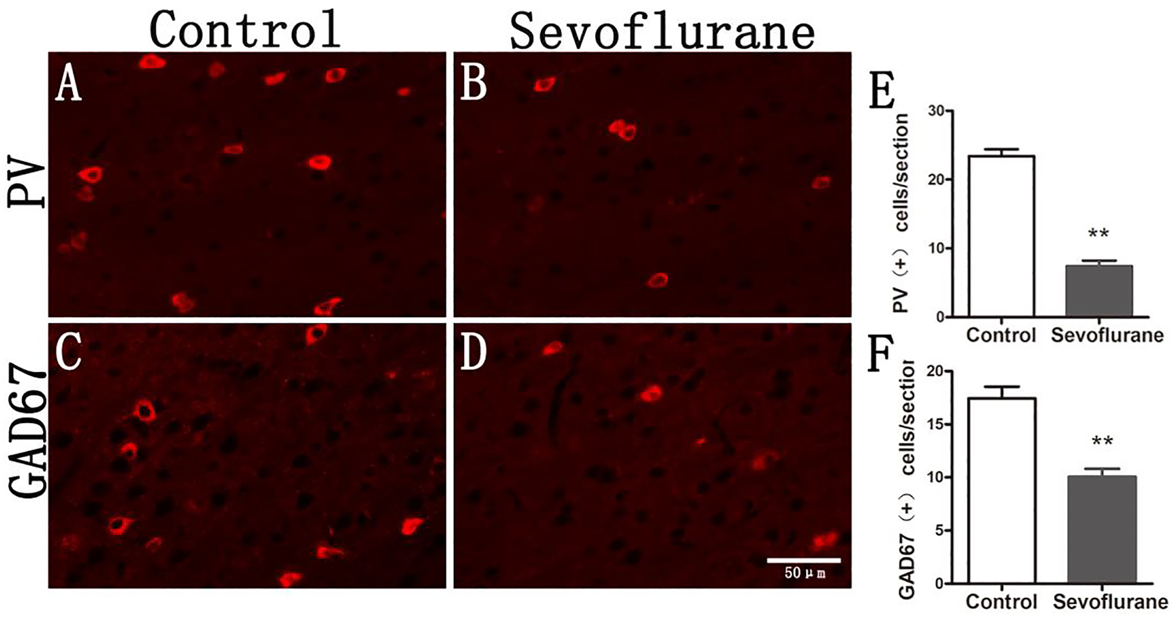
 ± 1.01, SeV group: 7.41 ± 0.82, p < 0.01) (Fig. 2). The number of GAD67 positive cells in the SeV group decreased significantly (control group: 17.44 ± 4.63, SeV group: 10.05 ± 3.17, p < 0.01) (Fig. 2).

Fig. 2.

The GABAergic interneurons of offspring in the LII/LIII entorhinal cortex were significantly reduced. PV and GAD67 positive cells were stained with red fluorescence, so in the inverted fluorescence microscope at 20X magnification, these cells are indicated by red fluorescence. (A) The PV positive cells in the LII/LIII entorhinal cortex of the P30 offspring in the control group. (B) PV immunofluorescence (red) on a P30 offspring rat in the sevoflurane group. (C) GAD67 immunofluorescence (red) on a P30 offspring rat in the control group. (D) GAD67 immunofluorescence (red) on a P30 offspring rat in the sevoflurane group. (E) The numbers of PV cells were significantly decreased in the sevoflurane compared to the control group. (F) The numbers of GAD67 cells were significantly decreased in the sevoflurane group compared to the control group. Note: The symbol ‘**’ represented there was significant difference between groups under the significant level of 0.01.

3.3 Prenatal exposure to sevoflurane leads to the abnormal expressions of NMDA receptor subunits in LII/LIII of the ECT of offspring

The results were as follows (Fig. 3): compared to the control group, the number of NR2A positive cells in the SeV group was decreased (control group: 24.00 ± 10.83, SeV group: 11.40 ± 10.10, p < 0.05) while the number of NR2B positive cells was significantly increased (control group: 15.61 ± 5.14, SeV group: 38.21 ± 8.50, p < 0.01).

Fig. 3.

Maternal exposure of sevoflurane leads to impaired maturation of NMDA receptors in offspring. NR2A and NR2B positive cells are indicated by green fluorescence. (A) The NR2A positive cells in the LII/LIII entorhinal cortex of the P30 offspring in the control group. (B) NR2A immunofluorescence (green) on a P30 offspring rat in the sevoflurane group. (C) NR2B immunofluorescence (green) on a P30 offspring rat in the control group. (D) NR2B immunofluorescence (green) on a P30 offspring rat in the sevoflurane group. (E) The numbers of NR2A cells were significantly decreased in the sevoflurane compared to the control group. (F) The numbers of NR2B cells were significantly increased in the sevoflurane compared to the control group. Note: The symbol ‘**’ represented there was significant difference between groups under the significant level of 0.01.

3.4 Prenatal sevoflurane exposure results in the abnormal development of the dendrites of pyramidal neurons

The results showed that compared to that in the control group, the number of dendritic spines of the pyramidal neurons in LII/LIII of the ECT in the SeV group was significantly increased (control group: 8.88
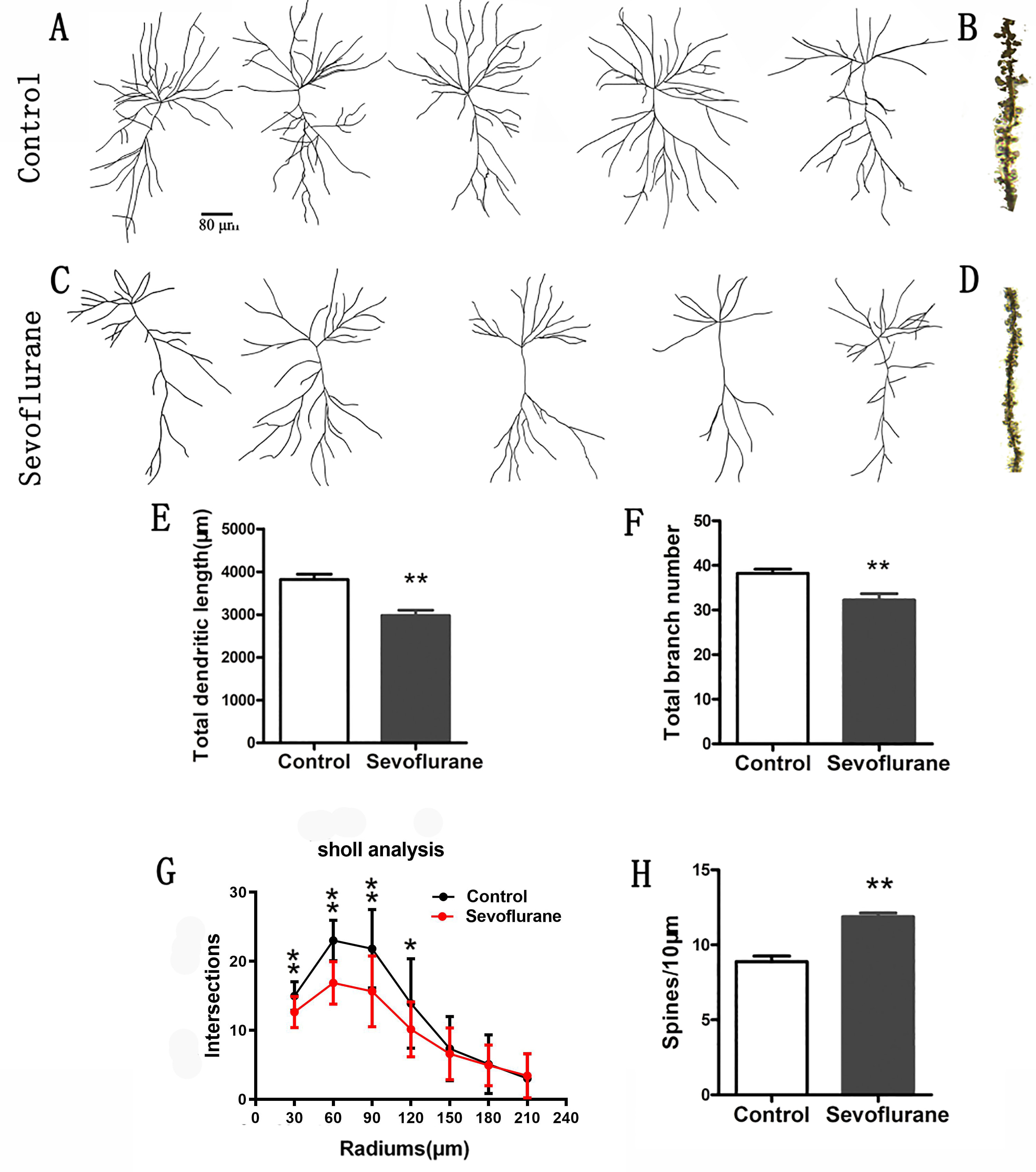
 ± 1.83, SeV group: 11.86 ± 1.27, p < 0.01), the total length of dendrites in the SeV group was significantly lower than that in the control group (control group: 3819.32 ± 614.99, SeV: 2978.45 ± 577.31, p < 0.01), and the number of dendrite branches in the SeV group was significantly lower than that in control group (control group: 38.24 ± 4.66, SeV group: 32.22 ± 6.88, p < 0.01) (see Fig. 4). Sholl analysis showed that the spatial distribution of dendrites was abnormal (p < 0.05, Fig. 4).

Fig. 4.

Sevoflurane exposure disturbs the maturation of pyramidal neurons in the LII/LIII entorhinal cortex of offspring. Six P30 brains in each group were processed for Golgi–Cox impregnation, and pyramidal neurons in the LII/LIII entorhinal cortex were studied in the control (examples in A) and sevoflurane (examples in C) groups. The left panels (A and C) show two examples of Golgi-impregnated neurons. The total branch number and dendritic length were significantly decreased in the sevoflurane group compared to the control group (E and F) (n = 24 neurons in each group). In addition, higher spine density was observed in the sevoflurane group had than the control group (see B, D, and H) (n = 24 dendrites in each group). Sholl analysis showed that the complexity of dendritic trees was lower in the sevoflurane group than the control group (G). Note: The symbol ‘*’ represented there was significant difference between groups under the significant level of 0.05. The symbol ‘**’ represented there was significant difference between groups under the significant level of 0.01.

4. Discussion

We previously speculated that prenatal sevoflurane, as an exogenous GABAA receptor agonist, down regulated the NRG1–ErbB4 signaling pathway and that this change could lead to the disturbance of interneurons and the abnormal development of the dendrites of pyramidal neurons. The results of this study basically confirmed our hypothesis. This could be helpful for the standardization of the clinical use of sevoflurane and its toxicity prevention and treatment.

With the development of surgical technology and fetal surgery, an increasing number of pregnant women need to be exposed to general anesthetics. Sevoflurane mainly acts as GABAA receptor agonist/enhancer and has sedative, analgesic, and muscle relaxant effects. Bartolini et al. [29] found that the inhalation of sevoflurane in 2MAC can inhibit uterine muscle contraction in a dose-dependent manner. Thus, based on its inhibition of uterine contraction, which can prevent premature delivery, sevoflurane is the most widely used inhalation anesthetic during pregnancy. Sevoflurane easily impacts fetuses though the placenta [30]. Zheng et al. [9] found that with the inhalation of 2.5% sevoflurane for 2 hours and 4.1% sevoflurane for 6 hours, the offspring of pregnant rat showed decreased learning and memory ability, accompanied by the release of inflammatory agents in the hippocampus and abnormal synaptic development. In recent years, a large number of studies have shown that sevoflurane it neurotoxic to the developing brain, which can lead to increased neuronal apoptosis, the inhibition of proliferation, neuronal development disorders, and long-term neurobehavioral abnormalities [31, 32]. However, most of the authors of these studies were focus on the hippocampus, which is related to learning and memory, and mainly studies the ultrastructural and functional impairment of projection neurons. Little attention has been paid to LII/LIII of the ECT, which is the key area of hippocampal information input. In LII/LIII of the ECT, interneurons, characterized as GABAergic neurons, could be regulated by sevoflurane. Impacts on the NRG1–ErbB4 pathway in interneurons may further regulate interneurons themselves and pyramidal neurons and affect the information transmission functions of the ECT.

In this study, we simulated clinical practice. The pregnant rats were anesthetized with 4% sevoflurane for 3 hours at 14.5 days of gestation to reach a MAC of 1.8 (lower than normal rats, considering that pregnant rats are more sensitive to sevoflurane), which was equivalent to that of most surgical operations for pregnant females. We found that sevoflurane, as an exogenous GABAA receptor agonist, could down regulate the NRG1–ErbB4 signaling pathway (Fig. 2). NRG1 is highly expressed in mammalian embryos and decreases with age. It involves many aspects of neural development, including neuronal migration, survival, axon projection, myelin sheath development, synaptic formation and the regulation of neurotransmitter receptor expression [13, 16, 33, 34, 35, 36, 37]. Previous studies have revealed that the NRG1–ErbB4 pathway is necessary for the generation and migration of intermediate neurons originating from the medial ganglionic eminence (MGE) region [16, 38, 39, 40]. The impairment of the NRG1–ErbB4 pathway can affect the migration of PV interneurons from the MGE region. PV interneurons account for 40% of interneurons, and there was a decrease of 30–50% of interneurons in ErbB4 gene knockout rats. We administered sevoflurane anesthesia during middle pregnancy, which is the critical period of the development of the generation and migration of the embryonic interneurons. In this period, the MGE area produces PV interneurons, which migrate to the edge of the cortex and subventricular area and then radially to the target cortex [29]. Our results also showed that the NRG1–ErbB4 pathway was down regulated along with a decrease in the number of interneurons. Therefore, the decrease in the number of interneurons in LII/LIII of the ECT in the SeV group could have been due to the inhibition of the formation of interneurons by the decreased NRG1–ErbB4 level. At the same time, the decreased NRG1–ErbB4 blocked interneurons’ migration to LII/LIII of the ECT.

The NMDA receptor (NMDAR) is mainly composed of two NR1 and two NR2 subunits. The distribution of NMDAR subunits (NR1, NR2, NR3) also changes with the process of neurodevelopment. NR1 begins to increase after birth until puberty and reaches a peak level in the third week after birth. NR2B and NR2D are the main subunits of NR2 in the embryonic stage. The expression of NR2 changes significantly in the two weeks after birth. NR2A begins to partially replace NR2B until the brain matures. The development of NMDAR subunits marks the transition from immature to mature neurons and is closely related to the development of learning and memory ability. In our experiment, exposure to sevoflurane was found to decrease the expression of NR2A and increase the expression of NR2B in offspring, which is consistent with the findings of previous studies [31, 32]. NRG1–ErbB4 changes could increase the phosphorylation of NR2B by Fyn and, thus, reduce the internalization of NR2B and delay the transition from NR2B to NR2A. NRG1–ErbB4 can regulate not only the release and activity of GABAergic neurotransmitters but also the excitatory synapses on inhibitory neurons (interneurons). The intracellular segment of the ErbB4 protein contains post-synaptic postsynaptic dens 95 (PSD-95)/DiscsLarge/zonula occludens protein-1 (ZO1) (PDZ) domains, which are anchored to the postsynaptic membrane by interacting with other proteins (such as PSD-95) that also contain PDZ [41, 42, 43]. Thus, ErbB4 regulates the function of the NMDAR by the connection of PDZ [41, 42, 43]. The NRG1–ErbB4 pathway phosphorylates the phosphorylation site of NR2B through a member of the Src family of Fyn (SRC/Fyn), which blocks the internalization of NR2B by protein AP-2 [44, 45]. In this way, the NRG1–ErbB4 pathway increases the expression of NR2B on the postsynaptic membrane [44, 45]. Thus, the decreased NRG1–ErbB4 level results in increased NR2B and decreased NR2A levels (Fig. 3). This alteration may lead to the abnormal development of learning and memory ability.

Pyramidal neurons are responsible for information transmission, and their function is very important. Therefore, it has always been a research hotspot in the topic of the neurotoxicity of general anesthetics during development. Interneurons play an important role in regulating pyramidal neurons. We found that prenatal sevoflurane exposure in middle pregnancy resulted in a significant increase in the number of dendritic spines, a significant decrease in the total length of dendrites, and an abnormal spatial distribution of dendrites in offspring (Fig. 4). There could be three possible mechanism behind the increased number of dendritic spines. One explanation is that the decrease in the number of interneurons leads to the weakening of their regulation of pyramidal neurons, resulting in abnormal numbers of dendritic spines. The second explanation is that the change of the NRG1–ErbB4 pathway could regulate the dendritic spines of pyramidal neurons. Barros et al. [35] found that the number of pyramidal dendritic spines decreased after ErbB4 and ErbB2 knockout in the nervous system. However, the number of pyramidal dendritic spines did not decrease in the experiment of ErbB4 knockout by pyramidal cells [44, 46]. It is suggested that ErbB2 may play a role of functional compensation, and there is an over-compensation in the number of pyramidal dendritic spines [37, 47]. The third explanation is that the NR2B subunit of NMDAR combines with PSD-95 and calmodulin-dependent kinase II (CaMKII), which activates a series of downstream signaling pathways and leads to an increase in the number of dendritic spines [48]. The NR2B/PSD95/kalirin-7 pathway is very important in the development of neuronal dendritic spines. Recent studies have shown that PSD-95, kalirin-7, and NR2B form a complex with a postsynaptic membrane through the PDZ domain. NR2B can activate kalirin-7, and then activate Rac1, a downstream RhoGTPase family member [49]. In this way, NR2B could dynamically regulate actin cytoskeleton rearrangement, promote the growth of dendritic spines, induce the formation of spinous structures in neuronal bodies, and cause the excessive formation of dendritic spines in pyramidal neurons and interneurons [49]. The decrease of total length and branches of dendrites in offspring may be explained by the change of the NRG1–ErbB4 signaling pathway and NR2B subunit, which mainly involve the growth and pruning of dendrites [36, 37]. Further, the plasticity of dendritic spines affects learning and memory function, especially the formation of long-term memory [50, 51].

The initial experimental hypothesis has been verified in this study. However, to test the hypothesis, this study focused on the study of the ECT with a concentration on the memory transfer station. Thus, this study did not involve an investigation of the dentate gyrus area of the hippocampus, which was directly projected by pyramidal neurons in the ECT. The time (3 h) for sevoflurane exposure may be too long in actual clinical situation. However, despite the hard exploratory work we’ve done, there is a few results we assumed except for that sevoflurane exposed to fetal brain for at least 3 hours or neonatal brains for more than 6 hours were neurotoxic. These results suggested that the safe concentration and exposure time in most clinical practice is safe.

5. Conclusions

Prenatal sevoflurane exposure in middle pregnancy could lead to the disorder of the interneurons by activating the GABAA receptor and its NRG1–ErbB4 pathway. In this way, prenatal sevoflurane leads to the abnormal dendrite development of the pyramidal neurons in the ECT of offspring rats and, thus, may interfere with the process of information transmission from the ECT to the hippocampus. This study indicated a possible novel neurotoxic pathway in the influence of sevoflurane on ECT of rat offspring. In clinical practice, the concentration of sevoflurane is much lower than the concentration in this study because of the addition of other auxiliary drugs such as opioid sedatives. Secondly, because of the advancement of surgical procedures, most non-obstetric procedures during pregnancy do not require 3 hours. Therefore, the current clinical use of sevoflurane is safe. However, prolonged exposure to high concentrations of sevoflurane still needs to be alert to neurotoxicity.

Abbreviations

NMDA, N-methyl-D-aspartate; GABA, gamma-aminobutyric acid; CNS, central nervous system; GABAA, GABA type A; LII and LIII, layers II and III; NRG1, Neuregulin-1; ErbB4, epidermal growth factor receptor-4; PV, parvalbumin; SD, Sprague–Dawley; SeV, sevoflurane; MAC, minimum alveolar concentration; P30, the day 30 of postpartum; PBS, phosphate buffered saline; GAD67, glutamic acid decarboxylase 67; NR2A, NMDA receptor subunit 2A; NR2B, NMDA receptor subunit 2B; BSA, bovine serum albumin; MGE, medial ganglionic eminence region; NMDAR, NMDA receptor; PDZ, PSD-95/DiscsLarge/ZO1; PSD-95, postsynaptic dens 95; ZO1, onula occludens protein-1; SRC/Fyn, Src family of Fyn; CaMKII, calmodulin-dependent kinase II.

Author contributions

XS, YG and TZ designed research; YG, TZ, YC, ZS, ZS and JL performed experiments; YC analyzed data; YG wrote the paper; TZ and XS critically revised the paper.

Ethics approval and consent to participate

This experiment was approved by the Animal Ethics Committee of Guangzhou Medical University, and the researchers strictly followed the relevant provisions of the “Guidelines for the Care and Use of Laboratory Animals” issued by the National Institutes of Health in 1996 (ethic code: 2016-029).

Acknowledgment

We thank Xiaolong Zeng for assistance in manuscript revision preparation.

Funding

This work was granted by the National Natural Science Foundation of China (Granted no. 81870823).

Conflict of interest

The authors declare no conflict of interest.

References
[1]
Okeagu CN, Anandi P, Gennuso S, Hyatali F, Stark CW, Prabhakar A, et al. Clinical management of the pregnant patient undergoing non-obstetric surgery: Review of guidelines. Best Practice & Research Clinical Anaesthesiology. 2020; 34: 269–281.
[2]
Jevtovic-Todorovic V, Absalom AR, Blomgren K, Brambrink A, Crosby G, Culley DJ, et al. Anaesthetic neurotoxicity and neuroplasticity: an expert group report and statement based on the BJA Salzburg Seminar. British Journal of Anaesthesia. 2013; 111: 143–151.
[3]
Grasshoff C, Antkowiak B. Propofol and Sevoflurane Depress Spinal Neurons in Vitro via Different Molecular Targets. Anesthesiology. 2004; 101: 1167–1176.
[4]
Ben-Ari Y. Excitatory actions of gaba during development: the nature of the nurture. Nature Reviews Neuroscience. 2002; 3: 728–739.
[5]
Wang DD, Kriegstein AR. Defining the role of GABA in cortical development. Journal of Physiology. 2009; 587: 1873–1879.
[6]
Palanisamy A. Maternal anesthesia and fetal neurodevelopment. International Journal of Obstetric Anesthesia. 2012; 21: 152–162.
[7]
Bjarnadottir M, Misner DL, Haverfield-Gross S, Bruun S, Helgason VG, Stefansson H, et al. Neuregulin1 (NRG1) Signaling through Fyn Modulates NMDA Receptor Phosphorylation: Differential Synaptic Function in NRG1+/- Knock-Outs Compared with Wild-Type Mice. Journal of Neuroscience. 2007; 27: 4519–4529.
[8]
Istaphanous GK, Ward CG, Nan X, Hughes EA, McCann JC, McAuliffe JJ, et al. Characterization and quantification of isoflurane-induced developmental apoptotic cell death in mouse cerebral cortex. Anesthesia and Analgesia. 2013; 116: 845–854.
[9]
Zheng H, Dong Y, Xu Z, Crosby G, Culley D, Zhang Y, et al. Sevoflurane Anesthesia in Pregnant Mice Induces Neurotoxicity in Fetal and Offspring Mice. Anesthesiology. 2013; 118: 516–526.
[10]
Briner A, De Roo M, Dayer A, Muller D, Habre W, Vutskits L. Volatile anesthetics rapidly increase dendritic spine density in the rat medial prefrontal cortex during synaptogenesis. Anesthesiology. 2010; 112: 546–556.
[11]
Zhao T, Li Y, Wei W, Savage S, Zhou L, Ma D. Ketamine administered to pregnant rats in the second trimester causes long-lasting behavioral disorders in offspring. Neurobiology of Disease. 2014; 68: 145–155.
[12]
Corfas G, Roy K, Buxbaum JD. Neuregulin 1-erbB signaling and the molecular/cellular basis of schizophrenia. Nature Neuroscience. 2004; 7: 575–580.
[13]
Mei L, Xiong W. Neuregulin 1 in neural development, synaptic plasticity and schizophrenia. Nature Reviews. Neuroscience. 2008; 9: 437–452.
[14]
Liang D, Fan F, Ding W, Fang Y, Hu L, Lei B, et al. Increased Seizure Susceptibility for Rats Subject to Early Life Hypoxia might be Associated with Brain Dysfunction of NRG1-ErbB4 Signaling in Parvalbumin Interneurons. Molecular Neurobiology. 2020; 57: 5276–5285.
[15]
Harrison PJ, Weinberger DR. Schizophrenia genes, gene expression, and neuropathology: on the matter of their convergence. Molecular Psychiatry. 2005; 10: 40–68.
[16]
Anton ES, Ghashghaei HT, Weber JL, McCann C, Fischer TM, Cheung ID, et al. Receptor tyrosine kinase ErbB4 modulates neuroblast migration and placement in the adult forebrain. Nature Neuroscience. 2004; 7: 1319–1328.
[17]
Ozaki M, Tohyama K, Kishida H, Buonanno A, Yano R, Hashikawa T. Roles of neuregulin in synaptogenesis between mossy fibers and cerebellar granule cells. Journal of Neuroscience Research. 2000; 59: 612–623.
[18]
Zhao JJ, Lemke G. Selective disruption of neuregulin-1 function in vertebrate embryos using ribozyme-tRNA transgenes. Development. 1998; 125: 1899–1907.
[19]
Gu Z, Jiang Q, Fu AKY, Ip NY, Yan Z. Regulation of NMDA receptors by neuregulin signaling in prefrontal cortex. Journal of Neuroscience. 2005; 25: 4974–4984.
[20]
Ozaki M, Sasner M, Yano R, Lu HS, Buonanno A. Neuregulin-beta induces expression of an NMDA-receptor subunit. Nature. 1997; 390: 691–694.
[21]
Konradi C, Heckers S. Molecular aspects of glutamate dysregulation: implications for schizophrenia and its treatment. Pharmacology & Therapeutics. 2003; 97: 153–179.
[22]
Gabrieli D, Schumm SN, Vigilante NF, Meaney DF. NMDA Receptor Alterations after Mild Traumatic Brain Injury Induce Deficits in Memory Acquisition and Recall. Neural Computation. 2021; 33: 67–95.
[23]
Howe T, Blockeel AJ, Taylor H, Jones MW, Bazhenov M, Malerba P. NMDA receptors promote hippocampal sharp-wave ripples and the associated coactivity of CA1 pyramidal cells. Hippocampus. 2020; 30: 1356–1370.
[24]
Bartolini G, Ciceri G, Marín O. Integration of GABAergic interneurons into cortical cell assemblies: lessons from embryos and adults. Neuron. 2013; 79: 849–864.
[25]
Li X, Su F, Ji M, Zhang G, Qiu L, Jia M, et al. Disruption of hippocampal neuregulin 1-ErbB4 signaling contributes to the hippocampus-dependent cognitive impairment induced by isoflurane in aged mice. Anesthesiology. 2014; 121: 79–88.
[26]
Clancy B, Darlington RB, Finlay BL. Translating developmental time across mammalian species. Neuroscience. 2001; 105: 7–17.
[27]
Ihmsen H, Schywalsky M, Plettke R, Priller M, Walz F, Schwilden H. Concentration-effect relations, prediction probabilities (Pk), and signal-to-noise ratios of different electroencephalographic parameters during administration of desflurane, isoflurane, and sevoflurane in rats. Anesthesiology. 2008; 108: 276–285.
[28]
Sholl DA. Dendritic organization in the neurons of the visual and motor cortices of the cat. Journal of Anatomy. 1953; 87: 387–406.
[29]
Bartolini G, Ciceri G, Marín O. Integration of GABAergic interneurons into cortical cell assemblies: lessons from embryos and adults. Neuron. 2013; 79: 849–864.
[30]
Chai D, Cheng Y, Jiang H. Fundamentals of fetal toxicity relevant to sevoflurane exposures during pregnancy. International Journal of Developmental Neuroscience. 2019; 72: 31–35.
[31]
Wang W, Jia L, Luo Y, Zhang H, Cai F, Mao H, et al. Location- and Subunit-Specific NMDA Receptors Determine the Developmental Sevoflurane Neurotoxicity through ERK1/2 Signaling. Molecular Neurobiology. 2016; 53: 216–230.
[32]
Zhang X, Shen F, Xu D, Zhao X. A lasting effect of postnatal sevoflurane anesthesia on the composition of NMDA receptor subunits in rat prefrontal cortex. International Journal of Developmental Neuroscience. 2016; 54: 62–69.
[33]
Falls DL. Neuregulins: functions, forms, and signaling strategies. Experimental Cell Research. 2003; 284: 14–30.
[34]
Buonanno A, Fischbach GD. Neuregulin and ErbB receptor signaling pathways in the nervous system. Current Opinion in Neurobiology. 2001; 11: 287–296.
[35]
Barros CS, Calabrese B, Chamero P, Roberts AJ, Korzus E, Lloyd K, et al. Impaired maturation of dendritic spines without disorganization of cortical cell layers in mice lacking NRG1/ErbB signaling in the central nervous system. Proceedings of the National Academy of Sciences. 2009; 106: 4507–4512.
[36]
Fazzari P, Paternain AV, Valiente M, Pla R, Luján R, Lloyd K, et al. Control of cortical GABA circuitry development by Nrg1 and ErbB4 signalling. Nature. 2010; 464: 1376–1380.
[37]
Cooper MA, Koleske AJ. ErbB4 localization to interneurons: clearer insights into schizophrenia pathology. Biological Psychiatry. 2011; 70: 602–603.
[38]
Buonanno A. The neuregulin signaling pathway and schizophrenia: from genes to synapses and neural circuits. Brain Research Bulletin. 2010; 83: 122–131.
[39]
Fisahn A, Neddens J, Yan L, Buonanno A. Neuregulin-1 modulates hippocampal gamma oscillations: implications for schizophrenia. Cerebral Cortex. 2009; 19: 612–618.
[40]
Flames N, Long JE, Garratt AN, Fischer TM, Gassmann M, Birchmeier C, et al. Short- and long-range attraction of cortical GABAergic interneurons by neuregulin-1. Neuron. 2004; 44: 251–261.
[41]
Garcia RA, Vasudevan K, Buonanno A. The neuregulin receptor ErbB-4 interacts with PDZ-containing proteins at neuronal synapses. Proceedings of the National Academy of Sciences. 2000; 97: 3596–3601.
[42]
Krivosheya D, Tapia L, Levinson JN, Huang K, Kang Y, Hines R, et al. ErbB4-neuregulin signaling modulates synapse development and dendritic arborization through distinct mechanisms. Journal of Biological Chemistry. 2008; 283: 32944–32956.
[43]
Huang YZ, Won S, Ali DW, Wang Q, Tanowitz M, Du QS, et al. Regulation of neuregulin signaling by PSD-95 interacting with ErbB4 at CNS synapses. Neuron. 2000; 26: 443–455.
[44]
Wang JQ, Guo M, Jin D, Xue B, Fibuch EE, Mao L. Roles of subunit phosphorylation in regulating glutamate receptor function. European Journal of Pharmacology. 2014; 728: 183–187.
[45]
Nakazawa T, Komai S, Tezuka T, Hisatsune C, Umemori H, Semba K, et al. Characterization of Fyn-mediated tyrosine phosphorylation sites on GluR epsilon 2 (NR2B) subunit of the N-methyl-D-aspartate receptor. Journal of Biological Chemistry. 2001; 276: 693–699.
[46]
Alkire MT, Gruver R, Miller J, McReynolds JR, Hahn EL, Cahill L. Neuroimaging analysis of an anesthetic gas that blocks human emotional memory. Proceedings of the National Academy of Sciences. 2008; 105: 17221727.
[47]
Yin D, Sun X, Bean JC, Lin TW, Sathyamurthy A, Xiong W, et al. Regulation of spine formation by ErbB4 in PV-positive interneurons. Journal of Neuroscience. 2013; 33: 19295–19303.
[48]
Tolias KF, Bikoff JB, Burette A, Paradis S, Harrar D, Tavazoie S, et al. The Rac1-GEF Tiam1 couples the NMDA receptor to the activity-dependent development of dendritic arbors and spines. Neuron. 2005; 45: 525–538.
[49]
Penzes P, Jones KA. Dendritic spine dynamics–a key role for kalirin-7. Trends in Neurosciences. 2008; 31: 419–427.
[50]
Xu T, Yu X, Perlik AJ, Tobin WF, Zweig JA, Tennant K, et al. Rapid formation and selective stabilization of synapses for enduring motor memories. Nature. 2009; 462: 915–919.
[51]
Tschida K, Mooney R. Deafening Drives Cell-Type-Specific Changes to Dendritic Spines in a Sensorimotor Nucleus Important to Learned Vocalizations. Neuron. 2012; 73: 1028–1039.
Share
Back to top