IMR Press / FBL / Volume 27 / Issue 4 / DOI: 10.31083/j.fbl2704121
Open Access Review
CAR-T Cells Targeting Immune Checkpoint Pathway Players
Show Less
1 Promab Biotechnologies, Richmond, CA 94806, USA
*Correspondence: vita.gol@promab.com (Vita Golubovskaya)
Academic Editor: Graham Pawelec
Front. Biosci. (Landmark Ed) 2022, 27(4), 121; https://doi.org/10.31083/j.fbl2704121
Submitted: 26 January 2022 | Revised: 15 March 2022 | Accepted: 17 March 2022 | Published: 2 April 2022
Copyright: © 2022 The Author(s). Published by IMR Press.
This is an open access article under the CC BY 4.0 license.
Abstract

CAR (Chimeric antigen receptor)-T cell therapy has become a very promising type of immunotherapy against hematological cancers. This report is focused on CAR-T cells targeting immune checkpoint proteins expressed on tumor cells. The CD70, CD47, CD80, CD86, B7H3, B7H4, PDL-1, TIGIT CAR-T cells and other CAR-T cells are discussed as an effective approach to deplete tumor cells expressing checkpoint proteins. CAR-T cell therapy targeting checkpoint pathways is a promising therapy to decrease inhibitory signaling pathways. The review highlights future directions and perspectives in CAR-T cells targeting immune checkpoint pathways.

Keywords
CAR-T cell
tumor
checkpoint
immunotherapy
review
1. Introduction

CAR-T cell therapy is a therapy using antibody-based chimeric antigen receptor expressing T cells targeting cell surface proteins on tumor cells. CAR-T cell therapy is an effective approach against hematological and solid cancers [1, 2, 3, 4, 5, 6]. CAR-T cell or T body cell was first described by Zelig Eshhar and his colleagues at the Weizmann Institute of Science in Israel [7, 8, 9, 10, 11, 12]. Several CAR-T cells targeting CD19 and BCMA for hematological cancers were recently approved by FDA for usage in clinic [13]. CAR includes a single chain variable fragment (ScFv) of antibody which binds tumor antigen [14]. CAR-T cells transduce anti-tumor activity through costimulatory domain and activation domain signaling [3, 15, 16].

Once CAR-T cell binds to a tumor antigen, the CAR-T cell proliferation and expansion is activated, turning on CAR-T cell cytotoxic functions and signaling accompanied with secretion of cytokines, granzymes causing tumor cell death [5, 12]. There are several challenges for CAR-T cell use against solid tumors such as repressive microenvironment, exhaustion of CAR-T cells, upregulation of inhibitory checkpoint pathways and delivery of CAR-T cells to the tumor site. This review describes CAR-T cells targeting checkpoint pathway molecules to effectively kill tumors.

There are many antigens expressed in hematological cancers which are targeted by CAR-T cells such as CD19, CD22, CD37, CD20, CD33, BCMA [6] and in solid cancers - mesothelin, EGFR (Epidermal growth factor receptor), Her-2 (human epidermal growth factor receptor 2), PSMA (prostate-specific membrane antigen), CD133, CD47, ROR-1 (receptor tyrosine kinase-like orphan receptor 1), MUC-1 (mucin 1), CEA (carcinoembryonic antigen) [17] and other [1]. Many tumors overexpress immune checkpoint protein players such as PD-L1, CD112, CD155, B7H3, B7H4, CD80, CD86 affecting immune response signaling as reviewed in [18, 19]. In this review we will describe main checkpoint proteins and specifically focus on CAR-T cells targeting these checkpoint proteins.

We will also highlight several CAR-T cells targeting checkpoint molecules on other types of immune cells such as macrophages, Treg or NK cells. We will provide future directions for the field.

2. CAR Structure

The CAR structure includes ScFv, hinge, transmembrane, co-stimulatory (CD28, CD137 (4-1BB), and activation domain such as CD3 zeta (Fig. 1). The ScFv consists of variable light (VL) and heavy (VH) chains of antibody fused in frame with the linker either in VL-linker-VH or VH-linker-VL format. The first generation of CAR had one CD3 domain; the second generation of CAR had two costimulatory domains and one CD3 activation domain; and the third generation of CAR had two costimulatory domains and CD3 zeta activation domain, as shown in Fig. 1.

Fig. 1.

CAR-T cell structures. First, second, third, fourth and fifth generation of CARs. ScFv, single chain variable fragment; VH-variable heavy chain; VL, variable light chain, hinge, transmembrane, CD28, 4-1BB or other costimulatory domains, CD3 zeta activation domain. The first generation of CAR included one CD3 zeta activation domain; the second generation of CAR included one costimulatory domain and one CD3 zeta activation domain. The third generation of CAR includes two costimulatory domains (for example, CD28 and 4-1BB) and one activation CD3 zeta domain. The fourth generation of CAR included second generation of CAR with secreted cytokine or chemokine protein. The fifth generation of CAR included additional expression of receptor domain for example IL-2 receptor chain beta stimulating STAT3/STAT5 signaling.

The fourth generation of CAR includes addition of secreted or membrane-bound cytokines such as IL-15, IL-12, IL-18, IL-21, and IL-23 using either T2A, F2A, P2A self-cleaving peptides, or adding IRES (internal ribosome entry site), using a separate promoter [20, 21, 22, 23] or using gene-editing technology to insert additional cargo players [24]. These CAR-T cells can express cytokine proteins such as IL-12. The fifth generation CAR-T cells includes additional intracellular domain of cytokine receptors such as IL-2 receptor beta chain (IL-2 R β) inducing intracellular signaling such as STAT3/5 [25, 26].

The future designs of CAR will include CARs with more safety and higher efficacy. For example, CAR-T cells can secrete bispecific antibody to increase their efficacy by targeting tumor and tumor microenvironment [27, 28]. One elegant study used CAR-T cells secreting BITE bispecific antibody targeting fibroblast activation protein alpha, FAP with pro-tumorigenic role in tumor microenvironment [28].

One of the challenges in targeting solid tumors is on-target off-tumor toxicity due to expression of tumor antigens at low levels in normal tissues [29]. Thus, different “off switches” can be introduced to the CAR structure in order to deplete CAR-T cells in case of toxicity. The CAR structure can include different safety switches such as inducible caspase-9 suicide switch [30, 31], truncated EGFR (t-EGFR), RQR8 [32] or other switches to increase CAR-T cell safety. As an example, the inducible caspase-9 switch can eliminate majority of CAR-T cells within 30 minutes providing increased safety in clinic [33]. The switches are important for decreasing on-target off-tumor potential toxic effects in clinical studies.

3. Immune Checkpoints

T cells receive either costimulatory or inhibiting immune signals through interaction of immune receptors and ligands, which are called immune checkpoints. The costimulatory or inhibitory checkpoint signaling interaction and pathways between tumor and immune cells are shown on Fig. 2. The main costimulatory immune checkpoints include: CD80, CD86 ligands and CD28 receptor; CD70 ligand and CD27 receptor; OX40 ligand and OX40 receptor; CD137 or 41BB ligand and CD137 (4-1BB) receptor, ICOS ligand and ICOS receptors (Fig. 2, left panel). The main inhibitory immune checkpoints are VISTA; B7H3; B7H4 ligands/receptors; PD-L1/PD-L2/PD-1; CD80, CD86/CTLA-4; CD112/CD155/TIGIT; Gal-9/TIM3; MHC-peptide/LAG-3/KIR (Fig. 2). The different antibodies targeting inhibitory checkpoint pathways were developed in clinic such as FDA-approved Pembrolizumab (Keytruda), Nivolumab (Opdivo) targeting PD-1; ipilimumab (Yervoy) targeting CTLA-4; Avelumab (Bavencio), Atezolizumab (Tecentriq) targeting PD-L1. Tumors use the checkpoint pathways to avoid autoimmunity during normal conditions [34].
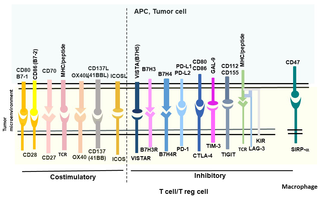
Fig. 2.

Costimulatory and inhibitory checkpoint pathways. T/Treg cells and macrophages are immune cells with checkpoint receptors, and tumor cells express ligands.

The tumor microenvironment (TEM) includes vascular, stromal cells, myeloid cells, fibroblasts and extracellular matrix (ECM) proteins [34]. The combination therapy approach to target tumor and also tumor microenvironment is an effective approach for anti-cancer therapy [35]. In addition, combination of different checkpoint inhibitors such as FDA-approved anti-CTLA-4 and anti-PD1 have been proven to be more effective in anti-cancer therapy in clinic [18, 36, 37].

The combination of CAR-T cells with checkpoint inhibitors such as PD-1 or PD-L1 can be used to overcome inhibitory tumor microenvironment by incorporating either PD-1 with CD28 or other co-stimulatory domains or dominant-negative PD-1 receptor with deleted transmembrane and inhibitory intracellular domains [35] or using secreted PD-1 ScFv [38] or silenced PD-1 expression. PD1 dominant-negative receptor engineered into mesothelin CAR structure competed with endogenous PD-1 and saturated PD-L1 expressed on tumor cells preventing inhibitory PD-1/PD-L1 signaling [35].

The “don’t eat me”, CD47/SIRP-1 alpha macrophage immune checkpoint [39, 40, 41, 42, 43, 44, 45, 46, 47, 48] is a very important pathway for regulating survival signaling. The detailed description of each checkpoint signaling regulating recognition of “self” and “not-self” between immune cells and tumor cells is included in the review [18] and in several other reports [34, 49, 50, 51, 52].

4. CAR-T Cells Targeting Immune Costimulatory Checkpoints
4.1 CD80, CD86/CD28 Co-Stimulatory and CD80, CD86/CTLA-4 Inhibitory Signaling and CAR-T Cells

The detailed CD80, CD86, CD28, CTLA-4 signaling is discussed in [53, 54, 55, 56, 57]. CD80 (B7-1) is a 33 kDa, 288 amino acid immunoglobulin protein encoded by CD80 gene. CD86 (B7-2) is a 70 kDa, 329 amino-acid immunoglobulin protein encoded by CD86 gene. CD80 and CD86 have been shown to be overexpressed in several types of cancer [54, 58]. The costimulatory ligands CD80 (B7-1) and CD86 (B7-2) are expressed on antigen presenting cells (APC) mediating either costimulatory signaling through CD28 receptor or inhibitory signaling through the cytotoxic T lymphocyte-associated antigen (CTLA-4) (CD152) receptor [59, 60] (Fig. 2). Expression of CD80 and CD86 has been shown to be regulated by epigenetic mechanism such as promoter methylation [54]. Recent report demonstrates that T regulatory cells (or Treg cells) constitutively expressed a cytotoxic T-lymphocyte-associated antigen-4 (CTLA-4) which down-regulated expression of CD80/CD86 co-stimulatory molecules on antigen-presenting cells by CTLA-4-dependent trogocytosis [61]. In addition, decreased CD80 expression by Treg-membranous CTLA-4 blocked cisCD80/PD-L1 heterodimers [61]. The authors suggested that simultaneous inhibition of CTLA-4 and PD-1/PD-L1 can enhance immune responses [61].

A recent study detected that CTLA-4-CD28-CAR-T cells effectively targeted CD80/CD86-positive cancer cells [62]. CTLA4-CAR-T cells effectively killed CD80- or CD86-positive tumor cells (Raji, RL, and NALM6), but not CD80/CD86-negative K562 cells [62]. CTLA-4-CAR-T cells decreased xenograft tumor growth associated with targeting of myeloid-derived suppressor cells (MDSCs) without cytokine release syndrome (CRS) [62]. Thus, authors managed to convert negative CTLA-4-CD80/CD86 signaling to tumor-killing mechanism.

4.2 CD70, CD27 Signaling and CD70-CAR-T Cells

CD70, a member of the tumor necrosis factor superfamily, has been expressed in hematological and solid tumors [63]. CD27 is a receptor for CD70 and provides costimulatory signaling to T cells by inducing cellular proliferation and survival signaling [64, 65, 66, 67]. CD70 has been shown to be overexpressed in mantle cell lymphoma, follicular lymphoma, diffuse large B-cell lymphoma, Hodgkin’s lymphoma, Waldenström macroglobulinemia, and multiple myeloma, but not in normal B cells or T cells [68, 69]. In addition, immunohistochemical staining analysis of CD70 expression in 25 different solid tumor types (N = 496 tissue samples) demonstrated that 43% of solid tumors had highest CD70 expression in renal cell carcinoma (79.5% positivity) and no expression in Kaposi sarcoma [69]. Lymphoma samples demonstrated 58% positivity [69].

In one report CD70-CAR-T cells were generated and demonstrated cytotoxic activity against CD19-negative lymphoma [68]. In other reports CD70-CAR-T cells effectively targeted glioblastoma tumors [70], and also melanoma [71]. Recent report demonstrated effective targeting of CD70 by CD70-CAR-T cells in acute myeloid leukemia (AML) samples [72]. Thus, CD70 can be targeted by CAR-T cells in different hematological and solid tumors.

4.3 MHC-Peptide/LAG-3/KIR and CAR-T Cells

Intracellular tumor-related antigens can be presented as peptides in the major histocompatibility complex (MHC) on the cell surface interacting with the T cell receptors (TCR) on T cells. Many studies reported targeting MHC-peptide-TCR interactions to block tumor growth using different therapies such as T cell recruiting antibodies, TCR mimicking antibodies, antibody conjugates or autologous genetically-modified effector T cells [73, 74, 75, 76, 77]. The TCR complex contains TCR variable heterodimer (TCR alpha, beta), which binds to the MHC-peptide complex, and a CD3 component which includes gamma, delta, two epsilon and two zeta subunits [78]. Most TCRs tested in clinic were HLA-A2-based and directed against melanoma-associated antigens recognized by T cells such as (MART-1), glycoprotein (gp) 100, carcinoembryonic antigen (CEA), melanoma-associated antigen (MAGE-A3), New York esophageal squamous cell carcinoma antigen (NY-ESO1), or p53 [77]. The TCR mimicking antibodies were generated by either screening of phage display library or through immunizing mice with recombinant MHC-peptide complex which were then used for generating TCR-like CAR-T cells. Several TCR-like CAR-T cells were reported such as MAGE-A1 [79], ESO-1 [80], WT-1 [81], GP100 [82], HMHA1 [83]. CAR-T cells targeting alpha-fetoprotein (AFP)-MHC complex effectively decreased liver cancer growth [84, 85]. AFP-CAR-T cells had high efficacy using Hep G2 tumor xenograft NSG mice model [84]. AFP-CAR-T cells represent an example of CAR-T cells targeting intracellular proteins presented as peptides on the surface of MHC complex. This approach can be applied to other targets.

LAG-3 (CD223) is expressed on T cells, NK cells and Treg cells, binds to the MHC II-peptide complex and blocks function of immune cells [86, 87, 88]. The LAG3 gene is located on chromosome 12 in humans and on chromosome 6 in mice [88]. LAG-3 consists of four extracellular immunoglobulin superfamily-like domains (D1-D4) with additional 30 amino-acid loop in the D1 domain which binds to MHC class II molecules with 100-fold higher affinity than CD4 protein [88]. The extracellular domain of LAG-3 and also cytoplasmic domain have been shown to be involved in the inhibitory signaling of T cells [88, 89].

T cells and primarily NK cells express killer-cell inhibitory receptors (KIR) on their cell surface to bind MHC class I molecules (Fig. 2) [90]. Interaction of KIR with MHC I leads to cell immune surveillance and NK cell-mediated cytotoxicity [90]. Many types of tumors often down-regulate human leukocyte antigen HLA I to escape immune recognition [91]. Recently inhibitory CAR with KIR extracellular domain and intracellular PD-1 domain was introduced into CD19-CAR to decrease 19-CAR-T cell toxicity or “on-target off-tumor” effects [92]. These KIR/PD-1-based CAR-T cells had high efficacy and less toxicity against CD19-positive HLA-C1-negative Burkitt’s lymphoma and did not affect CD19-positive/HLA-C1-positive human B cells [92].

5. CAR-T Cells Targeting Inhibitory Immune Checkpoints
5.1 B7H3-CAR-T Cells

B7H3 (CD276) ligand has been reported to be overexpressed in hematological and solid tumors [93, 94, 95, 96, 97, 98]. B7H3 has been shown to be overexpressed in both tumor and stromal tissues, tumor vasculature, tumor-infiltrating dendritic cells, and macrophages [94]. B7H3 is a 316 amino-acid cell surface protein which is encoded by CD276 gene on chromosome 15 in humans and chromosome 9 in mice [99, 100]. B7H3-CAR-T cells demonstrated significant anti-tumor activity against pediatric solid tumors using Ewing sarcoma, osteosarcoma, or medulloblastoma mouse xenograft models in vivo [101]. In addition, B7H3-CAR-T cells also effectively targeted AML and melanoma tumors in vitro and in vivo using mouse xenograft models [102]. B7H3-CAR-T cells which were administered intracerebroventricularly or intratumorally and demonstrated effective anti-tumor activity against cerebral atypical teratoid/rhabdoid tumors (ATRTs) which are incurable tumors arising in the central nervous system of children under 3 years of age [103]. In addition, B7H3-CAR-T cells were highly effective in a clinical study by inhibiting glioblastoma tumor growth [104]. B7H3-CAR-T cells were also effective against skull chordomas [105]. Another study showed that both B7H3-CAR-T cells and bispecific B7H3-CD3 BITE antibody killed a rare and aggressive subtype of non-Hodgkin lymphoma, extranodal nasal natural killer/T cell lymphoma [106]. The authors compared effects of B7H3-CAR-T cells with B7H3-CD3 BITE antibody and demonstrated very promising results for both treatments.

5.2 B7H4-CAR-T Cells

B7H4 protein belongs to the B7 family proteins and encoded by VTCN1 gene [107]. The hB7H4 sequence encodes a putative protein of 282 amino acids containing several N-glycosylation sites in the extracellular domain [107]. The hB7H4 protein contains a large transmembrane domain and a very short two amino-acid intracellular domain [107]. B7H4 is often overexpressed in many types of solid tumors such as breast, glioma, lung, prostate, melanoma, esophageal, bladder, and particularly in ovarian tumors [108, 109, 110, 111, 112, 113]. B7H4 has very limited expression in normal tissues making it an attractive therapeutic target [109, 114]. B7H4 binding to B7H4 receptor leads to inhibiting T cell proliferation and blocking secretion of cytokines [107, 115]. The B7H4-CAR-T cells inhibited ovarian tumor growth using xenograft mouse model [108]. The authors demonstrate that B7H4-CAR T cells mediated off-tumor toxicity at later time points and suggested that this could be due to expression of B7H4 in healthy mouse tissues [108]. B7H4-CAR-T cells demonstrated high multiorgan infiltration of lymphocyte cells in B7H4-treated mouse model in vivo [108].

5.3 PD-L1/PD-1-CAR-T Cells

PD-L1 (B7-H1), programmed death ligand-1, 40 kDa transmembrane protein [55]. PD-L1 (B7-H1 or CD274) is overexpressed in many types of tumors such as gastric, lung, breast, ovarian, pancreatic, oral, head and neck, colorectal, brain, thyroid, liver, kidney, renal, melanoma, skin and hematological cancers [116, 117]. Overexpression of PD-L1 is correlated with worse prognosis in many cancers [117]. PD-1 (CD279) receptor is expressed on lymphocytes, NK cells, dendritic cells, B cells [118]. PD-1 receptor has two ligands of B7 family members, PD-L1 and PD-L2 [116]. The anti-PD-L1-CAR-T cells were generated with CAR vector containing PD-1 extracellular and transmembrane domains, 4-1BB and TLR2 costimulatory and CD3 zeta activation domains and also with CAR vector containing PD-L1 ScFv with the same costimulatory domains and CD3 zeta cytoplasmic domains significantly decreased tumor growth of PD-L1-positive solid tumors [119]. Both dominant-negative PD-1 CAR-T cells and PD-L1-CAR-T cells had synergistic effect in vivo. In addition, PD-L1-CAR-T cells also lysed PD-L1-positive T cells in xenograft model [119]. Another report demonstrated that dominant-negative PD-1 lacking transmembrane and inhibitory cytoplasmic domain engineered within CAR had high efficacy against solid tumors by blocking PD-L1/PD-L2 pathways [35]. In another report bispecific PD-L1/c-Met-CAR-T cells have been shown to effectively inhibit growth of hepatocellular carcinoma [120]. c-Met is a tyrosine kinase Met or hepatocyte growth factor receptor (HGFR) which is often overexpressed in many types of cancer including hepatocellular carcinoma [121, 122, 123, 124, 125]. One report showed that c-Met up-regulated transcription of PD-L1 through the MAPK/NF-kappa B pathway promoting the progress of hepatocellular carcinoma [124]. The bispecific PD-L1/c-Met-CAR-T cells targeting two antigens PD-L1 and c-Met had specific activity against PD-L1-positive, c-Met-positive hepatocellular carcinoma xenograft tumors [120]. Another report showed that PD-L1-CAR-T cells effectively inhibited PD-L1high non-small cell lung carcinoma (NSCLC) growth [126].

PD-L1 is overexpressed not only on tumors but also on tumor microenvironment cells such as infiltrating lymphocytes and myeloid cells [14]. The authors used nanobody which consisted of the variable regions of heavy-chain VHH to generate PD-L1-CAR-T cells. Nanobodies are small camelid-derived single domain antibodies which are small and stable with similar to ScFv affinity [14]. VHH doesn’t need folding and linker optimization between VH and VL that can be required for ScFv [14]. VHH is also less immunogenic than mouse ScFv and due to their small size can bind different epitopes [14] The authors demonstrated high efficacy of VHH-based PD-L1-CAR-T cells by targeting PD-L1 on tumor microenvironment using syngeneic melanoma B16 and colon adenocarcinoma MC38 xenograft mice models [14]. Thus, PD-L1 can be targeted with CAR-T cells on both tumors and tumor microenvironment. Generation of the PD-L1-targeted CAR-T cells did not result in fratricide, possibly due to sequestering of the PD-L1 by PD-1 on the T cell surface or due to low level of PD-L1 expression on T cells [14]. The authors were able to use immunocompetent mouse xenograft model with tumor and tumor microenvironment signaling and demonstrate high efficacy of the CAR-T cells.

5.4 CD112, CD155/TIGIT-CAR-T Cells

TIGIT (T cell immunoreceptor with Ig and ITIM domains that can be named as VSig9, Vstm3, or WUCAM) is a 244 amino-acid checkpoint molecule that belongs to the poliovirus receptor (PVR)/nectin family [127]. TIGIT binds and competes with immune-activator CD226 receptor (DNAM-1) for binding to CD112 and CD155 ligands [128, 129]. The TIGIT mediates immunosuppressive effects by binding to poliovirus receptor (CD155) and by modulating cytokine secretion by dendritic cells [127]. TIGIT is a promising target for cell therapy as it is overexpressed in multiple cancer types including colorectal cancer, melanoma, lung adenocarcinoma, breast, pancreatic, ovarian cancer, and glioblastoma and its expression is correlated with poor prognosis [130]. Dual PD-1/TIGIT inhibition effectively increased CD8+ T cell expansion and function in vitro and promoted tumor rejection in mouse tumor models [129]. Recent report demonstrated that a TIGIT-based chimeric costimulatory switch receptor (TIGIT-28) improved T cell-mediated anti-tumor function [130].

5.5 TIM-3-CAR-T Cells

T cell immunoglobulin and mucin domain-containing protein 3 (TIM3) encoded by HAVCR2 gene is a member of the TIM family of immunoregulatory proteins (reviewed in [131]). TIM3 has several ligands such as Galectin 9, CEACAM-1 and high mobility group protein B1 [131]. Galectin-9-TIM-3 interaction has been shown to inhibit immune response [131]. TIM3 is expressed on T cells, NK cells and Treg cells [131]. In addition, TIM-3 was shown to be overexpressed in hematopoietic tissues of acute myeloid leukemia (AML) patients [132]. TIM-3 was identified as an AML stem cell surface marker which more highly expressed in leukemia stem cells compared to normal bone marrow hematopoietic stem cells [132]. Recently bispecific CAR-T cells targeting CD13 and TIM3 have been shown to eliminate acute myeloid leukemia cells [133]. The bispecific CAR-T cells (Biss CAR-T cells) targeting CD13 and TIM3 had reduced toxicity compared to single CD13-CAR-T cells [133]. These CAR-T cells targeted tumor cells which expressed both TIM-3 and CD13 proteins but did not kill normal cells that only expressed CD13 [133].

6. Other CAR Immune Cells Targeting Immune Checkpoints

The same approaches can be applied to other immune cell types (macrophages, Treg cells, NK cells, Gamma Delta T cells, dendritic cells, allogenic T/NK cells) with their own pathways [134, 135].

One of the macrophage-related signaling pathways is a CD47-SIRP-alpha pathway. For example, recently CD47-CAR-T cells targeted CD47-positive pancreatic, ovarian and melanoma tumors [17, 136]. Another approach is to engineer CAR-macrophages against solid tumors [137]. The advantage of using macrophages is their effective homing to solid tumors and phagocytotic activity [137].

The same checkpoint pathways can be targeted with CAR-NK cells such as PD-L1/PD-1, KIR, CTLA-4, IL1R8 [138], LAG-3, NKG2A, TIGIT, TIM-3, Siglec 3,9, VISTA and CD161 (reviewed in [139, 140, 141, 142]). All immune checkpoint pathways of NK cells are reviewed in [139]. For example, PD-L1-CAR-NK have been shown to be effective against human and mouse head and neck cancers [143]. The authors demonstrated that PD-L1-CAR NK cells decreased myeloid cells expressing high level of PD-L1 in peripheral blood from patients with head and neck cancer [143].

CAR-gamma delta T cells demonstrated high efficacy against solid and hematological cancers without GHVD (graft vs host disease), and all checkpoint targets can be used for designing CARs similarly as for regular CAR-T cells [144]. Allogenic CAR-T and NK cells without GHVD can be generated using knock-out TRAC, B2M or PD-1 pathways with CRISPR, TALEN or other gene-editing technology [145]. CAR-Treg cells used for therapy are discussed in [146]. There is a cross-talk between T cells, dendritic cells and NK cells that needs to be explored in future studies. Combination therapy approach combining therapies targeting checkpoint pathways has been shown to be very effective to block tumor growth [147, 148, 149].

7. Conclusions and Perspectives

This report summarizes main checkpoint proteins and CAR-T cells targeting immune checkpoint pathways such as PD-L1/PD-1, B7H3, B7H4, MHC-peptide/LAG3/KIR, TIM-3, CD70 and other shown in Fig. 2. We presented CAR-T cells blocking PD-L1/PD-1, CD112, CD155/TIGIT, B7H3, HLA-peptide, B7H4 and TIM-3 pathways that effectively killed tumors. We presented CAR-T cells which targeted both tumors and tumor microenvironment. Different approaches were discussed such as increasing safety of CAR-T cells using different switches, designing fourth and fifth generation of CAR-T cells by increasing their efficacy by adding cytokine and chemokine secretion, knocking-out checkpoint receptor signaling either with secreted antibodies, ScFv, using dominant-negative checkpoint molecules or silencing RNA. We provided review of other immune cells such as gamma-delta T cells, NK cells, DC and macrophages with similar checkpoint pathways that can be used for designing CARs in these immune cells.

There are several examples of clinical trials that are recruiting patients or active in USA, China and other countries that involve CAR-T cells targeting immune checkpoint pathways (www.clinicaltrials.gov) such as CD19, with PD-1 knock-out engineered CAR-T cells against lymphoma (NCT04213469), mesothelin CAR-T cells with CRISPR-Cas-9 mediated PD-1 and TCR genes knocked-out against relapsed or refractory advanced solid malignancies (NCT03545815). The clinical trial study with B7H3-CAR-T cells recruits patients in Seattle Children’s Hospital, USA with diffuse midline glioma and recurrent or refractory pediatric central nervous system tumors (NCT04185038). Another B7H3-CAR-T cell study recruits patients in Lineberger Cancer Center, Chapel Hill, NC with ovarian cancer (NCT04670068). Several clinical trials using B7H3-CAR-T cells either alone or in combination with temozolomide recruit patients with recurrent glioblastoma in China (NCT05241392), (NCT04077866). Another study recruits patients with CD70-CAR-T cells against hematological cancers in China (NCT04662294).

Future clinical trials will provide more data on CAR-T cells targeting checkpoint pathways and reveal novel biomarkers involved in these pathways. The screening off-tumor on-target effects will be carefully evaluated by different genomics, proteomics, protein arrays and other approaches. The single cell RNAseq report demonstrated importance of genomics approaches to reveal on-target neurotoxicity of CD19-CAR-T cells [150]. The single cell genomic profiling of CAR-T cells before and after treatment will allow to develop biomarkers of response to therapy, resistance players and novel signaling players.

The novel humanized mouse models will be developed to address differences in signaling and targets between murine and human models. The biodistribution of target molecules and biomarkers needs to be carefully examined to more effectively kill solid tumors and provide the best route of CAR-T cell administration (for example, intratumoral, intralymph nodal, intraperitoneal or intravenous). The analysis of mechanisms and signaling driven by tumor microenvironment, involvement of different immune cell types such as NK cells, Treg, macrophages and dendritic cells and checkpoint molecular players will provide basis for more effective tumor killing. Developing biomarkers of response to CAR-T cells targeting checkpoint signaling will require combination of different therapy approaches.

The new ligands for immune receptors on tumor cells are discovered which can be used for CAR-T cells therapy. Novel different CAR-T cells targeting the immune checkpoint ligands on tumors and blocking receptors on immune cells will be developed. The combination of CAR-T cells with checkpoint inhibitors [151, 152], chemotherapy and epigenetics and genetics players will be used to increase efficacy of these therapies.

Author Contributions

VG conceptualized and wrote the manuscript.

Ethics Approval and Consent to Participate

Not applicable.

Acknowledgment

We would like to acknowledge all authors of reports whose studies were not included in this review. We would like to thank John Sienkiewicz for professional editing of the manuscript.

Funding

This was funded by Promab Biotechnologies.

Conflict of Interest

VG is an employee of Promab Biotechnologies. VG is serving as the guest editor of this journal. We declare that VG had no involvement in the peer review of this article and has no access to information regarding its peer review. Full responsibility for the editorial process for this article was delegated to GP.

References
[1]
Eshhar Z, Waks T, Gross G. The Emergence of T-Bodies/CAR T Cells. The Cancer Journal. 2014; 20: 123–126.
[2]
Maus MV. Designing CAR T cells for glioblastoma. OncoImmunology. 2015; 4: e1048956.
[3]
Maus MV, Grupp SA, Porter DL, June CH. Antibody-modified T cells: CARs take the front seat for hematologic malignancies. Blood. 2014; 123: 2625–2635.
[4]
Maus MV, Haas AR, Beatty GL, Albelda SM, Levine BL, Liu X, et al. T Cells Expressing Chimeric Antigen Receptors can Cause Anaphylaxis in Humans. Cancer Immunology Research. 2013; 1: 26–31.
[5]
Maus MV, June CH. Zoom Zoom: Racing CARs for Multiple Myeloma. Clinical Cancer Research. 2013; 19: 1917–1919.
[6]
Cheadle EJ, Gornall H, Baldan V, Hanson V, Hawkins RE, Gilham DE. CAR T cells: driving the road from the laboratory to the clinic. Immunological Reviews. 2014; 257: 91–106.
[7]
Gross G, Waks T, Eshhar Z. Expression of immunoglobulin-T-cell receptor chimeric molecules as functional receptors with antibody-type specificity. Proceedings of the National Academy of Sciences. 1989; 86: 10024–10028.
[8]
Gross G, Eshhar Z. Therapeutic Potential of T Cell Chimeric Antigen Receptors (CARs) in Cancer Treatment: Counteracting off-Tumor Toxicities for Safe CAR T Cell Therapy. Annual Review of Pharmacology and Toxicology. 2016; 56: 59–83.
[9]
Lanitis E, Poussin M, Hagemann IS, Coukos G, Sandaltzopoulos R, Scholler N, et al. Redirected antitumor activity of primary human lymphocytes transduced with a fully human anti-mesothelin chimeric receptor. Molecular Therapy. 2012; 20: 633–643.
[10]
Lanitis E, Poussin M, Klattenhoff AW, Song D, Sandaltzopoulos R, June CH, et al. Chimeric antigen receptor T Cells with dissociated signaling domains exhibit focused antitumor activity with reduced potential for toxicity in vivo. Cancer Immunology Research. 2013; 1: 43–53.
[11]
Klebanoff CA, Gattinoni L, Restifo NP. Sorting through subsets: which T-cell populations mediate highly effective adoptive immunotherapy? Journal of Immunotherapy. 2012; 35: 651–660.
[12]
Klebanoff CA, Yamamoto TN, Restifo NP. Immunotherapy: Treatment of aggressive lymphomas with anti-CD19 CAR T cells. Nature Reviews Clinical Oncology. 2014; 11: 685–686.
[13]
Marple AH, Bonifant CL, Shah NN. Improving CAR T-cells: the next generation. Seminars in Hematology. 2020; 57: 115–121.
[14]
Xie YJ, Dougan M, Jailkhani N, Ingram J, Fang T, Kummer L, et al. Nanobody-based CAR T cells that target the tumor microenvironment inhibit the growth of solid tumors in immunocompetent mice. Proceedings of the National Academy of Sciences. 2019; 116: 7624–7631.
[15]
June CH, O’Connor RS, Kawalekar OU, Ghassemi S, Milone MC. CAR T cell immunotherapy for human cancer. Science. 2018; 359: 1361–1365.
[16]
Golubovskaya V, Wu L. Different Subsets of T Cells, Memory, Effector Functions, and CAR-T Immunotherapy. Cancers. 2016; 8: 36.
[17]
Golubovskaya V, Berahovich R, Zhou H, Xu S, Harto H, Li L, et al. CD47-CAR-T Cells Effectively Kill Target Cancer Cells and Block Pancreatic Tumor Growth. Cancers. 2017; 9: 139.
[18]
Wykes MN, Lewin SR. Immune checkpoint blockade in infectious diseases. Nature Reviews Immunology. 2018; 18: 91–104.
[19]
Pardoll DM. The blockade of immune checkpoints in cancer immunotherapy. Nature Reviews Cancer. 2012; 4: 252–264.
[20]
Huang Y, Li D, Qin D, Gou H, Wei W, Wang Y, et al. Interleukin-armed chimeric antigen receptor-modified T cells for cancer immunotherapy. Gene Therapy. 2018; 25: 192–197.
[21]
Huang Y, Li D, Zhang P, Liu M, Liang X, Yang X, et al. IL‐18R‐dependent and independent pathways account for IL‐18‐enhanced antitumor ability of CAR‐T cells. The FASEB Journal. 2018; 34: 1768–1782.
[22]
Chmielewski M, Abken H. TRUCKs: the fourth generation of CARs. Expert Opinion on Biological Therapy. 2015; 15: 1145–1154.
[23]
Chmielewski M, Kopecky C, Hombach AA, Abken H. IL-12 release by engineered T cells expressing chimeric antigen receptors can effectively Muster an antigen-independent macrophage response on tumor cells that have shut down tumor antigen expression. Cancer Research. 2011; 71: 5697–5706.
[24]
Zhang Z, Miao L, Ren Z, Tang F, Li Y. Gene-Edited Interleukin CAR-T Cells Therapy in the Treatment of Malignancies: Present and Future. Frontiers in Immunology. 2021; 12: 718686.
[25]
Fu Z, Zhou J, Chen R, Jin Y, Ni T, Qian L, et al. Cluster of differentiation 19 chimeric antigen receptor T-cell therapy in pediatric acute lymphoblastic leukemia. Oncology Letters. 2020; 20: 36.
[26]
Abrantes R, Duarte HO, Gomes C, Wälchli S, Reis CA. CAR‐Ts: new perspectives in cancer therapy. FEBS Letters. 2022; 596: 403–416.
[27]
Choi BD, Yu X, Castano AP, Bouffard AA, Schmidts A, Larson RC, et al. CAR-T cells secreting BiTEs circumvent antigen escape without detectable toxicity. Nature Biotechnology. 2019; 37: 1049–1058.
[28]
Khanali J, Azangou-Khyavy M, Boroomand-Saboor M, Ghasemi M, Niknejad H. JAK/STAT-Dependent Chimeric Antigen Receptor (CAR) Expression: A Design Benefiting From a Dual AND/OR Gate Aiming to Increase Specificity, Reduce Tumor Escape and Affect Tumor Microenvironment. Frontiers in Immunology. 2021; 12: 638639.
[29]
Sterner RC, Sterner RM. CAR-T cell therapy: current limitations and potential strategies. Blood Cancer Journal. 2021; 11: 69.
[30]
Diaconu I, Ballard B, Zhang M, Chen Y, West J, Dotti G, et al. Inducible Caspase-9 Selectively Modulates the Toxicities of CD19-Specific Chimeric Antigen Receptor-Modified T Cells. Molecular Therapy. 2017; 25: 580–592.
[31]
Gargett T, Brown MP. The inducible caspase-9 suicide gene system as a “safety switch” to limit on-target, off-tumor toxicities of chimeric antigen receptor T cells. Frontiers in Pharmacology. 2014; 5: 235.
[32]
Mosti L, Langner LM, Chmielewski KO, Arbuthnot P, Alzubi J, Cathomen T. Targeted multi-epitope switching enables straightforward positive/negative selection of CAR T cells. Gene Therapy. 2021; 28: 602–612.
[33]
Di Stasi A, Tey S, Dotti G, Fujita Y, Kennedy-Nasser A, Martinez C, et al. Inducible apoptosis as a safety switch for adoptive cell therapy. The New England Journal of Medicine. 2011; 365: 1673–1683.
[34]
Tang H, Qiao J, Fu Y. Immunotherapy and tumor microenvironment. Cancer Letters. 2016; 370: 85–90.
[35]
Chen N, Morello A, Tano Z, Adusumilli PS. CAR T-cell intrinsic PD-1 checkpoint blockade: a two-in-one approach for solid tumor immunotherapy. Oncoimmunology. 2017; 6: e1273302.
[36]
Niyongere S, Saltos A, Gray JE. Immunotherapy combination strategies (non-chemotherapy) in non-small cell lung cancer. Journal of Thoracic Disease. 2018; 10: S433–S450.
[37]
Puzzoni M, Silvestris N, Leone F, Giampieri R, Faloppi L, Demurtas L, et al. The Immune Revolution in Gastrointestinal Tumours: Leading the Way or Just Following? Targeted Oncology. 2016; 11: 593–603.
[38]
Rafiq S, Yeku OO, Jackson HJ, Purdon TJ, van Leeuwen DG, Drakes DJ, et al. Targeted delivery of a PD-1-blocking scFv by CAR-T cells enhances anti-tumor efficacy in vivo. Nature Biotechnology. 2018; 36: 847–856.
[39]
Betancur PA, Abraham BJ, Yiu YY, Willingham SB, Khameneh F, Zarnegar M, et al. A CD47-associated super-enhancer links pro-inflammatory signalling to CD47 upregulation in breast cancer. Nature Communications. 2017; 8: 14802.
[40]
Brightwell RM, Grzankowski KS, Lele S, Eng K, Arshad M, Chen H, et al. The CD47 “don’t eat me signal” is highly expressed in human ovarian cancer. Gynecologic Oncology. 2016; 143: 393–397.
[41]
Chao MP, Alizadeh AA, Tang C, Myklebust JH, Varghese B, Gill S, et al. Anti-CD47 antibody synergizes with rituximab to promote phagocytosis and eradicate non-Hodgkin lymphoma. Cell. 2010; 142: 699–713.
[42]
Cioffi M, Trabulo S, Hidalgo M, Costello E, Greenhalf W, Erkan M, et al. Inhibition of CD47 Effectively Targets Pancreatic Cancer Stem Cells via Dual Mechanisms. Clinical Cancer Research. 2015; 21: 2325–2337.
[43]
Hatherley D, Harlos K, Dunlop DC, Stuart DI, Barclay AN. The structure of the macrophage signal regulatory protein alpha (SIRPalpha) inhibitory receptor reveals a binding face reminiscent of that used by T cell receptors. The Journal of Biological Chemistry. 2007; 282: 14567–14575.
[44]
Liu J, Wang L, Zhao F, Tseng S, Narayanan C, Shura L, et al. Pre-Clinical Development of a Humanized Anti-CD47 Antibody with Anti-Cancer Therapeutic Potential. PLoS ONE. 2015; 10: e0137345.
[45]
Majeti R, Chao MP, Alizadeh AA, Pang WW, Jaiswal S, Gibbs KD, et al. CD47 is an Adverse Prognostic Factor and Therapeutic Antibody Target on Human Acute Myeloid Leukemia Stem Cells. Cell. 2009; 138: 286–299.
[46]
Tseng D, Volkmer J, Willingham SB, Contreras-Trujillo H, Fathman JW, Fernhoff NB, et al. Anti-CD47 antibody–mediated phagocytosis of cancer by macrophages primes an effective antitumor T-cell response. Proceedings of the National Academy of Sciences. 2013; 110: 11103–11108.
[47]
Weiskopf K, Jahchan NS, Schnorr PJ, Cristea S, Ring AM, Maute RL, et al. CD47-blocking immunotherapies stimulate macrophage-mediated destruction of small-cell lung cancer. Journal of Clinical Investigation. 2016; 126: 2610–2620.
[48]
Zeng D, Sun Q, Chen A, Fan J, Yang X, Xu L, et al. A fully human anti-CD47 blocking antibody with therapeutic potential for cancer. Oncotarget. 2016; 7: 83040–83050.
[49]
Toews K, Grunewald L, Schwiebert S, Klaus A, Winkler A, Ali S, et al. Central memory phenotype drives success of checkpoint inhibition in combination with CAR T cells. Molecular Carcinogenesis. 2020; 59: 724–735.
[50]
Roberts SS, Chou AJ, Cheung NK. Immunotherapy of Childhood Sarcomas. Frontiers in Oncology. 2015; 5: 181.
[51]
Seyedin SN, Schoenhals JE, Lee DA, Cortez MA, Wang X, Niknam S, et al. Strategies for combining immunotherapy with radiation for anticancer therapy. Immunotherapy. 2015; 7: 967–980.
[52]
O’Byrne K. Stimulating immune responses to fight cancer: Basic biology and mechanisms. Asia-Pacific Journal of Clinical Oncology. 2015; 11: 9–15.
[53]
Collins M, Ling V, Carreno BM. The B7 family of immune-regulatory ligands. Genome Biology. 2005; 6: 223.
[54]
de Vos L, Grünwald I, Bawden EG, Dietrich J, Scheckenbach K, Wiek C, et al. The landscape of CD28, CD80, CD86, CTLA4, and ICOS DNA methylation in head and neck squamous cell carcinomas. Epigenetics. 2020; 15: 1195–1212.
[55]
Greaves P, Gribben JG. The role of B7 family molecules in hematologic malignancy. Blood. 2013; 121: 734–744.
[56]
Slavik JM, Hutchcroft JE, Bierer BE. CD28/CTLA-4 and CD80/CD86 families: signaling and function. Immunologic Research. 1999; 19: 1–24.
[57]
van Gool SW, Barcy S, Devos S, Vandenberghe P, Ceuppens JL, Thielemans K, et al. CD80 (B7-1) and CD86 (B7-2): potential targets for immunotherapy? Research in Immunology. 1995; 146: 183–196.
[58]
Ren XY, Chen XJ, Chen XB, Wang CY, Liu Q, Pan X, et al. Immune Landscape of the B7 and TNFR Families in Oral Squamous Cell Carcinoma. Journal of Dental Research. 2020; 23: 109–117.
[59]
Harding FA, McArthur JG, Gross JA, Raulet DH, Allison JP. CD28-mediated signalling co-stimulates murine T cells and prevents induction of anergy in T-cell clones. Nature. 1992; 356: 607–609.
[60]
Lenschow DJ, Sperling AI, Cooke MP, Freeman G, Rhee L, Decker DC, et al. Differential up-regulation of the B7-1 and B7-2 costimulatory molecules after Ig receptor engagement by antigen. Journal of Immunology. 1994; 153: 1990–1997.
[61]
Tekguc M, Wing JB, Osaki M, Long J, Sakaguchi S. Treg-expressed CTLA-4 depletes CD80/CD86 by trogocytosis, releasing free PD-L1 on antigen-presenting cells. Proceedings of the National Academy of Sciences. 2021; 118.
[62]
Lin S, Cheng L, Ye W, Li S, Zheng D, Qin L, et al. Chimeric CTLA4-CD28-CD3z T Cells Potentiate Antitumor Activity Against CD80/CD86-Positive B Cell Malignancies. Frontiers in Immunology. 2021; 12: 642528.
[63]
Grewal IS. CD70 as a therapeutic target in human malignancies. Expert Opinion on Therapeutic Targets. 2008; 12: 341–351.
[64]
Hintzen RQ, de Jong R, Lens SM, van Lier RA. CD27: marker and mediator of T-cell activation? Immunology Today. 1994; 15: 307–311.
[65]
Jacquot S. CD27/CD70 interactions regulate T dependent B cell differentiation. Immunologic Research. 2000; 21: 23–30.
[66]
Lens SM, de Jong R, Hintzen RQ, Koopman G, van Lier RA, van Oers RH. CD27-CD70 interaction: unravelling its implication in normal and neoplastic B-cell growth. Leukemia & Lymphoma. 1995; 18: 51–59.
[67]
Wajant H. Therapeutic targeting of CD70 and CD27. Expert Opinion on Therapeutic Targets. 2016; 20: 959–973.
[68]
Deng W, Chen P, Lei W, Xu Y, Xu N, Pu JJ, et al. CD70‐targeting CAR‐T cells have potential activity against CD19‐negative B‐cell Lymphoma. Cancer Communications. 2021; 41: 925–929.
[69]
Flieswasser T, Camara-Clayette V, Danu A, Bosq J, Ribrag V, Zabrocki P, et al. Screening a Broad Range of Solid and Haematological Tumour Types for CD70 Expression Using a Uniform IHC Methodology as Potential Patient Stratification Method. Cancers. 2019; 11: 10.
[70]
Jin L, Ge H, Long Y, Yang C, Chang YE, Mu L, et al. CD70, a novel target of CAR T-cell therapy for gliomas. Neuro-Oncology. 2018; 20: 55–65.
[71]
Razavi A, Keshavarz-Fathi M, Pawelek J, Rezaei N. Chimeric antigen receptor T-cell therapy for melanoma. Expert Review of Clinical Immunology. 2021; 17: 209–223.
[72]
Sauer T, Parikh K, Sharma S, Omer B, Sedloev D, Chen Q, et al. CD70-specific CAR T cells have potent activity against acute myeloid leukemia without HSC toxicity. Blood. 2021; 138: 318–330.
[73]
Cohen CJ, Denkberg G, Lev A, Epel M, Reiter Y. Recombinant antibodies with MHC-restricted, peptide-specific, T-cell receptor-like specificity: new tools to study antigen presentation and TCR-peptide-MHC interactions. Journal of Molecular Recognition. 2003; 16: 324–332.
[74]
Weidle UH, Georges G, Tiefenthaler G. TCR-MHC/peptide interaction: prospects for new anti-tumoral agents. Cancer Genomics and Proteomics. 2014; 11: 267–277.
[75]
Dahan R, Reiter Y. T-cell-receptor-like antibodies-generation, function and applications. Expert Reviews in Molecular Medicine. 2012; 14: e6.
[76]
Kosor E, Gagro A, Drazenović V, Kuzman I, Jeren T, Rakusić S, et al. MHC tetramers: tracking specific immunity. Acta Medica Croatica. 2003; 57: 255–259.
[77]
Kunert A, Straetemans T, Govers C, Lamers C, Mathijssen R, Sleijfer S, et al. TCR-Engineered T Cells Meet New Challenges to Treat Solid Tumors: Choice of Antigen, T Cell Fitness, and Sensitization of Tumor Milieu. Frontiers in Immunology. 2013; 4: 363.
[78]
Bhattacharyya ND, Feng CG. Regulation of T Helper Cell Fate by TCR Signal Strength. Frontiers in Immunology. 2020; 11: 624.
[79]
Willemsen RA, Ronteltap C, Chames P, Debets R, Bolhuis RLH. T cell retargeting with MHC class i-restricted antibodies: the CD28 costimulatory domain enhances antigen-specific cytotoxicity and cytokine production. Journal of Immunology. 2005; 174: 7853–7858.
[80]
Oren R, Hod-Marco M, Haus-Cohen M, Thomas S, Blat D, Duvshani N, et al. Functional comparison of engineered T cells carrying a native TCR versus TCR-like antibody-based chimeric antigen receptors indicates affinity/avidity thresholds. Journal of Immunology. 2014; 193: 5733–5743.
[81]
Akahori Y, Wang L, Yoneyama M, Seo N, Okumura S, Miyahara Y, et al. Antitumor activity of CAR-T cells targeting the intracellular oncoprotein WT1 can be enhanced by vaccination. Blood. 2018; 132: 1134–1145.
[82]
Zhang G, Wang L, Cui H, Wang X, Zhang G, Ma J, et al. Anti-melanoma activity of T cells redirected with a TCR-like chimeric antigen receptor. Scientific Reports. 2014; 4: 3571.
[83]
Inaguma Y, Akahori Y, Murayama Y, Shiraishi K, Tsuzuki-Iba S, Endoh A, et al. Construction and molecular characterization of a T-cell receptor-like antibody and CAR-T cells specific for minor histocompatibility antigen HA-1H. Gene Therapy. 2014; 21: 575–584.
[84]
Liu H, Xu Y, Xiang J, Long L, Green S, Yang Z, et al. Targeting Alpha-Fetoprotein (AFP)-MHC Complex with CAR T-Cell Therapy for Liver Cancer. Clinical Cancer Research. 2017; 23: 478–488.
[85]
Akatsuka Y. TCR-Like CAR-T Cells Targeting MHC-Bound Minor Histocompatibility Antigens. Frontiers in Immunology. 2020; 11: 257.
[86]
Nguyen LT, Ohashi PS. Clinical blockade of PD1 and LAG3–potential mechanisms of action. Nature Reviews Immunology. 2015; 15: 45–56.
[87]
Huang R, Eppolito C, Lele S, Shrikant P, Matsuzaki J, Odunsi K. LAG3 and PD1 co-inhibitory molecules collaborate to limit CD8+ T cell signaling and dampen antitumor immunity in a murine ovarian cancer model. Oncotarget. 2015; 6: 27359–27377.
[88]
Andrews LP, Marciscano AE, Drake CG, Vignali DAA. LAG3 (CD223) as a cancer immunotherapy target. Immunological Reviews. 2017; 276: 80–96.
[89]
Workman CJ, Dugger KJ, Vignali DAA. Cutting edge: molecular analysis of the negative regulatory function of lymphocyte activation gene-3. Journal of Immunology. 2002; 169: 5392–5395.
[90]
Vély F, Olcese L, Bléry M, Vivier E. Function of killer cell inhibitory receptors for MHC class i molecules. Immunology Letters. 1996; 54: 145–150.
[91]
Khong HT, Restifo NP. Natural selection of tumor variants in the generation of “tumor escape” phenotypes. Nature Immunology. 2002; 3: 999–1005.
[92]
Tao L, Farooq MA, Gao Y, Zhang L, Niu C, I. Ajmal I, et al. CD19-CAR-T Cells Bearing a KIR/PD-1-Based Inhibitory CAR Eradicate CD19(+) HLA-C1(-) Malignant B Cells While Sparing CD19(+) HLA-C1(+) Healthy B Cells. Cancers. 2020; 12: 2612.
[93]
Huang Y, Zhang H, Li Z, Du T, Chen Y, Wang Y, et al. FUT8-mediated aberrant N-glycosylation of B7H3 suppresses the immune response in triple-negative breast cancer. Nature Communications. 2021; 12: 2672.
[94]
Seaman S, Zhu Z, Saha S, Zhang XM, Yang MY, Hilton MB, et al. Eradication of Tumors through Simultaneous Ablation of CD276/B7-H3-Positive Tumor Cells and Tumor Vasculature. Cancer Cell. 2017; 31: 501–515.e8.
[95]
Tang B, Zhu J, Zhao Z, Lu C, Liu S, Fang S, et al. Diagnosis and prognosis models for hepatocellular carcinoma patient’s management based on tumor mutation burden. Journal of Advanced Research. 2021; 33: 153–165.
[96]
Wu J, Wang F, Liu X, Zhang T, Liu F, Ge X, et al. Correlation of IDH1 and B7H3 expression with prognosis of CRC patients. European Journal of Surgical Oncology. 2018; 44: 1254–1260.
[97]
Xu Z, Zhang Y, Wang L, Li F, Man H, Li P, et al. B7‑H3 promotes malignant progression of muscle‑invasive bladder cancer. Oncology Reports. 2018; 40: 2722–2733.
[98]
Xylinas E, Robinson BD, Kluth LA, Volkmer BG, Hautmann R, Küfer R, et al. Association of T-cell co-regulatory protein expression with clinical outcomes following radical cystectomy for urothelial carcinoma of the bladder. European Journal of Surgical Oncology. 2014; 40: 121–127.
[99]
Chapoval AI, Ni J, Lau JS, Wilcox RA, Flies DB, Liu D, et al. B7-H3: a costimulatory molecule for T cell activation and IFN-gamma production. Nature Immunology. 2001; 2: 269–274.
[100]
Li G, Quan Y, Che F, Wang L. B7-H3 in tumors: friend or foe for tumor immunity? Cancer Chemotherapy and Pharmacology. 2018; 81: 245–253.
[101]
Majzner RG, Theruvath JL, Nellan A, Heitzeneder S, Cui Y, Mount CW, et al. CAR T Cells Targeting B7-H3, a Pan-Cancer Antigen, Demonstrate Potent Preclinical Activity against Pediatric Solid Tumors and Brain Tumors. Clinical Cancer Research. 2019; 25: 2560–2574.
[102]
Zhang Z, Jiang C, Liu Z, Yang M, Tang X, Wang Y, et al. B7-H3-Targeted CAR-T Cells Exhibit Potent Antitumor Effects on Hematologic and Solid Tumors. Molecular Therapy Oncolytics. 2020; 17: 180–189.
[103]
Theruvath J, Sotillo E, Mount CW, Graef CM, Delaidelli A, Heitzeneder S, et al. Locoregionally administered B7-H3-targeted CAR T cells for treatment of atypical teratoid/rhabdoid tumors. Nature Medicine. 2020; 26: 712–719.
[104]
Tang X, Wang Y, Huang J, Zhang Z, Liu F, Xu J, et al. Administration of B7-H3 targeted chimeric antigen receptor-T cells induce regression of glioblastoma. Signal Transduction and Targeted Therapy. 2021; 6: 125.
[105]
Long C, Li G, Zhang C, Jiang T, Li Y, Duan X, et al. B7-H3 as a Target for CAR-T Cell Therapy in Skull Base Chordoma. Frontiers in Oncology. 2021; 11: 659662.
[106]
Zheng M, Yu L, Hu J, Zhang Z, Wang H, Lu D, et al. Efficacy of B7-H3-Redirected BiTE and CAR-T Immunotherapies against Extranodal Nasal Natural Killer/T Cell Lymphoma. Translational Oncology. 2020; 13: 100770.
[107]
Sica GL, Choi IH, Zhu G, Tamada K, Wang SD, Tamura H, et al. B7-H4, a molecule of the B7 family, negatively regulates T cell immunity. Immunity. 2003; 18: 849–861.
[108]
Smith JB, Lanitis E, Dangaj D, Buza E, Poussin M, Stashwick C, et al. Tumor Regression and Delayed Onset Toxicity Following B7-H4 CAR T Cell Therapy. Molecular Therapy. 2016; 24: 1987–1999.
[109]
Cui L, Gao B, Cao Z, Chen X, Zhang S, Zhang W. Downregulation of B7-H4 in the MHCC97-H hepatocellular carcinoma cell line by arsenic trioxide. Molecular Medicine Reports. 2016; 13: 2032–2038.
[110]
Liu L, Li D, Chen S, Zhao R, Pang D, Li D, et al. B7-H4 expression in human infiltrating ductal carcinoma-associated macrophages. Molecular Medicine Reports. 2016; 14: 2135–2142.
[111]
Piao L, Yang Z, Jin J, Ni W, Qi W, Xuan Y. B7H4 is associated with stemness and cancer progression in esophageal squamous cell carcinoma. Human Pathology. 2018; 80: 152–162.
[112]
Yuan L, Dong L, Yu G, Fan W, Zhang L, Wang P, et al. Aberrant expression of B7-H4 may contribute to the development of hepatocellular carcinoma. Molecular Medicine Reports. 2016; 14: 5015–5024.
[113]
Zhou L, Ruan M, Liu Y, Zhu Y, Fu D, Wu K, et al. B7H4 expression in tumor cells impairs CD8 T cell responses and tumor immunity. Cancer Immunology, Immunotherapy. 2020; 69: 163–174.
[114]
MacGregor HL, Ohashi PS. Molecular Pathways: Evaluating the Potential for B7-H4 as an Immunoregulatory Target. Clinical Cancer Research. 2017; 23: 2934–2941.
[115]
Podojil JR, Liu LN, Marshall SA, Chiang M, Goings GE, Chen L, et al. B7-H4Ig inhibits mouse and human T-cell function and treats EAE via IL-10/Treg-dependent mechanisms. Journal of Autoimmunity. 2013; 44: 71–81.
[116]
Tsirigotis P, Savani BN, Nagler A. Programmed death-1 immune checkpoint blockade in the treatment of hematological malignancies. Annals of Medicine. 2016; 48: 428–439.
[117]
Afreen S, Dermime S. The immunoinhibitory B7-H1 molecule as a potential target in cancer: killing many birds with one stone. Hematology/Oncology and Stem Cell Therapy. 2014; 7: 1–17.
[118]
Blank C, Gajewski TF, Mackensen A. Interaction of PD-L1 on tumor cells with PD-1 on tumor-specific T cells as a mechanism of immune evasion: implications for tumor immunotherapy. Cancer Immunology, Immunotherapy. 2005; 54: 307–314.
[119]
Qin L, Zhao R, Chen D, Wei X, Wu Q, Long Y, et al. Chimeric antigen receptor T cells targeting PD-L1 suppress tumor growth. Biomarker Research. 2020; 8: 19.
[120]
Jiang W, Li T, Guo J, Wang J, Jia L, Shi X, et al. Bispecific c-Met/PD-L1 CAR-T Cells Have Enhanced Therapeutic Effects on Hepatocellular Carcinoma. Frontiers in Oncology. 2021; 11, 546586.
[121]
Jung M, Lee S, Moon KC. c-Met and EPHA7 Receptor Tyrosine Kinases Are Related to Prognosis in Clear Cell Renal Cell Carcinoma: Focusing on the Association with Myoferlin Expression. Cancers. 2022; 14: 1095.
[122]
Lindner AK, Pichler M, Thurnher M, Pichler R. Targeting c-Met to Improve Immune Checkpoint Inhibition in Metastatic Renal Cell Carcinoma. European Urology. 2022; 81: 1–2.
[123]
Morgan RD, Ferreras C, Peset I, Avizienyte E, Renehan AG, Edmondson RJ, et al. C-MET/VEGFR-2 co-localisation impacts on survival following bevacizumab therapy in epithelial ovarian cancer: an exploratory biomarker study of the phase 3 ICON7 trial. BMC Medicine. 2022; 20: 59.
[124]
Xu R, Liu X, Li A, Song L, Liang J, Gao J, et al. c-Met up-regulates the expression of PD-L1 through MAPK/NF-kappaBp65 pathway. Journal of Molecular Medicine. 2022. (in press)
[125]
Yang X, Liao H, Zhang H. Roles of MET in human cancer. Clinica Chimica Acta. 2022; 525: 69–83.
[126]
Helping CAR T cells reach tumors. Cancer Discovery. 2015; 5: OF6.
[127]
Yu X, Harden K, Gonzalez LC, Francesco M, Chiang E, Irving B, et al. The surface protein TIGIT suppresses T cell activation by promoting the generation of mature immunoregulatory dendritic cells. Nature Immunology. 2009; 10: 48–57.
[128]
Solomon BL, Garrido-Laguna I. TIGIT: a novel immunotherapy target moving from bench to bedside. Cancer Immunology, Immunotherapy. 2018; 67: 1659–1667.
[129]
Chauvin J, Zarour HM. TIGIT in cancer immunotherapy. Journal for ImmunoTherapy of Cancer. 2020; 8: e000957.
[130]
Hoogi S, Eisenberg V, Mayer S, Shamul A, Barliya T, Cohen CJ. A TIGIT-based chimeric co-stimulatory switch receptor improves T-cell anti-tumor function. Journal for ImmunoTherapy of Cancer. 2019; 7: 243.
[131]
Wolf Y, Anderson AC, Kuchroo VK. TIM3 comes of age as an inhibitory receptor. Nature Reviews Immunology. 2020; 20: 173–185.
[132]
Jan M, Chao MP, Cha AC, Alizadeh AA, Gentles AJ, Weissman IL, et al. Prospective separation of normal and leukemic stem cells based on differential expression of TIM3, a human acute myeloid leukemia stem cell marker. Proceedings of the National Academy of Sciences of the United States of America. 2011; 108: 5009–5014.
[133]
He X, Feng Z, Ma J, Ling S, Cao Y, Gurung B, et al. Bispecific and split CAR T cells targeting CD13 and TIM3 eradicate acute myeloid leukemia. Blood. 2020; 135: 713–723.
[134]
Titov A, Valiullina A, Zmievskaya E, Zaikova E, Petukhov A, Miftakhova R, et al. Advancing CAR T-Cell Therapy for Solid Tumors: Lessons Learned from Lymphoma Treatment. Cancers. 2020; 12: 125.
[135]
Titov A, Zmievskaya E, Ganeeva I, Valiullina A, Petukhov A, Rakhmatullina A, et al. Adoptive Immunotherapy beyond CAR T-Cells. Cancers. 2021; 13: 743.
[136]
Jacobs B, Gebel V, Heger L, Grèze V, Schild H, Dudziak D, et al. Characterization and Manipulation of the Crosstalk Between Dendritic and Natural Killer Cells Within the Tumor Microenvironment. Frontiers in Immunology. 2021; 12: 670540.
[137]
Abdin SM, Paasch D, Morgan M, Lachmann N. CARs and beyond: tailoring macrophage-based cell therapeutics to combat solid malignancies. Journal for ImmunoTherapy of Cancer. 2021; 9: e002741.
[138]
Molgora M, Bonavita E, Ponzetta A, Riva F, Barbagallo M, Jaillon S, et al. IL-1R8 is a checkpoint in NK cells regulating anti-tumour and anti-viral activity. Nature. 2018; 551: 110–114.
[139]
Buckle I, Guillerey C. Inhibitory Receptors and Immune Checkpoints Regulating Natural Killer Cell Responses to Cancer. Cancers. 2021; 13: 4263.
[140]
Huntington ND, Cursons J, Rautela J. The cancer–natural killer cell immunity cycle. Nature Reviews Cancer. 2020; 20: 437–454.
[141]
Franks SE, Wolfson B, Hodge JW. Natural Born Killers: NK Cells in Cancer Therapy. Cancers. 2020; 12: 2131.
[142]
Han J, Chu J, Keung Chan W, Zhang J, Wang Y, Cohen JB, et al. CAR-Engineered NK Cells Targeting Wild-Type EGFR and EGFRvIII Enhance Killing of Glioblastoma and Patient-Derived Glioblastoma Stem Cells. Scientific Reports. 2015; 5: 11483.
[143]
Robbins Y, Greene S, Friedman J, Clavijo PE, Van Waes C, Fabian KP, et al. Tumor control via targeting PD-L1 with chimeric antigen receptor modified NK cells. Elife. 2020; 9: e54854.
[144]
Rafia C, Harly C, Scotet E. Beyond CAR T cells: Engineered Vgamma9Vdelta2 T cells to fight solid tumors. Immunological Reviews. 2020; 298: 117–133.
[145]
Ghaffari S, Khalili N, Rezaei N. CRISPR/Cas9 revitalizes adoptive T-cell therapy for cancer immunotherapy. Journal of Experimental & Clinical Cancer Research. 2021; 40: 269.
[146]
Mohseni YR, Saleem A, Tung SL, Dudreuilh C, Lang C, Peng Q, et al. Chimeric antigen receptor‐modified human regulatory T cells that constitutively express IL‐10 maintain their phenotype and are potently suppressive. European Journal of Immunology. 2021; 51: 2522–2530.
[147]
Ding L, Dong HY, Zhou TR, Wang YH, Yan T, Li JC, et al. PD‐1/PD‐L1 inhibitors‐based treatment for advanced renal cell carcinoma: Mechanisms affecting efficacy and combination therapies. Cancer Medicine. 2021; 10: 6384–6401.
[148]
Dempke WCM, Fenchel K, Uciechowski P, Dale SP. Second- and third-generation drugs for immuno-oncology treatment—the more the better? European Journal of Cancer. 2017; 74: 55–72.
[149]
Anagnostou T, Ansell SM. Immunomodulators in Lymphoma. Current Treatment Options in Oncology. 2020; 21: 28.
[150]
Parker KR, Migliorini D, Perkey E, Yost KE, Bhaduri A, Bagga P, et al. Single-Cell Analyses Identify Brain Mural Cells Expressing CD19 as Potential off-Tumor Targets for CAR-T Immunotherapies. Cell. 2020; 183: 126–142.e17.
[151]
Li X, Berahovich R, Zhou H, Liu X, Li F, Xu S, et al. PLAP -CAR T cells mediate high specific cytotoxicity against colon cancer cells. Frontiers in Bioscience - Landmark. 2020; 25: 1765–1786.
[152]
Dyck L, Mills KHG. Immune checkpoints and their inhibition in cancer and infectious diseases. European Journal of Immunology. 2017; 47: 765–779.
Share
Back to top