IMR Press / FBL / Volume 26 / Issue 10 / DOI: 10.52586/4992
Open Access Review
Nanomaterials in Alzheimer’s disease treatment: a comprehensive review
Show Less
1 Department of Bioengineering, Integral University, Kursi Road, 226026 Lucknow, Uttar Pradesh, India
2 Department of Chemistry, Indian Institute of Technology Madras, 600036 Chennai, Tamil Nadu, India
3 Dhanvantari Nano Ayushadi Pvt Ltd, 600017 Chennai, Tamil Nadu, India
4 Department of Biotechnology, School of Biosciences & Bioengineering, Lovely Professional University, 144411 Phagwara, Punjab, India
5 School of Biological and Environment Sciences, Shoolini University, 173229 Solan, Himachal Pradesh, India
6 Department of Biotechnology, School of Engineering & Technology (SET), Sharda University, 201310 Greater Noida, Uttar Pradesh, India
7 Department of Life Sciences, School of Pharmacy, International Medical University (IMU), 57000 Bukit Jalil, Kuala Lumpur, Malaysia
8 School of Pharmaceutical Sciences, Shoolini University of Biotechnology and Management Sciences, 173212 Solan, Himachal Pradesh, India
9 Discipline of Pharmacy, Graduate School of Health, University of Technology Sydney, 2007 NSW, Australia
10 Department of Applied Physics, School of Science, Aalto University, 00076 Espoo, Finland
11 Department of Life Sciences, School of Basic Sciences and Research (SBSR), Sharda University, Knowledge Park III, 201310 Greater Noida, Uttar Pradesh, India
*Correspondence: dr.piyushkgupta@gmail.com (Piyush Kumar Gupta); kavindra.kesari@aalto.fi (Kavindra Kumar Kesari)
Front. Biosci. (Landmark Ed) 2021, 26(10), 851–865; https://doi.org/10.52586/4992
Submitted: 16 July 2021 | Revised: 19 August 2021 | Accepted: 17 September 2021 | Published: 30 October 2021
Copyright: © 2021 The Author(s). Published by BRI.
This is an open access article under the CC BY 4.0 license (https://creativecommons.org/licenses/by/4.0/).
Abstract

Alzheimer’s, a progressive neurodegenerative disease affects brain and neurons through enormous reduction in nerve cell regenerative capacity. Dementia and impairment of cognitive functions are more prevalent in Alzheimer’s disease (AD) patients in both industrialized and non-industrialized countries. Various factors play significant role in molecular cascades that leads to neuronal inflammation, dementia and thereby AD progression. Current medications are symptomatic that alleviates pain while lack in absolute cure, urging researchers to explore targets and therapeutics. Interestingly, nanomedicines developed due to the onset of nanotechnology, are being extensively investigated for the treatment of AD. This review presents the advancement in nanotherapeutic strategies, involving the emergence of nanomaterials that offers advantage to pass through the blood-brain barrier and acts as a therapeutic modality against AD.

Keywords
Dementia
Alzheimer’s disease
Nanoparticles
Nanomaterials
Therapeutics
2. Introduction

The neurodegenerative disorder, AD is the sixth leading cause of death that affects 5.8 million Americans and is estimated to reach 14 million by 2050. According to the Alzheimer’s association, AD or associated dementia victimize 1 in 3 senior patient which is higher than cancer disease (breast and prostate cancer) [1]. The economic burden levied by this disease and other form dementia is expected to rise to 1.1 trillion dollars by 2050 from 290 billion dollars in 2019 [1]. AD adversely progresses with age as the major risk factor, the disease doubles exponentially every five years after the age of 65 [2, 3, 4]. The persistent lacuna remains in the diagnosis of AD that fails to establish a full-proof diagnosis except for post-mortem identification of AD characteristics such as NFTs and SPs [5, 6]. Premortem reports of the neurological, cognitive and neurophysiological tests as well as in vivo brain imaging in addition to patient’s clinical history, have proven to provide maximal accuracy of 85% [5]. The trajectory of AD begins with healthy aging, preclinical AD that progresses to MCI and ultimately leads to dementia. These stages in trajectory are distinguished by associated symptoms represented in Fig. 1A. Though, the disease remains difficult to be diagnosed and distinguished between healthy aging, AD patients are characterized with specific hallmark features of brain.

Fig. 1.

Alzheimer disease and its pathogenesis. (A) Characteristic symptoms at various stages of AD. (B) The hallmarks and causatives of Alzheimer disease.

The cascade of AD resulted in progressive loss of cognitive function of the brain. This impairment further leads to dysfunction of nerve synapsis. The principal cause of AD on genetic evaluation found to be amyloid β-protein aggregation that culminated in neural network failure and has been imaged through various techniques as triple fluorescent confocal microscopy and 3D reconstruct [7]. The amyloid plagues and tau tangles circumvent the keen interest of the researchers to unravel the mystery. Amyloid protein and its precursor found to be pleomorphic and various techniques inclusive of bioimaging and other spectral techniques has been implicated to analyze the structural changes of the protein. Hayden and Teplow elucidated in a review about the cellular and molecular structural changes when studied in different platforms including in vivo, ex vivo, in vitro, and in silico. A clear scrutinized data revealed that use of antibodies in animal models enhance the AD mutations within the sequence of β-amyloid (Aβ) precursor region. Furthermore, the simulated study also has been observed and concluded that the protein is highly dynamic in nature with bulk of controversies and requires new approach towards mechanistic science in furtherance of therapeutics [8]. Recently, amyloid-β and tau protein soluble aggregates in cerebrospinal fluid (CSF) have been assessed in small number of mild cognitive impaired AD patients, the controls and the AD patients through atomic force microscopy (AFM) and through super-resolution imaging. The effect of aggregates to penetrate the lipid membrane has been studied and demonstrated that larger fraction of aggregates penetrates in mild impaired AD patients whereas the size of the soluble aggregates becomes larger in case of established patients leads to neuroinflammation that confirms the structural potency of the amyloid protein [9]. Kotler and co-workers elaborated the effect of amyloid toxicity and mechanism of cell membrane distortion along misfolding pathway of amyloid β-protein [10]. The hypothesis of amyloid effect in AD patients described very precisely that amyloid monomers change its structural form from low N-oligomers to fibrils accelerating the membrane disruption through nonspecific membrane binding, ion channels, cellular factors, oxidative stress, and signaling pathway. Hence agitate the cellular homeostasis finally led to synaptic and neural deterioration. In addition, an alternative pathway for amyloid β-protein clumps depending on gangliosides interaction while membrane distortion process has also been elucidated [11]. Additionally, Dobson studied the molecular basis of the amyloid protein along its interactions with the main chain and variable side chain as well concluded that the side chain found to be responsible for the initiation of amyloid plaque formation. Furthermore, a collection of data has been given in reference to amyloid formation, homeostasis terminated in outbreak of various neurodegenerative and other disorders. Few of the therapeutics inclusive of molecular chaperon that effectively suppress the formation highly toxic oligomers in AD [12] as well as kinetic analysis of chaperons [13] have been described significantly [14]. Ivanova and coworkers discussed the knockdown approaches of the biophysical process found to be responsible of cross-seeding in amyloid clumps. The human islet of amyloid polypeptide (IAPP) triggers the formation of toxic heterocomplexes along with amyloid beta has been discussed thoroughly [15]. In a bioinformatic study, the molecular simulations have been performed in accordance to find the structural disorganization of Aβ, tau, and the α-synuclein protein in aqueous solution using different computer models and successfully a novel PEP-FOLD structure for abovesaid three protein has been described [16].

Next, the blood vessels play a vital role in delivering oxygen rich blood and nutrients to all the tissues and organs of the body. The central nervous system (CNS) is also vascularized by the blood vessels that hold a unique property of allowing movement of ions, molecules, and cells in a regulated manner between the blood and the brain. This tight regulation having the unique property is termed as blood-brain barrier (BBB). The BBB maintains the homeostasis of the CNS and thus helps in proper functioning of neurons and protecting the tissues making up the neurons from toxins and pathogenic attack. It also helps in preventing the progression of various neurological diseases [17]. The BBB is known to have a major impact in the AD pathogenesis. It is a highly selectively semipermeable membrane which acts as a structural and chemical barrier to prevent the entry of any foreign substance that aims to invade the brain tissues. Dysfunction of BBB is known to induce the hindrance or failure in transporting beta-amyloid protein from brain to the peripheral circulation through the BBB [18].

Nanomaterials have been extensively used in the field of medicine and healthcare over the past two decades because of their tiny size and their extraordinary characteristics. Such nano-sized materials have been fabricated into various nanoparticles (NPs) that can cross easily BBB. These NPs have the ability to act on molecular structures and cellular components. These structures may be nucleic acids, cellular membranous tissues, proteins, and peptides causing unexpected changes in the functioning of biological processes in cells and tissues. The therapeutic approach based on NPs is gaining attention continuously [19]. In AD, the amyloid-β (Aβ) can be considered as the primary target by these NPs. The large number of potent molecules is being known while doing therapeutic research to treat AD [20]. The formation of amyloid protein is hindered by NPs as they offer high sensitivity in molecular detection as well as help in targeting the drug in an effective manner. The NPs also help in preventing the Aβ accumulation while the drug is being delivered to the cells targeted to treat AD.

In this review, we comprehensively discussed different hallmarks of brain that are affected during AD progression, the role of various factors in the pathological development of AD, therapeutic modalities in the treatment of AD, and different types of nanomaterials used to deliver drugs via crossing BBB to the brain of AD patients.

3. Hallmarks of brain affected by Alzheimer’s disease

Fig. 1B shows the schematic representation of the various causes and hallmarks associated with AD. Two cellular features are hallmarks of an AD patients’ brain: formation of Amyloid Plaques and formation of Tau Protein tangles. Amyloid precursor protein (APP) is a transmembrane protein which is cleaved by two enzymes β-secretase and γ-secretase [21]. Abnormal cleavage of APP produces β-amyloid which is 42 amino acid residue long protein fragment and sticky in nature [22]. However, it was later discovered that proteolytic cleavage site of APP is determined by the gene APP that codes for the γ-secretase proteins: PSEN1 and PSEN2 and mutation in these two genes is often regarded as the major cause for amyloid beta production [21]. On cleavage, β-amyloids aggregates to form clusters called oligomers which further interact to form more complex microscopic structure referred to as fibrils. These fibrils arrange themselves to form mat-like structure called beta-sheets which clump together to produce plaques combining various other substances. Deposition of Aβ induce hindrance in the flow of impulse from one neuron to other thereby changing the redox balance of the body resulting in activation of reactive oxygen species (ROS) leading to inflammation.

4. Role of various factors in the pathological development of Alzheimer’s disease
4.1 Lipid

Cerebral lipids are one of the major biological components of brain constitutes of about 50% of total brain weight. It is well documented that both genetic and non-genetic factors that affect the lipids in the brain. Aging, race/ethnicity, gender, and lifestyle are some of the non-genetic (demographic) risk factors for AD [23].

The significance of lipids in AD came to light following the identification of apolipoprotein E (ApoE), variant E4, one of the prominent genetic risk factors for AD [24]. It has been shown to play a key role in the transport of lipids and metabolic pathways associated with it. Genome-wide association studies revealed that the list of other genes involved in lipid metabolism, which are connected with AD pathology. To name a few, there are APOC1, CLU, APOC2, APOC4, ABCA7, ABCA1 and many others [25]. Alterations in fatty acids at the level of lipid rafts and cerebral lipid peroxidation were found at the early stage of AD [26].

Dysregulated lipid metabolism is the most common symptom for Late-onset AD. This was concluded based on studies with fibroblast and peripheral blood mononuclear cells of peoples affected by AD [27]. More importantly, the amyloid precursor protein has been shown to regulate the pathways that are central to lipid synthesis, mainly cholesterol [27]. Among the omega-3 fatty acids, the levels of docosahexaenoic acid (DHA) in hippocampus region of brain were found to be reduced in AD patients [28]. Besides, the levels of numerous fatty acids found to change with onset of AD [23].

4.2 Metals

Proteins are known to bind to essential metal cofactors and its binding to protein is very competitive. For maintaining neuronal functions, it is important to regulate the homeostasis of metal ions. At the same time, heavy metals are known to induce epigenetic changes and the AD associated pathological conditions [29]. Dysregulated metal homeostasis and exposure to toxic metals such as mercury, lead, aluminium, and cadmium aggravate the pathogenesis of AD [30]. During the progression of AD, there exists a good connecting link between the imbalance in the biologically significant metals such as magnesium, zinc, copper, calcium, manganese, iron and the abnormal expression of genes that code for endogenous proteins to carry out the metal transport [30]. It is also known that metal ions augment the reactive oxygen species production in the brain, which hinder the functions of neurons. Alternatively, fluctuations in the metal ion concentration have been shown to affect the Aβ synthesis, enzymatic degradation of Aβ, aggregation of Tau proteins and its clearance [29].

De Toma and coworkers gave an elaborative review describing the interactions of metal with amyloid peptides and islet of amyloid polypeptide (IAPP) that affect the structural, catalytic and signaling function of the body. Various essential metal ions as copper and zinc helps in neural synapsis and the balance of these metals should be maintained for the proper functioning of the brain. Higher concentration of metal ions reported in amyloid plaques found in AD about 15 μM and 300 μM concentration has been reported in amyloid aggregation which further generated ROS responsible for oxidative stress of the cell membrane [31]. Nevertheless, the amyloid aggregation interaction with the metal chelators and ROS regulation also have been disclosed with the mechanism [32]. Two derivative of di-phenyl propyne has been evaluated for interactions with amyloid species using UV-visible spectroscopy, nuclear magnetic resonance, spectroscopic, and simulation techniques. It has been scrutinized that dipeptidyl peptidase-2 (DPP2) showed more reactivity as compared to DPP1 and can be used for metal-amyloid interactions studies further [33].

4.3 Macromolecular crowding

Macromolecular crowding is an important aspect shown to influence the Aβ aggregation phenomena which is commonly referred as amyloidogenesis. With the support of coarse-grained simulations, it has been shown that an increase in total crowder surface area promotes the rate of fibrils formation from oligomers and as a result, fibrils growth proceeds at an accelerating rate [34]. Another group have investigated the effect of macromolecular crowding on protein aggregation kinetics which provided a complete view on the aging effects towards development of neurodegenerative diseases [35]. This study elucidates the link between the aggregation of peptides/proteins and the symptoms associated with neurodegenerative disorders. The effect of crowding polymers such as dextran and Ficoll on the Aβ fibrillation, a clinical hallmark in the pathogenesis of AD was investigated under both shaking and non-shaking conditions [36]. Results indicated that viscosity and the surfactant activity of the polymer influences the macromolecular crowding.

5. Therapeutic modalities in the treatment of Alzheimer’s disease

As an attempt to combat the AD, novel therapeutic approaches mainly the development of (i) small molecule inhibitors which blocks oligomerization step and (ii) catalytic antibodies for the hydrolysis of Aβ aggregates are devised [37]. Some of the US FDA approved medications used in the treatment of AD are donepezil (Aricept), galantamine (Razadyne), and rivastigmine (Exelon) which comes under cholinesterase inhibitors while memantine (Namenda) affects glutamatergic system [38]. Contilisant, a neuroprotectant demonstrated significant inhibitory effects against monoamine oxidases and cholinesterases and also augments the cognitive functions impaired due to aggregation of Aβ 42 [39]. Polyphenolic compounds are good examples of inhibitors of Aβ oligomers, which are naturally present in black tea extracts, red wine, and olive oil. Habchi et al. [40] reported the screening of small molecules as inhibitors of Aβ 42 aggregation, based on the aggregation rate measurements. As an immunotherapeutic strategy to fight against AD, antibodies are employed to clear the accumulated Aβ plaques in the cerebrum. For instance, SDPM1, an Aβ antibody which is made up of 20 amino acids, prevents the aggregation of Aβ amyloids through binding to Aβ40 and Aβ40 tetramers. Some of the antibody-based Aβ inhibitors which are currently under clinical trials are intravenous immunoglobulin (IVIG), gantenerumab, solanezumab, crenezumab, which binds soluble peptides and improve the cognitive functions [41, 42, 43]. Gantenerumab, a monoclonal antibody that selectively targets the central and N-terminus of Aβ [44] while the solanezumab interacts with a larger mid-portion of Aβ as an epitope [45]. Besides, there are few examples of intravenous immunoglobulins. For instance, IgG and 2E6 have been shown to inhibit aggregation of amyloid fibrils through interaction with spatial epitopes of oligomers of Aβ peptide [46, 47].

The lack of no full-proof treatment and diagnosis against this dreadful disease urges the researchers to explore various therapeutic approaches that focus to ease the diagnosis and therapy for AD [48]. The current pharmacological and non-pharmacological therapies adopted by physicians aim at alleviating the symptoms and improving the quality of life in patients [49]. The clinical trials underway mainly targets towards symptomatic therapy, by attaining minimum production and reduction of pathology within brain [50]. The ideal hallmark target that effectively halts or slows down the progression of neurological disease is still under investigation [51].

Nanomaterials have gained considerable attention because of their relevant characteristics such as biocompatibility, and low toxic nature. Besides, these nanomaterials can be tailored by facile chemical modification to impart unique and desirable properties suitable for biomedical applications [52, 53, 54]. Nanotechnology promisingly revolutionizes drug manufacturing, drug delivery, medical diagnostics and treatments. Targeting of the drug and enhanced safety profile is the prime advantage of using NP approaches [55]. Further, in the next section, we describe the various nanomaterials including magnetic NPs, dendrimers, liposomes, carbon nanotubes, nanopores, and fullerene to combat AD progression.

6. Nanomaterials as therapeutic tools to combat AD

With the advent of nanotechnology, the wide use of NPs as front-line tool in biomedical sciences is vastly recognized. In the last decade, a wide spectrum of organic and inorganic nanomaterials based nanocarriers viz., fullerenes, carbon nanotubes, quantum dots (QDs), dendrimers, liposomes (LIPs), magnetic NPs have been investigated, as potential means for targeted drug delivery, diagnostics, tissue regeneration, cell culture, biosensors, etc., in the field of biomedicine [56]. These nanocarriers can easily cross the blood brain barrier (BBB) or bypass the BBB and reach to the target region in the brain of AD patients. Due to which, these nanosized vehicles may display their improved clinical outcomes (Fig. 2). The importance of these nanomaterials in the design and development of therapeutic agent against the progression of the AD is discussed in the next section and summarized in Table 1 (Ref. [19, 57, 58, 59, 60, 61, 62, 63, 64, 65, 66, 67, 68, 69, 70, 71, 72, 73, 74, 75,]).
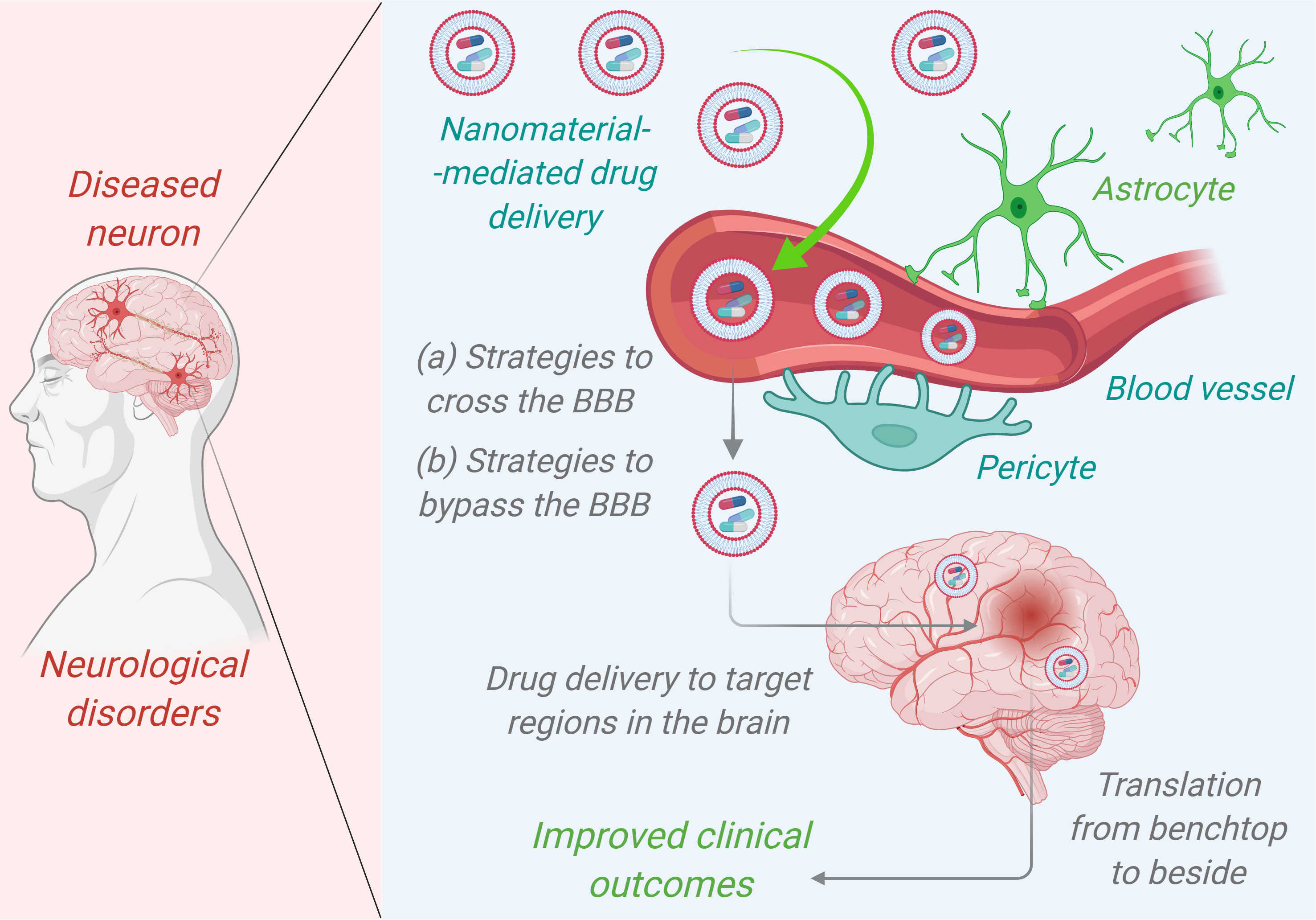
Fig. 2.

Nanomaterials mediated drug delivery of therapeutic agents in targeting Alzheimer disease patients’ brain to improve clinical outcomes.

Table 1.Nanodrug carriers delivering anti-AD drug in AD brain.
Nanodrug carrier Anti-AD drug Preclinical or clinical References
Polymeric Nanoparticles
PLGA-b-PEG Galantamine In vitro & In vivo [57]
PLGA Donepezil In vitro [58]
PLGA Withaferin In vitro [59]
PEG–PLGA Memantine In vitro & In vivo [60]
PAAM-Cardiolipin-PLGA Rosmarinic acid & Curcumin In vitro [61]
Solid-lipid Nanoparticles
SLN-DSPE-ApoE Resveratrol In vitro [62]
SLN-Palmitate-ApoE
S80-, PS-, PA-SNP Nicotinamide In vitro & In vivo [63]
S80-SNP Piperine In vitro & In vivo [64]
Liposomes
mApoE-PA-LIP Modified ApoE-derived peptide In vitro & In vivo [65]
GSH-PEG-EYPC-LIP VHH-pa2H In vitro & In vivo [66]
CPP-LIP Rivastigmine In vitro & In vivo [67]
Carbon Nanotubes
MWCNTs Berberine In vitro & In vivo [68]
Dendrimers
PAMAM-Lf Memantine In vitro & In vivo [19]
PAMAM-DG4.0, DG4.5 Tacrine In vitro & In vivo [69]
Pyridylphenylene —— In vitro [70]
Magnetic Nanoparticles
MNPs-PEG-PLA Curcumin In vitro & In vivo [71]
Au NPs Anthocyanin In vitro & In vivo [72]
Nanodiscs
4F Nanodiscs —— In vitro [73]
rHDL-rApoJ Nanodiscs rApoJ In vitro & In vivo [74]
Carbon Dots
CUR-Fe3O4@CDs Curcumin In vitro [75]
CDs, Carbon Dots; CPP, Cell penetrating peptide; CUR, Curcumin; DG, Dendrimer generation; DSPE, 1,2-Distearoyl-sn-glycero-3-phosphorylethanolamine; EYPC, Egg yolk phosphatidylcholine; GSH, Glutathione; Lf, Lactoferrin; LIP, Liposomes; mApoE, Modified ApoE protein; MNPs, Magnetic Nanoparticles; MWCNTs, Multi-walled carbon nanotubes; PA, Phosphatidic acid; PAAM, Polyacrylamide; PAMAM, Poly(amidoamine); PEG, Poly(ethylene glycol); PLA, Poly(lactic acid); PLGA, Poly(lactic-co-glycolic) acid; PS, Phosphatidylserine; rApoJ, Recombinant apolipoprotein J; rHDL, Recombinant high-density lipoprotein; S80, Polysorbate 80; SLN, Solid lipid Nanoparticles; VHH-pa2H, Amyloid beta binding llama single domain antibody fragments.
6.1 Fullerenes

Buckyballs or Buckminster fullerenes is a carbon allotrope with diameter of about 7Å constituting 60 carbon atoms in a geometry called truncated icosahedrons [76]. Depending on application, fullerenes are classified into three types: endohedral metallofullerenes, exohedral fullerenes and heterofullerenes. Endohedral metallofullerenes is composed of radioactive metal within the Buckyball and used for diagnostic purposes such as magnetic resonance imaging (MRI) and other imaging procedures employing radiocontrast media. Being less toxic and safer, these can be utilized as radioactive tracers for imaging organs [77]. Exohedral fullerenes are produced by chemical reaction between fullerenes and other chemical entities. These are derived from certain modifications of fullerene, also known as functionalized fullerenes. These are used as photosensitizers in photodynamic therapy where it produces harmful reactive oxygen species on stimulation by light, induced the programmed cell death [78, 79, 80]. Heterofullerenes contain other atoms like boron, nitrogen and few others in the place of one or more carbon present in fullerene compounds. A large number of conjugated double bonds present in the core of the fullerenes which scavenge free radicals and protect mitochondria from the attack by free radical species [81]. Oxidative stress induced as a result of free radicals generates demyelination of neurons, mitochondrial dysfunction, damage to microtubules, and apoptosis [82]. Ehrich et al. [83] investigated nanomaterials made of fullerene derivatives and showed the potential to counteract the toxic effect produced by organophosphatase-induced AChE inhibition, suggesting the antioxidant property. Furthermore, fullerenes are believed to activate the host immune response and generate antibodies specific to fullerenes [84]. REMD simulation studies by Xie et al. [85] proved that C-60 fullerene NPs (where molar ratio of fullerene: peptide was greater than 1:8) has immense ability to halt β-sheet formation of Aβ (16–22 peptides). Fullerene composed of 3C-60 molecules with much smaller surface area and unpredicted stronger inhibitory effect on the formation of β-sheet of the Aβ (16–22 peptides) was evident through REMD studies. Strong inhibition occurs as a result of hydrophobic interaction and aromatic-stack interactions present between the hexagonal rings, where phenyl rings relate to pentagonal rings that leads to weaker peptides responsible for holding β-sheet, and subsequently decreases the Aβ (16–22 peptides) fibril formation [85]. Fullerene exhibit unique contrasting characteristics of promoting ROS generation in UV or Visible light as well as scavenging ROS under dark conditions. Based on this contrasting property, Du et al. [86] designed UCNP@C60-pep (UCNP: up conversion nanoparticle, pep: Aβ-target peptide KLVFF) for AD therapy as it becomes active in the presence of NIR light. This hybrid nanoparticle generates ROS upon illumination with NIR light and photooxygenize Aβ peptides and hampers Aβ aggregation and as a result, consequent cytotoxicity is lessened. This near-infrared switchable fullerene-based synergy therapy to treat AD, serves as “image-guided therapy” used for UCL and MRI [86].

6.2 Nanotubes

Nanotubes are tube-like structures on which graphite is rolled and buckyballs present either at one or both the ends. These are of two types: SWCNT with an internal diameter of 1–2 nm and MWCNT having 2–25 nm diameter and 0.36 nm spacing between the two layers. Its length is of few micrometers [87]. Nanotubes enter the cell membrane through endocytosis or direct insertion or diffusion phenomena. To make it accessible within the cell, facile carboxylic or ammonium group can be introduced over this tube-like nanostructure. Thus, it serves as a means to deliver peptides, nucleic acids and other therapeutic molecules into the cell. Nanotubes have been explored in gene silencing therapy by conjugation of siRNA to the nanotube. It is advantageous over other means of transfer since it is non-immunogenic. Another way of targeting specific diseased cell is by conjugating antibodies along with radiolabeled or fluorescent tagged isotope [88, 89, 90]. SWNTs which were actually F-CNTs have been used by Yang et al. [91] to target the brain cells. It was orally administered to mice for continuous 10 days. When observed under electron microscopy, SWNTs were present in traces in absorptive cells, macrophages and neurons as well as in other organs such as heart, liver and brain. Improvement in learning and memory and other cognitive functions were observed as a result of acetylcholine transport with the help of SWNTs in a mouse model with induced AD. MWCNTs loaded with berberine (BRB) and coated with polysorbate and phospholipid formed complex of 186 nm, showed exceptional reclamation of memory up to 201th day. This complex maintained the biomolecules level in brain and thereby reduces Aβ fibrils responsible for the onset and later progression of AD [68].

6.3 Quantum dots

QDs (2–10 nm) are composed of core-shell made of inorganic substances while aqueous organic substance serve as coating to which biomolecule can be attached that has the ability to target several biomarkers. Upon activation by light, it emits fluorescence light and size of this nanocrystal determines the color of fluorescence [92]. Functionalization of QDs increases the particle size that restricts it to cross and filter through renal capillaries, and thus failed to get eliminated in order to overcome the toxicity of accumulation of QDs within body. In this regard, in vivo studies related to the metabolism and excretion of QDs are scarce [92]. Quan et al. [93] designed quantum dot nano-vehicle that have the ability to target surface cells and plays an important role in the detection of AD. Nanoformulated probe comprise of fluorescent QDs producing red light from the core which is enclosed in a shell made of polyethylene glycol (PEG)-conjugated with benzotriazole (BTA). This QD-PEG-BTA probe has shown 4 times more sensitivity for the detection of AD when compared to the conventional thioflavin derivatives. The success rate is high due to the fusion of high impact red fluorescence, presence of multivalent binding, and reduced background signal and non-specific binding. As a result, QDs impart increased sensitivity for the detection of amyloid-β in the progression of AD [93]. Apoe4 gene mutation has been detected using curcumin-graphene QDs layered on the transparent indium tin oxide (ITO) electrode. Amperometry studies showed ultrasensitive behavior and detected the DNA complex formation, in addition to repeatability, reproducibility, selectivity and long term storage stability of the complex [94]. S100β is another AD biomarker detected by an immunoassay based on photoelectrochemical sensing device using ITO electrode. The ITO electrode altered due to the incorporation of nanosized rGO and gold particles, and later casted as sol-gel film composed of isocyanate functional groups (-N=C=O). The primary antibody is immobilized on the rGO-Au/ITO electrode and the CdS QDs labeled antibody developed against S100β act as secondary antibody. Electrochemical properties and functional activities were observed to read the AD biomarker in the fluid. Tabrizi et al. [95] used this ITO modified and gold nanocomposite-CdS labeled antibody based immunosensor for the diagnosis of S100β.

6.4 Magnetic nanoparticles

Delivery of SPMNs under the influence of magnetic field have shown profound applications in non-invasive MRI. Certain magnetic nanoparticles (MNPs) have shown the potential to cross few biological and physical barriers, like BBB studied using molecular dynamics (MD) approach and deduced the association between BBB and NPs [96]. In a finding by Pansieri et al. [97] MNPs were investigated as a tool to efficiently diagnose the amyloidosis through imaging the amyloidogenic plaque or fibril depositions. This technique was reported to be safe and non-toxic when used under optimized conditions. However, the assessment of free or functionalized MNPs for biocompatibility with medical relevance remains to be investigated [97]. Nasr et al. [98] worked to achieve AD diagnosis in vivo and designed magnetic nanoparticle which cross BBB and detect the presence of Aβ plaques. Here, NPs functionalized with bovine serum albumin (BSA) which was further decorated with sialic acid (NP-BSAX-Sia) exhibited biocompatibility, high magnetic relaxivities for MRI and high selectivity to target Aβ plaques when examined in human AD transgenic mice. NP-BSAX-Sia can act as a promising detection tool for non-invasive diagnosis and examination of Aβ plaques in vivo [98]. Amin et al. [99] elaborated a method to deliver FMNPs in normal mice brain with the help of functionalized magnetic field produced by electromagnetic coils. FMNPs showed the ability to reach cortex and hippocampus of brain through BBB. It was extended to target Aβ1-42 in mice with Fe3O4 MNPs coated with dextran bearing osmotin. It was found effective in reducing synaptic loss as a result of Aβ1-42 accumulation, expression of BACE-1 as well as hyper phosphorylation of tau proteins [99].

6.5 Dendrimers

Dendrimers are tree-shaped nanosized structure comprises of three parts viz., central core, branches and functional groups which are present at the outer surface of macromolecule. These functional groups determine the efficacy of macromolecular complex which is composed of nucleic acid or entrapped drug. Dendrimers are multivalent molecules with a definite size and known structure with flexible features to modify the surface functional moieties to meet the requirements [100, 101]. It has lower viscosity as compared to the linear polymer equivalents and also exhibited good water solubility due to the presence of hydrophilic functional moieties on the surface. Further, engineering dendrimers with diverse chemical modification such as organic and inorganic groups at the branched site are also known [101, 102, 103]. As a replacement to conventional viral vectors, dendrimers are used for gene therapy. Dendrimers have shown promising results when tested in mammalian cell types and animal models. It enters the cells by endocytosis and transports DNA into nucleus for transcription of the desired gene and gene product [56]. The interesting advantage of dendrimer-based therapy is that it lacks the stimulation of immune reaction [56, 68].

6.6 Liposomes

Considered to be the original model of drug delivery vehicles, spherical in shape, composed of lipid bilayer membrane, may be unilamellar or multilamellar having aqueous interior environment, liposomes (LIP) hold a promising approach against AD. LIP facilitate loading of hydrophilic drug in aqueous compartment and lipophilic drug in LIP’s membrane for rapid delivery and efficacy (Fig. 3) [104]. To overcome the macrophage attack and opsonization in in vivo system, LIPs are coated by a layer of biomaterial with stealth properties such as polyoxyethylene, cholesterol, polyvinylpyrollidone polyacrylamide lipids, distearoyl phosphatidylcholine to form stealth LIPs. These coatings enhance the duration of drug action by prolonging its circulation time and protecting from immune attack [105, 106, 107, 108, 109, 110]. Some of the modified LIPs include immuno-LIPs, antibody-directed enzyme–prodrug therapy (ADEPT), and ligand bearing LIPs. In principle, LIPs are conjugated with an antibody targeted against the desired site and enzymes that activates prodrug and ligand specific for the target structure. It offers advantages like reduction in undesired effects and harm to normal cells, increases targeted drug delivery thereby boosts the drug’s efficacy and safety level [111, 112, 113, 114]. Owing to the failure of conventional small drugs or biological molecules to reach clinical trials, targeted nano-LIPs as drug delivery vehicle are promising modalities for AD [115]. So far, it has not reached clinical trials but it is found to be biocompatible, flexible with excellent property of carrying various types of therapeutic agents to cross the BBB and reach brain cells. LIPs can be designed for single therapeutic target or multiple pathways/cascades as targets. Various transformations utilizing peptides that can cross BBB, combined LIP-ligand complex involving phosphatidic acid, curcumin, and a retro-inverted peptide have been designed to target and inhibit Aβaggregation [115]. The therapeutic aspect of LIP conjugated with cardiolipin carrying curcumin (CRM)-cardiolipin (CL)/LIP and nerve growth factor (NGF) was evaluated in the presence of β-amyloid peptide in Wistar rats. The conjugated LIP surface covered with agglutinin showed decreased expression of phosphorylated p38, p-JNK and p-tau protein present at serine 202 and averted the neurodegeneration of SK-N-MC cells. The liposomal complex, NGF-CL/LIP also improved the expression of p-neurotrophic tyrosine kinase receptor type 1 and p-extracellular signal-regulated kinase 5 which rescues neuronal loss [116].

Fig. 3.

Liposomal nanodrug carrier in the rapid delivery of both hydrophilic and hydrophobic drugs targeting Alzheimer disease brain.

Kuo et al. [117] synthesized LIP containing cardiolipin and phosphatidic acid which provides target specificity against tau protein in hyperphosphorylated state. Trans-activator of transcription (TAT) peptide facilitated the ease of transport across BBB. LIP was loaded with NGF, rosmarinic acid (RA), curcumin (CURC), quercetin (QU), and phospholipid. The optimized TAT-NGF-RA-CURC-QU-CL/PA-LIP complex was found efficient in downregulating the expressions of pERK1/2 under phosphorylated state which is controlled by external signals such as c-Jun protein kinase present at N-terminal, p38, tau protein found at serine 202 and Caspase 3. This complex also enhanced the expression of p-ERK5 and p-cyclic adenosine monophosphate response element-binding protein [117]. Thus, LIPs are a promising delivery vehicle that pass-through BBB and protect nerve cell against the accumulated amyloid plaques.

6.7 Nanodiscs

Nanodiscs are a disc type structure having potential applications in proteomics and biomedicine. It is around 7–50 nm in diameter and consists of two main components: (i) phospholipids which are either of artificial origin or from the cell membrane and (ii) stabilizing agent which is belt shaped and holds the phospholipids together. Stabilizing agents can be protein or synthetic polymers [118]. Nanodiscs aims to mimic the cellular phospholipids for structural and functional studies of target molecules which are membrane proteins and peptides including amyloids. Membrane proteins and membrane interacting peptides are involved in numerous vital biological processes and are important targets for drug development [119]. There is a great utility of nanodiscs in the study of cellular signaling processes assembling on a membrane surface, by providing a well-defined and structured bilayer surface. Klein developed nanodiscs that allow unbiased high throughput screens that target binding sites for Alzheimer’s-associated Aβ oligomers and facilitate drug discovery for membrane protein targets [120, 121]. Sahoo et al. [73] established that apolipoprotein mimetic 4F nanodiscs retards beta-amyloid aggregation by using Alzheimer’s amyloid-beta (Aβ40) peptide as an example. β-amyloid forms short and thick fibers in the presence of 4F nanodiscs and the structural study reveals a ternary association between Aβ40 and 4F nanodiscs [73]. High-density lipoprotein (rHDL) nanodiscs and apolipoprotein J (ApoJ) have been constituted for the potential treatment of cerebral β-amyloidosis, a major feature of AD. Therapies based on rHDL-rApoJ nanodiscs have a potential use to treat neurological disorders associated with cerebral Aβ deposition. Polymethacrylate-copolymer (PMA) encased lipid-nanodiscs have been investigated to characterize and study the structure and toxicity of the Alzheimer’s Aβ intermediates [74].

6.8 Carbon dots

Carbon dots (CDs) are 0D carbon-based fluorescent nanomaterials less than 10 nm in size and are generally classified into carbon quantum dots (CQDs), carbonized polymer dots (CPDs), graphene quantum dots (GQDs) and carbon nitride dots (CNDs) [122]. CDs were first discovered in 2004 during the purification of SWCNTs via preparative electrophoresis [123]. Synthetic methodologies of CDs consist of top-down and bottom-up approaches with optimized conditions and precursors. CDs have demonstrated the abilities to penetrate the BBB due to their special characteristics, such as low toxicity, high biocompatibility, surface functional group modifications, excellent photoluminescence (PL), and size distribution [124]. CDs have been surface functionalized with amine and carboxyl groups to conjugate with various CNS drugs and also act as carriers to deliver drugs into the CNS to treat AD [125]. Recently, Kuang et al. [75] fabricated CUR-Fe3O4@CDs nanocomposite. This curcumin drug delivery system showed the strong affinity towards Aβ and inhibited extracellular Aβ fibrillation. Further, the nanocarriers inhibited ROS and neurotoxicity in PC12 cells. Thus, the CUR-Fe3O4@CDs nanocarrier restored the damaged nerve and can be a promising nanomaterial for AD treatment [75].

7. Conclusions and future outlook

AD remain as the prime cause of dementia which has many uncommon risk factors and pathologies associated with it. The research progress directed towards unravelling the disease mechanism and developing therapeutics against AD has been remarkable. Integrative analysis of AD diagnostic pathways that vary between patients affected by different causatives is warranted for better understanding of the underlying mechanisms. Identification of AD biomarkers and other observable pathological mechanism such as aberrant inflammation, processing of beta-amyloid protein and tau proteins, neurotrophic functions, etc. enables development of advanced and new approaches that pave way for the early diagnosis and also to identify the most appropriate targets for therapy. The therapeutic efficacy of various inhibitors, antibodies, and other modalities have been limited due to BBB. To overcome this limitation, various nanoformulations have been designed and investigated their crossing across BBB and studied their therapeutic efficiency against AD (Fig. 4).
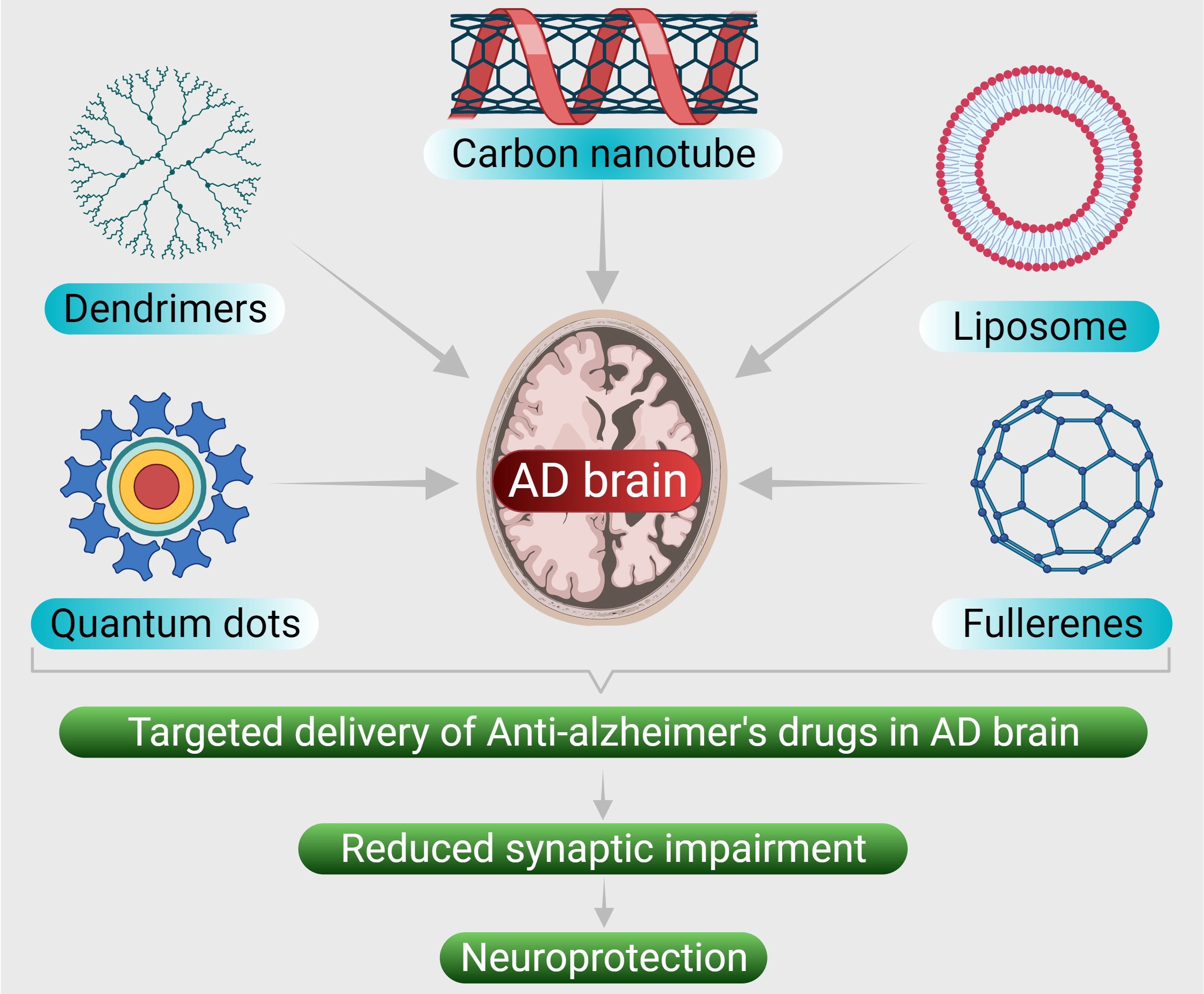
Fig. 4.

Various nanoformulations that have been designed to deliver the anti-AD drugs in Alzheimer disease brain.

Further, the other major limiting factor in AD research is the lack of appropriate animal model that can be assigned as closely mimicking the human AD, which is imperative to evaluate the clinical performance of the designed nanoformulations targeted against AD. Due to this ongoing limitation, the translation of AD targeted nanoformulation-based drug delivery systems to clinics is delayed. Thus, successful translation of AD therapeutic modality prerequisite development of animal model that meticulously investigates the therapeutic potential as well as serve to apprehend the complex disease mechanism of AD. Systematic clinical studies involving animal models and humans conducted under the regulatory framework would be vital to collect information about the efficacy, toxicity and pharmacological aspects of these nanoparticle-based AD therapeutics.

8. Author contributions

MF and MAG contributed in the collection of the literature, writing and editing the manuscript drafts. SA, SK, NKJ, BF, DD, DKC, PN, and KD contributed in editing the draft and provided the critical inputs in the review discussion. PKG and KKK conceptualized, planned, edited, and finalized the manuscript.

9. Ethics approval and consent to participate

Not applicable.

10. Acknowledgment

Piyush Kumar Gupta is thankful to the Department of Life Sciences, Sharda University, Greater Noida for providing the infrastructure and facility for research.

11. Funding

This research received no external funding.

12. Conflict of interest

The authors declare no conflict of interest.

Abbreviations

AAT, Alpha-1 antitrypsin; AChE, Acetylcholinesterase; AD, Alzheimer’s Disease; ADEPT, Antibody-Directed Enzyme Prodrug Therapy; APOE, Apolipoprotein E; APP, Amyloid Precursor Protein; BACE1, β-site Amyloid Precursor Protein Cleaving Enzyme 1; BBB, Blood Brain Barrier; BRB, Berberine; BSA, Bovine Serum Albumin; BTA, Benzotriazole; CLSM, Confocal Laser Scanning Microscopy; CSF, Cerebrospinal Fluid; CUR, Curcumin; FESEM, Field-Emission Scanning Electron Microscope; FMNPs, Fluorescent Carboxyl Magnetic Nile Red Particles; ITO, Indium-Tin Oxide; LIP, Liposome; MCI, Mild Cognitive Impairment; MD, Molecular Dynamic; MNs, Magnetic Nanoparticles; MRI, Magnetic Resonance Imaging; MWCNT, Multi-Walled Carbon Nanotubes; NFTs, Neurofibrillary Tangles; NGF, Nerve Growth Factor; NGF, Nerve Growth Factor; NIR, Near Infrared; NMR, Nuclear Magnetic Resonance; PAMAM, Polyamidoamine; PDPP, Polyvalent-directed Peptide Polymer; PEG, Polyethylene Glycol; pERK 1/2, phosphorylated Extracellular Regulated Kinase 1/2; p-JNK, Phosphorylated c-Jun N-terminal kinase; PSEN1, Presenilin 1; PSEN2, Presenilin 2; QDs, Quantum Dots; QU, Quercetin; RA, Rosmarinic Acid; REMD, Replica Exchange Molecular Dynamics; rGO, Reduced Graphene Oxide; ROS, Reactive Oxygen Species; SEM, Standard Error Mean; siRNA, Small Interfering RNA; SPMNs, Superparamagnetic Nanoparticles; SPs, Senile Plaques; SWCNT, Single-Walled Carbon Nanotubes; TAT, Transactivator of Transcription; TEM, Transmission Electron Microscope; TMS, Triple-helix Molecular Switch; UCP, Up Conversion Luminescence.

References
[1]
Gaugler J, James B, Johnson T, Scholz K, Weuve J. Alzheimer’s disease facts and figures. Alzheimer’s & Dementia. 2019; 15: 321–387.
[2]
Ott A, Breteler MMB, van Harskamp F, Claus JJ, van der Cammen TJM, Grobbee DE, et al. Prevalence of Alzheimer’s disease and vascular dementia: association with education. The Rotterdam study. British Medical Journal. 1995; 310: 970–973.
[3]
Querfurth HW, LaFerla FM. Alzheimer’s Disease. New England Journal of Medicine. 2010; 362: 329–344.
[4]
Rizzi L, Rosset I, Roriz-Cruz M. Global Epidemiology of Dementia: Alzheimer’s and Vascular Types. BioMed Research International. 2014; 2014: 1–8.
[5]
Fazili N, Naeem A, Ashraf G, Gan S, Kamal M. Therapeutic Interventions for the Suppression of Alzheimer’s Disease: Quest for a Remedy. Current Drug Metabolism. 2015; 16: 346–353.
[6]
Mirza Z, Ali A, Ashraf G, Kamal M, Abuzenadah A, Choudhary A, et al. Proteomics Approaches to Understand Linkage between Alzheimer’s Disease and Type 2 Diabetes Mellitus. CNS & Neurological Disorders - Drug Targets. 2014; 13: 213–225.
[7]
Knowles RB, Wyart C, Buldyrev SV, Cruz L, Urbanc B, Hasselmo ME, et al. Plaque-induced neurite abnormalities: Implications for disruption of neural networks in Alzheimer’s disease. Proceedings of the National Academy of Sciences. 1999; 96: 5274–5279.
[8]
Hayden EY, Teplow DB. Amyloid β-protein oligomers and Alzheimer’s disease. Alzheimer’S Research & Therapy. 2013; 5: 60.
[9]
De S, Whiten DR, Ruggeri FS, Hughes C, Rodrigues M, Sideris DI, et al. Soluble aggregates present in cerebrospinal fluid change in size and mechanism of toxicity during Alzheimer’s disease progression. Acta Neuropathologica Communications. 2019; 7: 120.
[10]
Knowles TPJ, Vendruscolo M, Dobson CM. The amyloid state and its association with protein misfolding diseases. Nature Reviews Molecular Cell Biology. 2014; 15: 384–396.
[11]
Kotler SA, Walsh P, Brender JR, Ramamoorthy A. Differences between amyloid-β aggregation in solution and on the membrane: insights into elucidation of the mechanistic details of Alzheimer’s disease. Chemical Society Reviews. 2014; 43: 6692–6700.
[12]
Cohen SIA, Arosio P, Presto J, Kurudenkandy FR, Biverstal H, Dolfe L, et al. A molecular chaperone breaks the catalytic cycle that generates toxic aβ oligomers. Nature Structural & Molecular Biology. 2015; 22: 207–213.
[13]
Arosio P, Michaels TCT, Linse S, Månsson C, Emanuelsson C, Presto J, et al. Kinetic analysis reveals the diversity of microscopic mechanisms through which molecular chaperones suppress amyloid formation. Nature Communications. 2016; 7: 10948.
[14]
Dobson CM. The Amyloid Phenomenon and its Links with Human Disease. Cold Spring Harbor Perspectives in Biology. 2017; 9: a023648.
[15]
Ivanova MI, Lin Y, Lee Y, Zheng J, Ramamoorthy A. Biophysical processes underlying cross-seeding in amyloid aggregation and implications in amyloid pathology. Biophysical Chemistry. 2021; 269: 106507.
[16]
Nguyen PH, Derreumaux P. Structures of the intrinsically disordered aβ, tau and α-synuclein proteins in aqueous solution from computer simulations. Biophysical Chemistry. 2020; 264: 106421.
[17]
Daneman R, Prat A. The Blood–Brain Barrier. Cold Spring Harbor Perspectives in Biology. 2015; 7: a020412.
[18]
Cao J, Hou J, Ping J, Cai D. Advances in developing novel therapeutic strategies for Alzheimer’s disease. Molecular Neurodegeneration. 2018; 13: 64.
[19]
Gothwal A, Kumar H, Nakhate KT, Ajazuddin, Dutta A, Borah A, et al. Lactoferrin Coupled Lower Generation PAMAM Dendrimers for Brain Targeted Delivery of Memantine in Aluminum-Chloride-Induced Alzheimer’s Disease in Mice. Bioconjugate Chemistry. 2019; 30: 2573–2583.
[20]
Sonawane SK, Ahmad A, Chinnathambi S. Protein-Capped Metal Nanoparticles Inhibit Tau Aggregation in Alzheimer’s Disease. ACS Omega. 2019; 4: 12833–12840.
[21]
Makin S. The amyloid hypothesis on trial. Nature. 2018; 559: S4–S7.
[22]
Zheng H, Koo EH. Biology and pathophysiology of the amyloid precursor protein. Molecular Neurodegeneration. 2011; 6: 27.
[23]
Chew H, Solomon VA, Fonteh AN. Involvement of lipids in Alzheimer’s disease pathology and potential therapies. Frontiers in Physiology. 2020; 11: 598.
[24]
Poirier J, Davignon J, Bouthillier D, Kogan S, Bertrand P, Gauthier S. Apolipoprotein E polymorphism and Alzheimer’s disease. Lancet. 1993; 342: 697–699.
[25]
Jones L, Holmans PA, Hamshere ML, Harold D, Moskvina V, Ivanov D, et al. Genetic evidence implicates the immune system and cholesterol metabolism in the aetiology of Alzheimer’s disease. PLoS ONE. 2010; 5: e13950.
[26]
Kao YC, Ho PC, Tu YK, Jou I, Tsai KJ. Lipids and Alzheimer’s disease. International Journal of Molecular Sciences. 2020; 21: 1505.
[27]
Castello MA. Lipid Regulation as a Critical Factor in the Development of Alzheimer’s Disease. Loma Linda University Electronic Theses, Dissertations & Projects. 2014; 260.
[28]
Belkouch M, Hachem M, Elgot A, Lo Van A, Picq M, Guichardant M, et al. The pleiotropic effects of omega-3 docosahexaenoic acid on the hallmarks of Alzheimer’s disease. the Journal of Nutritional Biochemistry. 2016; 38: 1–11.
[29]
Sastre M, Ritchie C. W, Hajji N. Metal ions in Alzheimer’s disease brain. JSM Alzheimer’s Diseases Related Dementia. 2015; 2: 1014.
[30]
Li Y, Jiao Q, Xu H, Du X, Shi L, Jia F, et al. Biometal dyshomeostasis and toxic metal accumulations in the development of Alzheimer’s disease. Frontiers Molecular Neuroscience. 2017; 10: 339.
[31]
Garai K, Sengupta P, Sahoo B, Maiti S. Selective destabilization of soluble amyloid β oligomers by divalent metal ions. Biochemical and Biophysical Research Communications. 2006; 345: 210–215.
[32]
DeToma AS, Salamekh S, Ramamoorthy A, Lim MH. Misfolded proteins in Alzheimer’s disease and type II diabetes. Chemical Society Reviews. 2012; 41: 608–621.
[33]
Pithadia AS, Kochi A, Soper MT, Beck MW, Liu Y, Lee S, et al. Reactivity of diphenylpropynone derivatives toward metal-associated amyloid-β species. Inorganic Chemistry. 2012; 51: 12959–12967.
[34]
Latshaw DC, Cheon M, Hall CK. Effects of macromolecular crowding on amyloid beta (16–22) aggregation using coarse-grained simulations. The Journal of Physical Chemistry B. 2014; 118: 13513–13526.
[35]
Minton AP. The effect of time-dependent macromolecular crowding on the kinetics of protein aggregation: a simple model for the onset of age-related neurodegenerative disease. Frontiers in Physics. 2014; 2: 48.
[36]
Lee CF, Bird S, Shaw M, Jean L, Vaux DJ. Combined effects of agitation, macromolecular crowding, and interfaces on amyloidogenesis. The Journal of Biological Chemistry. 2012; 287: 38006–38019.
[37]
Chen G, Xu T, Yan Y, Zhou Y, Jiang Y, Melcher K, et al. Amyloid beta: structure, biology and structure-based therapeutic development. Acta Pharmacologica Sinica. 2017; 38: 1205–1235.
[38]
Chowdhury SR, Xie F, Gu J, Fu L. Small-molecule Amyloid Aeta-Aggregation inhibitors in Alzheimer’s Disease drug development. Pharmaceutical Fronts. 2019; 1: 22–32.
[39]
Bautista-Aguilera ÓM, Budni J, Mina F, Medeiros EB, Deuther-Conrad W, Entrena JM, et al. Contilisant, a Tetratarget Small Molecule for Alzheimer’s Disease Therapy Combining Cholinesterase, Monoamine Oxidase Inhibition, and H3R Antagonism with S1R Agonism Profile. Journal of Medicinal Chemistry. 2018; 61: 6937–6943.
[40]
Habchi J, Chia S, Limbocker R, Mannini B, Ahn M, Perni M, et al. Systematic development of small molecules to inhibit specific microscopic steps of aβ42 aggregation in Alzheimer’s disease. Proceedings of the National Academy of Sciences. 2017; 114: E200–E208.
[41]
DeMattos RB, Bales KR, Cummins DJ, Dodart J-, Paul SM, Holtzman DM. Peripheral anti-Aβ antibody alters CNS and plasma Aβ clearance and decreases brain Aβ burden in a mouse model of Alzheimer’s disease. Proceedings of the National Academy of Sciences. 2001; 98: 8850–8855.
[42]
Jia Q, Deng Y, Qing H. Potential therapeutic strategies for Alzheimer’s disease targeting or beyond β-amyloid: insights from clinical trials. BioMed Research International. 2014; 2014: 837157.
[43]
Lannfelt L, Relkin NR, Siemers ER. Amyloid-ß-directed immunotherapy for Alzheimer’s disease. Journal of Internal Medicine. 2014; 275: 284–295.
[44]
Novakovic D, Feligioni M, Scaccianoce S, Caruso A, Piccinin S, Schepisi C, et al. Profile of gantenerumab and its potential in the treatment of Alzheimer’s disease. Drug Design, Development and Therapy. 2013; 7: 1359–1364.
[45]
Crespi GAN, Hermans SJ, Parker MW, Miles LA. Molecular basis for mid-region amyloid-β capture by leading Alzheimer’s disease immunotherapies. Scientific Reports. 2015; 5: 9649.
[46]
Dodel R, Hampel H, Depboylu C, Lin S, Gao F, Schock S, et al. Human antibodies against amyloid β peptide: a potential treatment for Alzheimer’s disease. Annals of Neurology. 2002; 52: 253–256.
[47]
Nishiyama Y, Taguchi H, Hara M, Planque SA, Mitsuda Y, Paul S. Metal-dependent amyloid β-degrading catalytic antibody construct. Journal of Biotechnology. 2014; 180: 17–22.
[48]
Zvěřová M. Alzheime disease and blood-based biomarkers and ash; potential contexts of use. Neuropsychiatric Disease and Treatment. 2018; 14: 1877–1882.
[49]
Zucchella C, Sinforiani E, Tamburin S, Federico A, Mantovani E, Bernini S, et al. The Multidisciplinary Approach to Alzheimer’s Disease and Dementia. A Narrative Review of Non-Pharmacological Treatment. Frontiers in Neurology. 2018; 9: 1058.
[50]
Weller J, Budson A. Current understanding of Alzheimer’s disease diagnosis and treatment. F1000Research. 2018; 7: 1161.
[51]
Derakhshankhah H, Sajadimajd S, Jafari S, Izadi Z, Sarvari S, Sharifi M, et al. Novel therapeutic strategies for Alzheimer’s disease: Implications from cell-based therapy and nanotherapy. Nanomedicine. 2020; 24: 102149.
[52]
Balaji AB, Pakalapati H, Khalid M, Walvekar R, Siddiqui H. Natural and synthetic biocompatible and biodegradable polymers. Biodegradable and Biocompatible Polymer Composites. 2018; 286: 3–32.
[53]
Bassas-Galia M, Follonier S, Pusnik M, Zinn M. Natural polymers. Bioresorbable Polymers for Biomedical Applications. 2017; 7: 31–64.
[54]
Swierczewska M, Han HS, Kim K, Park JH, Lee S. Polysaccharide-based nanoparticles for theranostic nanomedicine. Advanced Drug Delivery Reviews. 2016; 99: 70–84.
[55]
Patra JK, Das G, Fraceto LF, Campos EVR, Rodriguez-Torres MDP, Acosta-Torres LS, et al. Nano based drug delivery systems: recent developments and future prospects. Journal of Nanobiotechnology. 2018; 16: 71.
[56]
Zdrojewicz Z, Waracki M, Bugaj B, Pypno D, Cabała K. Medical applications of nanotechnology. Postepy Higieny I Medycyny Doswiadczalnej. 2016; 69: 1196–1204.
[57]
Gajbhiye KR, Gajbhiye V, Siddiqui IA, Pilla S, Soni V. Ascorbic acid tethered polymeric nanoparticles enable efficient brain delivery of galantamine: an in vitro-in vivo study. Scientific Reports. 2017; 7: 11086.
[58]
Tripathi SK, Patel B, Shukla S, Pachouri C, Pathak S, Pandey A. Donepezil loaded PLGA Nanoparticles, from Modified Nano-Precipitation, an Advanced Drug Delivery System to treat Alzheimer Disease. Journal of Physics: Conference Series. 2021; 1849: 012001.
[59]
Madhu S, Komala M, Pandian P. Formulation Development and Characterization of Withaferin-a Loaded Polymeric Nanoparticles for Alzheimer’s Disease. BioNanoScience. 2021; 11: 559–566.
[60]
Sánchez-López E, Ettcheto M, Egea MA, Espina M, Cano A, Calpena AC, et al. Memantine loaded PLGA PEGylated nanoparticles for Alzheimer’s disease: in vitro and in vivo characterization. Journal of Nanobiotechnology. 2018; 16: 32.
[61]
Kuo YC, Tsai HC. Rosmarinic acid- and curcumin-loaded polyacrylamide-cardiolipin-poly(lactide-co-glycolide) nanoparticles with conjugated monoclonal antibody to protect β-amyloid-insulted neurons. Materials Science and Engineering: C. 2018; 91: 445–457.
[62]
Neves AR, Queiroz JF, Reis S. Brain-targeted delivery of resveratrol using solid lipid nanoparticles functionalized with apolipoprotein E. Journal of Nanobiotechnology. 2016; 14: 27.
[63]
Vakilinezhad MA, Amini A, Akbari Javar H, Baha’addini Beigi Zarandi BF, Montaseri H, Dinarvand R. Nicotinamide loaded functionalized solid lipid nanoparticles improves cognition in Alzheimer’s disease animal model by reducing Tau hyperphosphorylation. Journal of Faculty of Pharmacy, Tehran University of Medical Sciences. 2018; 26: 165–177.
[64]
Yusuf M, Khan M, Khan RA, Ahmed B. Preparation, characterization, in vivo and biochemical evaluation of brain targeted Piperine solid lipid nanoparticles in an experimentally induced Alzheimer’s disease model. Journal of Drug Targeting. 2013; 21: 300–311.
[65]
Bana L, Minniti S, Salvati E, Sesana S, Zambelli V, Cagnotto A, et al. Liposomes bi-functionalized with phosphatidic acid and an ApoE-derived peptide affect aβ aggregation features and cross the blood–brain-barrier: implications for therapy of Alzheimer disease. Nanomedicine: Nanotechnology, Biology, and Medicine. 2014; 10: 1583–1590.
[66]
Rotman M, Welling MM, Bunschoten A, de Backer ME, Rip J, Nabuurs RJA, et al. Enhanced glutathione PEGylated liposomal brain delivery of an anti-amyloid single domain antibody fragment in a mouse model for Alzheimer’s disease. Journal of Controlled Release. 2015; 203: 40–50.
[67]
Yang Z, Zhang Y, Wang Z, Wu K, Lou J, Qi X. Enhanced brain distribution and pharmacodynamics of rivastigmine by liposomes following intranasal administration. International Journal of Pharmaceutics. 2013; 452: 344–354.
[68]
Lohan S, Raza K, Mehta SK, Bhatti GK, Saini S, Singh B. Anti-Alzheimer’s potential of berberine using surface decorated multi-walled carbon nanotubes: a preclinical evidence. International Journal of Pharmaceutics. 2017; 530: 263–278.
[69]
Igartúa DE, Martinez CS, del V. Alonso S, Prieto MJ. Combined Therapy for Alzheimer’s Disease: Tacrine and PAMAM Dendrimers Co-Administration Reduces the Side Effects of the Drug without Modifying its Activity. AAPS PharmSciTech. 2020; 21: 110.
[70]
Sorokina SA, Stroylova YY, Shifrina ZB, Muronetz VI. Disruption of Amyloid Prion Protein Aggregates by Cationic Pyridylphenylene Dendrimers. Macromolecular Bioscience. 2016; 16: 266–275.
[71]
Cheng KK, Chan PS, Fan S, Kwan SM, Yeung KL, Wáng YJ, et al. Curcumin-conjugated magnetic nanoparticles for detecting amyloid plaques in Alzheimer’s disease mice using magnetic resonance imaging (MRI). Biomaterials. 2015; 44: 155–172.
[72]
Ali T, Kim MJ, Rehman SU, Ahmad A, Kim MO. Anthocyanin-loaded PEG-Gold nanoparticles enhanced the neuroprotection of anthocyanins in an Aβ1–42 mouse model of Alzheimer’s Disease. Molecular Neurobiology. 2017; 54: 6490–6506.
[73]
Sahoo BR, Genjo T, Bekier M, Cox SJ, Stoddard AK, Ivanova M, et al. Alzheimer’s amyloid-beta intermediates generated using polymer-nanodiscs. Chemical Communications. 2018; 54: 12883–12886.
[74]
Fernández-de-Retana S, Cano-Sarabia M, Marazuela P, Sánchez-Quesada JL, Garcia-Leon A, Montañola A, et al. Characterization of ApoJ-reconstituted high-density lipoprotein (rHDL) nanodisc for the potential treatment of cerebral β-amyloidosis. Scientific Reports. 2017; 7: 14637.
[75]
Kuang Y, Zhang J, Xiong M, Zeng W, Lin X, Yi X, et al. A novel nanosystem realizing curcumin delivery based on Fe3O4@ carbon dots nanocomposite for Alzheimer’s disease therapy. Frontiers in Bioengineering and Biotechnology. 2020; 8: 614906.
[76]
Nimibofa A, Newton EA, Cyprain AY, Donbebe W. Fullerenes: Synthesis and applications. Journal of Materials Science Research. 2018; 7: 22–36.
[77]
Hashikawa Y, Murata M, Wakamiya A, Murata Y. Synthesis of Open-Cage Ketolactam Derivatives of Fullerene C60 Encapsulating a Hydrogen Molecule. Organic Letters. 2014; 16: 2970–2973.
[78]
Garcia-Diaz M, Huang Y, Hamblin MR. Use of fluorescent probes for ROS to tease apart Type I and Type II photochemical pathways in photodynamic therapy. Methods. 2016; 109: 158–166.
[79]
Huang L, Terakawa M, Zhiyentayev T, Huang Y, Sawayama Y, Jahnke A, et al. Innovative cationic fullerenes as broad-spectrum light-activated antimicrobials. Nanomedicine: Nanotechnology, Biology, and Medicine. 2010; 6: 442–452.
[80]
Maas M. Carbon nanomaterials as antibacterial colloids. Materials. 2016; 9: 617.
[81]
Fernández-Moriano C, González-Burgos E, Gómez-Serranillos MP. Mitochondria-Targeted Protective Compounds in Parkinson’s and Alzheimer’s Diseases. Oxidative Medicine and Cellular Longevity. 2015; 2015: 1–30.
[82]
Xiao WH, Bennett GJ. Effects of mitochondrial poisons on the neuropathic pain produced by the chemotherapeutic agents, paclitaxel and oxaliplatin. Pain. 2012; 153: 704–709.
[83]
Ehrich M, Van Tassell R, Li Y, Zhou Z, Kepley CL. Fullerene antioxidants decrease organophosphate-induced acetylcholinesterase inhibition in vitro. Toxicology in Vitro. 2011; 25: 301–307.
[84]
Hendrickson O, Fedyunina N, Zherdev A, Solopova O, Sveshnikov P, Dzantiev B. Production of monoclonal antibodies against fullerene C60 and development of a fullerene enzyme immunoassay. The Analyst. 2012; 137: 98–105.
[85]
Xie L, Luo Y, Lin D, Xi W, Yang X, Wei G. The molecular mechanism of fullerene-inhibited aggregation of Alzheimer’s β-amyloid peptide fragment. Nanoscale. 2014; 6: 9752–9762.
[86]
Du Z, Gao N, Wang X, Ren J, Qu X. Near-Infrared switchable fullerene-based synergy therapy for Alzheimer’s Disease. Small. 2018; 14: 1801852.
[87]
Ménard-Moyon C. Applications of Carbon Nanotubes in the Biomedical Field. Smart Nanoparticles for Biomedicine. 2018; 354: 83–101.
[88]
Elhissi AMA, Ahmed W, Hassan IU, Dhanak VR, D’Emanuele A. Carbon nanotubes in cancer therapy and drug delivery. Journal of Drug Delivery. 2012; 2012: 837327.
[89]
Jain KK. Advances in use of functionalized carbon nanotubes for drug design and discovery. Expert Opinion on Drug Discovery. 2012; 7: 1029–1037.
[90]
Li H, Hao Y, Wang N, Wang L, Jia S, Wang Y, et al. DOTAP functionalizing single-walled carbon nanotubes as non-viral vectors for efficient intracellular siRNA delivery. Drug Delivery. 2016; 23: 840–848.
[91]
Yang Z, Zhang Y, Yang Y, Sun L, Han D, Li H, et al. Pharmacological and toxicological target organelles and safe use of single-walled carbon nanotubes as drug carriers in treating Alzheimer disease. Nanomedicine: Nanotechnology, Biology, and Medicine. 2010; 6: 427–441.
[92]
Clift MJD, Stone V. Quantum dots: an insight and perspective of their biological interaction and how this relates to their relevance for clinical use. Theranostics. 2012; 2: 668–680.
[93]
Quan L, Wu J, Lane LA, Wang J, Lu Q, Gu Z, et al. Enhanced Detection Specificity and Sensitivity of Alzheimer’s Disease Using Amyloid-β-Targeted Quantum Dots. Bioconjugate Chemistry. 2016; 27: 809–814.
[94]
Mars A, Hamami M, Bechnak L, Patra D, Raouafi N. Curcumin-graphene quantum dots for dual mode sensing platform: Electrochemical and fluorescence detection of APOe4, responsible of Alzheimer’s disease. Analytica Chimica Acta. 2018; 1036: 141–146.
[95]
Tabrizi MA, Ferré-Borrull J, Kapruwan P, Marsal LF. A photoelectrochemical sandwich immunoassay for protein S100β, a biomarker for Alzheimer’s disease, using an ITO electrode modified with a reduced graphene oxide-gold conjugate and CdS-labeled secondary antibody. Mikrochimica Acta. 2019; 186: 117.
[96]
Pedram MZ, Shamloo A, Alasty A, Ghafar-Zadeh E. Optimal Magnetic Field for Crossing Super-Para-Magnetic Nanoparticles through the Brain Blood Barrier: a Computational Approach. Biosensors. 2016; 6: 25.
[97]
Pansieri J, Gerstenmayer M, Lux F, Mériaux S, Tillement, O, Forge V, et al. Magnetic Nanoparticles Applications for Amyloidosis Study and Detection: A Review. Nanomaterials. 2018; 8: 740.
[98]
Nasr SH, Kouyoumdjian H, Mallett C, Ramadan S, Zhu DC, Shapiro EM, et al. Detection of β-Amyloid by sialic acid coated Bovine serum albumin magnetic nanoparticles in a nouse model of Alzheimer’s Disease. Small. 2018; 14: 1701828.
[99]
Amin FU, Hoshiar AK, Do TD, Noh Y, Shah SA, Khan MS, et al. Osmotin-loaded magnetic nanoparticles with electromagnetic guidance for the treatment of Alzheimer’s disease. Nanoscale. 2017; 9: 10619–10632.
[100]
Mignani S, El Kazzouli S, Bousmina M, Majoral J. Expand classical drug administration ways by emerging routes using dendrimer drug delivery systems: a concise overview. Advanced Drug Delivery Reviews. 2013; 65: 1316–1330.
[101]
Yu H, Schlüter AD, Zhang B. Synthesis of High Generation Dendronized Polymers and Quantification of their Structure Perfection. Macromolecules. 2014; 47: 4127–4135.
[102]
Aliev G, Ashraf GM, Tarasov VV, Chubarev VN, Leszek J, Gasiorowski K, et al. Alzheimer’s Disease–Future Therapy Based on Dendrimers. Current Neuropharmacology. 2019; 17: 288–294.
[103]
Caminati G, Turro NJ, Tomalia DA. Photophysical investigation of starburst dendrimers and their interactions with anionic and cationic surfactants. Journal of the American Chemical Society. 1990; 112: 8515–8522.
[104]
Gregoriadis G. Liposomes in drug delivery: How it all happened. Pharmaceutics. 2016; 8: 19.
[105]
Cui J, Li C, Wang C, Li Y, Zhang L, Zhang L, et al. Development of pegylated liposomal vincristine using novel sulfobutyl ether cyclodextrin gradient: is improved drug retention sufficient to surpass DSPE-PEG-induced drug leakage? Journal of Pharmaceutical Sciences. 2011; 100: 2835–2848.
[106]
Ge X, Wei M, He S, Yuan WE. Advances of non-ionic surfactant vesicles (niosomes) and their application in drug delivery. Pharmaceutics. 2019; 11: 55.
[107]
Kouchakzadeh H, Shojaosadati SA, Maghsoudi A, Vasheghani Farahani E. Optimization of PEGylation conditions for BSA nanoparticles using response surface methodology. AAPS PharmSciTech. 2010; 11: 1206–1211.
[108]
Lim W, Tardi PG, Dos Santos N, Xie X, Fan M, Liboiron BD, et al. Leukemia-selective uptake and cytotoxicity of CPX-351, a synergistic fixed-ratio cytarabine: daunorubicin formulation, in bone marrow xenografts. Leukemia Research. 2010; 34: 1214–1223.
[109]
Shelley H, Babu RJ. Role of Cyclodextrins in Nanoparticle-Based Drug Delivery Systems. Journal of Pharmaceutical Sciences. 2018; 107: 1741–1753.
[110]
Torchilin VP. Multifunctional nanocarriers. Advanced Drug Delivery Reviews. 2012; 64:1532–1555.
[111]
Hung BY, Kuthati Y, Kankala RK, Kankala S, Deng JP, Liu CL, et al. Utilization of enzyme-immobilized mesoporous silica nanocontainers (IBN-4) in prodrug-activated cancer theranostics. Nanomaterials. 2015; 5: 2169–2191.
[112]
Pandey H, Rani R, Agarwal V. Liposome and their applications in cancer therapy. Brazilian Archives of Biology and Technology. 2016; 59: e16150477.
[113]
Sharma SK, Bagshawe KD. Antibody Directed Enzyme Prodrug Therapy (ADEPT): Trials and tribulations. Advanced Drug Delivery Reviews. 2017; 118: 2–7.
[114]
Stephanopoulos N, Tong GJ, Hsiao SC, Francis MB. Dual-surface modified virus capsids for targeted delivery of photodynamic agents to cancer cells. ACS Nano. 2010; 4: 6014–6020.
[115]
Ross C, Taylor M, Fullwood N, Allsop D. Liposome delivery systems for the treatment of Alzheimer’s disease. International Journal of Nanomedicine. 2018; 13: 8507–8522.
[116]
Kuo Y, Lin C, Li J, Lou Y. Wheat germ agglutinin-conjugated liposomes incorporated with cardiolipin to improve neuronal survival in Alzheimer’s disease treatment. International Journal of Nanomedicine. 2017; 12: 1757–1774.
[117]
Kuo Y, Chen C, Rajesh R. Optimized liposomes with transactivator of transcription peptide and anti-apoptotic drugs to target hippocampal neurons and prevent tau-hyperphosphorylated neurodegeneration. Acta Biomaterialia. 2019; 87: 207–222.
[118]
Bayburt TH, Sligar SG. Single-molecule height measurements on microsomal cytochrome P450 in nanometer-scale phospholipid bilayer disks. Proceedings of the National Academy of Sciences of the United States of America. 2002; 99: 6725–6730.
[119]
Sligar SG, Denisov IG. Nanodiscs: a toolkit for membrane protein science. Protein Science. 2021; 30: 297–315.
[120]
Lacor PN, Buniel MC, Furlow PW, Sanz Clemente A, Velasco PT, Wood M, et al. Aβ oligomer-induced aberrations in synapse composition, shape, and density provide a molecular basis for loss of connectivity in Alzheimer’s disease. Journal of Neuroscience. 2007; 27: 796–807.
[121]
Wilcox KC, Marunde MR, Das A, Velasco PT, Kuhns BD, Marty MT, et al. Nanoscale Synaptic Membrane Mimetic Allows Unbiased High Throughput Screen that Targets Binding Sites for Alzheimer’s-Associated aβ Oligomers. PLoS ONE. 2015; 10: e0125263.
[122]
Tejwan N, Saha SK, Das J. Multifaceted applications of green carbon dots synthesized from renewable sources. Advances in Colloid and Interface Science. 2020; 275: 102046.
[123]
Xu X, Ray R, Gu Y, Ploehn HJ, Gearheart L, Raker K, et al. Electrophoretic analysis and purification of fluorescent single-walled carbon nanotube fragments. Journal of the American Chemical Society. 2004; 126: 12736–12737.
[124]
Zhang W, Sigdel G, Mintz KJ, Seven ES, Zhou Y, Wang C, et al. Carbon dots: A future Blood–Brain Barrier penetrating nanomedicine and drug nanocarrier. International Journal of Nanomedicine. 2021; 16: 5003.
[125]
Du J, Xu N, Fan J, Sun W, Peng X. Carbon Dots for in Vivo Bioimaging and Theranostics. Small. 2019; 15: 1805087.
Share
Back to top