IMR Press / FBL / Volume 26 / Issue 10 / DOI: 10.52586/4979
Open Access Commentary
Is miR therapeutic targeting still a miRage?
Show Less
1 Harvard Medical School, Boston, MA 02115, USA
2 Beth Israel Deaconess Medical Center, Boston, MA 02215, USA
3 Harvard Stem Cell Institute, Cambridge, MA 02138, USA
4 Institute of Biomedical Technologies, National Research Council (CNR), Area della Ricerca di Pisa, 56124 Pisa, Italy
*Correspondence: elevanti@bidmc.harvard.edu; elena.levantini@itb.cnr.it (Elena Levantini)
Front. Biosci. (Landmark Ed) 2021, 26(10), 680–692; https://doi.org/10.52586/4979
Submitted: 20 August 2021 | Revised: 5 October 2021 | Accepted: 7 October 2021 | Published: 30 October 2021
Copyright: © 2021 The Author(s). Published by BRI.
This is an open access article under the CC BY 4.0 license (https://creativecommons.org/licenses/by/4.0/).
Abstract

Since the discovery of the first microRNA (miR), almost three decades ago, the roles played by miRs under normal and diseased settings have been widely investigated. miRs are found to play crucial roles in cancer initiation and progression, as well as towards therapy response mechanisms. Therefore, they are relevant and attractive targets for therapeutic development. Many preclinical studies have demonstrated their promise as future anti-cancer tools. Recently, increasing number of early phase clinical trials have emerged. In this Commentary, we will summarize the major discoveries within the miR research field and highlight the status quo of current miR-therapeutics, which has prominent potential of impacting future cancer regimens given their massive dysregulation in oncogenic processes.

Keywords
RNA therapeutics
Biomarkers
miR-21
NSCLC
Cancer
2. Introduction

In 2021 Frontiers in Bioscience-Landmark has reached its 25th anniversary, and this Commentary presents an opportunity to acknowledge the success of one of its most highly-cited papers, i.e., the 2019 article by Dai et al. [1], which focuses on the role of microRNA (miR)-21 in Non-Small Cell Lung Cancer (NSCLC). miR-21 will serve as Ariadne’s thread in this mini-review, in which, miR-21 is adopted as a narrative guide to summarize the major discoveries obtained in the microRNA field, a term coined back in 2001 to classify this distinctive class of small untranslated RNAs [2, 3, 4, 5, 6, 7].

Since 1993, when the 22 nucleotide-long RNA lin-4 was first identified [8], thousands of other microRNAs have been discovered to possess post-transcriptional gene regulation properties in RNA-based gene silencing processes in eukaryotes. lin-4 is rightfully considered as the founding member of miR biomolecules, and its discovery has shifted the paradigm on gene regulation, which has been based on the “central dogma” of genetics where DNA is first transcribed into messenger RNA (mRNA), and then translated into protein.

Within the genome, miRs are often found as clusters from which they are transcribed as discrete polycistronic transcripts, or share the promoter of host genes and are spliced from their mRNA transcripts during biogenesis [9]. miR-21 was one of the first mammalian microRNAs identified [3] and the first to be elucidated on the mechanism of miRs processing starting from pri-microRNAs (pri-miRs) [10]. These ~1-kb-long transcripts form a characteristic hairpin structure that undergoes nuclear processing through cleavage by the Drosha/DGCR8 ribonuclease complex [11, 12]. Afterwards, they are released into the cytoplasm as ~60 nucleotides-long precursor hairpins called pre-miRs, via an Exportin 5 and Ran-GTP complex. Subsequently, their ends are cleaved by the Dicer/TRBP complex resulting in the mature double-stranded miR duplex [13]. At this stage, only one strand of the miR duplex, the guide strand, is loaded onto Argonaute (Ago) to form the RNA-induced silencing complex RISC [14, 15]. This dynamic process eventually results in the production of mature miR that physically interacts with a complementary or partially complementary sequence usually located in the 3UTR (Untranslated Region) of a target mRNA to mediate gene silencing [16]. In order for miRs to exert their action, partial or full complementarity between the seed region of miR and (primarily) the 3UTR is required [16, 17], although additional mechanisms of interactions have been reported [18]. This allows for a single miR to possess the ability to bind to multiple mRNAs. In a nutshell, these interactions are the raison d’être of miR biomolecules which are responsable for triggering mRNA destabilization/decay or translational inhibition, although mRNA degradation appears to be the dominant silencing effect [16, 19, 20, 21, 22, 23, 24, 25].

3. miRs and cancer

Given that a single miR can regulate a variety of target genes, and that a single protein-coding gene usually contains several miR seed sequences, the miR regulatory system stands en bloc as a complex mechanism regulating eukaryotic gene expression. It is estimated that miRs may regulate >30% of eukaryotic genes [26]. Therefore, it is unsurprising that alterations in this composite post-transcriptional regulatory machine have been frequently linked to tumorigenesis [27]. There is considerable evidence to indicate that miRs and their biogenesis machinery are involved in the development of cancer. miRs are frequently altered owing to genomic events, such as mutations, deletion, amplification, or biogenesis defects due to mutations or downregulation of miR biogenesis regulating enzymes [16, 27, 28, 29].

Cancer is defined by abnormal cell division and differentiation, and can arise not only from alteration of various molecular mechanisms including dysregulation of several protein coding genes but also global changes in miR profiles. Despite miRs being the most widely studied class of non-coding RNAs, other components of the non-coding machinery such as the recently-discovered RNA species small nucleolar RNAs (snoRNAs), small interfering RNAs (siRNAs), transcription initiation RNAs (tiRNAs), PIWI-interacting RNAs (piRNAs), transcribed ultra-conserved regions (t-UCRs) and large intergenic noncoding RNAs (lincRNAs) [30, 31, 32] will certainly be identified as significant players in our war against cancer, once we can adequately delineate their complex biology.

The first evidence showing that miRs were dysregulated in human cancer came from studies on B-cell chronic lymphocytic leukemia (CLL), in which a critical region on chromosome 13q14 has been found to be frequently deleted in CLL, and that that this deleted region contained two microRNA genes (miR-15a and miR16-1) [33]. Subsequently, microRNA genes were identified at fragile sites and genomic regions involved in chromosomal alterations identified in various human tumors [34], and defects in miR-mediated regulation can noticeably lead to human cancers [35]. Several other mechanisms controlling altered miR expression in cancer have been reported comprising mutations [36], small nucleotide polymorphisms (SNPs) as described in lung cancer [37], DNA methylation (consistently half of the genomic sequences of miR genes are associated with CpG islands) [38], DNA hypomethylation leading to miR upregulation [39, 40] and histone acetylation [41, 42]. Not surprisingly, another dysregulated regulatory mechanism entails that some oncogenic mRNAs carry mutations or SNPs in their 3UTR thus eluding control through miR binding, and consequently avoiding negative regulation. An example is the oncogene K-RAS in lung cancer where a SNP in the let-7 binding site precludes its correct regulation which leads to higher tumor occurrence [43]. Remarkably, proliferating cells have been observed to express mRNAs with shortened 3UTRs and, as a result, contain fewer miRs’ binding sites [44]. This suggests almost infinite regulatory possibilities of dysregulation at/during tumorigenesis. Interestingly, it is recently shown that a very complex network comprising both coding and non-coding RNAs bears the same miR response elements (MREs) on their 3UTRs, and can compete with each other for the same limited pool of miRs. Such RNAs are called competing endogenous RNAs (ceRNAs) and they are capable of confiscating miRs, thereby acting as natural microRNA sponges. Thus, by competitive binding for shared miRs, they affect each other’s expression and can co-regulate each other in the highly complex ceRNA networks to support miR inhibition [45, 46]. Needless to say, alterations in ceRNA networks have important roles in different aspects of tumor etiology [47].

4. miR-21 the par excellence onco-miR

Large-scale profiling of gene expression in multiple human cancers have shown that one of the most frequently dysregulated gene in cancer is miR-21, frequently found overexpressed in all tumor types including lung cancer [48, 49, 50]. miR-21 expression within the tumor microenvironment (TME) is currently under-investigated when compared to tumor cells per se. However, we are beginning to comprehend its key relevance in the TME, which is to be considered as the most tumor-intermingled “organ” and symbiotic with cancer cells. In tumor-infiltrating macrophages, miR-21 inhibition for example, is pivotal in regulating tumor progression [51]. In addition, miR-21 (and miR-29a) which is secreted by tumor cells in exosomes, better defined as small extracellular vesicles (sEVs) [52, 53], can interact with members of the TLR (Toll-like receptors) family to stimulate TLR-mediated pro-metastatic inflammatory response on tumor associated macrophages (TAMs), thus suggesting a new mechanism of miR-TLR mediated intercellular communication between the TME and cancer cells [54]. Interestingly, sEVs produced within the TME by cancer-associated adipocytes (CAAs), TAMs, and cancer-associated fibroblasts (CAFs) contain miR-21, and sEVs-mediated miR-21 delivery into cancer cells is shown to confer therapeutic resistance in multiple cancers [55, 56], suggesting apart from cancer cells, the TME should also be targeted with anti-miR-21 therapeutics.

A role for miR-21 as an oncogenic miR (oncomiR) was demonstrated in vivo using a doxycycline-inducible miR-21-LoxSTOPLox-Tet-off mouse model [57], that upon induction, displayed malignant pre-B cell lymphoid-like phenotype. A parallel study demonstrated a pro-tumorigenic role of miR-21 in NSCLC where deletion of miR-21 in a KRAS-driven lung cancer model reduced tumorigenesis, while mir-21 overexpression showed the opposite effect [58]. Additionally, amplification of chromosomal 17q23.2 region, which includes miR-21, has been observed in breast, lung, hepatocellular, ovarian and prostate cancers [59]. Further, TCGA data demonstrate the locus containing miR-21 is amplified, and that such amplification can act as a prognostic marker in pulmonary adenocarcinomas [60]. miR-21 expression can also be upregulated via transcription factors that are upregulated in cancer such as AP-1 that acts down-stream of RAS and binds to miR-21 promoter [61, 62]. Other factors implicated in miR-21 upregulation are STAT3 [63] whose high expression is associated with poor patient outcomes [64, 65, 66] and NF-kB [67]. However, the complexity of the predicted promoter region of pri-miR-21 [61, 68] and occurrence of alternative transcription start sites [69] suggest that regulation of miR-21 transcription may not be a straight forward process [70]. Not surprisingly, miR-21 is upregulated by one of the most lethal oncogenes, i.e., BMI-1. BMI-1 is responsible for maintaining the self-renewal ability of cancer stem cells in virtually all tumors, including lung cancer [71, 72, 73]. In gastric cancer, BMI1 can upregulate miR-34a, which appears to have opposite effects than miR-21, hinting the presence of a negative feedback loop between BMI1 and miR-34a, through which BMI1 can regulate its own function [71]. The interplay between BMI1 and miR-21 in lung cancer is still undefined; however, BMI1 may form regulatory networks with other relevant miRs. For example, we identified miR-192 as one of the main upregulated miRs upon treating lung cancer cells with PTC-209, a compound capable of inhibiting BMI1 activity. This prevents cell cycle progression by arresting cells in G0. miR-34a was upregulated, although at a lower extent than miR-192, whereas, miR-21 levels remained unaffected (Maroni and Levantini, personal communication) thus implying that different miRs, other than miR-21, may constitute the BMI1 oncogenic regulatory network under different conditions.

5. miR signatures as potential biomarkers

Given miRs are highly dysregulated in cancer, the potential for miR signatures to distinguish not only between tumor and normal tissue, but also to identify different subgroups of tumors and predict outcome or response to therapy treatments has been intensely investigated. Our ability to adopt them as biomarkers, however, is still in the infancy stage. Inconsistent results were frequently reported by different research groups and these may arise from differences in stage and treatment received by patients of the analyzed samples. Therefore, large scale studies are needed and preferably should be carried out at multicenter locations to ensure higher patient recruitment, with strict screening criteria and higher statistical power when assessing sensitivity, specificity and pertinence of miRs as future biomarkers. However, successful observations have been reproduced and one evidence that has been stratifying over the years, is the role that miR-21 plays in restricting response to therapy and causing drug resistance by regulating expression of resistance-related factors in multiple cancer types. One of the first reports describing miR-21 involvement in chemo-resistance was shown in cholangiocarcinoma cells, in which its (and miR-200b’s) inhibition led to increased sensitivity to gemcitabine [74]. miR-21 overexpression is related to development of Multi Drug Resistance in breast cancer [75], in which its downregulation improves the chemotherapeutic effect of Taxol [76]. miR-21 inhibition is shown to be directly responsible for PTEN expression and re-sensitivity to doxorubicin via increased caspase-related apoptosis [77]; and its inhibition, associated with co-delivery of the cytostatic compound docetaxel within “chitosomes”, improves chemo-sensitivity to the compound [78]. Near-infrared-radiation-responsive hollow gold nanoparticles have been adopted to achieve sequential delivery of miR-21 inhibitor and doxorubicin, in breast cancer xenograft models. They displayed successful tumor delivery followed by effective accumulation, that resulted in good therapeutic efficacy, showing the potentiality of the “sequential delivery concept” for cancer therapeutics settings [79]. Similarly, simultaneous systemic administration in exosome nanocarriers of the cytostatic drug 5-FU and a miR-21 inhibitor oligonucleotide demonstrated major anti-tumor effects in a colon cancer mouse model [80]. In gastric cancer, the same combination of 5-FU and miR-21 inhibitor was delivered in trastuzumab-conjugated nanoparticles, which increased trastuzumab targeting and antibody-dependent cellular cytotoxicity while enhancing sensitivity of gastric cancer cells to trastuzumab and 5-FU [81]. There is growing evidence that delivery of miR therapeutics together with chemotherapeutics can improve treatment efficiency, and miR-21 is one of the most extensively studied miR therapeutics in combination-delivery settings, given its protagonist role in various tumorigenic settings [78, 79, 80, 81].

Higher expression of miR-21 has been reported in plasma [82] and tissues [83] from NSCLC patients, as compared to healthy donors. Additionally, it has been identified as a useful predictor for early detection and chemo-sensitivity biomarker in plasma from patients with advanced NSCLC [84, 85]. Further, miR-21 has been implicated in the development of gefitinib resistance in EGFR mutant NSCLCs [86]; as well as resistance to cisplatin, gemcitabine, 5-fluorouracil and teniposide in various cancers [75, 87, 88, 89, 90, 91, 92, 93, 94, 95]. It has alone been correlated with the development of brain metastases in NSCLC patients [96] and its inhibition could improve sensitivity to radiotherapy in the NSCLC cell line A549, mediated by the PI3K/Akt pathway [97]. Consistently, miR-21is a part of a defined signature (containing seven miRs) capable of predicting response to platinum-based treatments [98]. As biomarker, miR-21, belongs to a novel panel of serum-based miRs capable of discriminating breast cancer patients and healthy controls [99]. miR-21 possesses the highest specificity and sensitivity in NSCLC patients’ sputum or plasma [84, 100]; and it is a common component to several molecular signatures for NSCLC diagnosis [101, 102, 103] where high plasma sEV miR-21 levels are shown to associate with poor overall survival (OS) [104]. On the basis of the above considerations, miR-21 can serve as an attractive candidate as a potentially relevant biomarker for diagnosis, prediction, and prognosis [105, 106, 107, 108] though, it has not been adopted in the clinic presently [109]. However, it should also be taken into account that not all that glitters is gold, and that miR-21’s utility as a biomarker may be a double-edged sword. miR-21 ubiquitous overexpression in virtually all cancers represents the biggest challenge which is specificity. miR-21 may not be the most suitable biomarker for a specific type of cancer but rather a broad “alarm-miR” which is indicative on the presence of oncogenesis and may require coupling with more tissue/organ-specific biomarkers. The evidence presented suggest a great possibility that miR-21 to be adopted in the screening for lung cancer, which is the deadliest malignancy worldwide.

NSCLC, that we investigate in my laboratory, accounts for more than 80% of all lung cancer cases [110]. It is essentially untreatable, although many strategies have been proposed to improve patients’ survival. Most patients are diagnosed at an advanced stage and half of cases have distant metastatic disease at initial diagnosis, with poor prognosis. As a result, there is a major need to identify faster and non-invasive ways to detect lung cancer at early stages. miRs, as biomarkers, may fulfil this request after we have successfully understood how to employ them in the correct manner. Aberrant miR signatures are commonly observed in NSCLC and they are most frequently associated to upregulation rather than downregulation [111, 112, 113, 114, 115, 116, 117, 118, 119, 120, 121, 123]. These abnormally overexpressed miRs represent a valuable repertoire from which the proper panel of biomarkers can be selected for association to “alarm-miR-21” in the design of non-invasive tests for next-generation NSCLC detection.

Certainly, additional basic research is required before miR-21 can be extensively adopted as an approved biomarker. However, its upregulation in numerous types of cancer and association with cancer-related pathways renders it a promising molecule for RNA-based therapies. miR-21 regulates various downstream effectors associated with tumor pathogenesis during all stages of carcinogenesis as it is involved in regulating signaling pathways implicated in apoptosis, cell growth, proliferation, survival, angiogenesis, migration, extravasation, invasion and metastasis, and also chemo- and radio-resistance pathways [57, 58, 124, 125, 126, 127, 128, 129, 130, 131, 132, 133, 134, 135, 136, 137, 138, 139, 140, 141, 142, 143, 144]. Dai et al. [1] corroborated the existence of an inverse relation between miR-21 and PTEN in NSCLC, that is also observed in other cancers [143, 145, 146, 147, 148, 149, 150]. Specifically, they demonstrated that miR-21 regulates lung cancer proliferation, cell cycle progression and EMT, through the PTEN/Akt/GSK3β signaling cascades [1], and they also showed the relevance of down-regulating miR-21 as a new therapeutic strategy for human lung cancer.

Several experiments aimed at affecting miR-21 have been performed over the years. When a miR-21 antagomir was used in breast cancer cells, tumor growth was restricted through inhibiting cell proliferation and inducing apoptosis [151]. Additionally, synthetic circular RNAs (circRNAs) that could function as miR-21 sponges, thus acting as competitive inhibitors, have been developed to inhibit miR-21 activity which displayed inhibition of cell proliferation in gastric cancer [152]. Intra-venous administration of MKAD-21 (a chemically modified antisense oligo against miR-21) in xenograft models could dose-dependently suppressed bladder cancer growth through regulation of the PPP2R2A-ERK network, which is a mediator of miR-21-induced oncogenesis in bladder cancer [153]. Anti-miR-21(AM-21), composed of two cationic lipids, has been therapeutically used in a preclinical murine model of lung cancer, in which it displayed reduced tumor growth with prolonged survival without toxicity [154].

6. miRs as therapeutic targets

As a naturally occurring class of gene regulators, microRNAs have attracted great attention as promising targets for therapeutic development, given their knack for targeting virtually any gene of interest at the post-transcriptional level. Tumor progression is caused by the deregulation of multiple cellular pathways, therefore the combinatorial ability miRs display to target multiple disease-related genes concurrently, may provide an advantage, as compared to traditional small molecule therapeutics. Both viral and non-viral carriers, have been utilized to develop miR-based therapies aimed at either reducing the expression of specific oncomiRs (by means of antimiRs/antagomiRs based on antisense oligonucleotides (ASO), locked nucleic acid (LNA) antimiRs, or tiny LNA antimiRs) or at replenishing rare tumor suppressing-miRs (by means of synthetic and/or chemically-modified miR mimics). Nowadays, the miR therapy road is still paved with several challenges (Fig. 1) that need to be addressed (mainly identification of potential off target effects and improved delivery methods) before their effective translation into future therapeutic applications can take place.
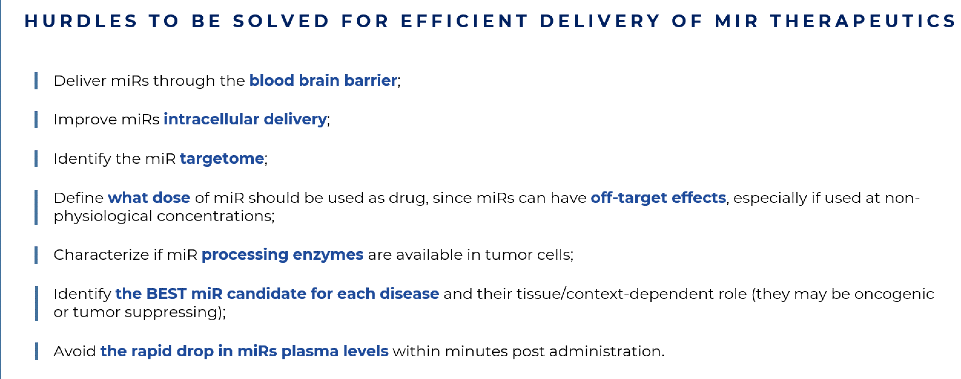
Fig. 1.

Hurdles to be solved for efficient delivery of miR therapeutics.

In primis, in order to truly enter the era of RNA therapeutics, it is necessary to identify the best miR candidates or miR targets for each disease, as well as understand if a specific miR (and its targets) follows a unidirectional function/behavior of oncomiR or tumor suppressor, or if their role changes in different tissues/conditions. This information is relevant to decide whether miR inhibitory or miR mimics therapeutic strategies can be utilized in each specific disease-driven scenario. At the present stage, we should also consider an extra level of complexity; i.e., the major heterogeneity displayed by tumors and their intermixed TMEs, which can be patient-specific and represents a major obstacle in identifying the relevant target miRs to adopt each time. Despite tumor cellular heterogeneity, functional heterogeneity governed by factors such as hypoxia and inflammation can cause dynamic localized heterogeneities [28, 155, 156, 157, 158, 159]. miR biogenesis enzymes such as Drosha, Dicer and AGO2 are downregulated by hypoxia, further destabilizing miR expression [157, 158, 160, 161] and complicating the identification of candidate miRs.

Currently, the molecular characterization of tumors is predominantly derived from analyses of bulk tissues, and the application of single cell RNA sequencing (scRNAseq) techniques should bring major advances in our understanding of how transcripts (both non-coding and coding) perform at the single-cell molecular level, and how they are affected in contexts as various as tumor initiation, response to drug treatment, and development of therapy resistance. Given miRs’ mechanisms of action, the amount of miR within a cell can dictate the impact on gene expression. Therefore, quantifying single cell miRs levels will be great interest to study their expression in defined subpopulations (identified by scRNAseq as distinct transcriptional clusters) contained within tumors and TMEs. As mentioned above, miRs can essentially exert dual roles and function as both oncogenic or tumor suppressive units. However, they can even act in a context-dependent manner and exert different roles in suppressing or enhancing tumor progression. miR-141, for example, was reported to be a tumor-suppressive miR by counteracting Tregs recruitment and immune escape in advanced-stage NSCLC patients with malignant pleural effusion [162]. However, the same miR is shown to be oncogenic by targeting tumor suppressor genes PHLPP1 and PHLPP2 in NSCLC and inducing the proliferation of cancer cells [163]. Similarly, members of the miR-29 family, comprising three isoforms (miR-29a, miR-29b and miR-29c) can act as tumor-suppressors in mantle-cell lymphoma, acute myeloid leukemia (AML), lung cancer, diffuse large B cell lymphoma and Burkitt lymphoma, and oncogenic miRs in indolent human B cell chronic lymphocytic leukemia, AML and metastatic breast cancer [164, 165]. The miR143/145 cluster is a tumor suppressor in cancer cells however, it can also induce neo-angiogenesis in the TME leading to increased tumor growth [166]. To achieve a better response to treatments, through miR-based cancer therapy, it is anticipated that miRs are required to be downregulated or upregulated in a tissue- or context-dependent manner. Regrettably, this level of accuracy is not achievable at this present stage.

Further, miRs affect multiple target genes hence it is difficult to predict and evaluate all their potential off-target effects [167]. For example, when a target gene has functions that can either promote or suppress the carcinogenic process [168] such as the miR-21 target BCL2 [169], the effectiveness of a miR-based therapeutic option is questioned. Also, the ability of miRs to target several genes involved in disease is a bonus as it helps with the targeting of the entire lineage of disease-related genes, but this intrinsic feature may also favor the introduction of off-target effects [170]. Thus, elucidating the entire and extensive network of interactions displayed by miR-target genes (the miR targetome) is the gold standard to take miRs one step further towards therapeutic application. Only when such knowledge is available, well-designed therapeutic methods can be implemented to unambiguously slow down tumor growth, and preventing drug resistance and metastases, even in personalized medicine protocols.

An additional hurdle to consider is the ability to design appropriate parenteral delivery systems capable of specifically and effectively target cell types/tissues/organs and to ensure high stability, efficient uptakes by the cells and proper localization of the miR sequence into the cytoplasm [171, 172, 173]. Another concern is the use of miR drugs at non-physiological concentrations as they may exhibit a different targeting scenario than usual and can dysregulate gene expression in unanticipated manners. An even more critical factor required for the activity of miR drug processing is the amount of physiological RISC complex available, given that RISC proteins are abnormally downregulated during hypoxia [174], which is frequently associated to tumorigenesis hence creating a catch-22 situation. Sufficient expression of RISC complex proteins should be first evaluated while treating with exogenous miRs, considering that limited RISC expression may create competition between exogenous and endogenous miRs [175] thus diminishing the effectiveness of treatment overall. Finally, the complex extracellular matrix surrounding solid tumors may impede the delivery of any compound to cancer cells, and non-malignant cells may concomitantly sequester miR drugs from the tumor area. All these parameters can significantly contribute to a reduced presence of miR therapeutics in the TME, and consequently lead to inefficient uptakes into tumor cells. The ability to directly target dysregulated miRs in tumor cells or in tumor-infiltrating cells and/or cancer-associated fibroblasts or endothelial cells would have significantly improved treatment regimens however, such delivery technique is a chimera that we are still chasing. Overall, some progress has been made, but we still have a lengthy journey ahead before miR therapies are fully optimized for cancer treatment [176, 177].

miRs are water-soluble which endow properties that are appropriate for parenteral administration. Unfortunately, following i.v. injection, naked miRs are rapidly degraded by the abundant nucleases present in the extracellular and plasma environment [178] and removed from the circulation [175], thus causing a rapid drop in plasma levels within minutes post administration. Delivering miRs via systemic administration by using antibodies, ligands, and nanoparticles, or even by directly injecting the drug at pathogenic site, can enhance target specificity and efficacy, as well as minimize side effects [175, 179]. However, the latter is impractical for treating metastasizing tumors and leukemias [180]. Another challenge is the ability of delivering miRs through the blood brain barrier (BBB). Partial success has been obtained by adopting intranasal administration and modified micelle, liposomes, or nanoparticles, given only lipid-soluble small molecules are able to cross the BBB [181, 182]. Recent findings have revealed that miRs can be drugged with small molecules, i.e., small-molecule inhibitors of miRs (SMIRs) and small-molecule degraders (SMDs) that target miRs directly. The first SMIR was developed against miR-21 [183]. SMIRs bind to miRs and inhibit their biogenesis and maturation, while SMDs bind to miRs and induce their degradation [184, 185]. These small molecules will pave the way for the development of novel therapeutics against miRs that only recently were considered druggable.

Presently, despite some phase I/II clinical trials, the era of phase III compounds is still a moving target. A prominent example of a miR drug that encountered problems in clinical trials is MRX34, a mimic of naturally occurring miR-34, that was terminated at the phase I study due to serious immune-related adverse events and later on, a planned phase II study in melanoma patients was withdrawn [186]. A more favorable outcome occurred to the first TargomiR (miR-15/16 mimics packaged in nano-cells targeted with EGFR antibodies) that was tested in a phase I study on patients with malignant pleural mesothelioma [187], showing an acceptable safety profile thus supporting additional studies of TargomiRs in combination with chemotherapy or immune checkpoint inhibitors [188]. Similarly, Cobomarsen (MRG-106), a locked nuclear acid-based anti-miR, which targets miR-155 in multiple hematological malignancies, is shown to be safe in a phase I study.

This small excursus on miR-based medicine shows that the possibility to adopt them as pharmaceuticals is still in its early stages and that several strategies have been designed to efficiently deliver them (Fig. 2).

Fig. 2.

Strategies to treat patients with miR therapeutics. Both single molecule (left panel) and combination strategies (right panel) are tested in pre-clinical settings to design efficient and proper delivery in patients of miRs or miRs combined to chemotherapeutics.

7. miRs as future therapies in pulmonary diseases

There is considerable interest in using miR drugs in pulmonary diseases. Direct intrapulmonary administration, with or without aerosolization, has been adopted in murine models of pulmonary hypertension [189, 190, 191, 192, 193]. A meritorious paper from Schlosser et al. [194], systematically investigated the relative pros of intratracheal and intranasal routes of administration, with and without aerosolization, and comparing them with intravenous, intraperitoneal and subcutaneous injection. Pulmonary levels of the miR mimic showed variations by up to 4 orders of magnitude between different delivery methods thus demonstrating the impact of both the route of administration and mimic formulation. Although all methods showed elevated lung levels, intratracheal and intranasal routes of administration were clearly superior for lung-selective targeting. This selectivity was evidenced by pulmonary levels that were at least 10-fold, and up to 10,000-fold, higher than levels observed in other tissues. In applications where lung-targeted mimic delivery is not an essential requirement, intraperitoneal and subcutaneous administration could serve as alternative methods to increase lung miR levels without the need for anesthesia [194]. Previous studies have indicated that some aerosolized materials may be distributed more uniformly within the lungs [195, 196]; and in a proof-of-concept study, the delivery of miR-17 to bronchial epithelial cells (BECs) using nebulized lipid-polymer hybrid nanoparticles (LPNs) showed efficient and well-tolerated delivery of miR mimics to BECs [197].

In lung cancer, miR-21 has been considered as a potential gold standard for personalized therapy given its heavily-documented oncogenic role. Advancements in RNA chemistry and delivery technologies are encouraging for a potential future use of miR-21 as part of the standard treatment for NSCLC [198]. Recently, a nano-cellular (minicell) delivery vehicle loaded with miR-16-5p mimics was designed to target tumors via minicell-surface attached bispecific EGFR-targeting antibodies (EnGeneIC Dream Vectors - EDVs), upon intravenous injection [199]. Preliminary data presented by Van Zandwijk et al. [188] from phase I clinical trials for patients with Malignant Pleural Mesothelioma and advanced NSCLC showed manageable safety profiles in 5 patients [199].

Early phase clinical trials, aimed at suppressing miR-21, are currently ongoing. RG-012, a chemically-modified oligonucleotide capable of binding miR-21, is currently being tested in phase I on patients affected by Alport syndrome, and initial data showed that the rate of progression of renal fibrosis is reduced (ClinicalTrials.gov: NCT03373786). In addition, an interventional clinical trial is currently being conducted that involves the study of six miRs (including miR-21) to determine whether a patient with stage II colon cancer is a candidate to receive adjuvant chemotherapy based on OS and disease-free survival (DFS) measurements (ClinicalTrials.gov: NCT02466113).

Overall, experimental and clinical evidence demonstrate that miR-21 is a promising biomarker (for diagnosis, prognosis and prediction) in tumorigenesis, as it is commonly dysregulated in virtually all types of cancer. Identification of its target genes and the effects of their downregulation remains to be fully elucidated. In vitro and in vivo studies have shown that its inhibition has anti-tumor effects, thus advocating for its promising therapeutic function [200]. Presently, clinical trials and toxicity pharmacokinetic evaluations are being conducted for miR-21 inhibitors however several limitations for its use still exists such as the lack of knowledge on dose requirements as well as delivery strategies and identification of side effects. Work on these limitations is currently underway and current techniques are being designed to mitigate such issues, and to maximize therapeutic efficacy. The overall progress in developing miR therapeutics within the short time frame, since their discovery, is noteworthy [201]. Nevertheless, several well-designed preclinical studies, coupled to novel delivery platforms, are strictly required to explore the promising potential of RNA therapy. These findings will serve as catalysts for the new era of RNA-based therapeutics, especially for miRs, that are markedly dysregulated in cancers.

8. Ethics approval and consent to participate

Not applicable.

9. Acknowledgment

EL acknowledges Azhar Ali, Igea D’Agnano, Giorgia Maroni, for helpful discussion and Elisabetta Pisano (Communication Office, Institute of Clinical Physiology, CNR, Pisa, Italy) for graphical design.

10. Funding

This work is funded by MIUR (Ministry of Education, University and Research) Flagship Interomics Project 20/2017 to EL, and sponsored research support from PTC Therapeutics to EL.

11. Conflict of interest

The author declares no conflict of interest. EL is serving as one of the Editorial Board members of this journal. We declare that EL had no involvement in the peer review of this article and has no access to information regarding its peer review. Full responsibility for the editorial process for this article was delegated to GP.

References
[1]
Cretoiu D. MiR-21 regulates growth and EMT in lung cancer cells via PTEN Akt GSK3 beta signaling. Frontiers in Bioscience. 2019; 24: 1426–1439.
[2]
Ambros V, Bartel B, Bartel DP, Burge CB, Carrington JC, Chen X, et al. A uniform system for microRNA annotation. RNA. 2003; 9: 277–279.
[3]
Lagos-Quintana M, Rauhut R, Lendeckel W, Tuschl T. Identification of novel genes coding for small expressed RNAs. Science. 2001; 294: 853–858.
[4]
Ambros V. MicroRNAs: tiny regulators with great potential. Cell. 2001; 107: 823–826.
[5]
Lee RC, Ambros V. An extensive class of small RNAs in Caenorhabditis elegans. Science. 2001; 294: 862–864.
[6]
Lau NC, Lim LP, Weinstein EG, Bartel DP. An abundant class of tiny RNAs with probable regulatory roles in Caenorhabditis elegans. Science. 2001; 294: 858–862.
[7]
Smallridge R. A small fortune. Nature Reviews. Molecular Cell Biology. 2001; 2: 867.
[8]
Lee RC, Feinbaum RL, Ambros V. The C. elegans heterochronic gene lin-4 encodes small RNAs with antisense complementarity to lin-14. Cell. 1993; 75: 843–854.
[9]
Tanzer A, Stadler PF. Evolution of microRNAs. Methods in Molecular Biology. 2006; 342: 335–350.
[10]
CAI X. Human microRNAs are processed from capped, polyadenylated transcripts that can also function as mRNAs. RNA. 2004; 10: 1957–1966.
[11]
Lee Y, Kim M, Han J, Yeom K, Lee S, Baek SH, et al. MicroRNA genes are transcribed by RNA polymerase II. The EMBO Journal. 2004; 23: 4051–4060.
[12]
Denli AM, Tops BBJ, Plasterk RHA, Ketting RF, Hannon GJ. Processing of primary microRNAs by the Microprocessor complex. Nature. 2004; 432: 231–235.
[13]
Wilson RC, Tambe A, Kidwell MA, Noland CL, Schneider CP, Doudna JA. Dicer-TRBP complex formation ensures accurate mammalian microRNA biogenesis. Molecular Cell. 2015; 57: 397–407.
[14]
Bartel DP. MicroRNAs: Target Recognition and Regulatory Functions. Cell. 2009; 136: 215–233.
[15]
Schirle NT, Sheu-Gruttadauria J, MacRae IJ. Structural basis for microRNA targeting. Science. 2014; 346: 608–613.
[16]
Bartel DP. MicroRNAs: genomics, biogenesis, mechanism, and function. Cell. 2004; 116: 281–297.
[17]
Felekkis K, Touvana E, Stefanou C, Deltas C. MicroRNAs: a newly described class of encoded molecules that play a role in health and disease. Hippokratia. 2010; 14: 236–240.
[18]
Londin E, Loher P, Telonis AG, Quann K, Clark P, Jing Y, et al. Analysis of 13 cell types reveals evidence for the expression of numerous novel primate- and tissue-specific microRNAs. Proceedings of the National Academy of Sciences of the United States of America. 2015; 112: E1106–E1115.
[19]
Filipowicz W, Bhattacharyya SN, Sonenberg N. Mechanisms of post-transcriptional regulation by microRNAs: are the answers in sight? Nature Reviews. Genetics. 2008; 9: 102–114.
[20]
Ameres SL, Zamore PD. Diversifying microRNA sequence and function. Nature Reviews. Molecular Cell Biology. 2013; 14: 475–488.
[21]
Desvignes T, Batzel P, Berezikov E, Eilbeck K, Eppig JT, McAndrews MS, et al. MiRNA Nomenclature: a View Incorporating Genetic Origins, Biosynthetic Pathways, and Sequence Variants. Trends in Genetics. 2015; 31: 613–626.
[22]
Eichhorn SW, Guo H, McGeary SE, Rodriguez-Mias RA, Shin C, Baek D, et al. MRNA destabilization is the dominant effect of mammalian microRNAs by the time substantial repression ensues. Molecular Cell. 2014; 56: 104–115.
[23]
Kamenska A, Lu W, Kubacka D, Broomhead H, Minshall N, Bushell M, et al. Human 4E-T represses translation of bound mRNAs and enhances microRNA-mediated silencing. Nucleic Acids Research. 2014; 42: 3298–3313.
[24]
Bartel DP. Metazoan MicroRNAs. Cell. 2018; 173: 20–51.
[25]
Esquela-Kerscher A, Slack FJ. Oncomirs - microRNAs with a role in cancer. Nature Reviews. Cancer. 2006; 6: 259–269.
[26]
Wilson RC, Doudna JA. Molecular mechanisms of RNA interference. Annual Review of Biophysics. 2013; 42: 217–239.
[27]
Ha M, Kim VN. Regulation of microRNA biogenesis. Nature Reviews. Molecular Cell Biology. 2014; 15: 509–524.
[28]
Rupaimoole R, Calin GA, Lopez-Berestein G, Sood AK. MiRNA Deregulation in Cancer Cells and the Tumor Microenvironment. Cancer Discovery. 2016; 6: 235–246.
[29]
Lin S, Gregory RI. MicroRNA biogenesis pathways in cancer. Nature Reviews. Cancer. 2015; 15: 321–333.
[30]
Mercer TR, Dinger ME, Mattick JS. Long non-coding RNAs: insights into functions. Nature Reviews Genetics. 2009; 10: 155–159.
[31]
Taft RJ, Glazov EA, Cloonan N, Simons C, Stephen S, Faulkner GJ, et al. Tiny RNAs associated with transcription start sites in animals. Nature Genetics. 2009; 41: 572–578.
[32]
Huang T, Alvarez A, Hu B, Cheng S. Noncoding RNAs in cancer and cancer stem cells. Chinese Journal of Cancer. 2013; 32: 582–593.
[33]
Calin GA, Sevignani C, Dumitru CD, Hyslop T, Noch E, Yendamuri S, et al. Human microRNA genes are frequently located at fragile sites and genomic regions involved in cancers. Proceedings of the National Academy of Sciences of the United States of America. 2004; 101: 2999–3004.
[34]
Calin GA, Dumitru CD, Shimizu M, Bichi R, Zupo S, Noch E, et al. Frequent deletions and down-regulation of micro- RNA genes miR15 and miR16 at 13q14 in chronic lymphocytic leukemia. Proceedings of the National Academy of Sciences of the United States of America. 2002; 99: 15524–15529.
[35]
Iorio MV, Croce CM. MicroRNA dysregulation in cancer: diagnostics, monitoring and therapeutics. a comprehensive review. EMBO Molecular Medicine. 2012; 4: 143–159.
[36]
Raveche ES, Salerno E, Scaglione BJ, Manohar V, Abbasi F, Lin Y, et al. Abnormal microRNA-16 locus with synteny to human 13q14 linked to CLL in NZB mice. Blood. 2007; 109: 5079–5086.
[37]
Shen H, Hu Z, Chen J, Tian T. Genetic variants of miRNA sequences and non small cell lung cancer survival. European Journal of Cancer Supplements. 2008; 6: 10–11.
[38]
Weber B, Stresemann C, Brueckner B, Lyko F. Methylation of human microRNA genes in normal and neoplastic cells. Cell Cycle. 2007; 6: 1001–1005.
[39]
Brueckner B, Stresemann C, Kuner R, Mund C, Musch T, Meister M, et al. The human let-7a-3 locus contains an epigenetically regulated microRNA gene with oncogenic function. Cancer Research. 2007; 67: 1419–1423.
[40]
Iorio MV, Visone R, Di Leva G, Donati V, Petrocca F, Casalini P, et al. MicroRNA signatures in human ovarian cancer. Cancer Research. 2007; 67: 8699–8707.
[41]
Scott GK, Mattie MD, Berger CE, Benz SC, Benz CC. Rapid alteration of microRNA levels by histone deacetylase inhibition. Cancer Research. 2006; 66: 1277–1281.
[42]
Shin S, Lee E, Cha HJ, Bae S, Jung JH, Lee S, et al. MicroRNAs that respond to histone deacetylase inhibitor SAHA and p53 in HCT116 human colon carcinoma cells. International Journal of Oncology. 2009; 35: 1343–1352.
[43]
Chin LJ, Ratner E, Leng S, Zhai R, Nallur S, Babar I, et al. A SNP in a let-7 microRNA complementary site in the KRAS 3’ untranslated region increases non-small cell lung cancer risk. Cancer Research. 2008; 68: 8535–8540.
[44]
Sandberg R, Neilson JR, Sarma A, Sharp PA, Burge CB. Proliferating cells express mRNAs with shortened 3’ untranslated regions and fewer microRNA target sites. Science. 2008; 320: 1643–1647.
[45]
Cheng D, Xiang Y, Ji L, Lu X. Competing endogenous RNA interplay in cancer: mechanism, methodology, and perspectives. Tumour Biology. 2015; 36: 479–488.
[46]
Salmena L, Poliseno L, Tay Y, Kats L, Pandolfi PP. A ceRNA hypothesis: the Rosetta Stone of a hidden RNA language? Cell. 2011; 146: 353–358.
[47]
Wang Y, Hou J, He D, Sun M, Zhang P, Yu Y, et al. The Emerging Function and Mechanism of ceRNAs in Cancer. Trends in Genetics : TIG. 2016; 32: 211–224.
[48]
Volinia S, Calin GA, Liu C, Ambs S, Cimmino A, Petrocca F, et al. A microRNA expression signature of human solid tumors defines cancer gene targets. Proceedings of the National Academy of Sciences. 2006; 103: 2257–2261.
[49]
Liu L, Chen Q, Lai R, Wu X, Wu X, Liu F, et al. Elevated expression of mature miR-21 and miR-155 in cancerous gastric tissues from Chinese patients with gastric cancer. Journal of Biomedical Research. 2010; 24: 187–197.
[50]
Huang C, Yu W, Cui H, Wang Y, Zhang L, Han F, et al. Increased expression of miR-21 predicts poor prognosis in patients with hepatocellular carcinoma. International Journal of Clinical and Experimental Pathology. 2015; 8: 7234–7238.
[51]
Sahraei M, Chaube B, Liu Y, Sun J, Kaplan A, Price NL, et al. Suppressing miR-21 activity in tumor-associated macrophages promotes an antitumor immune response. Journal of Clinical Investigation. 2019; 129: 5518–5536.
[52]
Théry C, Witwer KW, Aikawa E, Alcaraz MJ, Anderson JD, Andriantsitohaina R, et al. Minimal information for studies of extracellular vesicles 2018 (MISEV2018): a position statement of the International Society for Extracellular Vesicles and update of the MISEV2014 guidelines. Journal of Extracellular Vesicles. 2018; 7: 1535750.
[53]
D’Agnano I, Berardi AC. Extracellular Vesicles, A Possible Theranostic Platform Strategy for Hepatocellular Carcinoma-An Overview. Cancers. 2020; 12: 261.
[54]
Fabbri M, Paone A, Calore F, Galli R, Gaudio E, Santhanam R, et al. MicroRNAs bind to Toll-like receptors to induce prometastatic inflammatory response. Proceedings of the National Academy of Sciences of the United States of America. 2012; 109: E2110–E2116.
[55]
Au Yeung CL, Co N, Tsuruga T, Yeung T, Kwan S, Leung CS, et al. Exosomal transfer of stroma-derived miR21 confers paclitaxel resistance in ovarian cancer cells through targeting APAF1. Nature Communications. 2016; 7: 11150.
[56]
Zheng P, Chen L, Yuan X, Luo Q, Liu Y, Xie G, et al. Exosomal transfer of tumor-associated macrophage-derived miR-21 confers cisplatin resistance in gastric cancer cells. Journal of Experimental & Clinical Cancer Research. 2017; 36: 53.
[57]
Medina PP, Nolde M, Slack FJ. OncomiR addiction in an in vivo model of microRNA-21-induced pre-B-cell lymphoma. Nature. 2010; 467: 86–90.
[58]
Hatley ME, Patrick DM, Garcia MR, Richardson JA, Bassel-Duby R, van Rooij E, et al. Modulation of K-Ras-dependent lung tumorigenesis by MicroRNA-21. Cancer Cell. 2010; 18: 282–293.
[59]
Krichevsky AM, Gabriely G. miR-21: a small multi-faceted RNA. Journal of Cellular and Molecular Medicine. 2009; 13: 39–53.
[60]
Campbell JD, Alexandrov A, Kim J, Wala J, Berger AH, Pedamallu CS, et al. Distinct patterns of somatic genome alterations in lung adenocarcinomas and squamous cell carcinomas. Nature Genetics. 2016; 48: 607–616.
[61]
Fujita S, Ito T, Mizutani T, Minoguchi S, Yamamichi N, Sakurai K, et al. MiR-21 Gene expression triggered by AP-1 is sustained through a double-negative feedback mechanism. Journal of Molecular Biology. 2008; 378: 492–504.
[62]
Talotta F, Cimmino A, Matarazzo MR, Casalino L, De Vita G, D’Esposito M, et al. An autoregulatory loop mediated by miR-21 and PDCD4 controls the AP-1 activity in RAS transformation. Oncogene. 2009; 28: 73–84.
[63]
Iliopoulos D, Jaeger SA, Hirsch HA, Bulyk ML, Struhl K. STAT3 Activation of miR-21 and miR-181b-1 via PTEN and CYLD are Part of the Epigenetic Switch Linking Inflammation to Cancer. Molecular Cell. 2010; 39: 493–506.
[64]
Yu H, Lee H, Herrmann A, Buettner R, Jove R. Revisiting STAT3 signalling in cancer: new and unexpected biological functions. Nature Reviews. Cancer. 2014; 14: 736–746.
[65]
Johnson DE, O’Keefe RA, Grandis JR. Targeting the IL-6/JAK/STAT3 signalling axis in cancer. Nature Reviews. Clinical Oncology. 2018; 15: 234–248.
[66]
Hillmer EJ, Zhang H, Li HS, Watowich SS. STAT3 signaling in immunity. Cytokine & Growth Factor Reviews. 2016; 31: 1–15.
[67]
Sheedy FJ, Palsson-McDermott E, Hennessy EJ, Martin C, O’Leary JJ, Ruan Q, et al. Negative regulation of TLR4 via targeting of the proinflammatory tumor suppressor PDCD4 by the microRNA miR-21. Nature Immunology. 2010; 11: 141–147.
[68]
Löffler D, Brocke-Heidrich K, Pfeifer G, Stocsits C, Hackermüller J, Kretzschmar AK, et al. Interleukin-6 dependent survival of multiple myeloma cells involves the Stat3-mediated induction of microRNA-21 through a highly conserved enhancer. Blood. 2007; 110: 1330–1333.
[69]
Ribas J, Ni X, Castanares M, Liu MM, Esopi D, Yegnasubramanian S, et al. A novel source for miR-21 expression through the alternative polyadenylation of VMP1 gene transcripts. Nucleic Acids Research. 2012; 40: 6821–6833.
[70]
Sheedy FJ. Turning 21: Induction of miR-21 as a Key Switch in the Inflammatory Response. Frontiers in Immunology. 2015; 6: 19.
[71]
Wang X, Wang C, Zhang X, Hua R, Gan L, Huang M, et al. Bmi-1 regulates stem cell-like properties of gastric cancer cells via modulating miRNAs. Journal of Hematology & Oncology. 2016; 9: 90.
[72]
Maroni G, Bassal MA, Krishnan I, Fhu CW, Savova V, Zilionis R, et al. Identification of a targetable KRAS-mutant epithelial population in non-small cell lung cancer. Communications Biology. 2021; 4: 370.
[73]
Yong KJ, Basseres DS, Welner RS, Zhang WC, Yang H, Yan B, et al. Targeted BMI1 inhibition impairs tumor growth in lung adenocarcinomas with low CEBPα expression. Sci Transl Med. 2016; 8: 350ra104.
[74]
Meng F, Henson R, Lang M, Wehbe H, Maheshwari S, Mendell JT, et al. Involvement of human micro-RNA in growth and response to chemotherapy in human cholangiocarcinoma cell lines. Gastroenterology. 2006; 130: 2113–2129.
[75]
Najjary S, Mohammadzadeh R, Mokhtarzadeh A, Mohammadi A, Kojabad AB, Baradaran B. Role of miR-21 as an authentic oncogene in mediating drug resistance in breast cancer. Gene. 2020; 738: 144453.
[76]
Mei M, Ren Y, Zhou X, Yuan X, Han L, Wang G, et al. Downregulation of miR-21 Enhances Chemotherapeutic Effect of Taxol in Breast Carcinoma Cells. Technology in Cancer Research & Treatment. 2010; 9: 77–86.
[77]
Wang Z, Lu B, Wang H, Cheng Z, Yin Y. MicroRNA-21 Modulates Chemosensitivity of Breast Cancer Cells to Doxorubicin by Targeting PTEN. Archives of Medical Research. 2011; 42: 281–290.
[78]
Sun X, Xu H, Huang T, Zhang C, Wu J, Luo S. Simultaneous delivery of anti-miRNA and docetaxel with supramolecular self-assembled “chitosome” for improving chemosensitivity of triple negative breast cancer cells. Drug Delivery and Translational Research. 2021; 11: 192–204.
[79]
Ren Y, Wang R, Gao L, Li K, Zhou X, Guo H, et al. Sequential co-delivery of miR-21 inhibitor followed by burst release doxorubicin using NIR-responsive hollow gold nanoparticle to enhance anticancer efficacy. Journal of Controlled Release. 2016; 228: 74–86.
[80]
Liang G, Zhu Y, Ali DJ, Tian T, Xu H, Si K, et al. Engineered exosomes for targeted co-delivery of miR-21 inhibitor and chemotherapeutics to reverse drug resistance in colon cancer. Journal of Nanobiotechnology. 2020; 18: 10.
[81]
Hu N, Yin JF, Ji Z, Hong Y, Wu P, Bian B, et al. Strengthening Gastric Cancer Therapy by Trastuzumab-Conjugated Nanoparticles with Simultaneous Encapsulation of Anti-MiR-21 and 5-Fluorouridine. Cellular Physiology and Biochemistry : International Journal of Experimental Cellular Physiology, Biochemistry, and Pharmacology. 2017; 44: 2158–2173.
[82]
Markou A, Sourvinou I, Vorkas PA, Yousef GM, Lianidou E. Clinical evaluation of microRNA expression profiling in non small cell lung cancer. Lung Cancer. 2013; 81: 388–396.
[83]
Jiang M, Zhang P, Hu G, Xiao Z, Xu F, Zhong T, et al. Relative expressions of miR-205-5p, miR-205-3p, and miR-21 in tissues and serum of non-small cell lung cancer patients. Molecular and Cellular Biochemistry. 2013; 383: 67–75.
[84]
Wei J, Gao W, Zhu C, Liu Y, Mei Z, Cheng T, et al. Identification of plasma microRNA-21 as a biomarker for early detection and chemosensitivity of non-small cell lung cancer. Chinese Journal of Cancer. 2011; 30: 407–414.
[85]
Shen Y, Tang D, Yao R, Wang M, Wang Y, Yao Y, et al. MicroRNA expression profiles associated with survival, disease progression, and response to gefitinib in completely resected non-small-cell lung cancer with EGFR mutation. Medical Oncology. 2013; 30: 750.
[86]
Shen H, Zhu F, Liu J, Xu T, Pei D, Wang R, et al. Alteration in Mir-21/PTEN expression modulates gefitinib resistance in non-small cell lung cancer. PLoS ONE. 2014; 9: e103305.
[87]
Geretto M, Pulliero A, Rosano C, Zhabayeva D, Bersimbaev R, Izzotti A. Resistance to cancer chemotherapeutic drugs is determined by pivotal microRNA regulators. American Journal of Cancer Research. 2017; 7: 1350–1371.
[88]
Gaudelot K, Gibier J, Pottier N, Hémon B, Van Seuningen I, Glowacki F, et al. Targeting miR-21 decreases expression of multi-drug resistant genes and promotes chemosensitivity of renal carcinoma. Tumor Biology. 2017; 39: 101042831770737.
[89]
Hong L, Han Y, Zhang Y, Zhang H, Zhao Q, Wu K, et al. MicroRNA-21: a therapeutic target for reversing drug resistance in cancer. Expert Opinion on Therapeutic Targets. 2013; 17: 1073–1080.
[90]
Echevarría-Vargas IM, Valiyeva F, Vivas-Mejía PE. Upregulation of miR-21 in cisplatin resistant ovarian cancer via JNK-1/c-Jun pathway. PLoS ONE. 2014; 9: e97094.
[91]
Wu Z, Tao Z, Zhang J, Li T, Ni C, Xie J, et al. MiRNA-21 induces epithelial to mesenchymal transition and gemcitabine resistance via the PTEN/AKT pathway in breast cancer. Tumour Biology. 2016; 37: 7245–7254.
[92]
Paik WH, Kim HR, Park JK, Song BJ, Lee SH, Hwang J. Chemosensitivity induced by down-regulation of microRNA-21 in gemcitabine-resistant pancreatic cancer cells by indole-3-carbinol. Anticancer Research. 2013; 33: 1473–1481.
[93]
Li Y, Li W, Yang Y, Lu Y, He C, Hu G, et al. MicroRNA-21 targets LRRFIP1 and contributes to VM-26 resistance in glioblastoma multiforme. Brain Research. 2009; 1286: 13–18.
[94]
Zhu J, Qi Y, Wu J, Shi M, Feng J, Chen L. Evaluation of plasma microRNA levels to predict insensitivity of patients with advanced lung adenocarcinomas to pemetrexed and platinum. Oncology Letters. 2016; 12: 4829–4837.
[95]
Valeri N, Gasparini P, Braconi C, Paone A, Lovat F, Fabbri M, et al. MicroRNA-21 induces resistance to 5-fluorouracil by down-regulating human DNA MutS homolog 2 (hMSH2). Proceedings of the National Academy of Sciences of the United States of America. 2010; 107: 21098–21103.
[96]
Dong J, Zhang Z, Gu T, Xu S, Dong L, Li X, et al. The role of microRNA-21 in predicting brain metastases from non-small cell lung cancer. OncoTargets and Therapy. 2017; 10: 185–194.
[97]
Ma Y, Xia H, Liu Y, Li M. Silencing miR-21 Sensitizes Non-Small Cell Lung Cancer a549 Cells to Ionizing Radiation through Inhibition of PI3K/Akt. BioMed Research International. 2014; 2014: 1–6.
[98]
Zhong S, Golpon H, Zardo P, Borlak J. MiRNAs in lung cancer. a systematic review identifies predictive and prognostic miRNA candidates for precision medicine in lung cancer. Translational Research. 2021; 230: 164–196.
[99]
Han J, Jiang Y, Zhang C, Yang Y, Pang D, Song Y, et al. A novel panel of serum miR-21/miR-155/miR-365 as a potential diagnostic biomarker for breast cancer. Annals of Surgical Treatment and Research. 2017; 92: 55–66.
[100]
Xie Y, Todd NW, Liu Z, Zhan M, Fang H, Peng H, et al. Altered miRNA expression in sputum for diagnosis of non-small cell lung cancer. Lung Cancer. 2010; 67: 170–176.
[101]
Zhang J, Li D, Zhang Y, Ding Z, Zheng Y, Chen S, et al. Integrative analysis of mRNA and miRNA expression profiles reveals seven potential diagnostic biomarkers for non-small cell lung cancer. Oncology Reports. 2020; 43: 99–112.
[102]
Geng Q, Fan T, Zhang B, Wang W, Xu Y, Hu H. Five microRNAs in plasma as novel biomarkers for screening of early-stage non-small cell lung cancer. Respiratory Research. 2014; 15: 149.
[103]
Zhang H, Mao F, Shen T, Luo Q, Ding Z, Qian L, et al. Plasma miR-145, miR-20a, miR-21 and miR-223 as novel biomarkers for screening early-stage non-small cell lung cancer. Oncology Letters. 2017; 13: 669–676.
[104]
Liu Q, Yu Z, Yuan S, Xie W, Li C, Hu Z, et al. Circulating exosomal microRNAs as prognostic biomarkers for non-small-cell lung cancer. Oncotarget. 2017; 8: 13048–13058.
[105]
Peng Q, Zhang X, Min M, Zou L, Shen P, Zhu Y. The clinical role of microRNA-21 as a promising biomarker in the diagnosis and prognosis of colorectal cancer: a systematic review and meta-analysis. Oncotarget. 2017; 8: 44893–44909.
[106]
Qu K, Lin T, Pang Q, Liu T, Wang Z, Tai M, et al. Extracellular miRNA-21 as a novel biomarker in glioma: evidence from meta-analysis, clinical validation and experimental investigations. Oncotarget. 2016; 7: 33994–34010.
[107]
Zhao W, Zhao J, Zhang L, Xu Q, Zhao Y, Shi X, et al. Serum miR-21 level: a potential diagnostic and prognostic biomarker for non-small cell lung cancer. International Journal of Clinical and Experimental Medicine. 2015; 8: 14759–14763.
[108]
Corcoran C, Friel AM, Duffy MJ, Crown J, O’Driscoll L. Intracellular and extracellular microRNAs in breast cancer. Clinical Chemistry. 2011; 57: 18–32.
[109]
Szejniuk WM, Robles AI, McCulloch T, Falkmer UGI, Røe OD. Epigenetic predictive biomarkers for response or outcome to platinum-based chemotherapy in non-small cell lung cancer, current state-of-art. The Pharmacogenomics Journal. 2019; 19: 5–14.
[110]
Wakelee H, Kelly K, Edelman MJ. 50 Years of progress in the systemic therapy of non-small cell lung cancer. American Society of Clinical Oncology Educational Book. American Society of Clinical Oncology. Annual Meeting. 2014; 177–189.
[111]
Mohammadi A, Mansoori B, Duijf PHG, Safarzadeh E, Tebbi L, Najafi S, et al. Restoration of miR-330 expression suppresses lung cancer cell viability, proliferation, and migration. Journal of Cellular Physiology. 2021; 236: 273–283.
[112]
Yuan Y, Liao H, Pu Q, Ke X, Hu X, Ma Y, et al. MiR-410 induces both epithelial–mesenchymal transition and radioresistance through activation of the PI3K/mTOR pathway in non-small cell lung cancer. Signal Transduction and Targeted Therapy. 2020; 5: 85.
[113]
Huang X, Xiao S, Zhu X, Yu Y, Cao M, Zhang X, et al. MiR-196b-5p-mediated downregulation of FAS promotes NSCLC progression by activating IL6-STAT3 signaling. Cell Death & Disease. 2020; 11: 785.
[114]
Liang G, Meng W, Huang X, Zhu W, Yin C, Wang C, et al. MiR-196b-5p–mediated downregulation of TSPAN12 and GATA6 promotes tumor progression in non-small cell lung cancer. Proceedings of the National Academy of Sciences. 2020; 117: 4347–4357.
[115]
Pal AS, Bains M, Agredo A, Kasinski AL. Identification of microRNAs that promote erlotinib resistance in non-small cell lung cancer. Biochemical Pharmacology. 2021; 189: 114154.
[116]
Peng Y, Dai Y, Hitchcock C, Yang X, Kassis ES, Liu L, et al. Insulin growth factor signaling is regulated by microRNA-486, an underexpressed microRNA in lung cancer. Proceedings of the National Academy of Sciences of the United States of America. 2013; 110: 15043–15048.
[117]
Cortez MA, Ivan C, Valdecanas D, Wang X, Peltier HJ, Ye Y, et al. PDL1 Regulation by p53 via miR-34. Journal of the National Cancer Institute. 2016; 108: djv303.
[118]
Qin X, Xu H, Gong W, Deng W. The Tumor Cytosol miRNAs, Fluid miRNAs, and Exosome miRNAs in Lung Cancer. Frontiers in Oncology. 2014; 4: 357.
[119]
Boeri M, Verri C, Conte D, Roz L, Modena P, Facchinetti F, et al. MicroRNA signatures in tissues and plasma predict development and prognosis of computed tomography detected lung cancer. Proceedings of the National Academy of Sciences of the United States of America. 2011; 108: 3713–3718.
[120]
Lou W, Liu J, Gao Y, Zhong G, Chen D, Shen J, et al. MicroRNAs in cancer metastasis and angiogenesis. Oncotarget. 2017; 8: 115787–115802.
[121]
Ma L. MicroRNA and Metastasis. Advances in Cancer Research. 2016; 132: 165–207.
[122]
Calin GA, Ferracin M, Cimmino A, Di Leva G, Shimizu M, Wojcik SE, et al. A MicroRNA signature associated with prognosis and progression in chronic lymphocytic leukemia. The New England Journal of Medicine. 2005; 353: 1793–1801.
[123]
Caldas C, Brenton JD. Sizing up miRNAs as cancer genes. Nature Medicine. 2005; 11: 712–714.
[124]
Park S, Park YS, Ahn JY, Do E, Kim D, Kim JE, et al. MiR 21-5p as a predictor of recurrence in young gastric cancer patients. Journal of Gastroenterology and Hepatology. 2016; 31: 1429–1435.
[125]
Riccioni R, Lulli V, Castelli G, Biffoni M, Tiberio R, Pelosi E, et al. MiR-21 is overexpressed in NPM1-mutant acute myeloid leukemias. Leukemia Research. 2015; 39: 221–228.
[126]
Oue N, Anami K, Schetter AJ, Moehler M, Okayama H, Khan MA, et al. High miR-21 expression from FFPE tissues is associated with poor survival and response to adjuvant chemotherapy in colon cancer. International Journal of Cancer. 2014; 134: 1926–1934.
[127]
Li T, Li R, Li Y, Zhong S, Chen Y, Zhang C, et al. MiR-21 as an independent biochemical recurrence predictor and potential therapeutic target for prostate cancer. The Journal of Urology. 2012; 187: 1466–1472.
[128]
Huang W, Kang X, Cen S, Wang Y, Chen X. High-Level Expression of microRNA-21 in Peripheral Blood Mononuclear Cells is a Diagnostic and Prognostic Marker in Prostate Cancer. Genetic Testing and Molecular Biomarkers. 2015; 19: 469–475.
[129]
Wang W, Zhang H, Wang L, Ma Y, Gao F, Zhang S, et al. MiR-21 expression predicts prognosis in hepatocellular carcinoma. Clinics and Research in Hepatology and Gastroenterology. 2016; 38: 715–719.
[130]
Markou A, Tsaroucha EG, Kaklamanis L, Fotinou M, Georgoulias V, Lianidou ES. Prognostic value of mature microRNA-21 and microRNA-205 overexpression in non-small cell lung cancer by quantitative real-time RT-PCR. Clinical Chemistry. 2008; 54: 1696–1704.
[131]
Chan JA, Krichevsky AM, Kosik KS. MicroRNA-21 is an antiapoptotic factor in human glioblastoma cells. Cancer Research. 2005; 65: 6029–6033.
[132]
Zhang L, Zhan X, Yan D, Wang Z. Circulating MicroRNA-21 is Involved in Lymph Node Metastasis in Cervical Cancer by Targeting RASA1. International Journal of Gynecological Cancer. 2016; 26: 810–816.
[133]
Liu LZ, Li C, Chen Q, Jing Y, Carpenter R, Jiang Y, et al. MiR-21 induced angiogenesis through AKT and ERK activation and HIF-1alpha expression. PLoS ONE. 2011; 6: e19139.
[134]
Guo X, Lv X, Lv X, Ma Y, Chen L, Chen Y. Circulating miR-21 serves as a serum biomarker for hepatocellular carcinoma and correlated with distant metastasis. Oncotarget. 2017; 8: 44050–44058.
[135]
Wang L, Wang J. MicroRNA-mediated breast cancer metastasis: from primary site to distant organs. Oncogene. 2012; 31: 2499–2511.
[136]
Zhang H, Li Y, Lai M. The microRNA network and tumor metastasis. Oncogene. 2010; 29: 937–948.
[137]
Asangani IA, Rasheed SAK, Nikolova DA, Leupold JH, Colburn NH, Post S, et al. MicroRNA-21 (miR-21) post-transcriptionally downregulates tumor suppressor Pdcd4 and stimulates invasion, intravasation and metastasis in colorectal cancer. Oncogene. 2008; 27: 2128–2136.
[138]
Zhu S, Wu H, Wu F, Nie D, Sheng S, Mo Y. MicroRNA-21 targets tumor suppressor genes in invasion and metastasis. Cell Research. 2008; 18: 350–359.
[139]
Cho WCS, Chow ASC, Au JSK. Restoration of tumour suppressor hsa-miR-145 inhibits cancer cell growth in lung adenocarcinoma patients with epidermal growth factor receptor mutation. European Journal of Cancer. 2009; 45: 2197–2206.
[140]
Hermansen SK, Nielsen BS, Aaberg-Jessen C, Kristensen BW. MiR-21 is Linked to Glioma Angiogenesis: a Co-Localization Study. The Journal of Histochemistry and Cytochemistry. 2016; 64: 138–148.
[141]
Xu Z, Liu X, Wang H, Li J, Dai L, Li J, et al. Lung adenocarcinoma cell-derived exosomal miR-21 facilitates osteoclastogenesis. Gene. 2018; 666: 116–122.
[142]
Feng Y, Tsao C. Emerging role of microRNA-21 in cancer. Biomedical Reports. 2016; 5: 395–402.
[143]
Wu Y, Song Y, Xiong Y, Wang X, Xu K, Han B, et al. MicroRNA-21 (Mir-21) Promotes Cell Growth and Invasion by Repressing Tumor Suppressor PTEN in Colorectal Cancer. Cellular Physiology and Biochemistry. 2017; 43: 945–958.
[144]
Liu Z, Wang H, Liu J, Wang Z. MicroRNA-21 (miR-21) expression promotes growth, metastasis, and chemo- or radioresistance in non-small cell lung cancer cells by targeting PTEN. Molecular and Cellular Biochemistry. 2013; 372: 35–45.
[145]
Zhang BG, Li JF, Yu BQ, Zhu ZG, Liu BY, Yan M. MicroRNA-21 promotes tumor proliferation and invasion in gastric cancer by targeting PTEN. Oncology Reports. 2012; 27: 1019–1026.
[146]
Qin X, Yan L, Zhao X, Li C, Fu Y. MicroRNA-21 overexpression contributes to cell proliferation by targeting PTEN in endometrioid endometrial cancer. Oncology Letters. 2012; 4: 1290–1296.
[147]
Ou H, Li Y, Kang M. Activation of miR-21 by STAT3 induces proliferation and suppresses apoptosis in nasopharyngeal carcinoma by targeting PTEN gene. PLoS ONE. 2014; 9: e109929.
[148]
Xiong B, Cheng Y, Ma L, Zhang C. MiR-21 regulates biological behavior through the PTEN/PI-3 K/Akt signaling pathway in human colorectal cancer cells. International Journal of Oncology. 2013; 42: 219–228.
[149]
Meng F, Henson R, Wehbe-Janek H, Ghoshal K, Jacob ST, Patel T. MicroRNA-21 regulates expression of the PTEN tumor suppressor gene in human hepatocellular cancer. Gastroenterology. 2007; 133: 647–658.
[150]
Peralta-Zaragoza O, Deas J, Meneses-Acosta A, De la O-Gómez F, Fernández-Tilapa G, Gómez-Cerón C, et al. Relevance of miR-21 in regulation of tumor suppressor gene PTEN in human cervical cancer cells. BMC Cancer. 2016; 16: 215.
[151]
Yan LX, Wu QN, Zhang Y, Li YY, Liao DZ, Hou JH, et al. Knockdown of miR-21 in human breast cancer cell lines inhibits proliferation, in vitro migration and in vivotumor growth. Breast Cancer Research. 2011; 13: R2.
[152]
Liu X, Abraham JM, Cheng Y, Wang Z, Wang Z, Zhang G, et al. Synthetic Circular RNA Functions as a miR-21 Sponge to Suppress Gastric Carcinoma Cell Proliferation. Molecular Therapy - Nucleic Acids. 2018; 13: 312–321.
[153]
Koutsioumpa M, Chen H, O’Brien N, Koinis F, Mahurkar-Joshi S, Vorvis C, et al. MKAD-21 Suppresses the Oncogenic Activity of the miR-21/PPP2R2a/ERK Molecular Network in Bladder Cancer. Molecular Cancer Therapeutics. 2018; 17: 1430–1440.
[154]
Florczuk M, Szpechcinski A, Chorostowska-Wynimko J. MiRNAs as Biomarkers and Therapeutic Targets in Non-Small Cell Lung Cancer: Current Perspectives. Targeted Oncology. 2017; 12: 179–200.
[155]
Rupaimoole R, Slack FJ. MicroRNA therapeutics: towards a new era for the management of cancer and other diseases. Nature Reviews. Drug Discovery. 2017; 16: 203–222.
[156]
Tili E, Michaille J, Croce CM. MicroRNAs play a central role in molecular dysfunctions linking inflammation with cancer. Immunological Reviews. 2013; 253: 167–184.
[157]
Rupaimoole R, Ivan C, Yang D, Gharpure KM, Wu SY, Pecot CV, et al. Hypoxia-upregulated microRNA-630 targets Dicer, leading to increased tumor progression. Oncogene. 2016; 35: 4312–4320.
[158]
van den Beucken T, Koch E, Chu K, Rupaimoole R, Prickaerts P, Adriaens M, et al. Hypoxia promotes stem cell phenotypes and poor prognosis through epigenetic regulation of DICER. Nature Communications. 2014; 5: 5203.
[159]
Rupaimoole R, Wu SY, Pradeep S, Ivan C, Pecot CV, Gharpure KM, et al. Hypoxia-mediated downregulation of miRNA biogenesis promotes tumour progression. Nature Communications. 2014; 5: 5202.
[160]
Shen J, Xia W, Khotskaya YB, Huo L, Nakanishi K, Lim S, et al. EGFR modulates microRNA maturation in response to hypoxia through phosphorylation of AGO2. Nature. 2013; 497: 383–387.
[161]
Bandara V, Michael MZ, Gleadle JM. Hypoxia represses microRNA biogenesis proteins in breast cancer cells. BMC Cancer. 2014; 14: 533.
[162]
Lv M, Xu Y, Tang R, Ren J, Shen S, Chen Y, et al. MiR141-CXCL1-CXCR2 signaling-induced Treg recruitment regulates metastases and survival of non-small cell lung cancer. Molecular Cancer Therapeutics. 2014; 13: 3152–3162.
[163]
Mei Z, He Y, Feng J, Shi J, Du Y, Qian L, et al. MicroRNA-141 promotes the proliferation of non-small cell lung cancer cells by regulating expression of PHLPP1 and PHLPP2. FEBS Letters. 2014; 588: 3055–3061.
[164]
Pekarsky Y, Croce CM. Is miR-29 an oncogene or tumor suppressor in CLL? Oncotarget. 2010; 1: 224–227.
[165]
Anastasiadou E, Boccellato F, Vincenti S, Rosato P, Bozzoni I, Frati L, et al. Epstein-Barr virus encoded LMP1 downregulates TCL1 oncogene through miR-29b. Oncogene. 2010; 29: 1316–1328.
[166]
Dimitrova N, Gocheva V, Bhutkar A, Resnick R, Jong RM, Miller KM, et al. Stromal Expression of miR-143/145 Promotes Neoangiogenesis in Lung Cancer Development. Cancer Discovery. 2016; 6: 188–201.
[167]
Dragomir M, Mafra ACP, Dias SMG, Vasilescu C, Calin GA. Using microRNA Networks to Understand Cancer. International Journal of Molecular Sciences. 2018; 19: 1871.
[168]
Svoronos AA, Engelman DM, Slack FJ. OncomiR or Tumor Suppressor? The Duplicity of MicroRNAs in Cancer. Cancer Research. 2016; 76: 3666–3670.
[169]
Buscaglia LEB, Li Y. Apoptosis and the target genes of microRNA-21. Chinese Journal of Cancer. 2011; 30: 371–380.
[170]
Pichler M, Calin GA. MicroRNAs in cancer: from developmental genes in worms to their clinical application in patients. British Journal of Cancer. 2015; 113: 569–573.
[171]
Crooke ST, Graham MJ, Zuckerman JE, Brooks D, Conklin BS, Cummins LL, et al. Pharmacokinetic properties of several novel oligonucleotide analogs in mice. The Journal of Pharmacology and Experimental Therapeutics. 1996; 277: 923–937.
[172]
Wahlestedt C, Salmi P, Good L, Kela J, Johnsson T, Hökfelt T, et al. Potent and nontoxic antisense oligonucleotides containing locked nucleic acids. Proceedings of the National Academy of Sciences of the United States of America. 2000; 97: 5633–5638.
[173]
van Rooij E, Kauppinen S. Development of microRNA therapeutics is coming of age. EMBO Molecular Medicine. 2014; 6: 851–864.
[174]
Ho JJD, Metcalf JL, Yan MS, Turgeon PJ, Wang JJ, Chalsev M, et al. Functional importance of Dicer protein in the adaptive cellular response to hypoxia. The Journal of Biological Chemistry. 2012; 287: 29003–29020.
[175]
Chen Y, Gao D, Huang L. In vivo delivery of miRNAs for cancer therapy: challenges and strategies. Advanced Drug Delivery Reviews. 2015; 81: 128–141.
[176]
Wen MM. Getting miRNA Therapeutics into the Target Cells for Neurodegenerative Diseases: A Mini-Review. Frontiers in Molecular Neuroscience. 2016; 9: 129.
[177]
Purow B. The elephant in the room: do microRNA-based therapies have a realistic chance of succeeding for brain tumors such as glioblastoma? Journal of Neuro-Oncology. 2011; 103: 429–436.
[178]
Haupenthal J, Baehr C, Kiermayer S, Zeuzem S, Piiper A. Inhibition of RNAse a family enzymes prevents degradation and loss of silencing activity of siRNAs in serum. Biochemical Pharmacology. 2006; 71: 702–710.
[179]
Mercatelli N, Coppola V, Bonci D, Miele F, Costantini A, Guadagnoli M, et al. The inhibition of the highly expressed miR-221 and miR-222 impairs the growth of prostate carcinoma xenografts in mice. PLoS ONE. 2008; 3: e4029.
[180]
Hanna J, Hossain GS, Kocerha J. The Potential for microRNA Therapeutics and Clinical Research. Frontiers in Genetics. 2019; 10: 478.
[181]
Garg T, Bhandari S, Rath G, Goyal AK. Current strategies for targeted delivery of bio-active drug molecules in the treatment of brain tumor. Journal of Drug Targeting. 2015; 23: 865–887.
[182]
Dong X. Current Strategies for Brain Drug Delivery. Theranostics. 2018; 8: 1481–1493.
[183]
Gumireddy K, Young DD, Xiong X, Hogenesch JB, Huang Q, Deiters A. Small-molecule inhibitors of microrna miR-21 function. Angewandte Chemie. 2008; 47: 7482–7484.
[184]
Liu D, Wan X, Shan X, Fan R, Zha W. Drugging the “undruggable” microRNAs. Cellular and Molecular Life Sciences. 2021; 78: 1861–1871.
[185]
Wen D, Danquah M, Chaudhary AK, Mahato RI. Small molecules targeting microRNA for cancer therapy: Promises and obstacles. Journal of Controlled Release. 2015; 219: 237–247.
[186]
Hong DS, Kang Y, Borad M, Sachdev J, Ejadi S, Lim HY, et al. Phase 1 study of MRX34, a liposomal miR-34a mimic, in patients with advanced solid tumours. British Journal of Cancer. 2020; 122: 1630–1637.
[187]
Reid G, Kao SC, Pavlakis N, Brahmbhatt H, MacDiarmid J, Clarke S, et al. Clinical development of TargomiRs, a miRNA mimic-based treatment for patients with recurrent thoracic cancer. Epigenomics. 2016; 8: 1079–1085.
[188]
van Zandwijk N, Pavlakis N, Kao SC, Linton A, Boyer MJ, Clarke S, et al. Safety and activity of microRNA-loaded minicells in patients with recurrent malignant pleural mesothelioma: a first-in-man, phase 1, open-label, dose-escalation study. The Lancet. Oncology. 2017; 18: 1386–1396.
[189]
Courboulin A, Paulin R, Giguère NJ, Saksouk N, Perreault T, Meloche J, et al. Role for miR-204 in human pulmonary arterial hypertension. The Journal of Experimental Medicine. 2011; 208: 535–548.
[190]
Bertero T, Lu Y, Annis S, Hale A, Bhat B, Saggar R, et al. Systems-level regulation of microRNA networks by miR-130/301 promotes pulmonary hypertension. The Journal of Clinical Investigation. 2014; 124: 3514–3528.
[191]
Rothman AMK, Arnold ND, Pickworth JA, Iremonger J, Ciuclan L, Allen RMH, et al. MicroRNA-140-5p and SMURF1 regulate pulmonary arterial hypertension. The Journal of Clinical Investigation. 2016; 126: 2495–2508.
[192]
Hong Z, Chen K, DasGupta A, Potus F, Dunham-Snary K, Bonnet S, et al. MicroRNA-138 and MicroRNA-25 down-regulate Mitochondrial Calcium Uniporter, Causing the Pulmonary Arterial Hypertension Cancer Phenotype. American Journal of Respiratory and Critical Care Medicine. 2017; 195: 515–529.
[193]
Kim J, Kang Y, Kojima Y, Lighthouse JK, Hu X, Aldred MA, et al. An endothelial apelin-FGF link mediated by miR-424 and miR-503 is disrupted in pulmonary arterial hypertension. Nature Medicine. 2013; 19: 74–82.
[194]
Schlosser K, Taha M, Stewart DJ. Systematic Assessment of Strategies for Lung-targeted Delivery of MicroRNA Mimics. Theranostics. 2018; 8: 1213–1226.
[195]
Brain JD, Knudson DE, Sorokin SP, Davis MA. Pulmonary distribution of particles given by intratracheal instillation or by aerosol inhalation. Environmental Research. 1976; 11: 13–33.
[196]
Pritchard JN, Holmes A, Evans JC, Evans N, Evans RJ, Morgan A. The distribution of dust in the rat lung following administration by inhalation and by single intratracheal instillation. Environmental Research. 1985; 36: 268–297.
[197]
Vencken S, Foged C, Ramsey JM, Sweeney L, Cryan S, MacLoughlin RJ, et al. Nebulised lipid-polymer hybrid nanoparticles for the delivery of a therapeutic anti-inflammatory microRNA to bronchial epithelial cells. ERJ Open Research. 2019; 5: 00161-2018.
[198]
Bica-Pop C, Cojocneanu-Petric R, Magdo L, Raduly L, Gulei D, Berindan-Neagoe I. Overview upon miR-21 in lung cancer: focus on NSCLC. Cellular and Molecular Life Sciences. 2018; 75: 3539–3551.
[199]
Shah MY, Ferrajoli A, Sood AK, Lopez-Berestein G, Calin GA. MicroRNA Therapeutics in Cancer - an Emerging Concept. EBioMedicine. 2016; 12: 34–42.
[200]
Bautista-Sánchez D, Arriaga-Canon C, Pedroza-Torres A, De La Rosa-Velázquez IA, González-Barrios R, Contreras-Espinosa L, et al. The Promising Role of miR-21 as a Cancer Biomarker and its Importance in RNA-Based Therapeutics. Molecular Therapy - Nucleic Acids. 2020; 20: 409–420.
[201]
Labatut AE, Mattheolabakis G. Non-viral based miR delivery and recent developments. European Journal of Pharmaceutics and Biopharmaceutics. 2018; 128: 82–90.
Share
Back to top