


 , Hojung Lee 3, Minji Seo 1, Peter Chang-Whan Lee 2,*,†
1 Department of Obstetrics and Gynecology, Nowon Eulji Medical Center, College of Medicine, Eulji University, 01830 Seoul, Republic of Korea
2 Department of Biomedical Sciences, University of Ulsan College of Medicine, Asan Medical Center, 05505 Seoul, Republic of Korea
3 Department of Pathology, Nowon Eulji Medical Center, College of Medicine, Eulji University, 01830 Seoul, Republic of Korea
†These authors contributed equally.
Academic Editor: Michael H. Dahan
Abstract
Background: Uterine adenomyosis is defined as the presence of ectopic
endometrial tissue in the myometrium of the uterus and is a known cause of
chronic pelvic pain, heavy menstrual bleeding, and subfertility. However, its
pathogenesis is not completely established. Several reports have suggested that
vascular endothelial cell growth factor (VEGF) may be associated with the
progression of adenomyosis. The goal of this study was to evaluate the role of
VEGF on pathophysiology of uterine adenomyosis by comparing expression of VEGF in
the same uterus and in the endometrium and myometrium, with patients’
adenomyosis. Methods: We analyzed 22 premenopausal patients with a focal
type of uterine adenomyosis who received an adenomyomectomy between December 2019
and April 2020 at our hospital. All patients were preoperatively treated with
gonadotropin-releasing hormone(GnRH) analogs. During these surgeries, samples
were obtained from the uterus of each patient which included the adenomyosis
lesion, the myometrium without adenomyosis, and endometrial tissue.
Immunohistochemistry stain of VEGF and real-time polymerase chain reaction
(RT-PCR) of VEGF expression were compared for each of three points in the uterus.
We also compared microvascular density in the adenomyosis lesion between the
ectopic endometrial gland and myometrial stroma. Results: VEGF
expression was found to be increased in adenomyotic lesions and myometrium
compared with the eutopic endometrium (p
Keywords
- adenomyosis
- uterus
- vascular endothelial growth factor (VEGF)
- pathogenesis
- myometrium
Uterine adenomyosis is a disease in which the endometrial gland invades the myometrium, commonly leading to chronic pelvic pain, heavy menstrual bleeding, and subfertility, affecting 19.5% of women of reproductive age [1]. Complete remission has been possible to date only by conducting a hysterectomy, as no other treatments have been developed that can eliminate the lesion and the pathogenesis of this disorder has not yet been fully elucidated. To achieve effective alternative treatment outcomes for uterine adenomyosis, its pathophysiologic characteristics, and their associated cofactors, need to be more fully understood [1, 2, 3].
Tissue injury and repair mechanisms can be used to explain the epithelial esenchymal transition (EMT) of the myometrium. Upon a microinjury at a weak point of the myometrium, induced by hyperperistalsis of the uterus, endometrial cells can invade this area. This type of injury thus induces inflammatory changes, and the hypothesis is that due to hyperestrogenism, endometrial tissue will proliferate in the myometrium, and EMT with fibrosis could form an adenomyotic lesion [4].
Another hypothesis for the onset of adenomyosis is that it originates from Müllerian remnants or adult stem cells. Müllerian ducts are primordial embryological structures that play a role in the development of the uterus, fallopian tubes and upper part of the vagina. It has been proposed that metaplastic changes to the Müllerian remnants in the myometrium may differentiate to de novo endometrial glands that then promote the progression of adenomyosis [5]. Permanent adult stem cells in the uterus have been identified in the endometrial basalis that may have the potential to differentiate into endometrial glands when activated [6].
Several previous reports have suggested that expansion of adenomyosis is related to increased vascular endothelial cell growth factor (VEGF) levels, regardless of whether they have a diffuse or focal pattern. It is known that VEGF has a crucial role in regulating tumor growth and that it facilitates metastasis by inducing neovascularization [7]. Adenomyosis has features that are similar to tumor growth, such as the expansion and invasiveness of endometrial tissues, and there have been some reports that VEGF is more highly expressed in the endometrium of an adenomyosis patient than in a uterus without adenomyosis. Li et al. [8] have described higher VEGF expression in normal (eutopic) and abnormal (ectopic) locations in the endometrium in adenomyosis, which suggests an important role in the development of this disease. Other authors have also detected an elevated serum VEGF level in patients with adenomyosis [9]. Moreover, Huang et al. [10] have described a higher expression of VEGF in a uterus with adenomyosis. In that particular study, it was speculated that VEGF is associated with the growth of adenomyotic tissue.
We analyzed 22 premenopausal patients with a focal type of uterine adenomyosis
who received an adenomyomectomy between December 2019 and April 2020 at our
hospital. Among them, two patients were excluded. One was excluded because severe
endometriosis had been found during the surgery, and the other was dropped when
immunohistochemistry of the endometrium yielded an inadequate sample at one of
the endometrial points. Patients were all in premenopausal age, and the mean age
of the patients was 38.3 (median 40, range 32–44) (Table 1). There were no
adverse surgical outcomes because of sample collection. To compare myometrium
that had not been invaded by deep infiltrating endometriosis, patients who had
confirmed adenomyosis without endometriosis were registered. All patients were
preoperatively treated with a GnRH agonist once, 3 weeks before their surgery.
Human uterine tissue was obtained during surgery from the three separate
compartments of the uterus from each study subject. Briefly, the uterus was
excised vertically to visualize the eutopic endometrium, myometrium, and
adenomyosis according to a previously reported conservative technique.
Approximately 5
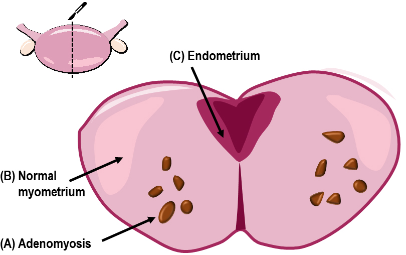 Fig. 1.
A schematic depiction of the uterine tissue sampling during surgery. (A) Adenomyotic tissue was sampled and collected from the middle of the lesion. (B) Myometrium with no adenomyotic involvement (designated as normal myometrium in the same patient). (C) Endometrial tissue sampling was conducted at the contralateral side of the adenomyosis.
| Patient no. | Type of surgical approach | Age | Parity | Main symptom | Main location | Total specimen (g) |
| 1 | Laparotomy | 34 | 0-0-1-0 | Dysmenorrhea | Anterior | 160 |
| 2 | Laparotomy | 41 | 0-1-1-0 | Dysmenorrhea, menorrhagia | Posterior | 77 |
| 3 | Laparoscopy | 43 | 2-0-0-2 | Menorrhagia | Anterior | 60 |
| 4 | Laparoscopy | 41 | 2-0-1-2 | Dysmenorrhea | Anterior | 27 |
| 5 | Laparotomy | 36 | 0-0-0-0 | Menorrhagia | Anterior | 80 |
| 6 | Laparotomy | 34 | 0-0-3-0 | Dysmenorrhea, menorrhagia | Anterior | 256 |
| 7 | Laparoscopy | 45 | 2-0-1-2 | Lower abdominal discomfort | Anterior | 53 |
| 8 | Laparotomy | 42 | 0-0-0-0 | Dysmenorrhea | Posterior | 178 |
| 9 | Laparoscopy | 33 | 0-0-0-0 | Dysmenorrhea | Posterior | 56 |
| 10 | Laparotomy | 32 | 0-0-0-0 | Dysmenorrhea | Posterior | 87 |
| 11 | Laparotomy | 29 | 0-0-0-0 | Dysmenorrhea | Posterior | 115 |
| 12 | Laparotomy | 44 | 0-0-0-0 | Palpable mass | Posterior | 99 |
| 13 | Laparotomy | 39 | 1-0-1-1 | Dysmenorrhea, menorrhagia | Posterior | 201 |
| 14 | Laparotomy | 41 | 2-0-1-2 | Dysmenorrhea, menorrhagia | Posterior | 301 |
| 15 | Laparotomy | 36 | 0-0-0-0 | Dysmenorrhea | Posterior | 87 |
| 16 | Laparotomy | 42 | 0-0-5-0 | Dysmenorrhea, recurrent abortion | Posterior | 188 |
| 17 | Laparotomy | 32 | 0-0-0-0 | Dysmenorrhea | Posterior | 88 |
| 18 | Laparotomy | 35 | 0-0-1-0 | Dysmenorrhea | Anterior | 83 |
| 19 | Laparotomy | 44 | 3-0-2-3 | Menorrhagia | Posterior | 124 |
| 20 | Laparoscopy | 43 | 0-0-2-0 | Dysmenorrhea, menorrhagia | Anterior | 107 |
When the tissue was collected from the three targeted points of the uteri in the operation room, each sample was divided into two pieces. One of these pieces was sent to the pathology department for immunochemical analyses, and the other was preserved in RNAlater™ Solution (Invitrogen by Thermo Fisher Scientific Corporation,Waltham, MA, USA) and stored at –70 ℃ until further use.
A designated pathologist interpreted all of the IHC results used in this study.
IHC for formalin-fixed, paraffin-embedded samples was performed as follows.
Following deparaffinization with xylene (2
Total RNAs were extracted from chopped tissues using the
easy-BLUE™ Total RNA Extraction Kit (iNtRON, #S17061). Following
this, 1
Data were statistically analyzed using SPSS 20.0 (IBM Corp., Chicago, IL, USA)
and GraphPad Prism 5 software (GraphPad Software, LLC, San Diego, CA, USA). If
the data sets showed normality, they were analyzed by one-way ANOVA via the
Student-Newman-Keuls test for all pairwise comparisons, and the Student’s
t-test was also used. In the case of the three datasets that rejected
normality, the Kruskal-Wallis test and Mann-Whitney test were used for the
analysis. p-values less than 0.05 were considered to indicate
statistical significance. Data were expressed as the mean
A total cohort of 22 adenomyosis patients were initially enrolled with informed consent prior to surgery. One was subsequently excluded because of severe endometriosis detected during the operation and another was not analyzed further owing to inadequate sample availability at the point of the endometrium for immunohistochemistry. The study participants were all premenopausal, with a mean age of 38.3 years (median 40, range 32–44) (Table 1). In this series, there were no adverse surgical outcomes from the tissue sampling.
Immunohistochemistry (IHC) results were analyzed for the eutopic endometrium,
myometrium without adenomyosis, and adenomyotic lesion (Fig. 2C) using the sum of
the quantitative and intensity scores (Q + I). There were significant differences
found among all three sampling regions (p
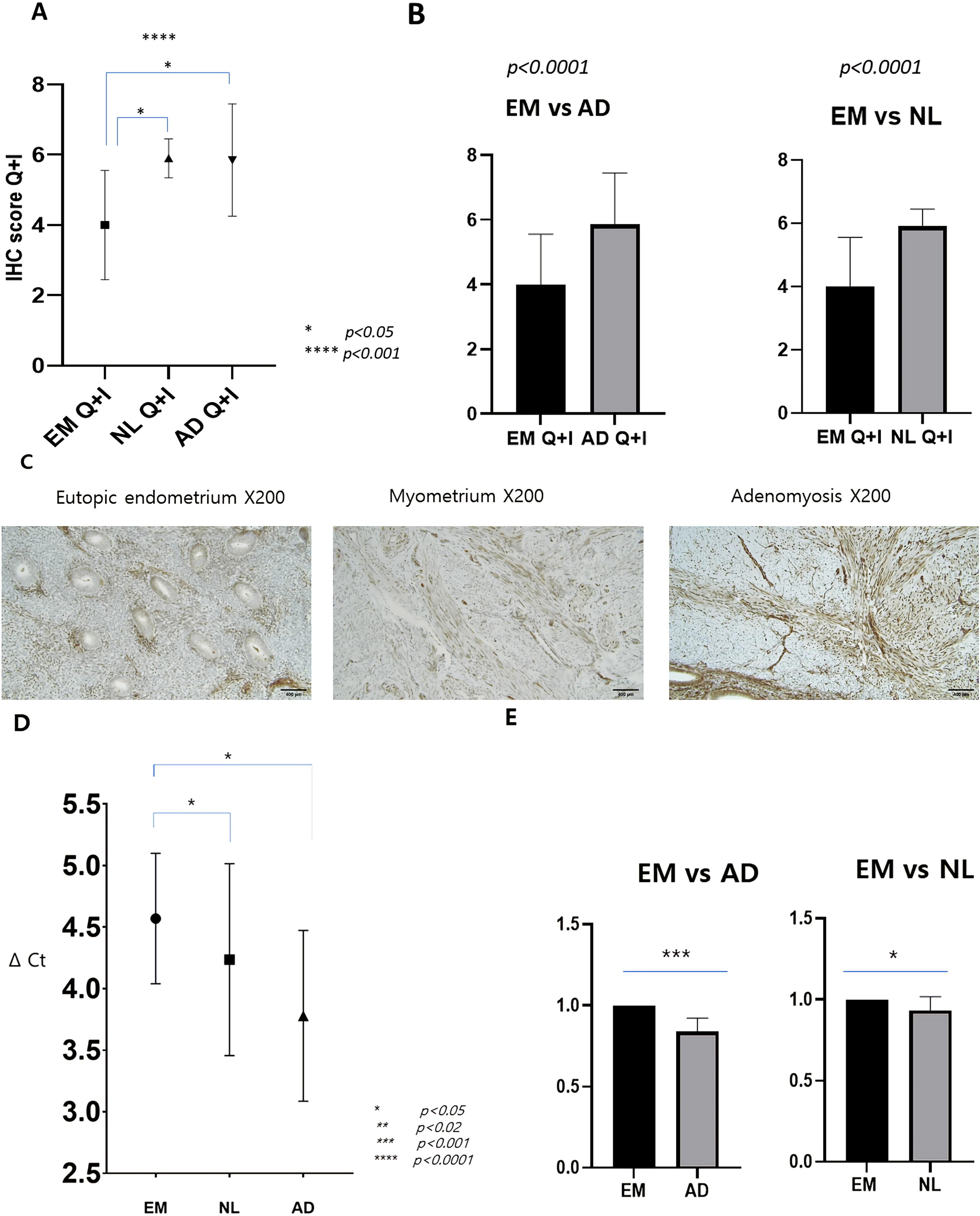 Fig. 2.
IHC and RT-PCR results for the eutopic endometrium, myometrium
without adenomyotic tissue, and adenomyotic lesion. Data were compiled from the
sum of quantitative and intensity scores (Q + I) and expressed as the mean
Real-time polymerase chain reaction (RT-PCR) was conducted to assay the VEGF-A transcript levels and showed statistically significant differences between the delta Ct values for the eutopic endometrial tissue, normal myometrium, and adenomyosis lesion (Fig. 2D). VEGF-A mRNA expression was detectable first in the adenomyosis lesion and was found to be higher in this tissue (p = 0.002) and in the myometrium (p = 0.018) compared with the endometrium (Fig. 2E).
The IHC and RT-PCR data indicated that VEGF expression was statistically higher in the adenomyotic lesion and myometrium than in the eutopic endometrium. To determine whether this was also the case for the microvessel counts, which reflect the function of VEGF, the microvessel densities (MVDs) were determined by CD31 staining of the same tissue slide samples (Fig. 3A). The adenomyotic lesions are composed of myometrial glands and myometrium, and the MVDs were thus separately counted for these samples at the stroma in the myometrium and in the ectopic endometrium in the identical area. There was no significant difference in these counts between ectopic glands existing in the adenomyotic tissue and the eutopic glands in the endometrium (p = 0.5; Fig. 3B). However, in the myometrium located in the stroma of the adenomyosis, microvessels were more prominent than in the eutopic endometrium (p = 0.02; Fig. 3C).
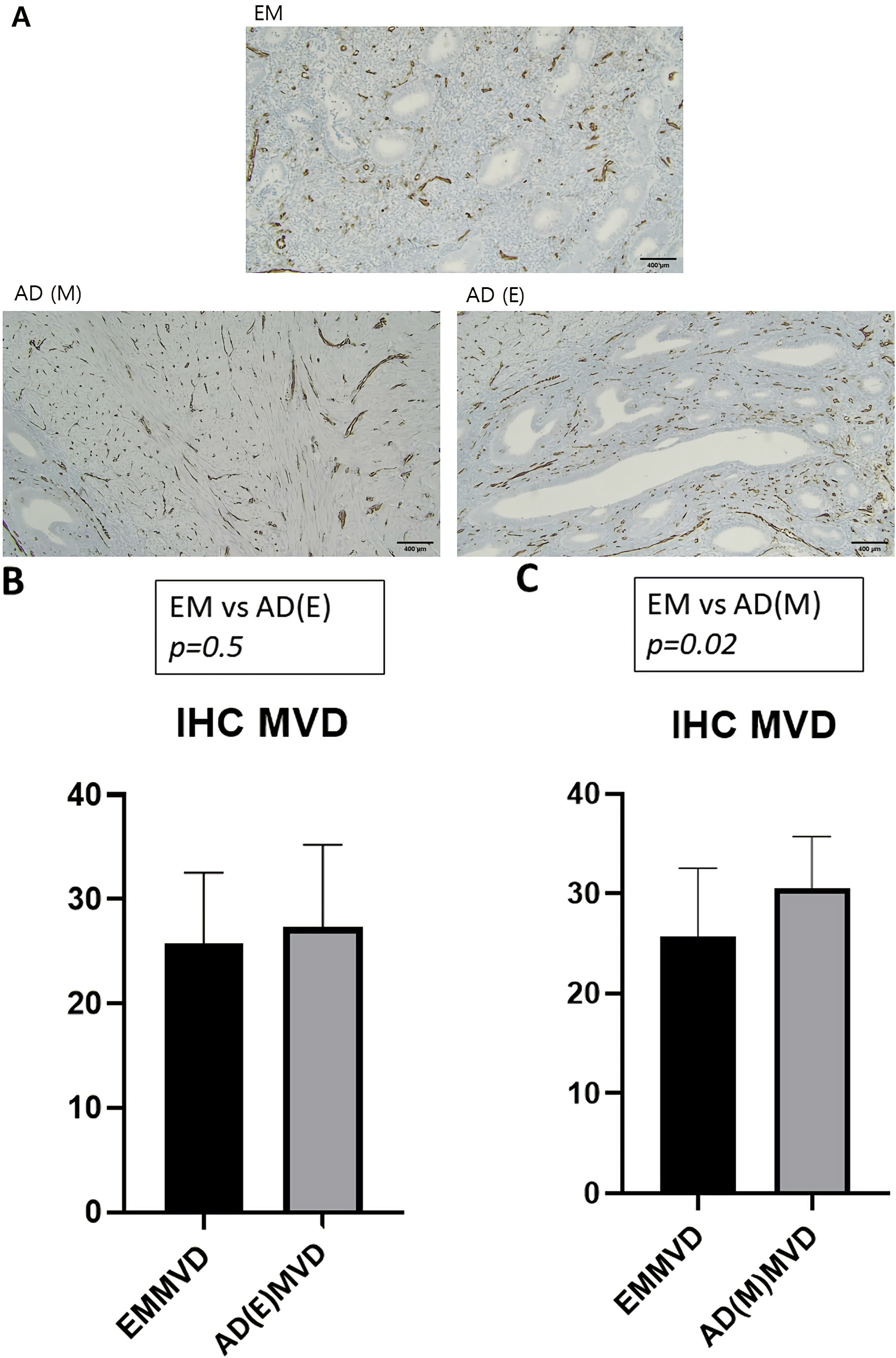 Fig. 3.
IHC and microvessel density analysis of eutopic endometrium,
myometrium without adenomyotic tissue, and adenomyotic lesion. (A)
Immunohistochemical counting of the microvascular density by IHC staining with a
CD31 probe in the endometrium and myometrium in the uteri with adenomyoses;
original magnification,
The uterus comprises three different histopathological components, which are the innermost endometrium, the more central myometrium, and the outermost serosa. Adenomyosis is pathologically defined as the presence of endometrial glands and stroma within the myometrium, but it is not clear how endometrial glands become located inside the myometrium in this disease. In our present analyses, we tested the proposition that VEGF plays a crucial role in the uterine myometrium by changing the features of the myometrial tissue rather than facilitating invasion from the endometrium. In uterine adenomyosis, the endometrial glands become located away from their original position, thus adopting an “ectopic” rather than their normal “eutopic” position.
Histologically, a mucosal-muscular interface, called the endometrial-myometrial interface (EMI), exists between the endometrium and myometrium but with no intervening basement membrane, thus differing from other mucosal organs. The invagination theory of adenomyosis proposes that when microtraumas occur, this structure can be easily disrupted, thus allowing endometrial tissue to grow from the inner side of the myometrium [11].
VEGF is one of the factors that promotes neoangiogenesis and plays a crucial role in tissue growth, thus representing a viable candidate for promoting adenomyosis [2]. Previous studies have reported that both the serum and histopathologic VEGF levels are elevated in the uterus upon the onset of adenomyosis, but few prior investigations have compared the endometrium and myometrium. Orazov et al. [12] previously conducted a histopathological review of adenomyosis patients and detected VEGF expression in both the epithelial cells of the glandular structure and in the stromal cells of the myometrium. These authors speculated that other kinds of growth factors could be stimulating the myofibroblasts other than VEGF, but no subsequent data have been presented to support this contention. Goteri et al. [13] found that VEGF expression was increased in both the ectopic and eutopic endometrium in the uterus of adenomyosis patients and concluded that it was focused on the function of the endometrial component in the adenomyosis lesion.
We further analyzed the potential role of VEGF in the progression of uterine adenomyosis by comparing its expression between the endometrium, myometrium (not involving the adenomyosis), and adenomyosis lesion in the same uterus. We found VEGF to be more highly upregulated in the myometrium and adenomyosis lesion than in the endometrium (Fig. 2). Moreover, in the same microscopic field as the adenomyosis, we observed that the MVD was higher in the stromal cells of the myometrial component than in the epithelial cells that comprised the endometrial glands (Fig. 3). This suggests that VEGF plays a significant role in the muscular component of the endometrium during the progression of adenomyosis. To our knowledge, our current study is the first to assess the VEGF expression profile in different histologic areas of the same uterus in a series of adenomyosis patients.
There were some limitations of note in this study. In the first instance, the VEGF expression distribution profiles were compared within the same uterus of individual patients with adenomyosis rather than with a normal control group. Even though we did analyze normal myometrial tissues, these were still derived from adenomyosis patients and may not be equivalent to normal. However, this was still a valid approach to understanding the role of VEGF in uterine adenomyosis. Additionally, variations in sample tissue quality cannot be excluded since our primary consideration when collecting samples during surgery was to do so in a manner that would not be harmful to the patient.
The strength of our current investigation in this regard was that our uterine samples would not have been affected by these cyclic changes because of the compensatory administration of a gonadotropin-releasing hormone (GnRH) agonist to every patient 2–3 weeks before surgery. This was done to reduce operative bleeding and to adjust for variations in the hormone levels of the patients because adenomyosis is an estrogen-dependent disease.
The established medical treatments of adenomyosis, which focus on blocking the effects of estrogen, can have side effects, including menopausal symptoms and unintended contraceptive impacts. Further study is needed to more fully elucidate the pathogenesis of uterine adenomyosis and develop future disease-specific treatments. Identifying novel subregulatory mechanisms that underlie the pathophysiology of adenomyosis, such as the modulation of VEGF expression, will likely help to find targets for new medical approaches.
Vascular endothelial growth factor contributes to the progression of uterine adenomyosis and may be more strongly activated in the stromal component of the myometrium than in the endometrial (eutopic or ectopic) compartment of the adenomyosis in the same uterus. This suggests that VEGF plays a significant role in the muscular component of the endometrium during the progression of adenomyosis.
Conceptualization—JK and YK; methodology—IJ and PL; formal analysis—JK; investigation of tissue—JK, IJ and HL; resources—MS; data curation—JK; writing — original draft preparation—JK and IJ; writing — review and editing—YK and PL; visualization—JK; supervision—YK and PL; project administration—JK, HL and PL; funding acquisition—JK, PL and YK. All authors have read and agreed the published version of the manuscript.
This study was approved by the Institutional Review Board of Nowon Eulji Medical Center (IRB No. 2019-07-023). Informed consent to participate was obtained from all patients, including tissue collection during the surgery.
Not applicable.
This study received financial support from Eulji University (2019EMBRISN0003). Also, this work was supported by Basic Science Research Program through the National Research Foundation of Korea (NRF) funded by the Korea government (MSIT) (NRF-2020R1F1A1070713, NRF-2020R1A4A1016029).
The authors declare no conflict of interest. YSK is serving as one of the Guest Editors of this journal. We declare that YSK had no involvement in the peer review of this article and has no access to information regarding its peer review. Full responsibility for the editorial process for this article was delegated to MHD.