

 , M. Sucu 1, E. Arslan 1
1 Department of Obstetrics and Gynecology, Perinatology Unit, School of Medicine, University of Çukurova, Adana, Turkey
Abstract
Introduction: Absence of the ductus venosus (ADV) is a rare vascular anomaly often associated with fetal cardiac/extracardiac anomalies, aneuploidies, and hydrops. This study assesses the abnormal venous circulation, associated malformations, and chromosomal anomalies of ADV. Materials and Methods: The authors performed a retrospective study of 14 cases with ADV diagnosed by the prenatal ultrasound in tertiary referral center from 2009 to 2017. Results: The authors detected 14 patients of ADV. Karyotyping was offered to all cases, and four patients accepted the procedure. Normal karyotype was found in three out of four cases, while one case had 45, X0. Five cases underwent termination of pregnancy, six cases died at neonatal period due to accompanying cardiac anomalies, and heart failure. Three cases survived (the oldest baby is 4-years-old now). Umbilical vein (UV) was connected to portal venous system (PVS) in two cases (intrahepatic drainage, one of them is surviving), while UV was connected to systemic venous circulation in 12 cases (extrahepatic drainage, two of them are surviving). In five cases there was no other reason that might explain cardiomegaly and hydrops except ADV (two of them is surviving). Three cases were diagnosed as cystic hygroma in first trimester additionally found ADV. All of surviving fetuses delivered after 35th weeks of gestation. Conclusion: The presence fetal hydrops, accompanying congenital anomalies, and prematurity are associated with poor prognosis in fetuses with ADV. In isolated cases, the prognosis is generally good.
Keywords
- Prenatal diagnoses
- Ductus venosus
- Umbilical vein
- Congenital abnormality
Ultrasonographic evaluation of fetal umbilical - portal venous system (PVS) has been increasing considerably in recent years [1]. It is important to identify the correct anatomic relationship of this venous structure to understand the effects on fetus.
The umbilical vein (UV) joins a confluence of vessels termed the portal sinus. The portal sinus is a conglomeration of structures including the intrahepatic portal vein, the extrahepatic portal vein, and the ductus venosus (DV). DV is a short venous structure which connects the umbilical and systemic venous circulation [2].
Blood flows to the portal sinus from the UV and then delivered to the liver via the portal veins and systemic circulation via the DV. The oxygenated blood coming from the placenta is conducted to the left heart and brain by ductus venosus [3].
DV can be considered a physiological valve. Approximately 50% of the umbilical venous return is shunted through the DV in early gestation and decreases to 20%-30% in the third trimester [4]. Therefore, in the absence of the DV (ADV), the physiology of fetal circulation changes. With ADV, the incidence of additional cardiac/extracardiac anomalies and genetic/chromosomal aberrations increase. In case of the anomalous fetus, it is necessary to evaluate PVS and DV because in order to make a correct prognosis.
This retrospective study was conducted at Çukurova University Hospital (academic tertiary referral center) prenatal ultrasound unit. All women diagnosed with fetal ADV from January 2009 to September 2017 were analysed. Data was collected from the digital patient recording system.
All of the sonographic evaluations were performed by one of the nine experienced authors, using instrumentations with a convex volumetric transducer (RAB 6-D 2-7 MHz and RAB2 5L) probe. The anatomical screening is performed between 18-22 gestational weeks in this clinic. The authors also evaluate the fetal anomalies referred to them. The DV was evaluated using B mode, colour or power Doppler in two planes in all of the cases which were suspected with cardiac/extra cardiac abnormalities, pleural/pericardial effusion or hydrops. With ADV, umbilical shunting type was determined according to UV connection. The connection with systemic venous system or PVS was classified as an extrahepatic or intrahepatic drainage, respectively. Fetal karyotyping and echocardiography were offered to all women with ADV.
Neonatal outcomes were obtained from electronic medical reports or the family was interrogated by phone call. The findings of all the cases with neonatal loss and termination of pregnancy were confirmed with autopsy examination except for first trimester terminations. All pregnant women were informed and written content was obtained. This study was approved by the Ethics Committee of University of Çukurova.
First trimester trisomy screening, second trimester anatomical survey, and fetal anomaly evaluation (referred to this clinic) were performed in total 18,500 patients over the nine-year period. The authors detected ADV in 14 (≈0.08%) patients.
The gestational age at diagnosis ranged from 12 to 35 weeks (Table 1). Four patients accepted the karyotype analyse. Normal karyotype was found in three out of four cases, while one case (case 9) had 45, X0 (Turner Syndrome). Five cases underwent termination of pregnancy, and six cases died at neonatal period due to accompanying cardiac anomalies and heart failure. Three cases survived (the oldest baby is now 4-years old). UV was connected to PVS in two cases (intrahepatic drainage, one of them is surviving), while UV was connected to systemic venous circulation in 12 cases (extrahepatic drainage, two of them are survived). In five cases (cases 4, 5, and 12-14), there was no other reason that might explain cardiomegaly and hydrops except ADV (two of them survived). Three cases (cases 6, 8, and 9) were diagnosed with cystic hygroma in first trimester additionally found with ADV.
| GA at diagnosis (weeks) | GA at delivery (weeks) | Associated anomalies | UV connection |
Karyotype | Outcomes |
|---|---|---|---|---|---|
| 1 28 | 35 | Cardiac: VSD, cardiomegaly, tricuspid insufficiency Extra cardiac: agenesis of corpus callosum |
RA | Not performed | NL |
| 2 35 | 39 | Ebstein anomaly, pulmonary stenosis | PVS | Not performed | NL |
| 3 24 | 24 | Cardiac: AVSD Extra cardiac: Dandy Walker anomaly, micrognathia |
VCI |
Not performed |
TOP |
| 4 30 | 30 | Cardiomegaly, hydrops | RA | Not performed | NL |
| 5 35 | 35 | Cardiomegaly | RA | Not performed | 4 years of life, survivor |
| 6 13 | 13 | Cystic hygroma, hydrops | RA | Not performed | TOP |
| 7 30 | 39 | Cardiac: cardiomegaly, VSD Extra cardiac: ventriculomegaly, pelvic kidney |
RA | Not performed | 3 years of life, survivor |
| 8 13 | 13 | Cystic hygroma | RA | Not performed | TOP |
| 9 12 | 12 | Cystic hygroma, AVSD | RA | 45, X0 | TOP |
| 10 25 | 37 | Cardiac: truncus arteriosus Extra cardiac: cerebellum hypoplasia |
RA | Normal | NL |
| 11 22 | 22 | Cardiac: heterotaxia Extra cardiac: ventriculomegaly |
VCI | Normal | TOP |
| 12 34 | 34 | Cardiomegaly, hydrops | VCI | Not performed | NL |
| 13 23 | 36 | Cardiomegaly | PVS | Normal | 8 months of life, survivor |
| 14 32 | 32 | Hydrops, cardiomegaly | RA | Not performed | NL |
AVSD: atrioventricular septal defect; GA: gestational age; NL: neonatal loss; PVS: portal venous system; RA: right atrium; TOP: termination of pregnancy; UV: umbilical vein; VCI: vena cava inferior; VSD: ventricular septal defect.
The authors analyzed a total of 14 cases of ADV. The overall neonatal survival was 21%. The poor prognosis depended on the presence of associated cardiac/extra cardiac congenital anomalies and hydrops.
Leonidas and et al. [5] were the first to report insertion of the UV directly into the IVC at the level of the iliac veins in a neonate with Noonan syndrome. Two different routes for umbilical venous return (extrahepatic and intrahepatic umbilical venous drainage) have been reported in fetuses with ADV: extrahepatic (bypassing the liver) or intrahepatic drainage (via the PVS) [6-8]. Achiron et al. [1] recently proposed a new classification of fetal umbilical - PVS shunts but that has not been yet widely accepted.
With extrahepatic drainage, UV can connect to the right atrium (RA- the most common type), coronary sinus, left atrium, IVC, iliac or renal veins [7, 9, 10]. These types of bypasses may lead to cardiac decompensation. Hydrops can results from massive blood overload on the heart. Additionally, genetic/chromosomal anomalies and cardiac/extracardiac anomalies can cause hydrops [8, 11-15]. In general, combination of these factors are the reasons for hydrops.
With intrahepatic drainage, UV connects to the PVS and overloading on the heart does not occur. Hydrops and cardiac failure may result from other mechanism, such as portal hypertension [9, 16]. Prognosis is relatively good in this subtype [13, 15, 17]. In the present series, there were two intrahepatic drainages, and one of this baby is now eight month. It must be kept in mind that ADV with intrahepatic drainage diagnosis is difficult than extrahepatic type and sonographers must be careful to evaluate venous structures.
In the present cohort, relevant to the literature, there is high association with ADV and other system anomalies. Prognosis mainly depends according to accompanying these anomalies. In five cases (two of them survived), there were no other reason for cardiac failure and hydrops except ADV. Prenatal diagnosis of ADV is linked with a high rate of pregnancy termination (35%). Correct evaluation of fetal anatomy is important to define prognosis for counselling the parents. All of the surviving fetuses delivered after 35 gestational weeks.
ADV is a rare disorder; in the present cohort, prevalence of ADV is approximately 0.8%. Because the hemodynamic changes are less significant in intrahepatic drainage subtype, the prevalence of ADV might be underestimated. ADV can also be under diagnosed in cases with structural anomalies, because ultrasonographers may pay attention to other malformations [8].
In the present institution, the authors routinely evaluate DV in cases with intrauterine growth retardation (IUGR), cardiac failure, hydrops, and additional anomalies. The authors believe that in these situations, DV assessment should be a part of the examination. DV can be evaluated in two planes: sagittal and transverse sections. Sagittal section is more appropriate, but when not available, transverse section can also be effective. Ultrasonographic image of a case with ADV is seen in Figure 1. DV views are between portal sinus and VCI. Doppler analysis is helpful to determine DV. Doppler sample gate should be between 0.5-1.0 mm according to gestational age. The aliasing area in DV is due to the narrow structure of this vessel. With pulsed Doppler, classic waveform of DV should be evaluated. The insonation angle sweep speed should be less than 30° and high (2-3 cm/s), respectively.
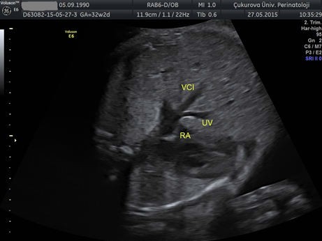 Figure 1.
— Ultrasonographic imaging of ductus venosus agenesis.
The incidences of chromosomal abnormalities in cases with ADV were reported between 17.4% to 42.3% [8, 1 8, 19]. Karyotype analysis should be offered [16]. The most frequent chromosome aberration of ADV is Turner and Noonan Syndrome [18]. In four cases who accepted the chromosome analysis, Turner Syndrome (45, X0) was diagnosed in one case. So, ADV must be kept in mind in these syndromes.
It is possible to detect ADV during first trimester [20, 21]. The association between cystic hygroma and ADV is known [16]. The routinely perform DV Doppler analysis in first trimester in order to increase the detection rate and decrease the false positive rate of combined test. Especially in cystic hygroma, it is important to evaluate DV. The present authors detect three cases of ADV in first trimester that are also diagnosed of cystic hygroma.
The retrospective design, heterogeneous sources of information, and limited data on long-term outcomes (one of surviving child four-years-old yet) are among the present study limitations. The study population was too small to determine the true incidence of the different types of ADV. The authors do not know which chromosomal/genetical disorders are more common in ADV. Because of the fetuses which were referred to their clinic had numerous anomalies, the authors may not determine the isolated cases. Prospective studies in large and unselected populations are needed. Strength of this study is that there is no loss of data in the follow up due to in a single referral center.
DV should be assessed in all anomalous fetus. In ADV, UV may connect to the inferior caval vein, right atrium, iliac vein or PVS. In intrahepatic type, findings may be unclear. Correct mapping of vessels is crucial to determine cardiovascular effects and prognosis. Cardiac failure and hydrops may develop due to overflowing on the heart or associated anomalies. Serial sonographic examinations are required in order to detect cardiac failure. Fetal hydrops and the presence of additional congenital anomalies are associated with poor prognosis. In isolated cases, the prognosis is generally good.